III. 3. THE CYSTINE/CYSTEINE CONTENT OF HEMOGLOBINS
T. H. J. Huisman*
Department of Pediatrics, State University of Groningen, The Netherlands
I. Introduction 153 II. The Purification of Hemoglobin Preparations 157
III. Comparison of the Different Methods Used for the Estimation of Hemo-
globin Concentration 159 IV. Estimation Methods for the Total Number of Half-cystine Residues . . 159
V. The Argentometric Amperometric Titration in Ammoniacal Solution with
a Mercury-Coated Platinum Electrode 162 VI. The Application of the PCMB Method in the Study of Reactive —SH
Groups of Hemoglobins 163 VII. A Comparison of the Final Data Obtained for the Cystine/Cysteine Con-
tent of Different Human Hemoglobins 165 VIII. Studies on the Cystine/Cysteine Content of Adult and Fetal Hemoglobins
of Some Animals 168
I. Introduction
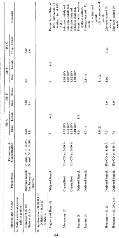
The reported numbers of residues of half-cystine and of —SH groups in various human hemoglobins (Hb-A, Hb-F, Hb-S, and Hb-C) vary from two to eight per molecule of hemoglobin. These observations are summa- rized in Table I. In this survey the results of the different analyses are arranged in order of the procedures used. Moreover, different types of preparations were analyzed and several methods for the estimation of the hemoglobin concentration were used ; these data are also presented. A num-
ber of different molecular weights (60000, 64000, 66000, 66700, 66800, 68- 000) have been employed by different authors in the research on the struc- ture of hemoglobins (1). Recent investigations (2) have shown that a mo- lecular weight of 66700-66800 is the most probable value for hemoglobin;
in order to facilitate the comparison of the different figures the reported
* The author is indebted to Dr. F. A. Hommes and Dr. H. J. van der Helm for their help and for many suggestions offered during the course of this work. He also wishes to thank Miss T. Sebens for her capable technical assistance.
153
TABLE I THE CYSTINE/CYSTEINE CONTENT OF DIFFERENT HUMAN HEMOGLOBINS'1 Method and Author Preparation Estimation of Concentration
Hb-A Hb-F Hb-S Hb-C Orig. Denat. Orig. Denat. Orig. Denat. Orig. Denat. Remarks A. Total cystine as cysteic acid in globins Hommes et al. (8) Stein et al. U) Dialyzed hemol. Pur. by starch electrol.
Ν anal. Ν = 16.8% 8.28 Ν anal. Ν = 16.8% 4.8 5.67 3.4 8.3 8.38 4.6 Â. Ag-titration in 0.03-0.1 M NH4OH + 0.05 M NH4NO3 Ingbar and Kass (J) Murayama (7) Ingram (S) Ingram (9)
Dialyzed hemol. Crystallized Crystallized Dialyzed hemol. Dialyzed hemol.
Hb-C Ο at 5400 A Hb-CO at 5400 Â Hommes et al. (3) Dialyzed hemol. Hb-CO at 4180 A Hommes et al. (10, 11) Dialyzed hemol. Hb-CO at 4180 Â
4.32 (0°) 3.41 (38°) 4.18 (0°) 8.01 (38°) 3.9 4.2 (1) 8.0 (2) 7.5 7.4
4-5 4.03 (0°) 1.90 (38°) 3.92 (0°) 7.91 (38°) 3.8 (1) 8.4 (2) 3.9 8.05 4.0
7.55
Denat. by addition of 20% methanol. Ti- tration in 0.85% NaCl. Solution undialyzed. Solution undialyzed. Solution dialyzed. Solution dialyzed. Denat. with sodium dodecyl sulfate. Mercury-coated Pt electr. (1) = first end point; (2) = second end point. Mercury-coated Pt elec * Mercury-coated Pt electr.
154
C. Ag-titration in tris buffer, pH 7.4. Benesch et al. (12) Murayama (13, 14) Hommes et al. (10 Stein et al. (4) Crystallized Crystallized Dialyzed hemol. Pur. by starch electrol.
Fe-content; Hb-Os at 5400 + 5760 Â Hb-CO at 5400 A Hb-CO at 4180 A Ν anal. Ν = 1è.8%
9.5 9.4
8.0 8.01 7.5 4.9 4.3 6.04 4.0 3.4
Denat. in 8 M urea 7.91 8.06 Denat. \n 8 M urea Denat. in 4 M urea 4.5 Denat. in 8 M urea D. Hg-titration according to Kolthoff et al. (15) Murayama (7) Murayama (14) Ingram (8) Hommes et al. (S)
Crystallized Hb-CO at 5400 A 3.26 (0°) 5.89 (38°) Crystallized Hb-CO at 5400 Â 5.89 2.2 Dialyzed hemol. Hb-CO at 4180 A 5.3 7.45
3.2 6.0 4.7
3.13 (0°) 5.02 (38°) 5.02 2.75 6.9 7.05
8.02 5.9
Presented as Hg atoms/4 Fe. Denat. with sodium dodecyl sulfate, i.l Denat. with alkali. Å. PCMB method Murayama (7) Spectrophotometric according to Boyer (16) Hommes et al. (10) Spectrophotometric according to Boyer (16) Benesch et al. (12) Equilibrium dialysis
Crystallized Hb-CO at 5400 Â 2.9 2.9 Dialyzed hemol. Hb-CO at 4180 Â 6.6 2.65 2.75 2.35
pH 6.8 pH 4.6 Crystallized Fe-content; Hb-02# 8 at 5400 + 5760 Â á Values from literature; calculated on molecular weight 66000 (globin) and 66800 (hemoglobin) and presented as residues of half-cystine or number of —SH groups.
155
156 T. H. J . HTJISMAN
values are, if necessary, converted to those based on a molecular weight of 66800.
The first results mentioned in Table I are those obtained by Hommes et al. (3) and by Stein et al. (4) analyzing the total quantities of half- cystine, determined as cysteic acid, of some human hemoglobin types.
The numbers of half-cystine residues reported by these two groups of in- vestigators are quite different and vary from 4.8 to 8.38 for the Hb's A, S, and C and from 3.4 to 5.67 for the Hb-F. The possibility is present that this discrepancy is due to differences in the methods employed or in the procedures used for the purification of the hemoglobin samples.
Different argentometric titration methods are available for the esti- mation of the number of free —SH groups in proteins. Several investiga- tors {3j 5-11) titrated human hemoglobins (in dialyzed hemolysates or after crystallization) amperometrically with silver nitrate in ammoniacal solution. In most cases 0.03 to 0.1 M N H4O H + 0 . 0 5 Μ N H4N 03 served as supporting electrolyte. In one case 0.85 per cent sodium chloride was added. Next to a dropping-mercury electrode, rotating platinum-wire electrodes were used, both with and without pretreatment with an am- moniacal HgCl2 solution. Different numbers of free — S H groups, varying between 1.9 and 8.05 per molecule of hemoglobin (Hb-A, Hb-S, and Hb-C) were established. Moreover, Murayama ( 7 ) observed variations in the number of titratable — S H groups of normal and sickle-cell hemoglobin, when the temperature of the solution titrated was changed from 0° to 38°.
The application of the argentometric titration procedure (12) using a tris(hydroxymethyl)aminomethane buffer (tris) solution of pH 7.4 had made it possible to titrate the —SH groups in proteins in neutral buffered solutions. This technique has also been used by some investigators (4, 10, 12-14) in the study of hemoglobins, especially after addition of 4 to 8 M of urea as denaturing agent. In dialyzed hemolysates as well as in crystal- lized preparations of the hemoglobins A, S, and C about 8 titratable —SH groups were found, while in fetal human hemoglobin 4 (10) and 6 (14) free — S H groups were established. Purification of the hemoglobins by starch slab electrophoresis seems to lower the titratable —SH groups to 4-5 in the hemoglobins A and S and to 3-4 in the fetal hemoglobin (4).
Mercurimetric-amperometric titration methods, as for instance de- scribed by Kolthoff et al. (15), are also employed by some authors (8, 7 , 8, 14). Again varied results were obtained. On account of his results ob- tained with the argentometric and mercurimetric-amperometric titration methods, Murayama (7, 14) has presented some schemes of spatial ar- rangements of titratable —SH groups in the human hemoglobins A,F,S, and C. In view of the great differences between the results obtained by
C o l l e c t o r
FIG. 1. The equipment for carboxymethylcellulose chromatography.
different authors, definite conclusions in this respect are difficult to draw at the present time.
The spectrophotometric procedure of Boyer (16), based on the reaction of protein — S H groups with p-chloromercuribenzoic acid ( P C M B ) , has also been applied to human hemoglobins (7, 10). At a pH of 6.8 about 3
—SH groups were reactive both in Hb-A and Hb-S (7), while at a pH of 4.6 over 6 —SH groups were found with this technique (10).
In view of the great variations in the data reported by several investi- gators, it seems important to study more closely some aspects of the dif- ferent procedures employed. As no general procedure for the estimation of the cystine/cysteine content of the hemoglobins can be advocated, more than one technique will be cited.
II. The Purification of Hemoglobin Preparations
Hemoglobin solutions were obtained in the usual way by lysing care- fully washed red cells with toluene or by freezing and thawing. The cell debris was removed in a high-speed centrifuge. The hemoglobin was con- verted into its monocarboxy form and finally dialyzed in the cold for 48 hours against a large volume of distilled water with four changes of the dialyzing fluid.
158 T. H. J. HUISMAN
As many reports appeared in the literature concerning the heterogene- ity of these hemoglobin preparations (summarized for instance in reference 17), it was found valuable to analyze hemoglobin preparations purified by a suitable method. Stein et al. (4) analyzed the main components of hemo- globins A, F, S, and C prepared by zone electrophoresis of the carbonmon- oxy derivatives on starch (18) and obtained data on the cystine/cysteine content of these hemoglobin preparations, which were markedly lower than those presented by other investigators for unpurified hemoglobins.
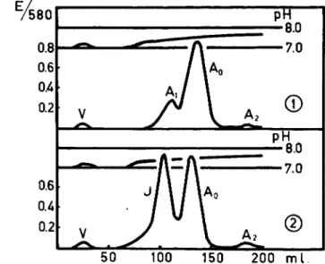
A similar heterogeneity of normal adult hemoglobin as found in starch slab electrophoresis (18) is also obtained by chromatography of this pro- tein on carboxymethylcellulose (19). The use of this adsorbent, which was prepared according to Peterson, Sober, et al. (19a, b), offers other possi- bilities for the separation of closely related hemoglobins and also for a study of inhomogeneity of this protein. The equipment necessary for this chromatographic procedure is presented in Fig. 1. AVith a dilute phosphate buffer of low ionic strength and of pH value gradually increasing from 6.8 to 8.0 as eluting solvent, normal adult hemoglobin is resolved into at least three different fractions (Fig. 2). The first fraction, called Hb-A, and present in amounts of about 10%, is eluted at a pH of 7.40; the main com- ponent (Hb-Ao) at a pH of 7.49; and the H b - A2 component, present in amounts varying between 0.5% and 2%, at a pH of 7.69. Paper electro-
FIG. 2. The separation of normal adult hemoglobin into three fractions (Ai, A0, A2) by carboxymethylcellulose chromatography. The chromatographic behavior of the Hb-Ai component is the same as that of the abnormal Hb-J.
phoretic analyses of these isolated compounds (19) have shown that these three fractions are closely related to, if not identical with, those obtained by Kunkel and Wallenius {18) on starch slab electrophoresis. With this chromatographic procedure it was found possible to prepare the H b - A0 on a large scale, as well as the Hb-F from cord blood and Hb-S and Hb-C from blood samples derived from patients homozygous for these abnormal hemoglobins. Thus, using different methods, enough material could be ob- tained for the analyses of the cystine/cysteine content of purified hemo- globins A0, F, S, and C. Although this preparative procedure is different from that used by Stein et al. (4), the separations of Hb-A into three com- ponents by the two procedures are so closely related that the analytical data obtained in this study may be compared with those published by these investigators.
III. Comparison of the Different Methods Used for the Estimation of Hemoglobin Concentration
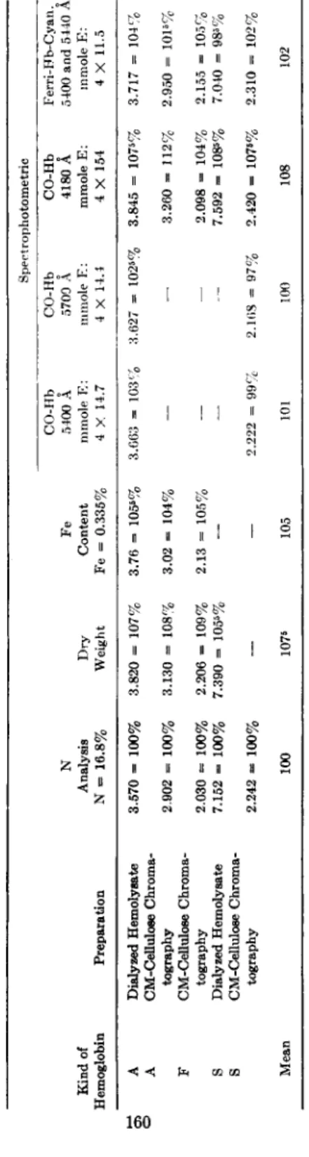
Different methods have been employed for the estimation of the amount of hemoglobin present in solution: nitrogen analysis (4), dry weight estimations, iron analysis (12) f and different spectrophotometric procedures (3, 5-14), which are based on the millimolar extinctions of oxy-Hb and of monocarboxy-Hb at different maxima (20). These meth- ods have been applied to some hemoglobin preparations, the results of these analyses being presented in Table II. The spectrophotometric anal- yses mentioned in this table have been carried out in the Beckman spec- trophotometer Model D U using a 1-cm. corex Beckman cell. In general a good agreement exists, and with the exception of dry weight analyses and of the spectrophotometric estimation of CO-Hb at 4180 Â the experimental error is limited to about 5%. It seems therefore that the use of different methods for the estimation of the hemoglobin concentration does not offer an explanation for the differences between the analytical data presented by several authors (Table I ) .
IV. Estimation Methods for the Total Number of Half-Cystine Residues An accurate method for the determination of the total quantity of half-cystine seems to be a chromatographic estimation of cysteic acid present in hydrolysates of oxidized proteins. Schram and co-workers (21) reported a chromatographic procedure for the estimation of the amount of cysteic acid using the anion exchanger Dowex 2 (200-400 mesh) as ad- sorbent and chloroacetic acid solutions (0.01 and 0.1 M) as eluting sol- vents. According to Kimmel et al. (22), the cysteic acid can also be iso-
TABLE II THE ESTIMATION OF THE HEMOGLOBIN CONCENTRATION WITH DIFFERENT METHODS0 Spect rophotometric CO-Hb CO-Hb CO-Hb Ferri-Hb-Cyan.o Ν Fe 5400 Â 5700 Â 4180 Â 5400 and 5440 Â Kind of Analysis Dry Content mmole E: mmole E: mmole E: mmole E: Hemoglobin Preparation Ν = 16.8% Weight Fe = 0.335% 4 X 14.7 4 X 14.4 4 X 154 4 X 11.5 A Dialyzed Hemolyeate 3.570 = 100% 3.820 = 107% 3.76 = 105s% 3.663 = 103% 3.627 =» 1025% 3.845 = 1075% 3.717 = 104% A CM-Celluloee Chroma- tography 2.902 = 100% 3.130 = 108% 3.02 = 104% — — 3.260 = 112% 2.950 = 1015% F CM-Cellulose Chroma- tography 2.030 = 100% 2.206 = 109% 2.13 = 105%, — — 2.098 = 104% 2.155 = 105% S Dialyzed Hemolysate 7.152 = 100% 7.390 = 1055% — — — 7.592 = 1085% 7.040 = 985% S CM-Cellulose Chroma- tography 2.242 = 100% — — 2.222 = 99%, 2.168 = 97% 2.420 = 107«% 2.310 = 102% Mean 100 1076 105 101 100 108 102 á The values are presented as mg./ml. and are calculated on a molecular weight of 66800.
160
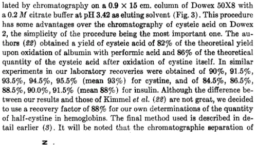
CYSTINE/CYSTEINE CONTENT OF HEMOGLOBINS 161 lated by chromatography on a 0.9 X 15 cm. column of Dowex 50X8 with a 0.2 M citrate buffer at pH 3.42 as eluting solvent (Fig. 3 ) . This procedure has some advantages over the chromatography of cysteic acid on Dowex 2, the simplicity of the procedure being the most important one. The au- thors (22) obtained a yield of cysteic acid of 82% of the theoretical yield upon oxidation of albumin with performic acid and 86% of the theoretical quantity of the cysteic acid after oxidation of cystine itself. In similar experiments in our laboratory recoveries were obtained of 90%, 91.5%, 93.5%, 94.5%, 95.5% (mean 93%) for cystine, and of 84.5%, 86.5%, 88.5%, 90.0%, 91.5% (mean 88%) for insulin. Although the difference be- tween our results and those of Kimmel et al. (22) are not great, we decided to use a recovery factor of 88% for our own determinations of the quantity of half-cystine in hemoglobins. The final method used is described in de- tail earlier (3). It will be noted that the chromatographic separation of
FIG. 3 . The chromatographic separation of cysteic acid on Dowex 5 0 X 8 , 2 0 0 - 4 0 0 mesh; 0.2 M citrate, pH 3.42.
cysteic acid on Dowex 50 has the disadvantage that this acid is not ab- sorbed at a pH of 3.42. Thus, a frontal elution of cysteic acid occurs. Any other ninhydrin-positive substance, formed during oxidation and hydroly- sis of the oxidized protein and with the same chromatographic behavior, may interfere and be calculated as cysteic acid.
Following the estimation of the total quantity of half-cystine in the different globins, the total number of S-atoms per mole of globin was de- termined. It is worth mentioning that the total amount of sulfur in hemo- globin is rather low. Thus, the estimation has a limited accuracy. After subtraction of the methionine values from the total sulfur the differences amount to the moles of half-cystine present in the hemoglobins. According to the recent analyses of the amino acid compositions (4, 23-26) there are six methionine residues in hemoglobins A, S, and C and eight in Hb-F (4).
Our own values of seven methionine residues in Hb-A and of nine methio- nine residues in Hb-F (25) seem to be one residue too high.
4 8 12 16 20 24 28 32 36 40 ml.
200-400 mesh.
0.2Λ/Citrate pH 342
162 T. H . J . HTJISMAN
V. The Argentometric-Amperometric Titration in Ammoniacal Solution with a Mercury-Coated Platinum Electrode
In a recent paper (11), we presented a detailed description of the ar- gentometric titration procedure with a rotating mercury-coated platinum wire electrode and with a mixture of 0.05 Μ N H4N 03 and 0.1 M N H4O H as supporting electrolyte. Pretreatment of the platinum electrode with an ammoniacal HgCU solution (30-45 minutes at —0.2 volt versus a standard calomel electrode) before the argentometric titrations of the hemoglobin samples was found necessary since repetition of the titration experiments with a formerly mercury-coated platinum electrode, without repeating this pretreatment, resulted in a gradual shift of the titration curves to
3 2 4 6 8 10 12 2 4 6 8 10 12
MOLES AG/MOLES HB
FIG. 4 . The argentometric titration curves of human adult and fetal hemoglobins.
(Mercury coated platinum electrode; supporting electrolyte: 0.1 M N H4O H + 0 . 0 5 M NH4NO3).
lower values. In most cases titration curves of normal adult hemoglobin were obtained with only one sharp end point. Sometimes two end points (Fig. 4) were found, the first corresponding with about four —SH groups and the second with eight titratable —SH groups in normal Hb-A. It will be noted that the titration curves of Hb-A, both in the dialyzed red cell hemolysates as well as in the solutions obtained after the purification pro- cedure with carboxymethylcellulose chromatography, gave at times the two end points. It may be that rather small changes in the titration condi- tions make the first end point in some cases almost invisible. In purified fetal hemoglobin only one end point corresponding to about four titratable
—SH groups was obtained (Fig. 4). Although the use of a tris buffer has some advantages over the ammonia buffer (12), the method of Benesch et al. (12) was not used in the experiments presented here. The data obtained
6 0
5 0
2 3 0 0 2 4 0 0 2 5 0 0 2 6 0 0 2 7 0 0 2 8 0 0 2 9 0 0 3 0 0 0 A
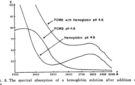
FIG. 5. The spectral absorption of a hemoglobin solution after addition of PCMB.
with argentometric titration procedure in ammoniacal solution are in ac- cordance with the values obtained with the method of Benesch et al. when 8 M urea was added to the reaction mixture (10).
VI. The Application of the PCMB Method in the Study of Reactive — S H Groups of Hemoglobin
The spectrophotometric determination of —SH groups in proteins as developed by Boyer (16), is based on the increase in absorption through interaction of the p-chloromercuribenzoate (PCMB) with —SH groups leading to much higher extinction readings at 2500 to 2550 Â. The number of reactive — S H groups can be estimated from the increment over the calculated absorption at the chosen wave length. The reaction is usually performed at the pH 4.6 and pH 7.0; it is found that in general the — S H groups of native proteins react more sluggishly at a pH of 7.0. As men- tioned already in Table I this procedure has also been applied to human hemoglobins, the results of these analyses being divergent.
Employing this technique for the estimation of the reactive —SH groups in human hemoglobins the following procedure was followed: vari- ous amounts (0.05-0.7 ml.) of a P C M B solution (4.24 Χ 1 0 ~4 M /liter) were added from a syringe microburette to constant amounts of hemo- globin (2.25 Χ ΙΟ"8 M H b - 02 dissolved in 3 ml. of a 0.1 M buffer solution of special p H ) . The solutions were mixed carefully with a small siliconized glass rod and allowed to stand at room temperature for some time, the length of which will be discussed later. The optical densities at 2500 Â
Ε.
100
9 0
ΘΟ
7 0
164 T. H. J . HTJISMAN
were determined against blanks, consisting of exactly the same amount of Hb in 3 ml., to which the corresponding volume (0.05-0.7 ml.) of distilled water was added. The increase of extinction as a result of P C M B binding to —SH groups was calculated from these readings and those of buffer blanks, to which the same amounts (0.05-0.7 ml.) of the P C M B solution were added. In most experiments a 0.1 M acetate buffer of pH 4.6 was used. From the ultraviolet absorption spectra of the hemoglobin solution, the P C M B solution, and the mixture of hemoglobin with P C M B (pre- sented in Fig. 5), it is evident that the wave length of 2500 Â is a suitable one for the estimation of the increase in extinction caused by the reaction of P C M B with hemoglobin.
Studying the velocity of the reaction at different pH values it was found that at a pH of 7.0 (0.1 M phosphate), at a pH of 9.0 (0.1 M borate), and at a pH of 11.2 (0.1 M glycine) the reaction is complete within 60 minutes (Fig. 6). Working at a pH of 4.6 (0.1 M acetate) it
0.2r
0.1
pH adjusted to 2.0
acetate p H 4 6 phosphate
pH 7.0
~ borate pH 9.0
^ - ^ rg l y c i n e pH 11.2
I ι
1
160 120 180 time in minutes FIG. 6. Increase in extinction from reaction of PCMB with normal adult hemo- globin in buffer solutions of different pH values.
lasted about 3 hours before no increase of absorption was observed (Fig.
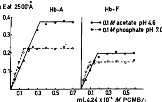
6). It will be noted that the increase in absorption caused by the interac- tion of the reagent with —SH groups is much higher at a lower pH, which is confirmed by the increase in absorption after adjusting the pH of the phosphate buffer of pH 7.0 to 2.0 (Fig. 6). It seems therefore that at a lower pH more —SH groups in hemoglobin are reactive with P C M B than are at a neutral or alkaline pH. This phenomenon is also illustrated- in Fig. 7, where the reactions, at pH 4.6 and pH 7.0, between P C M B and human Hb-A as well as human Hb-F are given (reaction time 3 hours at room temperature). The break in the curve illustrating the reaction of
CYSTINE/CYSTEINE CONTENT OF HEMOGLOBINS 165 Δ Eat 2500W ο Hb-A Hb-F
0.3
—•0.1 M acetate pH 4.6
•—•0.1 M phosphate pH 7.0
0.2 Λ.
ο
0.1
0.1 03 05 mU.24x10"4 M P C M B /L
FIG. 7. The estimation of —SH groups in human adult and fetal hemoglobins with PCMB (3.0 ml. Hb solution containing 2.25 X 10"8 M H b - 02 4 - 0 — 0.7 ml. 4.24 χ 10-* Af PCMB per liter. Ε estimated in 1 cm. cuvette at 2500Â.)
PCMB with Hb-A in a 0.1 M phosphate buffer of pH 7.0 occurs much earlier than does the break in the curve obtained at a pH of 4.6. Similar results were obtained for the fetal human hemoglobin, the amounts of P C M B bound by this protein being, however, lower than those bound by the Hb-A.
VII. A Comparison of the Final Data Obtained
for the Cystine/Cysteine Content of Different Human Hemoglobins The results of the different analyses are presented in Table III. N o significant differences were found to be present between the data obtained on dialyzed hemolysates of the freshly washed red cells and on the hemo- globin fractions obtained after purification by carboxymethylcellulose chromatography. The total half-cystine contents of the hemoglobins A, S, and C before as well as after purification were found to be 8 residues.
The total quantity of half-cystine in fetal human hemoglobin, purified by denaturation with alkali (8) as well as by chromatography on carboxy- methylcellulose, is 6 residues. These data are confirmed by the analyses of the total sulfur contents of the hemoglobins A and F ; in both proteins 14 S atoms per mole of hemoglobin were found to be present. After sub- traction of the numbers of methionine residues in the hemoglobins (see sub C) 8 half-cystine residues in Hb-A and 6 half-cystine residues in Hb-F are established.
Eight —SH groups per mole of Hb-A are titratable argentometrically with the mercury-coated rotating platinum-wire electrode for both un- purified and purified hemoglobin preparations. The number of the titra-
TABLE III THE CYSTINE/CYSTEINE CONTENT OF DIFFERENT HUMAN HEMOGLOBINS BEFORE AND AFTER PURIFICATION BY CHROMATOGRAPHY ON CARBOXYMETHYLCELLULOSE0 Hb-A Hb-F Hb-S Hb-C Method Before6 After < Before d Afte Before6 After e Beforeb After« Total cystine as cysteic acid 21· 8.4 3· 7.85 15· 5.55 4· 5.75 11' 8.1 2· 8.05 15· 8.45 — — Total S content — 14.1 — 13.5 — 13.6 — 13.7 — — — — — — — — Total S content minus number of methionine residues — 14 - 6 = 8 — 14 - 6 = 8 — 14 - 8 = 6 — — — — — — — — — — Ag-titration in 0.1 M NH4OH + 0.05 M NH4NO3 12 7.9 6 7.8 9 3.8 4 3.7 5 8.05 — — 5 7.55 — — Hg-titration 4 5.9 — — 4 4.6 — — 4 6.85
—
— 4 5.85 — — PCMB in phosphate, pH 7.0, 2500 Â 2 2.75 2 2.85 2 1.65 2 1.75 PCMB in acetate, pH 4.6, 2500 Â 3 6.05 3 5.95 3 3.35 2 3.6 3 6.1 2 6.0 — — 2 5.9 0 The values are presented as residues of half-cystine, number of S atoms and of —SH groups; molecular weight 66000 fglobin) and 66800 (hemoglobin). b Dialyzed hemolysates of freshly washed erythrocytes. c Fraction isolated by chromatography on carboxymethylcellulose. d Purified by denaturation with alkali. • Number of analyses.166
table — S H groups in H b - F again did not change after purification by carboxymethylcellulose chromatography. In both fetal hemoglobin prep- arations 4 — S H groups were established.
The number of molecules of P C M B bound at a pH of 4.6 by the hemo- globins A, S, and C in hemolysates as well as after purification are the same, namely 6 per mole of Hb. For fetal hemoglobin about 4 moles of P C M B per hemoglobin molecule are bound. At a pH of 7.0 these values are 3 and 2 for the Hb-A and H b - F respectively. The increment of — S H groups available for binding P C M B , when the pH of the solution is lowered from 7.0 to 4.6, may be due to an architectural alteration of the hemoglobin molecule caused by acid denaturation.
Summarizing, we confirm the results of our former analyses (3, 10) that the hemoglobins S, A, and C do contain 8 half-cystine residues, which all are present as fully reactive — S H groups. In fetal human hemoglobin only 6 half-cystine residues per mole are present, 4 of them being titratable with our amperometric-argentometric procedure. N o t all of these groups of the hemoglobins A, S, C, and F are reactive with P C M B ; so P C M B binding is not equivalent to silver binding. Our data suggest that there are three — S H groups of Hb-A, which are reactive at neutral pH. After acid denaturation again 3 — S H groups of Hb-A become available. For human fetal hemoglobin a similar picture is obtained; at a pH of 7.0, two
—SH groups are capable of binding P C M B , while this increases to four
—SH groups after acid denaturation. Our results are contradictory to data reported by some other investigators. Stein et al. (4) described values
for the total number of half-cystine residues and of titratable — S H groups in normal adult hemoglobin prepared by starch slab electrophoresis (18), which are much lower than those found in our studies (Table I ) . They suggested that "a bulk of an impurity rich in sulfhydryl groups was re- moved applying this procedure." Using the carboxymethylcellulose chrom- atography as purification procedure we did not obtain evidence for the presence of any substance rich in — S H groups. Although the two proce- dures are not completely the same they are closely related, since similar separations of normal Hb-A of dialyzed hemolysates into three compo- nents were observed (19). Our data obtained for human fetal Hb are contradictory to those described by Stein et al. (4) as well as to those given by Murayama (18). Both groups of investigators reported the pres- ence of the same number of titratable — S H groups (using silver titration in tris buffer after addition of 8 M urea) as of half-cystine residues, indi- cating that human fetal Hb, just as hemoglobins A, S, and C, does not contain any disulfide bonds. Our results suggest that in human Hb-F two
—SH groups are unreactive.
An explanation for these differences cannot be given. Without doubt
168 T. H. J. HUISMAN
the estimation of the total number of half-cystine residues both in Hb-A and in Hb-F by different chemical methods is highly important in order to remove the disagreement between our results and those given by Stein et al. (4). Application of different chromatographic procedures for the estimation of cysteic acid in the oxidized hydrolyzates of Hb-A and Hb-F seems to be the most promising way in this respect.
VIII. Studies on the Cystine/Cysteine Content of Adult and Fetal Hemoglobins of Some Animals
In order to obtain some information on a possible similarity in the amounts of half-cystine residues and the number of reactive —SH groups in adult and fetal hemoglobins of some species of mammals, we investi- gated the adult and fetal hemoglobins of the cow, the sheep, and the rabbit. The fetal hemoglobin of the cow was obtained from a five month old fetus, while the fetal pigments of the sheep and the rabbit were pre- pared from blood derived from newborn animals. The hemoglobin solu- tions, which were obtained in a way similar to that described earlier, were purified by carboxylmethylcellulose chromatography (19). After the esti- mation of the total half-cystine content, as cysteic acid, the proteins were analyzed for titratable —SH groups using the argentometric titration technique with the rotating mercury-coated platinum electrode, and for P C M B binding using the method mentioned before.
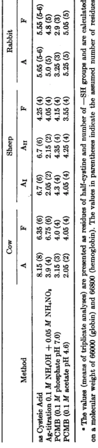
The results are presented in Table IV. The total half-cystine content of the adult Hb of the cow is 8 residues ; 4 are present as titratable —SH groups, while 3 are capable of binding P C M B at a pH of 7.0 and 2 at a pH of 4.6. The number of four available sulfhydryl groups is in accord- ance with the data presented by Ingram (8). In the fetal hemoglobin only 6 half-cystine residues are found, which are all present as titratable —SH groups. Four P C M B molecules are bound by this protein, both at a pH of 7.0 and at a pH of 4.6. In sheep we are confronted with the presence of two different adult hemoglobins, which show many physical and chemical differences (27). The total quantities of half cystine residues are the same in both proteins, i.e., about 6 residues, two of them being titratable with the argentometric titration procedure. This value is different from those reported by Benesch et al. (12) and by Ingram (8), who established 8 titratable —SH groups in the adult hemoglobin of the sheep. Again this discrepancy may be due to the different procedures used. Four molecules of P C M B are bound by these two adult hemoglobins, both at a pH of 7.0 and at a pH of 4.6. In fetal sheep hemoglobin, only 4 half-cystine residues were established, which are present as fully reactive —SH groups both in binding silver and P C M B (at a pH of 7.0 as well as at a pH of 4.6). N o
TABLE IV THE CYSTINE/CYSTEINE CONTENT OF DIFFERENT ANIMAL HEMOGLOBINS0 Cow Sheep Rabbit Method A F Áι Áð F A F as Cysteic Acid Ag-titration 0.1 M NH4OH + 0.05 M NH4NOs PCMB (0.1 M phosphate pH 7.0) PCMB (0.1 M acetate pH 4.6)
8.15 (8) 3.9 (4) 3.15 (3) 2.05 (2) 6.35 (6) 6.75 (6) 4.0 (4) 4.05 (4) 6.7 (6) 2.05 (2) 4.3 (4) 4.05 (4) 6.7 (6) 2.15 (2) 4.35 (4) 4.25 (4) 4.25 (4) 4.05 (4) 4.15 (4) 3.55 (4)
5.65 (5-6) 5.55 (5-6) 5.0(5) 4.8(5) 3.25(3) 2.9(3) 5.25 (5) 5.05 (5) â The values (means of triplicate analyses) are presented as residues of half-cystine and number of —SH groups and are calculated on a molecular weight of 66000 (globin) and 66800 (hemoglobin). The values in parentheses indicate the assumed number of residues of half-cystine and of —SH groups.
169
170 T. H. J . HUISMAN
differences were found between the hemoglobins of the adult and fetal rabbit in this respect; both hemoglobins do contain about 5 half-cystine residues. The number of titratable —SH groups was found to be 5 also, while 3 molecules of P C M B are bound per mole of hemoglobin at a pH of 7.0, and 5 molecules of PCMB at a pH of 4.6. This finding supports the hypothesis that the hemoglobins of the adult and newborn rabbit are strongly related to each other (17).
When compared with the results obtained for the human adult and fetal hemoglobins, it is evident that the fetal hemoglobins of the cow and the sheep also contain less half-cystine residues than the corresponding adult components. These half-cystine residues, however, are all present as fully reactive —SH groups. The adult components, on the contrary, contain lower amounts of titratable —SH groups than may be expected from the analyses of the total quantities of half-cystine. So the situation in the cow as well as in the sheep is opposite to that in man. It may be that the different arrangements of half-cystine residues in these hemo- globins are related to their resistance to denaturation by alkali. It will be noted that all hemoglobins in which the total half-cystine residues are present as reactive — S H groups (human Hb-A, cow Hb-F, sheep Hb-F, and rabbit Hb-A and Hb-F) show a lower resistance to alkali denaturation than do the corresponding fetal and adult hemoglobins. A connec- tion between the arrangement of the half-cystine residues and the stability is already known for other proteins (28). Especially proteins in which di- sulfide linkages are present are stable under most of the conditions of de- naturation. The possibility exists that similar linkages are present in those hemoglobins which possess a high resistance to denaturation by alkali.
REFERENCES
1. H. S. Rhinesmith, W. A. Schroeder, and L. Pauling, J. Am. Chem. Soc. 79, 609 (1957).
2. H. S. Rhinesmith, W. A. Schroeder, and L. Pauling, / . Am. Chem. Soc. 79, 4682 (1957).
8. F. A. Hommes, J. Santema-Drinkwaard, and T. H. J. Huisman, Biochim. et Biophys. Acta 20, 564 (1956).
4. W. H. Stein, H. G. Kunkel, R. D. Cole, D. H. Spackman, and S. Moore, Biochim.
et Biophys. Acta 22, 640 (1957).
5. S. H. Ingbar and Ε. H. Kass, Proc. Soc. ExpLl. Biol. Med. 77, 74 (1951).
6. M. Murayama, Federation Proc. 15, 318 (1956).
7. M. Murayama, J. Biol. Chem. 228,231 (1957).
8. V. M. Ingram, Biochem. J. 59, 653 (1955).
9. V. M. Ingram, Biochem. J. 65, 760 (1957).
10. F. A. Hommes, A. Dozy, and T. H. J. Huisman, Biochem. J. 68, 309 (1958),
CYSTINE/CYSTEINE CONTENT OF HEMOGLOBINS 171 11. F. A. Hommes and T. H. J. Huisman, Biochem. J. 68, 312 (1958).
12. R. E. Benesch, H. A. Lardy, and R. Benesch, J. Biol. Chem. 216, 663 (1955).
13. M. Murayama, Federation Proc. 16, 223 (1957).
14. M. Murayama, Federation Proc. 16, 756 (1957).
15. I. M. Kolthoff, W. Stricks, and L. Morren, Anal. Chem. 26, 366 (1954).
16. P. D . Boyer, / . Am. Chem. Soc. 76, 4331 (1954).
17. Τ. H. J. Huisman, Clin. Chim. Acta 3, 201 (1958).
18. H. G. Kunkel and G. Wallenius, Science 122, 288 (1955).
19. Τ. H. J. Huisman, E. A. Martis, and A. Dozy, ./. Lab. Clin. Med. 52, 312 (1958).
19a. E. A. Peterson and H. A. Sober, J Am. Chem. Soc. 78, 751 (1956).
19b. H. A. Sober, F. J. Gutter, M. M. Wyckoff, and E. A. Peterson, J. Am. Chem.
Soc. 78, 756 (1956).
20. R. Lemberg and J. W. Legge, "Hematin Compounds and Bile Pigments." Inter- science, New York, 1949.
21. E. Schräm, S. Moore, and E. J. Bigwood, Biochem. J. 57, 33 (1954).
22. J. R. Kimmel, E. O. P. Thompson, and E. L. Smith, J. Biol. Chem. 217, 151 (1955).
23. W. A. Schroeder, L. M. Kay, and I. C. Wells, J. Biol. Chem. 187, 221 (1950).
24. A. Rossi-Fanelli, D . Cavallini, and C. de Marco, Biochim. et Biophys. Acta 17, 377 (1955).
25. P. C. van der Schaaf and T. H. J. Huisman, Biochim. et Biophys. Acta 17, 81 (1955).
26. Τ. H. J. Huisman, P. C. van der Schaaf, and A. van der Sar, Blood 10, 1079 (1955).
27. H. J. van der Helm, G. van Vliet, and T. H. J. Huisman, Arch. Biochem.
Biophys. 72, 331 (1957).
28. C. B. Anfinsen and R. R. Redfield, Advances in Protein Chem. 11, 82 (1956).
Discussion
BENESCH: It seems that the main discrepancy between your results and those of Stein et al. is the cysteic acid values, since with this method Stein et al. find only 5-6 half-cystines, whereas you report 8. Although I have no personal experience with the cysteic acid method, I wonder how far this method can be relied upon to give absolute values?
HAUROWITZ : Some of the cystine in presence of iron is destroyed, and you end up with sulfate. You find some of the sulfur sometimes in the humin fraction. This I think is the explanation.
SWAN: The determination of cysteine plus cystine in a protein by oxidation of these residues followed by estimation of the resultant cysteic acid isolated on ion exchange resins has been studied in detail by Dr. E. O. P. Thompson in our labora- tory. He finds (Proc. Intern. Wool Textile Research Conj., Melbourne, 1955 C, p. 102, 1956) that although under certain conditions cysteine and cystine give quantitative recoveries, oxidation of the unhydrolyzed protein followed by hydrolysis always gives a low result. The best procedure was found to be partial hydrolysis of the protein in a mixture of 6 Ν HBr/98% formic acid, oxidation with KBrOa, and further hydrolysis to liberate the cysteic acid. This amino acid emerges first on subsequent chroma- tography on Dowex 50. However, even under the best conditions only about 88-94%
recoveries are obtained. I have used the Dowex 50 method for separating cysteic acid from hydrolyzates of performic acid-oxidized wools. In my hands, the base line was
172 T. H. J . HUISMAN
rather erratic and this is serious since the whole of the cysteic acid emerges in three or four tubes. If Dowex 2 is used, the cysteic acid emerges last and the analysis becomes very time consuming.
HOCH: The temperature at which argentometric titrations of sulfhydryl groups are carried out can be a deciding factor in the end point of the titrations, that is, in the accuracy of the determination. Cysteine, and the 3 dithiols which we have studied, all have apparently lower sulfhydryl contents at 2 3 ° than at 4 ° and yeast alcohol dehydrogenase shows the same phenomenon. I should like to ask if the temperatures at which the measurements of sulfhydryl groups of hemoglobins were made were con- trolled or varied.
KLOTZ : The temperatures listed by Murayama were the temperatures at titration.
When I heard your paper I immediately thought that your slope is exactly the re- verse of Murayama's.
HOCH : Yes, the end points of Dr. Murayama's silver titrations at 3 8 ° were twice the number he titrated at 0°, a change opposed in direction to the one we found. He did not report values obtained in the intermediate temperature ranges, however, so that it is not possible to state that the changes he observed with hemoglobin are con- tinuous with rising temperature as are those with yeast alcohol dehydrogenase, or that there is a discontinuity which might indicate a structural change in hemoglobin due to the temperature.
HUISMAN : We titrated normal adult hemoglobin with silver at 3 8 ° and at 0 ° and obtained exactly the same results. With our technique we could not confirm the find- ings of Dr. Murayama.
LORAND: I would like to raise a technical point. In comparing those pH curves on PCMB, Dr. Boyer's paper pointed out that an anion has reacted. Is it fair to compare solutions of two different anions and ascribe differences entirely to the pH?
BOYER: Anions present may markedly affect the rate of reaction. Whenever you want to measure the total sulfhydryl in a protein you have to choose conditions where all —SH groups will be available and will readily react. When you have slow reaction, you are liable not to get the total sulfhydryl. I don't think anyone who wants to compare results from one method to another should do so unless they have used conditions which will make all sulfhydryls react rapidly with the PCMB. Use of urea is helpful but has some disadvantage because it gives a slight spectral shift to the PCMB. In the spectrophotometric procedure you have to watch the base lines and make sure you are measuring the effect of sulfhydryl reaction.