ASSESSMENT OF SEVERITY AND TIME COURSE OF CRITICAL ILLNESS NEUROPATHY IN SEPTIC PATIENTS:
A PROSPECTIVE OBSERVATIONAL STUDY
NEMES Réka1, FÜLEP Zoltán1, LÁSZLÓ István1, SÁRKÁNY Péter1, FEKETE Klára2, MECHLER Ferenc2, FÜLESDI Béla1
1Debreceni Egyetem, Orvos- és Egészségtudományi Centrum, Aneszteziológiai és Intenzív Terápiás Tanszék, Debrecen
2Debreceni Egyetem, Orvos- és Egészségtudományi Centrum, Neurológiai Klinika, Debrecen
A KRITIKUS ÁLLAPOTÚ BETEGEK ESETÉBEN
JELENTKEZÔ NEUROPATHIA (CIP) SÚLYOSSÁGÁNAK ÉS LEFOLYÁSÁNAK VIZSGÁLATA SZEPTIKUS BETEGEK KÖRÉBEN: PROSPEKTÍV OBSZERVÁCIÓS VIZSGÁLAT Nemes R, MD; Fülep Z, MD; László I, MD; Sárkány P, MD;
Fekete K, MD; Mechler F, MD, PhD, DSc;
Fülesdi B, MD, PhD, DSc
Ideggyogy Sz 2015;68(1–2):30–36.
Prospektív megfigyeléses vizsgálatunkban kritikus állapotú betegek esetében vizsgáltuk a CIP elektrofiziológiai jellemzôit és összevetettük a klinikai képpel, valamint a betegek kimenetelével.
Betegek és módszerek– Huszonegy, kritikus állapotú (12 fölötti Acute Physiology and Chronic Health Evaluation II score súlyosságú) beteg esetében rutin elektroneurográfiát végeztünk mindkét oldali nervus medianuson és ulnarison, a felvétel utáni öt, egymást követô napon, majd ezt követôen hetente. A motoros és a szenzoros vezetési indexeket az APACHE II és a Simplified Acute Physiology Score II értékei - vel korreláltattuk.
Eredmények– Az elsô vizsgálat alkalmával 18/21 beteg esetében tapasztaltuk a motoros és szenzoros vezetési inde - xek 20%-ot meghaladó csökkenését. A klinikai súlyossági pontrendszerek szignifikáns negatív korrelációt mutattak a CMAP- és SNAP-amplitúdókkal és a kalkulált idegvezetési indexekkel (Spearman: p<0,001). A beutaláskor súlyos elektrofiziológiai eltéréseket vagy a kórlefolyásban nem javuló tendenciát mutató ENG-vel rendelkezô betegek mor- talitása nagyobb volt.
Következtetések– Kritikus állapotú betegek esetében az elektrofiziológiai eltérések a kórfolyamat korai fázisában kimutathatók. Ezek az eltérések erôs korrelációt mutatnak a beteg általános élettani állapotával. A súlyos elektrofizioló- giai eltérések elôre jelezhetik a mortalitást, ugyanakkor az esetek egy részében a folyamat reverzíbilis lehet.
Kulcsszavak: „critical illness” neuropathia;
elektroneurográfia; szepszis Objective– In this prospective observational study we inves-
tigated electrophysiological alterations in the early phase of critical illness and correlated electrophysiological findings with the clinical picture and outcome.
Methods– We enrolled 21 critically ill surgical patients having ≥12 Acute Physiology and Chronic Health Evaluation (APACHE) II scores on admission. Routine non-invasive bilat- eral electroneurography (ENG) examination of median and ulnar nerves was done on five consecutive days starting in two days after admission. Then weekly follow-up was per- formed. Motor and sensory nerve conduction indices were calculated and correlated with APACHE II and Simplified Acute Physiology Score II severity scores.
Results– On the first examination 18/21 patients had
>20% reduction in the motor and sensory nerve conduction indices. Severity score systems showed significant negative correlation with the daily change of CMAP and SNAP ampli- tudes and calculated nerve conduction indices (Spearman’s correlation, p<0,001). Mortality was higher in the patients with worse admission ENG and/or stagnant electrophysio- logical status or declining tendency in the first week.
Conclusions– Electrophysiological alterations appeared soon after the development of critical illness. Early phase alterations showed a strong correlation with patients’ gener- al condition and more severe electrophysiological alterations predisposed to higher mortality. In several cases early alter- ations proved to be reversible.
Keywords: critical illness polyneuropathy, electroneurography, sepsis
Correspondent: Béla FÜLESDI, Debreceni Egyetem, Orvos- és Egészségtudományi Centrum, Aneszteziológiai és Intenzív Terápiás Tanszék; 4032 Debrecen, Nagyerdei krt. 98. Telefon/fax: (06-52) 255-346. E-mail: fulesdi@dote.hu
Érkezett: 2013. május 17. Elfogadva: 2013. szeptember 11.
www.elitmed.hu
C
ritical illness polyneuropathy (CIP), myopathy (CIM) and neuromyopathy (CINM) has been investigated already for three decades, the first accounts of peripheral neuropathy complicating sep- sis and multi-organ failure (MOF) came in into light by Boltonet al. in the ‘80s1, 2. Investigations of the first 20 years focused mainly on its incidence, aetiol- ogy and classification. The pathophysiological back- ground is not known in all details. Recent experimen- tal evidence suggests that the responsible patho physiological alteration in the early phase is rather related to channelopathy than axonal degener- ation3. Latronico and co-workers found that early electrophysiological alterations are not necessarily accompanied by histological alterations. They spec- ulated that sepsis-related nerve failure caused an early impairment of axonal transport and transmem- brane potential and with the persistence of sepsis, the energy supply or use is not restored and histological alterations ensue4. In 2006 Z’Graggenand co-work- ers proposed that the chronic axonal membrane depo- larization due to hyperkalaemia and/or hypoperfu- sion may also be involved in the pathophysiology of the neuropathy in critically ill patients5.Most studies examining neuromuscular disor- ders (NMD) in critically ill patients have evaluated patients later in their intensive care unit (ICU) course when they were able to cooperate6. Hence, little is known about the time course of develop- ment of intensive care unit acquired neuromuscular dysfunction in the setting of severe sepsis. The first early-phase investigations were published in 2000 by Tennilaet al7then several other studies revealed that neuromuscular disorders develop early during the course of critical illness6–11. Electrophysiolo - gical alterations can appear as-early-as two days after admittance. CIM often precedes CIP, their coexistence is not rare and CIP has worse impact on final outcome8–9.
Along these lines, in the present study we aimed to prospectively assess the early electrophysiologi- cal characteristics of critical illness polyneuropathy in surgical patients showing signs of sepsis and sep- tic shock in a five day follow-up setting and to cor- relate electrophysiological findings with the clini- cal picture and outcome of the patients.
Materials and methods
This observational study was performed in a 20-bed surgical ICU of the University of Debrecen, Medical and Health Science Centre. The study pro- tocol was approved by the local ethics committee.
Informed consent was obtained from the patient or
the next relative. Patients aged >18 years with symptoms of sepsis, with or without multiple organ failure (MOF) showing Acute Physiology and Chronic Health Evaluation (APACHE) II score ≥12 on admission were enrolled in the study. Exclusion criteria were pre-existing central nervous system disorder or neuromuscular disease. All patients received standard surgical (if necessary) and med- ical treatment, including fluid resuscitation, vasoac- tive drugs, culture- and antibiogram- adjusted antimicrobial therapy, mechanical ventilation or renal replacement therapy when necessary and best supportive care. Neuromuscular blocking agents (NMBA) were only used for surgical interventions.
No patients were treated with continuous adminis- tration of NMBAs during the ICU stay. Severity of illness was monitored by daily rating of APACHE II, Simplified Acute Physiology Score (SAPS) II and Sequential Organ Failure Assessment (SOFA) severity scores12–14. The diagnostic criteria of sys- temic inflammatory response syndrome (SIRS) sep- sis, severe sepsis, septic shock and MOF15and CIP, CIM, CINM and ICU-aquired weakness16 have been previously described elsewhere.
NERVE CONDUCTION STUDIES
Baseline neurological examination and bilateral sen- sory and motor nerve conduction studies of the medi- an and ulnar nerves were performed within 48 hours of admission and then repeated on four consecutive days. In case of surgical intervention the measure- ment was postponed for the next day and repetitive stimulation of the ulnar nerve was performed to exclude effect of NMBAs. After the five measure- ments, additional weekly follow-up electroneurogra- phies (ENG) were performed. The measurements were carried out at the bedside using a transportable 2 channel Keypoint (Alpine Biomed Aps, Skov - lunde, Denmark) apparatus. Conventio nal techniques were applied using surface electrodes for stimulation and recording. For motor nerve conduction studies the median nerve was stimulated at wrist and at elbow; the ulnar nerve was stimulated at the wrist, below and above the elbow. For sensory nerve con- duction studies the nerves were stimulated at the bases of the fingers and orthodromic recording was performed at the wrist. Incremental stimulation of the nerve was applied until the best compound muscle action potential (CMAP) and sensory nerve action potential (SNAP) amplitudes were obtained. Skin temperature at the site of measurement was kept above 32 ºC. Serum electrolyte alterations were nor- malised prior to measurements. The serial studies for each patient were carried out by the same examiner
(RN). The lower limit for normal nerve conduction velocities in our laboratory for the median and ulnar nerves was 50 m/s. The lower limit of normal CMAP was 5 mV and 5 µV for SNAP in both nerves.
Sex and age-matched healthy controls (n=21) were included from the database the Electrophy - siology Laboratory of the Department of Neu - rology, University of Debrecen Medical and Health Science Centre.
In order to describe overall nerve function, motor (MNCI) and sensory (SNCI) nerve conduction indices were introduced, according to the sugges- tions of Wittet al17and calculated as follows: meas- ured amplitudes were divided by the laboratory ref- erence value, averaged and multiplied by one- hundred to give the performance percentage. These indices were used both for monitoring and for deter- mining correlation with clinical variables. We cor- related daily calculated severity scores with CMAP, SNAP amplitudes and calculated nerve function indices. Peroneal and sural nerve conduction stud- ies were performed in line with the median and ulnar ENGs according to the standard polyeneu- ropathy protocol. However, in several cases both peroneal CMAP and sural SNAP amplitudes became unrecordable. Hence these parameters were not used for calculation.
Motor and sensory nerve functions were handled separately, as it has been reported earlier that motor fibres are more prone to dysfunction in the critical- ly ill18, and we presumed that this can be relevant in early phase investigation of critical illness neuro- muscular dysfunction as well. In three patients only four measurements could be performed of the 5-day follow-up because two patients died and one addi- tional patient was transported to another facility for further treatment.
STATISTICAL ANALYSIS
Medians and interquartile ranges (IQR) are report- ed for all values. Before starting comparisons, normality test was performed in order to check the distribution of the data. Electrophysiological parameters were then compared with the appropri- ate Mann-Whitney U-test. The relationship between electrophysiological indices and severity score val- ues was tested with the Spearman test. A p<0.05 was accepted as statistically significant.
Results
Twenty-one critically ill (APACHE II score ≥12 on admission) patients were enrolled in this study.
The mean age was 63.52±16.9 years, male: female ratio was 15:6. On admittance the mean APACHE II score was 15.38±6.19, the mean SAPS II score was 37.95±12.43 and the mean SOFA score was 7.14 ± 3.28 (Table 1.). On admittance all patients showed the clinical signs of sepsis and had a posi- tive haemoculture. 10 of the 21 patients were in sep- tic shock. On the first examination 13/21 were ven- tilated and analgosedated. In these patients muscle strength could not be tested. Of the eight patients where muscle strength could be assessed, three patients were presented with generalized weakness, 5/8 patients had preserved muscle strength on admittance. Deep tendon reflexes were reduced in 8/21 patients, in six of these patients muscle strength could not be tested, 2/8 patients showed generalized weakness.
On the first examination18/21 (86%) patients had >20% reduction in MNCI and SNCI (Table 1.).
There was no difference between the left and right side measurements, hence their average was used for calculation. The CMAP amplitudes of both the median {median: 2.3 mV (IQR: 1.5/3.425) vs 8,4 mV (IQR: 6.7/9.4), p<0,001} and the ulnar nerves {2.9 mV (IQR: 2.18/3.88) vs 7.4 mV (IQR: 6.3/8.4), p<0,001} showed a significant reduction compared to the control values and the laboratory reference.
The same was true for the SNAP amplitudes of the median {median: 2,1 µV (IQR 1.2/4.33) vs 5.5 µV (IQR 4.8/8), p<0,001} and the ulnar {2 µV (IQR 1.23/4) vs 5.2 µV (IQR 4.1/6.4), p<0.001} nerves.
The median of the MNCI was 61% (IQR 40.63/72.38) and the median of the SNCI was 44%
(IQR 29.25/75.13) compared to the 153% (IQR 136.63/178.25) (p<0.001) MNCI and 110 % (IQR 92.63/140.99) (p<0.001) SNCI ratios of the control group (Figure 1.). 3/21 patients showed normal ENG on admission (100-80% MNCI and SNCI), 8/21 patients had mild decrease (80-50%) and 10/21 patients had severe (<50%) amplitude reductions (Table 1.). Conduction velocities were normal or near normal. Sensory responses could not be obtained in two patients for technical reasons.
During the 5-day follow-upfour patients showed improvement, seven patients showed no change, and 10 patients’ results worsened (Table 1.).
Weekly follow up was possible in eight patients.
Seven of the rest of the patients died before weekly follow-up could be completed and six of the patients were discharged from the ICU or transport- ed to other departments for further treatment.
During the weekly follow up 3/7 of the patients’
ENG results showed further improvement, 3/7 patients showed further decrease and 2/7 patients’
condition did not change.
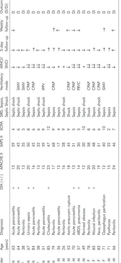
Table 1.Demographic data and gross electrophysiological evaluation of the patients Admission parametersFollow-up GenderAgeDiagnosisDM (+/-)APACHE IISAPS IISOFASIRS, Sepsis, Ventilatory MNCI/5-day Weekly Outcome (years)Septic ShockmodeSNCIfollow-upfollow-up(D/Di) 1 – M63Acute pancreatitis+15294Septic shock↓↓↓D 2 – M64Acute pancreatits+20426Septic shockSIMV↓↓D 3 – F38Peritonitis13355SepsisSIMV↓↑Di 4 – M67Urinary sepsis+20508Septic shockCPAP↓↓↑Di 5 – M84Acute pancreatits+16456SepsisCPAP↓↓→D 6 – F58Peritonitis13326Septic shock↓↓↑D 7 – M51Acute pancreatitis15399Septic shockIPPV↓↓↓D 8 – M85Cholecystitis+234712Sepsis↓→Di 9 – M79Peritonitis17689SepsisCPAP↓→→Di 10 – M36Acute pancreatitis12174Sepsis+→↑Di 11 – M26Peritonitis16289Septic shock+↓Di 12 – M53Aortic aneurysm rupture18459ShockCPAP↓↓↑D 13 – M70Acute pancreatitis+13315SepsisCPAP↓↓↓↓Di 14 – M57ARDS, pneumonia+14305SepsisPRVC↓↓↓↓Di 15 – M49Perineal abscess123010Septic shock+↓Di 16 – F78Peritonitis+20588ShockCPAP↓↓↓D 17 – F85Wound infection+20486SepsisCPAP↓↓→Di 18 – F83Ileus, peritonitis19516Septic shockCPAP↓↓↓D 19 – F71Oesophagus perforation124010SepsisSIMV↓→→D 20 – M71Pyothorax15322Sepsis↓↓→D 21 – M66Peritonitis24467Sepsis↓↑↑Di DM: diabetes; MNCI: Motor Nerve Conduction Index; SNCI: Sensory Nerve Conduction Index; +: 100-80% MNCI and SNCI; ↓: 80-50% MNCI and SNCI and decrease in the follow-up; ↓↓: <50% MNCI and SNCI; →: stagnation; ↑: improvement; D: died; Di: discharged, CPAP= Continouos positive airway pressure, IPPV= intermittent positive pressure ventilation, SIMV=syn- chronized intermittent mandatory ventilation, ARDS=adult respiratory distress syndrome
Relationship between electrophysiological find- ings obtained at admission and outcome of the patients: None of the patients died with normal ENG at admittance, but 4/8 (50%) patients from the group with mild ENG alterations, and 6/10 (60%) of patients with severely reduced ENG results.
Considering the five day follow up, 1/4 (25%) patient died of those who showed improvement, 3/7 (43%) of those whose electrophysiological status stagnated, and 6/10 (60%) of those whose ENG results worsened.
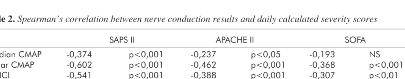
Relationship between clinical scoring systems and nerve conduction indices: SAPS II and APACHE II score systems showed significant neg- ative correlation with both median and ulnar CMAP and SNAP amplitudes and also with calculated motor and sensory nerve conduction indices. The SAPS II scores generally showed stronger correla- tion than the APACHE II scores (Table 2.). The strongest correlation was found between SAPS II scores and ulnar CMAP (Spearman’s r=-0.602) and MNCI (Spearman’s r=-0.541) (Figure 2.). Daily
calculated SOFA scores did not correlate with CMAP and SNAP amplitudes, and weak correlation was observed with MNCI as well.
Discussion
In the present cohort of 20, critically ill surgical patients electrophysiological alterations appeared soon after the development of critical illness. We found that in 85% of the patients electrophysiologi- cal signs of critical illness neuropathy could be obtained. These results are in agreement with those of previously published reports4, 6–11, 19.
Different pathophysiological mechanisms, such as of bioenergetic failure20, microvascular changes, electrical phenomena, inflammation and cellular metabolic changes including alterations in the exci- tation-contraction coupling as well as a shift in the balance between protein synthesis and breakdown are thought to be involved5, 21, 22. It is also clear from previous observations that histological appearance
Table 2.Spearman’s correlation between nerve conduction results and daily calculated severity scores
SAPS II APACHE II SOFA
Median CMAP -0,374 p<0,001 -0,237 p<0,05 -0,193 NS
Ulnar CMAP -0,602 p<0,001 -0,462 p<0,001 -0,368 p<0,001
MNCI -0,541 p<0,001 -0,388 p<0,001 -0,307 p<0,01
Median SNAP -0,462 p<0,001 -0,215 p<0,05 -0,141 NS
Ulnar SNAP -0,445 p<0,001 -0,325 p<0,001 -0,149 NS
SNCI -0,458 p<0,001 -0,285 p<0,01 -0,136 NS
CMAP: Compound Muscle Action Potential; SNAP: Sensory Nerve Action Potential; MNCI: Motor Nerve Conduction Index; SNCI:
Sensory Nerve Action Potential; NS: not significant
Figure 1.Motor and Sensory Nerve Conduction Indices on first measurement compared to age-matched healthy controls. The broken grey line indicates the lab referen- ce value
Figure 2.Correlation between Motor Nerve Conduction Indices and SAPS II severity scores
of failing nerves and muscles may be normal or pro- vide evidence of minimal muscle necrosis in the early phase of CIP despite reduced SNAPs, suggest- ing that the defect is mainly functional4.
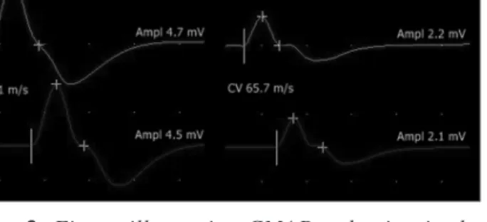
This may explain our results showing that although electrophysiological signs of CIP were present in the majority of our septic cases at admis- sion, 4 of patients’ CMAP and SNAP amplitudes improved (of whom 1 patient’s totally normalized) during the five day follow-up, and in the weekly fol- low-up 3 other patients showed improvement, one of whom entirely normalized. Similarly to our results, Ahlbeck et al found in a 1-month follow up study that the number of patients presenting signs of critical illness neuromuscular disturbances changed over time and also that patients showing signs of CIP could move toward showing addition- al signs of critical illness myopathy11. Figure 3 illustrates a typical case of electrophysiological worsening during follow-up (case 16).
Two previous studies showed that the develop- ment of CIP predisposed to higher mortality in sep- tic patients23, 24. Khanet al. stated that patients with abnormal baseline ENG is associated with a signifi- cantly increased mortality6. In the present study mortality showed an increasing tendency along with severity of baseline electrophysiological alterations and first week’s trend. That is, the more severe the early electrophysiological alteration was, the worse outcome it predisposed to. In all fairness, in several younger patients even severe electrophysiological alterations could normalise along with the improve- ment of general condition.
While evaluating data from the other sign of the
coin, we may say that the severity of illness25, 26and duration of multi-organ dysfunction syndrome with or without SIRS27, 28are known to be independent risk factors of CIP [22]. In previous prospective observational studies APACHE II and III scores at admission were important predictors of the devel- opment of critical illness associated neuromuscular disorders25, 26. In our study no relationship could be detected between the admittance severity score and the severity of baseline electrophysiological alter- ations. Additionally, baseline electrophysiological alterations did not predict electrophysiological alterations during the first week. This indicates that patients in worse condition on admittance did not necessarily develop more severe electrophysiologi- cal alterations during the course of the septic process. In contrast to this, negative correlation was found between daily calculated SAPS II and APACHE II scores and both motor and sensory nerve conduction indices measured at the same occasion, indicating that rather the patient’s general condition during the course of the disease is propor- tional to the performance of peripheral nerves. This may underline the importance of effective sepsis treatment in preventing the further progression of electrophysiological abnormalities and CIP.
Our observations suggest that changes in early electrophysiological alterations are good indicators of the body’s affection by the systemic inflammato- ry response and sepsis, and the body’s response to it. The early alterations warn us about the severe affection of the peripheral nerves to SIRS and sep- sis and may indicate the development of CIP.
Further studies are needed to determine the time frame when functional alterations turn into definite morphological laesions, that is when the diagnosis of CIP can be made. The results proving the reversibility of early alterations must encourage investigations aiming for better understanding of the pathophysiological background, hence new strategies for prevention of critical illness neuro- muscular abnormalities might be developed.
A kutatás az Európai Unió és Magyarország támogatásával a TÁMOP 4.2.4.A/2-11-1-2012- 0001 azonosító számú „Nemzeti Kiválóság Prog - ram – Hazai hallgatói, illetve kutatói személyi támogatást biztosító rendszer kidolgozása és mû - köd tetése konvergencia program” címû kiemelt projekt keretei között valósult meg.
Figure 3. Figure illustrating CMAP reduction in the median nerve from examination 1 (left side) to examina- tion 5 (right side) with preserved conduction velocities in a critically ill patient suffering from faecal peritonitis
REFERENCES
1.Bolton CF. The discovery of critical illness polyneuropa- thy: a memoir. Can J Neurol Sci 2010;37:431-8.
2.Bolton CF. Electrophysiologic studies of critically ill patients. Muscle Nerve 1987;10:129-35.
3.Novak KR, Nardelli P, Cope TC, Filatov G, Glass JD, Khan J, et al. Inactivation of sodium channels underlies reversible neuropathy during critical illness in rats. J Clin Inves 2009;119:1150-8.
4.Latronico N, Fenzi F, Recupero D, Guarneri B, Tomelleri G, Tonin P, et al. Critical illness myopathy and neuropa- thy. Lancet 1996;347:1579-82.
5.Z’Graggen WJ, Lin CS, Howard RS, Beale RJ, Bostock H.
Nerve excitability changes in critical illness polyneuropa- thy. Brain 2006;129:2461-70.
6.Khan J, Harrison TB, Rich MM, Moss M. Early develop- ment of critical illness myopathy and neuropathy in patients with severe sepsis. Neurology 2006;67:1421-5.
7.Tennilä A, Salmi T, Pettilä V, Roine RO, Varpula T, Takkunen O.Early signs of critical illness polyneuropathy in ICU patients with systemic inflammatory response syn- drome or sepsis. Intensive Care Med 2000;26:1360-3.
8.Koch S, Spuler S, Deja M, Bierbrauer J, Dimroth A, Behse F, et al. Critical illness myopathy is frequent: accompany- ing neuropathy protracts ICU discharge. J Neurol Neurosurg Psychiatry 2011;82:287-93.
9.Weber-Carstens S, Deja M, Koch S, Spranger J, Bubser F, Wernecke KD, et al.Risk factors in critical illness myopa- thy during the early course of critical illness: a prospective observational study. Crit Care 2010;14(3):R119.
10.Tepper M, Rakic S, Haas JA, Woittiez AJJ. Incidence and onset of critical illness polyneuropathy in patients with sep- tic shock. Neth J Med 2000;56:211-4.
11.Ahlbeck K, Fredriksson K, Rooyackers O, Maback G, Remahl S, Ansved T, et al. Signs of critical illness polyneu- ropathy and myopathy can be seen early in the ICU course.
Acta Anaesthesiol Scand 2009;53:717-23.
12.Knaus WA, Zimmerman JE, Wagner DP, Draper EA, Lawrence DE. APACHE-acute physiology and chronic health evaluation: a physiologically based classification system. Crit Care Med 1981;9:591-7.
13.Le Gall JR, Loirat P, Alperovitch A, Glaser P, Granthil C, Mathieu D, et al.A simplified acute physiology score for ICU patients. Crit Care Med 1984;12:975-7.
14.Vincent JL, Moreno R, Takala J, Willatts S, De Mendoca A, Bruining H, et al. The SOFA (Sepsis-related Organ Failure Assessment) score to describe organ dysfunction/
failure. On behalf of the Working Group on Sepsis-Related Problem of the European Society of International Care Medicine. Intensive Care Med 1996;22:707-10.
15.Dellinger RP. Surviving Sepsis Campaign: International guidelines for management of severe sepsis and septic shock: 2008. Crit Care Med 2008;36:296-327.
16.Stevens RD, Marshall SA, Cornblath DR, Hoke A, Needham DM, de Jonghe B, et al. A framework for diag- nosing and classifying intensive care unit-acquired weak- ness. Crit Care Med 2009;37(Suppl 10):S299-308.
17.Witt NJ, Zochodne DW, Bolton CF, Grand’Maison F, Wells G, Young GB, et al. Peripheral nerve function in sepsis and multiple organ failure. Chest 1991;99:176-84.
18.Bolton CF. Neuromuscular manifestations of critical ill- ness. Muscle Nerve 2005;32:140-63.
19.Mohammadi B, Schedel I, Graf K, Teiwes A, Hecker H, Haameijer B, et al.Role of endotoxin in the pathogenesis of critical illness polyneuropathy. J Neurol 2008;255:265-72.
20.Latronico N, Bertolini G, Guarneri B, Botteri M, Peli E, Andreoletti S, et al.Simplified electrophysiological evalua- tion of peripheral nerves in critically ill patients: the Italian multi-centre CRIMYNE study. Crit Care 2007;11:R11.
21.Hermans G, Vanhorebeek I, Derde S, Van den Berghe G.
Metabolic aspects of critical illness polyneuromyopathy.
Crit Care Med 2009;37(Suppl 10):S391-7.
22.Latronico N, Bolton CF. Critical illness polyneuropathy and myopathy: a major cause of muscle weakness and paralysis. Lancet Neurol 2011;10:931-41.
23.Leijten FS, Harinck-de Weerd JE, Poortvliet DC, de Weerd AW.The role of polyneuropathy in motor convalescence after prolonged mechanical ventilation. JAMA 1995;274:
1221-5.
24.Garnacho-Montero J, Madrazo-Osuna J, García-Gar - mendia JL, Ortiz-Leyba C, Jiménez-Jiménez FJ, Barrero- Almodóvar A, et al. Critical illness polyneuropathy: risk factors and clinical consequences. A cohort study in septic patients. Intensive Care Med 2001;27:1288-96.
25.de Letter MA, Schmitz PI, Visser LH, Verheul FA, Schellens RL, Op de Coul DA, et al. Risk factors for the development of polyneuropathy and myopathy in critically ill patients.
Crit Care Med 2001;29:2281-6.
26.Nanas S, Kritikos K, Angelopoulos E, Siafaka A, Tsikriki S, Poriazi M, et al.Predisposing factors for critical illness polyneuromyopathy in a multidisciplinary intensive care unit. Acta Neurol Scand 2008;118:175-81.
27.De Jonghe B, Sharshar T, Lefaucheur JP, Authier FJ, Durand-Zaleski I, Boussarsar M, et al. Paresis acquired in the intensive care unit: a prospective multicenter study.
JAMA 2002;288:2859-67.
28.Bednarík J, Vondracek P, Dusek L, Moravcova E, Cundrle I. Risk factors for critical illness polyneuromyopathy. J Neurol 2005;252:343-51.