Lempel et al. 2021 Adhesive management of anterior tooth wear in combination with the Dahl concept – 27-months observational case-series. Op Dent.
Used by permission. © Operative Dentistry, Inc. Transmission or reproduction of protected items beyond that allowed by fair use requires the written permission of Operative Dentistry, Inc.
Adhesive management of anterior tooth wear in combination with the Dahl concept – 27-months observational case-series
--Manuscript Draft--
Manuscript Number: 20-190R1
Article Type: Clinical Research
Section/Category:
Keywords: ceramic veneers; resin composite veneers; Dahl concept; case-series
Corresponding Author: Edina Lempel, DMD, PhD
University of Pecs Pécs, HUNGARY
First Author: Edina Lempel, DMD, PhD, Habil
Order of Authors: Edina Lempel, DMD, PhD, Habil
Kinga Dorottya Németh, DMD
Bálint Viktor Lovász, DMD, PhD student József Szalma, DMD, PhD, Habil Manuscript Region of Origin: HUNGARY
Abstract: Localized anterior maxillary tooth wear caused by erosion and attrition with loss of interocclusal space is difficult to manage. This observational case-series study reports six cases with worn anterior dentition, treated with labial ceramic and palatal direct resin composite veneers at an increased vertical dimension of occlusion without restoration of unaffected posterior teeth. 36 palatal direct veneers were made in six patients from a nanohybrid resin composite with the help of a wax-up based template at an increased vertical dimension. After the complete re-establishment of posterior occlusion 40 labial lithium-disilicate ceramic veneers were fabricated with a mock-up guided method. The sandwich veneers were evaluated after a mean service time of 22.7 months according to the USPHS criteria. Re-establishment of posterior contacts as well as subjective patient satisfaction and function were evaluated. The overall success of the labial ceramic veneers was excellent. The quality of the palatal resin composite restorations was found to be good with predominantly ‘Alpha’ scores. The marginal quality (11.1% and 33.3% of integrity and discoloration, respectively) and surface roughness (16.7%) showed small deteriorations indicated by ‘Beta’ scores.
The resin composite showed, in general, signs of wear facets which resulted in ‘Beta’
scores in 44.4% of the cases. Posterior contacts re-established firmly within 4 weeks in all cases. Patient satisfaction was high concerning aesthetics and function. The short- term outcome of this non-invasive treatment option is favorable and promising.
Adhesive management of anterior tooth wear in combination withthe Dahl concept – 27- month observational case-series
Running title: Sandwich veneers with Dahl concept of worn teeth
Clinical relevance statement
Localized anterior tooth wear can be restored with adhesively bonded labial ceramic and palatal direct resin composite veneers at an increased vertical dimension of occlusion. The separated occlusal contacts re-established within 1 month. The ceramic veneers showed excellent performance and the requirement for maintenance of the palatal veneers was minimal over a period of 22.7 months. This additive technique provides maximal tooth preservation for worn dentition.
Abstract
Localized anterior maxillary tooth wear caused by erosion and attrition with loss of interocclusal space is difficult to manage.
This observational case-series study reports six cases with worn anterior dentition, treated with labial ceramic and palatal direct resin composite veneers at an increased vertical dimension of occlusion without restoration of unaffected posterior teeth.
36 palatal direct veneers were made in six patients from a nanohybrid resin composite with the help of a wax-up based template at an increased vertical dimension. After the complete re-establishment of posterior occlusion 40 labial lithium-disilicate ceramic veneers were fabricated with a mock-up guided method. The sandwich veneers were evaluated after a mean service time of 22.7 months according to the USPHS criteria. Re-establishment of posterior contacts as well as subjective patient satisfaction and function were evaluated. The overall success of the labial ceramic veneers was excellent. The quality of the palatal resin composite restorations was found to be good with predominantly ‘Alpha’ scores. The marginal quality (11.1% and 33.3% of integrity and discoloration, respectively) and surface roughness (16.7%) showed small deteriorations indicated by ‘Beta’ scores. The resin composite showed, in general, signs of wear facets which resulted in
‘Beta’ scores in 44.4% of the cases. Posterior contacts re-established firmly within 4 weeks in all
1 2 3 4 5 6 7 8 9 10 11 12 13 14 15 16 17 18 19 20 21 22 23 24 25 26 27 28 29 30 31 32 33 34 35 36 37 38 39 40 41 42 43 44 45 46 47 48 49 50 51 52 53 54 55 56 57 58 59
cases. Patient satisfaction was high concerning aesthetics and function. The short-term outcome of this non-invasive treatment option is favorable and promising.
INTRODUCTION
The general term for non-carious tooth loss is tooth wear, describing loss of dental hard tissue generated by different etiological factors such as chemical and mechanical processes.1 According to Loomans et al., tooth wear is pathological if it is atypical for the age of the patient, causing pain or discomfort, functional problems, or deterioration of aesthetic appearance, which, if it progresses, may give rise to undesirable complications of increasing complexity.2 Tooth wear can be identified as erosion, attrition, abrasion and abfraction.3 Clinical observations have demonstrated that wear mechanisms do not act alone but instead, cause loss of tooth surfaces by interacting with each other.4 An increased incidence of erosive wear is reported in young populations, thanks to the dietary habits, increased tendency of soft and energy drink consumption and the stressful effect of an accelerated lifestyle.5 Irrespective of etiological factors, tooth wear might influence oral health, personal comfort, may result in loss of vertical dimension of occlusion (VDO), tooth sensitivity and pulpal involvement. However, especially for young adult patients, aesthetic complaints are paramount.6,7 Depending on the progression, the incisal edges may also show signs of wear and shortening due to loss of enamel support and subsequent chipping. The rate of anterior erosive tooth wear could be severe, even at a young age, if bruxism or other parafunction takes place alone or concurrently with the erosion.8
The management of tooth wear may involve complex preventive and restorative care, possibly with full rehabilitation at an increased vertical dimension of occlusion.9-11 Currently, adhesive dentistry is undergoing considerable progress enabling advanced restoration techniques. In the restorative management of tooth wear there has been a tendency towards minimum-intervention approaches.2 According to the minimally invasive treatment concept, partial covering all-ceramic restorations and direct resin based composite (RBC) recently became recommended as preferred therapy.12-16 Mesko et al. included several papers in their systematic review dealing with the materials and techniques applied to restore teeth with severe tooth wear.17 They concluded that the absence of randomized clinical trials on the subject appears to be the main limitation of the existing evidence.
4 5 6 7 8 9 10 11 12 13 14 15 16 17 18 19 20 21 22 23 24 25 26 27 28 29 30 31 32 33 34 35 36 37 38 39 40 41 42 43 44 45 46 47 48 49 50 51 52 53 54 55 56 57 58 59
However, in line with Ahmed et al., Mesko et al. also found RBC to be a feasible option to restore teeth with severe wear and demonstrated a good performance of this material over short and medium-term follow-up.17,18 Unlike the traditional substractive techniques, with the help of reliable adhesion to the tooth, RBCs provide additive treatment solutions while preserving the remaining tooth structure.2 Vailati and Belser presented a three-step technique to reconstruct eroded anterior teeth with an additive adhesive approach, providing maximum preservation of tooth structure with predictable aesthetic and functional outcome.19-21 The applied sandwich veneers provide palatal guidance and buccal esthetics with the protection of the remaining tooth structure concurrently with the adhesive increase of VDO in the molar region.19-21
An ultraconservative and simplified treatment is proposed through the use of the combination of centric relation and Dahl principle in the solution of localized anterior tooth wear and erosion.22 In localized anterior tooth wear compensatory eruption may take place, maintaining the VDO despite the loss of anterior clinical crown height.23 The resultant loss of interocclusal space presents a challenge for restoration, especially when the posterior teeth are unaffected. The Dahl approach consists of creating interocclusal space through axial tooth movement with an appliance or restorations placed in supra-occlusion, and the subsequent re-establishment of full arch occlusal contacts over a period of time.24,25 The original Dahl appliance was based on a metal cobalt- chromium appliance cemented on the palatal surfaces of upper anterior teeth, however, more recently RBC restorations have been used to create the space with good short to medium term survival.25-28 Although, the clinical performance seems satisfactory, the limited mechanical and physical properties of RBC restorations demand maintenance.28 To overcome the aesthetic issue which arises from the deterioration of RBC, ceramic veneers can be used on the labial surfaces of anterior teeth in harmony with the perceived needs and concerns of the patient. Meanwhile, the RBC on the palatal surface can wear the increased functional stress at an increased VDO. At the level of case reports, this sandwich approach in combination with the Dahl principle was introduced by Magne et al., however, there is a lack of data about the short-, medium- and long-term performance of this biomechanically favorable treatment option.22
The aim of this prospective observational case-series study was to assess the short-term clinical performance and patient satisfaction of the sandwich approach used for the treatment of localized anterior tooth wear consisting of palatal resin composite direct build-ups and vestibular lithium-disilicate veneers in combination with the Dahl approach.
4 5 6 7 8 9 10 11 12 13 14 15 16 17 18 19 20 21 22 23 24 25 26 27 28 29 30 31 32 33 34 35 36 37 38 39 40 41 42 43 44 45 46 47 48 49 50 51 52 53 54 55 56 57 58 59
METHODS & MATERIALS
Patient selection
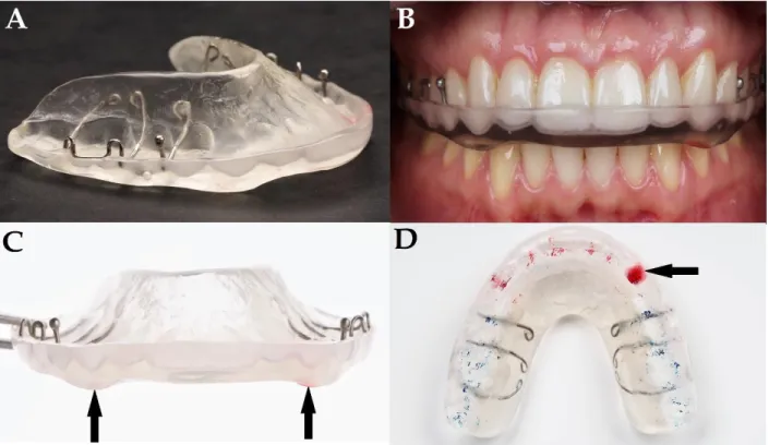
For this prospective observational case-series study a total of six patients (2 males and 4 females) with ages ranging from 22 and 34 years old were selected according to pre-determined inclusion criteria. The inclusion criteria employed comprised of the following: all participants were adults, able to read and sign the informed consent document, physically and psychologically able to tolerate the procedure. Furthermore, patients who were selected for the study had good oral hygiene, full dentition and normal occlusion without periodontal disease, as verified by the clinical and radiographic records. Moreover, patients had remained in continuous clinical follow-up, without attending other dentists. The minimal length of the follow-up time was 20 months. Reasons for placement of direct palatal RBC veneers and labial ceramic veneers were either localized anterior attrition or combination of erosion and attrition with relatively unaffected posterior teeth (Figure 1A, 1B). Tooth wear was determined to be clinically significant when dentin was exposed and when there was a reduction of crown height. Margins placed on enamel with a vital pulp were also requirements which had to be fulfilled in order for the placement of direct and indirect veneers.
The study protocol was approved by the Regional Research Ethics Committee of University of Pécs (3410.1/PTE).
The patientswere seeking advice for aesthetic reasons and wanted to improve their deteriorated and disadvantageous smile. Before the written informed consent was signed, patients were given a full explanation of the proposed treatment. They were treated at the Operative Dentistry Department of the University of Pécs by one operator. Cause of the localized anterior tooth wear was identified as much as possible based on a careful history and examination. The etiology was primarily attrition (bruxism related occlusal stress) in half of the cases and combined erosion/attrition (confirmed and treated gastoesophageal reflux disease in two cases and high rate of soft drink/energy drink consumption in one case with concurrent bruxism related wear) in the other half (Table 1).
Bruxism related occlusal stress was identified by self-report and clinical examination. The etiology of bruxism related to anxiety, insomnia and lifestyle-related stress. The elimination of the etiological factors was already ongoing or started to treat.
4 5 6 7 8 9 10 11 12 13 14 15 16 17 18 19 20 21 22 23 24 25 26 27 28 29 30 31 32 33 34 35 36 37 38 39 40 41 42 43 44 45 46 47 48 49 50 51 52 53 54 55 56 57 58 59
History taking was performed by a questionnaire survey according to Pintado et al. which focused on night or awake grinding, jaw fatigue or temporal headache on awakening.29 The clinical examination consisted of the assessment of tooth wear, chipping or abfraction, masticatory muscle hypertrophy or discomfort, teeth mobility, hypersensitivity, clicking of the temporomandibular joint and tongue or cheek indentation.30 Before starting the treatment, a consultation with an expert in temporomandibular disorder was provided. Diagnostic Criteria for Temporomandibular Disorders (DC/TMD) were clinically applied to determine the presence or absence of any temporomandibular disorder.31,32 The erosive status of the maxillary anterior teeth was evaluated with the help of the Anterior Clinical Erosive Classification (ACE).33 According to the clinical examinations, localized anterior loss of tooth structure, including dentin with minimum wear of mandibular incisors, relatively intact posterior occlusal surfaces and absence of temporomandibular disorder were diagnosed. According to self-reports and clinical signs, all the patients suffered from bruxism related occlusal stress. Table 1 shows the patient related factors, like age and gender and provides information about the etiology of tooth wear with its classification.
The suggested therapy, approved by the patients, was the sandwich veneer approach in combination with the Dahl’s concept. Patients received a total of 36 direct palatal RBC veneers on their vital maxillary anterior teeth (Table 1). This method can replace the eroded tooth structure with the additive approach at an increased VDO. The residual posterior interocclusal space is progressively eliminated by the passive eruption of the posterior teeth and slight intrusion of the anterior teeth reestablishing the occlusal contacts. A total of 42 lithium disilicate-reinforced pressed ceramic veneers were used on the labial surfaces to improve the aesthetic appearance and to preserve maximally the remaining tooth substance while simultaneously balancing the functional stresses on the anterior dentition (Table 1).
Restorative procedure
Upper and lower one-stage sandwich vinyl polysiloxane impressions (Variotime Easy Putty and Light Flow, Kulzer, Hanau, Germany) were taken. This was followed by bite registration (Variotime Bite, Kulzer, Hanau, Germany) recorded in centric relation obtained by a leaf gauge.
The difference between maximal intercuspation and centric relation was also determined. A
4 5 6 7 8 9 10 11 12 13 14 15 16 17 18 19 20 21 22 23 24 25 26 27 28 29 30 31 32 33 34 35 36 37 38 39 40 41 42 43 44 45 46 47 48 49 50 51 52 53 54 55 56 57 58 59
facebow was used to transfer the relation between the jaws and temporomandibular joint. The study casts were then mounted on an articulator in centric relation. A wax-up was carried out on the palatal, incisal and labial surfaces of the upper six anterior teeth and additionally on the labial surfaces of those premolars which were affected by the erosive effect. The wax-up was the basis for creating an intraoral mock-up (Structur 2 SC, VOCO, Cuxhaven, Germany), which was used as a visual aid to show the proposed treatment outcome and analyze the aesthetic and functional (anterior and canine guidance) changes. A silicone (Registrado Clear, VOCO, Cuxhaven, Germany or Variotime Easy Putty, Kulzer, Hanau, Germany) copyplast from the diagnostic wax-up was used as a trimmed palatal/incisal stent for the direct RBC build-ups to provide a desirable form for the palatal surface. Before the build-up procedure the teeth were isolated with a rubber dam and the palatal surfaces of the anterior teeth were air abraded with 29 µm Al2O3 powder with copious irrigation at 1 mm distance for 15 s (AquaCare Twin, Velopex, London, UK). Only sharp enamel edges were removed with diamond bur (No.890LF, Meisinger, Neuss, Germany). To prevent bonding to adjacent teeth, every second teeth were restored at the same time. Teeth were separated with slight interproximal stripping (Sof-Lex Finishing strips, 3M, St. Paul, MN, USA) and isolated interproximally by a teflon tape. Regarding the adhesive technique, a two-step etch-and-rinse system (Adper Single Bond, 3M, St. Paul, MN, USA) was used for each restoration. Every second tooth was conditioned with the total-etch technique by applying 37% phosphoric acid for 20 s (Ultra-etch, Ultradent, South Jordan, UT, USA), followed by 20 s rinsing and careful drying. The one step enamel-dentin adhesive was applied by rubbing the exposed dentin and enamel with a micro-brush soaked in the resin. In order to evaporate the solvent, gentle, 10 s air-drying was carried out, followed by 10 s polymerization with a light emitting diode (LED) curing unit (LED.D, Woodpecker, Guilin, China; λ = 420–480 nm) in standard mode, at an average tip irradiance of 1450 mW/cm2 with an 8 mm diameter fiberglass light guide. Nanohybrid RBC (Enamel Plus HRi Bio Function, Micerium S.p.A., Avegno, Italy; BF2 shade) was applied to the matrix in alternating order then the matrix was seated on the teeth and the excess material was removed from the labio- incisal aspect maintaining the planned new length as seen with the help of the stent (Figure 1C).
The RBC was light-cured for 40 s from an incisal direction. After removal of the stent, the palatal surface was directly light cured for a further 40 s to provide a high rate of polymerization.
Interproximal excess was removed carefully – without causing bleeding - with a metal interproximal strip (Kerr perforated fine diamond finishing strip, Kerr, Bioggio, Switzerland). The
4 5 6 7 8 9 10 11 12 13 14 15 16 17 18 19 20 21 22 23 24 25 26 27 28 29 30 31 32 33 34 35 36 37 38 39 40 41 42 43 44 45 46 47 48 49 50 51 52 53 54 55 56 57 58 59
restorative procedure was repeated for the remaining teeth. Once all the teeth have been bonded to the palatal aspect the labio-incisal surface was covered with one more layer of RBC to complete provisionally the anterior restoration at the desired length until the posterior contacts are re- established. The provisional increase of the length with direct RBC could provide a functional test for the re-established anterior and canine guidance. Patients were informed about the possible inconvenience caused by the posterior disclusion and color mismatch of the lengthened incisal edge before the final facial veneer fabrication. The finishing-polishing procedure was performed with football, needle shape red fine-grit diamond burs (No.330F and No.890LF, Meisinger, Neuss, Germany) followed by Enhance/PoGo system (Dentsply Sirona, York, PA, USA) and with aluminum oxide strips (Sof-Lex Finishing strips, 3M, St. Paul, MN, USA). Then the occlusion was refined according to the following principles to ensure even stable contacts on all anterior teeth of equal intensity in centric relation (static occlusion) and guidance provided by the incisors and canines (dynamic occlusion) (Figure 1D). The restored palatal surface and the increased incisal length must be in harmony with the envelope of function and should provide unobstructed movement. The palatal cingulum and slightly flat occlusal stop provide stable contact and centric freedom for the lower incisors. This smooth surface will help minimize future wear of the opposing lower incisors and directs forces down the long axis of the tooth, thereby reducing the possible labial tooth movement. During lateral excursion the canine guidance should provide incisal disclusion which is crucial to avoid the future fracture of the restored incisal edges. After the occlusal adjustment, final finishing, and polishing were performed by the Enhance/PoGo system and the surface gloss was enhanced by goat hair brush, felt disk/wheel and polishing pastes (Enamel Plus HFO Shiny Finishing Kit, Micerium S.p.A., Avegno, Italy). Posterior disclusion was measured from a wax record taken postoperatively between the first molar teeth (Table 1). The increase of VDO by the additional palatal layer is ranged between 1.0 to 2.0 mm.
Few days (~ 2-3) after the palatal restorative procedure, patients were scheduled for an anterior occlusal check-up to control the stable contacts and anterior guidance. It was also important to assess patient comfort and to test the phonetic implications of the extensive rehabilitation of the maxillary incisors. For a phonetic test, the ‘s’ sound is appropriate since it can be influenced by horizontal or vertical volume alterations on the anterior teeth.
Progression of the posterior tooth eruption was assessed weekly with an 8 µm Shim Stock foil (Arti-Fol, Bausch, Köln, Germany) (Figure 2A-D). After the detection of firm contacts on all
4 5 6 7 8 9 10 11 12 13 14 15 16 17 18 19 20 21 22 23 24 25 26 27 28 29 30 31 32 33 34 35 36 37 38 39 40 41 42 43 44 45 46 47 48 49 50 51 52 53 54 55 56 57 58 59
posterior teeth (four weeks after palatal build-up for all patients) the procedure for labial ceramic veneer fabrication was started. The first step was the mock-up preparation according to the initial diagnostic wax-up. As the new position of the occlusal plane and the increased VDO may be slightly different from what was initially planned, the length of the maxillary anterior teeth should be reconfirmed during this second mock-up session. Once the patient’s consensus on the final shape of the maxillary anterior teeth was obtained, the mock-up guided preparation with the help of a depth marker diamond cylinder bur (No.834, size 016, cutting depth 0.3 mm; size 021, cutting depth 0.5 mm; Komet, Besigheim, Germany) was performed (Figure 3A). Three shallow orientation grooves were cut through the mock-up RBC or/and enamel on the labial surface. Then the mock-up was removed and the orientation groove which left a mark on the enamel was highlighted with a water-resistant pen (1513 Multimark F permanent, Faber-Castell, Stein, Germany). Round ended tapered diamond (No.868 and 8868, Komet, Besigheim, Germany) was used to remove enamel between the marked grooves and to create definitive contouring and finishing. The color marking was not removed completely to ensure no more than the desired enamel was prepared. Supra/paragingival chamfer-like finishing line was prepared in a depth of 0.3 mm, which was extended interproximally as an elbow, involving the labial half of the contact area without breaking the contact. Butt-joint incisal preparation was performed, completely removing the length added by the provisional RBC build-up, leaving only the original length of the tooth (Figure 3B). The ceramic veneer would later re-establish the final length. The margin was placed in the volume of the palatal RBC (Figure 3B and Figure 4A-C). The prepared surface was polished with abrasive discs (Sof-Lex disks, 3M, St. Paul, MN, USA) and a gingival retraction cord (Size 0, UltraPak, Ultradent, South Jordan, UT, USA) was placed in the sulcus. Upper two-stage and lower one-stage sandwich vinyl polysiloxane impressions (Variotime Easy Putty and Light Flow, Kulzer, Hanau, Germany) were taken. This was followed by bite registration (Variotime Bite, Kulzer, Hanau, Germany) recorded in maximum intercuspation. The shade of the prepared teeth and also the desired shade were selected with the patient’s consensus. Provisionals were made with the help of the impression which was fabricated to perform the mock-up. At the next appointment the pressed lithium disilicate-reinforced glass ceramic laminate veneers (GC Initial LiSi Press, GC, Leuven, Belgium) were tried in the mouth to check the fit and try-in paste (NX3 Nexus Third Generation, Kerr, Bioggio, Switzerland) was used to select the proper shade of the adhesive luting cement (Figure 3C). To achieve durable bond-strength the internal surfaces of the
4 5 6 7 8 9 10 11 12 13 14 15 16 17 18 19 20 21 22 23 24 25 26 27 28 29 30 31 32 33 34 35 36 37 38 39 40 41 42 43 44 45 46 47 48 49 50 51 52 53 54 55 56 57 58 59
cleaned (Ivoclean, Ivoclar Vivadent, Schaan, Liechtenstein) veneers were etched with 9,5%
hydrofluoric acid (Porcelain Etch, Ultradent, South Jordan, UT, USA) for 20 s, followed by washing, drying and silane (Silane, Ultradent, South Jordan, UT, USA) application which was left to dry. The luting procedure started from both cleaned central incisors and progressed bilaterally.
The above described total-etch adhesive procedure was performed on the isolated teeth. To provide accurate fit of the veneer prior to light-curing the adhesive layer was thinned with air-drying for minimum 20 s and the removal of excess was helped by the vacuum of the high-volume evacuator’s tip. A clean microbrush was used to remove the gathered excess at the marginal bevel if the air- stream and the evacuator’s effect had to be completed. The light-curable adhesive resin cement (NX3 Nexus Third Generation, Kerr, Bioggio, Switzerland) was applied to the veneers, which were then gently seated with finger pressure. Excess cement was removed with a microbrush and a thin modelling brush. The above-mentioned LED curing unit was used to polymerize the cement for 4x20 s from labial, mesial, distal and incisal aspects with 5 s break to avoid heat damage of the pulp. The final restorative phase was completed by polishing the marginal areas with a silicone rubber point (PoGo system, Dentsply Sirona, York, PA, USA), after removal of excess set cement with a No.12 surgical scalpel (Figure 3D). Restorations were carefully checked for any occlusal interference (static and dynamic). After the one-week follow-up of the labial veneers, maintenance was provided in each half year.
Clinical evaluation
From the palatal build-up session, the progression of the compensatory eruption was recorded weekly by the evaluation of the opposing teeth contacts until maximum intercuspation was achieved between each posterior tooth. During the five-week follow-up appointment, the patient’s opinion on the aesthetic result, post-operative pain or discomfort and bruxism or TMD related symptoms were evaluated (Table 2).
In January of 2020, the anterior palatal direct RBC veneers and labial ceramic veneers were evaluated by two examiners independently using a dental mirror and explorer in accordance with modified United States Public Health Service (USPHS)27 criteria (Table 3). Particular attention was paid to the interface between the palatal direct veneer and the labial indirect veneer in terms of marginal integrity. The time and type of any intervention applied during the maintenance was
4 5 6 7 8 9 10 11 12 13 14 15 16 17 18 19 20 21 22 23 24 25 26 27 28 29 30 31 32 33 34 35 36 37 38 39 40 41 42 43 44 45 46 47 48 49 50 51 52 53 54 55 56 57 58 59
recorded. Besides the evaluation of the restoration, patients were questioned again about the aesthetic satisfaction, discomfort and bruxism or TMD related symptoms.
Data collection and statistical analysis were performed using SPSS for Windows 23.0 (SPSS, Chicago, IL, USA). Because of the low case number, only a descriptive statistical analysis was indicated and only percentages of the respective scores of the USPHS evaluation are given.
RESULTS
In the present observational case-series study a total of 36 direct RBC palatal veneers and 42 labial ceramic veneers were evaluated in 6 adult patients (mean age: 26.67 at the time of restoration placement) with the diagnosis of localized anterior tooth wear. The follow-up time varied from 20 to 27 months for palatal veneers and 18 to 25 months for labial veneers with a mean observation time of 23.7 and 21.7 months, respectively. The investigators were trained and calibrated before the evaluation. Cohen’s kappa statistic showed excellent intraobserver (kappa values of 0.82 and 0.80) and interobserver (a kappa value of 0.79) agreement. Figure 5 shows the evaluation of patients’ satisfaction with the aesthetic result, discomfort, re-establishment of posterior contacts and bruxism or TMD related symptoms at the five-week follow-up appointment (from the palatal build-ups to one week after the labial veneers are cemented). According to one participant’s report, discomfort was only present in the first few days, right after the palatal build-up. Phonetic implications - pronouncing the ‘s’ sound - occurred for all patients, but after a short conversation (five to ten minute adaptation), it was no longer perceptible. The progression time and order showed almost the same pattern for all participating patients which is visualized in Figure 2C. All patients had accepted the new VDO well, without showing symptoms of TMD. However, bruxism was still present in two patients (detected by self-report), although it had not intensified. To decrease the risk of bruxism-related failure, a supplemental protection with a night-guard (Michigan-type occlusal appliance) was fabricated for the above-mentioned patients at the five- week check-up (Figure 6). During the subsequent observation period there was no change in aesthetic satisfaction and the stabilized occlusion was reported to be comfortable. Based on the self-reports and clinical examinations of the other four patients there were no obvious signs of bruxism, despite being present at the start of treatment. The evaluation of the palatal direct RBC and labial ceramic restorations according to the USPHS criteria is presented in Figure 7 which
4 5 6 7 8 9 10 11 12 13 14 15 16 17 18 19 20 21 22 23 24 25 26 27 28 29 30 31 32 33 34 35 36 37 38 39 40 41 42 43 44 45 46 47 48 49 50 51 52 53 54 55 56 57 58 59
involved also the assessment of the marginal adaptation and discoloration of the palatal-labial veneer interface. During the observation period all the involved teeth were caries-free and remained vital. Of the overall 78 restorations none of them were considered unacceptable, the restorations were scored as ‘alpha’ or ‘bravo’ for all items evaluated according to the USPHS criteria. The survival during this short-term observation period for both types of restoration was 100%. Palatal direct restorations had higher rates of small deteriorations for almost every evaluated criteria (Figure 8A), except for anatomic form and color match. Slight wear on the palatal restoration was found to be the most frequent issue (44.4%), followed by slight marginal discoloration (33.3%).
Pitted surface roughness was found on all six palatal RBC (16.6%) in one patient, whereas the texture of all other palatal veneers showed a silky gloss appearance. Slight marginal deterioration was detected at only four palatal veneers (11.1%), which were managed with re-polishing. The labial-palatal veneer interface showed mild deterioration in marginal adaptation (16.7%) and marginal discoloration (22.2%). The labial ceramic veneers showed excellent performance, with slight marginal discoloration detected at only one veneer (Figure 8B).
DISCUSSION
In this prospective observational case-series study the short-term survival of anterior sandwich veneers as Dahl-restorations were evaluated in six patients who suffered from localized anterior erosion or attrition. Furthermore, patient’s satisfaction, discomfort, progression of posterior teeth eruption and bruxism related occlusal stress were also analyzed. Acceptable to excellent clinical performance was observed for the sandwich veneers - which consisted of direct palatal RBC and indirect labial ceramic restorations - after a mean observation period of 23.7 and 21.7 months, respectively. However, results and survival analysis should be interpreted with care because on one hand the case number is very limited, and on the other, no long-term data are available regarding this sandwich veneer approach. Our primary purpose was not only to describe the outcome of the sandwich approach after a short observation period, but also to generate hypotheses regarding the survival of this treatment method that subsequently can be tested in studies of greater methodological rigor.
Vailati and Belser introduced a three-step adhesive rehabilitation technique with a minimally invasive sandwich approach, which reconstructs the palatal surface first with direct or indirect resin
4 5 6 7 8 9 10 11 12 13 14 15 16 17 18 19 20 21 22 23 24 25 26 27 28 29 30 31 32 33 34 35 36 37 38 39 40 41 42 43 44 45 46 47 48 49 50 51 52 53 54 55 56 57 58 59
composite palatal veneers followed by the restoration of the labial aspect with ceramic veneers.19-
21 This adhesive technique allows preservation of the remaining tooth structure, especially with respect to the intact proximal surface. Subsequent work of the authors demonstrated, in a 6-year follow-up study, no major detectable failure of sandwich restorations.34 The adhesively bonded ceramic veneers with well-determined surface-treatment and cementation protocol represent a highly reliable and long-term treatment option regarding the esthetic and functional aspects, and is supported by several other researches.35-38 Due to the low number of cases, our results cannot be supported by statistical analysis, but in line with the findings of other studies, for each of the examined parameters, the labial ceramic veneers performed excellently during the short observation period. The original three-step adhesive rehabilitation technique restores the posterior region with overlays at an increased VDO, however our treatment combined the sandwich approach with the Dahl concept and used palatal veneers as anterior deprogrammers followed by a compensatory eruption for the relatively unaffected posterior teeth to reconstruct the functional occlusion. This combined method of adhesive techniques and the Dahl principle was introduced by Magne et al.22 The Dahl concept allows maximal tooth structure preservation, without the need for posterior overlay restorations, with special respect to the healthy molars and premolars. Poyser et al. collected the clinically relevant studies related to the Dahl concept, and found that the majority of the available literature originated from the United Kingdom.24 Although, case-reports are available, there is lack of international uptake of this technique.22,24 On the other hand, the quality of the level of evidence from these studies is medium to low, thus, randomized controlled clinical trials are necessary to provide evidence-based data.24 The original Dahl appliance is a removable localized palatal bite-raising metal device which can create the necessary space for the further full- coverage restorations of destructed anterior teeth.25 To overcome the problem, induced by excessive tooth preparation for full coverage crown, adhesive double-veneer (palatal metal and labial ceramic) was introduced by Bishop et al.39 The improvements and continuous development of adhesive dental materials ensure durable, ultraconservative solutions. The treatment of anterior tooth wear with direct RBC restoration, placed in supra-occlusion at an increased VDO, was described first by Darbar and Hemmings.40 Direct RBC replaces the eroded tooth structure with an additive approach and additionally can be used as an anterior deprogrammer to reposition the mandible in centric relation.8,22 Hemmings et al. reported 89.4% success rate of direct resin composite palatal veneers in the treatment of localized anterior tooth wear after 30-months follow-
4 5 6 7 8 9 10 11 12 13 14 15 16 17 18 19 20 21 22 23 24 25 26 27 28 29 30 31 32 33 34 35 36 37 38 39 40 41 42 43 44 45 46 47 48 49 50 51 52 53 54 55 56 57 58 59
up and the posterior occlusion satisfactorily restored after a mean duration of 4.6 months with good patient’s satisfaction.26 Redman et al., in their short to medium term study, confirmed that RBC restorations placed at an increased VDO are viable first-line options to treat anterior tooth wear, however, the authors concluded, that the probability of failures is increased after five years.27 Gulamali et al. concluded in their medium-term survival study, that more than 90% of the investigated direct and indirect RBC restorations exhibited minor or major failure over the preceding ten years and required intervention and maintenance.28 Mechanical limitations of RBCs can lead to marginal fracture and staining, meanwhile, failures such as bulk fracture, surface roughness and discoloration are uncommon.27 Regarding most deteriorations a similar tendency was detectable in our study also. However, survival of palatal restorations was found to be 100%.
Few restorations had minor defects after the short observation time, such as surface roughness, marginal discoloration, marginal adaptation and consequent gingival inflammation. In line with Redman et al., surface roughness took the form of pitting in one patient, suspected to be due to the exposure of tiny voids as the palatal RBC wore and erosive effects softened the external layer.27 In our cases, a nanohybrid RBC was applied directly to the silicon matrix and pressed to the palatal surface of the tooth. Probably, the absence of an intensive condensation phase is the reason for the voids and subsequent surface roughness. Although, a clinical study found, that the indirectly made palatal veneer had surprisingly higher incidence of roughness.27 Despite the short observation period, marked small wear facets appeared frequently (USPHS ‘Bravo’ score is 44.4%) on the palatal veneers, especially on the static occlusal contacts. All the six patients suffered from night or awake grinding before the initiation of the treatment, however only two patients reported continuing bruxism after the palatal build-up. They received night-guard splints to prevent fracture of the veneers. Though, based on the detected wear facets, it is assumed that some degree of occlusal stress was still present in all patients, the newly constructed anatomic palatal shape and designed guidance limited the parafunctional movements which was consequently less noticeable to the patients. Although only two of our participants needed night-guard splint, it is important to emphases, that regular check-ups and maintenance is suggested to detect in time the signs of increased activity of bruxism. As an alternative solution to decrease the risk of restoration’s failure a fabrication of a night-guard splint routinely could be beneficial at the end of the restorative treatment. It is considered advantageous to use RBC restorations opposite worn teeth, since RBCs have lower wear resistance than the opposing teeth, thus the wear rate of the antagonist could be
4 5 6 7 8 9 10 11 12 13 14 15 16 17 18 19 20 21 22 23 24 25 26 27 28 29 30 31 32 33 34 35 36 37 38 39 40 41 42 43 44 45 46 47 48 49 50 51 52 53 54 55 56 57 58 59
decreased.41,42 Further advantages of RBC as a Dahl restoration, are the maintainability, time- and cost-saving ability and preservation of healthy tooth structures.43 Moreover, it can protect exposed dentin from further wear and the reversible nature of this approach may allow repair and an alternative restoration in the future. This technique offers good patient compliance, and satisfaction, even when repair is needed. Our patients tolerated the increased VDO and posterior disclusion very well. They were maximally adapted within a short time, as it was reported in other studies as well.26,27
The palatal RBC restorations provided a deliberate occlusal plane and axial loading of the antagonist teeth, therefore no adverse tooth movements were detected on the anterior teeth. The fundamental importance of the reconstructed canine guidance lies in preventing the fracture of the labial veneer by separating the upper and lower incisors during lateral movements. Already in the design or mock-up phase, it is necessary to determine the future length of the incisors and align them with the available inter-incisal space provided by the planned canine guidance. Furthermore, it is assumed, that the stable anterior occlusion and guidance facilitate posterior teeth eruption by neuromuscular regulation. Post-eruptive tooth movement maintains the position in occlusion by compensation for occlusal tooth wear. Although, the proper mechanism of compensatory eruption is unclear, it is supposed that coordinated forces of the oro-facial muscles, under the influence of the central nervous system, are responsible for tooth eruption.44 Lund described that the control of mastication depends on sensory feedback which serves to regulate the muscle activity through all stages of mastication.45 Individual chewing cycle is divided into three stages: opening, fast-closing and slow-closing. Latter is associated with increased forces required for grinding the bolus. It is speculated, that when the posterior region is discluded, as in the case of only anterior contacts and guidance, the slow-closing phase and resultant increased elevator muscle activity is absent thus the food mastication function is confused. The central nervous system receives several feedbacks from muscles, periodontal receptors and touch receptors of the mucosa and consequently induce bone remodeling with consequent tooth eruption to maintain the physiologic function of the masticatory system. In our study, all the subjects had re-established posterior contacts within four weeks.
Redman et al. found in 31 patients 61% to have complete and 39% to have partial re-establishment of posterior occlusion within 2 to 18 months, with a mean of 7 months.27 It is likely that mandibular repositioning and the young age of our patients contributed to the rapid result.
4 5 6 7 8 9 10 11 12 13 14 15 16 17 18 19 20 21 22 23 24 25 26 27 28 29 30 31 32 33 34 35 36 37 38 39 40 41 42 43 44 45 46 47 48 49 50 51 52 53 54 55 56 57 58 59
Within the limitations of this observational case-series, it is concluded, that the adhesively bonded labial ceramic and palatal direct RBC veneer – namely sandwich veneer - with the combination of the Dahl concept is a promising method to treat localized anterior tooth wear. The minimally invasive additive approach and the compensatory eruption of posterior teeth reconstruct the aesthetics and function with maximal preservation of the remaining tooth structure. Although, mechanical limitations of the direct RBC can lead to wear, marginal breakdown, marginal discoloration and surface roughness, the maintenance of these palatal restorations is straightforward with refinishing or with localized repair. There is a high degree of patient satisfaction associated with sandwich restorations. The temporary posterior disclusion is well- tolerated and contacts are re-established within a very short period of time.
REFERENCES
1. Litonjua LA, Andreana S, Bush PJ & Cohen RE (2003) Tooth wear: Attrition, erosion, and abrasion Quintessence International 34(6) 435-46.
2. Loomans B, Opdam N, Attin T, Bartlett D, Edelhoff D, Frankenberger R, Benic G, Ramseyer S, Wetselaar P, Sterenborg B, Hickel R, Pallesen U, Mehta S, Banerji S, Lussi A & Wilson N (2017) Severe Tooth Wear: European Consensus Statement on Management Guidelines Journal of Adhesive Dentistry 19(2) 111-119.
3. Grippo JO (1991) Abfractions: a new classification of hard tissue lesions of teeth Journal of Esthetic Dentistry 3(1) 14-19.
4. Shellis RP & Addy M (2014) The interaction between attrition, abrasion and erosion in tooth wear Monographs in Oral Science 25 32-45.
5. Gambon DL, Brand HS & Veerman EC (2012) Dental erosion in the 21st century: what is happening to nutritional habits and lifestyle in our society? British Dental Journal 213(2) 55‐ 57.
6. Hanif A, Rashid H & Nasim M (2015) Tooth surface loss revisited: Classification, etiology and management Journal of Restorative Dentistry 3(2) 37-43.
7. Johansson A, Johansson AK, Omar R & Carlsson GE (2008) Rehabilitation of the worn dentition Journal of Oral Rehabilitation 35(7) 548‐ 66.
4 5 6 7 8 9 10 11 12 13 14 15 16 17 18 19 20 21 22 23 24 25 26 27 28 29 30 31 32 33 34 35 36 37 38 39 40 41 42 43 44 45 46 47 48 49 50 51 52 53 54 55 56 57 58 59
8. Mizrahi B (2004) A technique for simple and aesthetic treatment of anterior tooth wear Dental Update 31(2) 109-114.
9. Sundaram G, Wilson R, Watson TF, Bartlett D (2007) Clinical measurement of palatal tooth wear following coating by a resin sealing system Operative Dentistry 32(6) 539-543.
10. Mehta SB, Banerji S, Millar BJ & Suarez-Feito JM (2012) Current concepts on the management of tooth wear: Part 3. Active restorative care 2: the management of generalised tooth wear British Dental Journal 212(3) 121-127.
11. Mehta SB, Banerji S, Millar BJ & Suarez-Feito JM (2012) Current concepts on the management of tooth wear: Part 4. An overview of the restorative techniques and dental materials commonly applied for the management of tooth wear. British Dental Journal 212(4) 169-177.
12. Dietschi D & Argente A (2011) A comprehensive and conservative approach for the restoration of abrasion and erosion. Part II. Clinical procedures and case report European Journal of Esthetic Dentistry 6(2) 142-159.
13. Fradeani M, Barducci G, Bacherini L & Brennan M (2012) Esthetic rehabilitation of a severely worn dentition with minimally invasive prosthetic procedures (MIPP).
International Journal of Periodontics and Restorative Dentistry 32(2) 135‐ 147.
14. Meyers IA (2013) Minimum intervention dentistry and the management of tooth wear in general practice Australian Dental Journal 58(1) 60‐ 5.
15. Hansen TL, Schriwer C, Øilo M & Gjengedal H (2018) Monolithic zirconia crowns in the aesthetic zone in heavy grinders with severe tooth wear – An observational case-series Journal of Dentistry 72 14-20.
16. Schmidlin PR, Filli T, Imfeld C, Tepper S & Attin T (2009) Three-year evaluation of posterior bite reconstruction using direct resin composite – a case series Operative Dentistry 34(1) 102-108.
17. Mesko ME, Sarkis-Onofre R, Cenci MS, Opdam NJ, Loomans B & Pereira-Cenci T (2016) Rehabilitation of severely worn teeth: a systematic review Journal of Dentistry 48 9-15.
18. Ahmed KE & Murbay S (2016) Survival rates of anterior composites in managing tooth wear: systematic review Journal of Oral Rehabilitation 43(2) 145-53.
4 5 6 7 8 9 10 11 12 13 14 15 16 17 18 19 20 21 22 23 24 25 26 27 28 29 30 31 32 33 34 35 36 37 38 39 40 41 42 43 44 45 46 47 48 49 50 51 52 53 54 55 56 57 58 59
19. Vailati F & Belser UC (2008) Full-mouth adhesive rehabilitation of a severely eroded dentition: the three-step technique. Part 1 European Journal of Esthetic Dentistry 3(1) 30- 44.
20. Vailati F & Belser UC (2008) Full-mouth adhesive rehabilitation of a severely eroded dentition: the three-step technique. Part 2 European Journal of Esthetic Dentistry 3(2) 128- 146.
21. Vailati F & Belser UC (2008) Full-mouth adhesive rehabilitation of a severely eroded dentition: the three-step technique. Part 3 European Journal of Esthetic Dentistry 3(3) 236- 257.
22. Magne P, Magne M & Belser UC (2007) Adhesive restorations, centric relation, and the Dahl principle: minimally invasive approaches to localized anterior tooth erosion European Journal of Esthetic Dentistry 2(3) 260-273.
23. Berry DC & Poole DFG (1976) Attrition: possible mechanisms of compensation Journal of Oral Rehabilitation 3(3) 201-206.
24. Poyser NJ, Porter RW, Briggs PF, Chana HS & Kelleher MG (2005) The Dahl Concept:
past, present and future British Dental Journal 198(11) 669‐ 676.
25. Dahl B L, Krogstad O & Karlsen K (1975) An alternative treatment in cases with advanced localized attrition Journal of Oral Rehabilitation 2(3) 209-214.
26. Hemming KW, Darbar UR & Vaughan S (2000) Tooth wear treated with direct composite restorations at an increased vertical dimension: Results at 30 months Journal of Prosthetic Dentistry 83(3) 287-293.
27. Redman CDJ, Hemmings KW & Good JA (2003) The survival and clinical performance of resin-based composite restorations used to treat localized anterior tooth wear British Dental Journal 194(10) 566-572.
28. Gulamali AB, Hemmings KW, Tredwin CJ & Petrie A (2011) Survival analysis of composite Dahl restorations provided to manage localized anterior tooth wear (ten year follow-up) British Dental Journal 211(4) E9.
29. Pintado MR, Anderson GC, DeLong R & Douglas WH (1997) Variation in tooth wear in young adults over a two-year period Journal of Prosthetic Dentistry 77(3) 313–320.
30. Kato T, Thie NM, Montplaisir JY & Lavigne GJ (2001) Bruxism and orofacial movements during sleep Dental Clinics of North America 45(4) 657–684.
4 5 6 7 8 9 10 11 12 13 14 15 16 17 18 19 20 21 22 23 24 25 26 27 28 29 30 31 32 33 34 35 36 37 38 39 40 41 42 43 44 45 46 47 48 49 50 51 52 53 54 55 56 57 58 59
31. Schiffman E, Ohrbach R, Truelove E, Look J, Anderson G, Goulet JP et al. (2014) International RDC/TMD Consortium Network, International Association for Dental Research; Orofacial Pain Special Interest Group, International Association for the Study of Pain. Diagnostic Criteria for Temporomandibular Disorders (DC/TMD) for clinical and research applications: recommendations of the International DC/TMD Consortium Network and Orofacial Pain Special Interest Group. J Oral Facial Pain Headache 28(1) 6–
27.
32. Somoskövi I, Radácsi A, Nagy Ákos K, Radnai M (2018) Developing the Hungarian version of the Diagnostic Criteria for Temporomandibular Disorders (DC/TMD).
Fogorvosi Sz 111(2) 44-51 (Article in Hungarian).
33. Vailati F & Belser UC (2010) Classification and treatment of the anterior maxillary dentition affected by dental erosion: The ACE classification International Journal of Periodontics & Restorative Dentistry 30(6) 559-571.
34. Vailati F, Gruetter L & Belser UC (2013) Adhesively restored anterior maxillary dentitions affected by severe erosion: up to 6-year results of a prospective clinical study European Journal of Esthetic Dentistry 8(4) 506-530.
35. Peumans M, de Munck J, Fieuws S, Lambrechts P, Vanherle G & van Meerbeek B (2004) A prospective ten-year clinical trial of porcelain veneers Journal of Adhesive Dentistry 6(1) 65–76.
36. Magne P & Magne M (2006) Use of additive wax-up and direct intraoral mock-up for enamel preservation with porcelain laminate veneers European Journal of Esthetic Dentistry 1(1) 10-19.
37. Beier US, Kapferer I, Burtscher D & Dumfahrt H (2012) Clinical performance of porcelain laminate veneers for up to 20 years International Journal of Prosthodontics 25(1) 79–85.
38. Layton DM & Walton TR (2012) The up to 21-year clinical outcome and survival of feldspathic porcelain veneers: accounting for clustering International Journal of Prosthodontics 25(6) 604–612.
39. Bishop K, Bell M, Briggs P & Kelleher M (1996) Restoration of a worn dentition using a double-veneer technique British Dental Journal 180(1) 26–29.
4 5 6 7 8 9 10 11 12 13 14 15 16 17 18 19 20 21 22 23 24 25 26 27 28 29 30 31 32 33 34 35 36 37 38 39 40 41 42 43 44 45 46 47 48 49 50 51 52 53 54 55 56 57 58 59