74
V
ascular stiffness is an independent risk factor for mul- tiple negative cardiovascular outcomes in normally aging adults, including stroke, myocardial infarction, and sudden death.1 Whether vascular stiffness occurs in develop- mental cardiovascular disorders and portends similar adverse cardiovascular outcomes is not well documented. Previous investigations in mouse models have consistently linked hap- loinsufficiency for elastin, an extracellular matrix protein that provides recoil to elastic tissues, to increased vascular stiffness and hypertension.2 Comparable data on the effect of elastin deficiency on vascular stiffness in humans are lacking and, thus, warrant study in naturally occurring elastin defi- ciency disorders.Loss of function defects in the human elastin gene (ELN) cause focal arterial stenosis, generalized vascular narrowing, and hypertension in a rare condition called nonsyndromic supravalvular aortic stenosis3–5 (SVAS; MIM No. 185500, prevalence 1:20 000). Deletion of an entire ELN allele as part of the contiguous gene deletion disorder, Williams syndrome
(WS; MIM No. 194050, prevalence 1:8000–10 000), leads to the same vascular phenotype.6
To test the hypothesis that elastin insufficiency is associ- ated with vascular stiffness in humans, we initiated the multi- institutional Williams Syndrome-Skin And Vessel Elasticity (WS-SAVE) study. Using applanation tonometry, we collected pulse wave velocity (PWV) measurements in the single largest WS cohort studied to date, consisting of 77 affected individuals aged 7 to 62 years. Vascular stiffness normally increases with age and with comorbid conditions such as hypertension and diabetes mellitus.7 Consequently, this robust sample size and broad age range allowed us to evaluate the presence of vascu- lar stiffness in WS throughout the life span and also to identify covariates that modify arterial stiffness in this population. In particular, we investigate both pharmacological interventions and genetic alterations that influence arterial stiffness and iden- tify treatments associated with lower PWV. Differences in the WS deletion affecting copy number (CN) for NCF1, a nico- tinamide adenine dinucleotide phosphate (NADPH) oxidase Abstract—Williams syndrome is caused by the deletion of 26 to 28 genes, including elastin, on human chromosome 7.
Elastin insufficiency leads to the cardiovascular hallmarks of this condition, namely focal stenosis and hypertension.
Extrapolation from the Eln+/− mouse suggests that affected people may also have stiff vasculature, a risk factor for stroke, myocardial infarction, and cardiac death. NCF1, one of the variably deleted Williams genes, is a component of the nicotinamide adenine dinucleotide phosphate (NADPH) oxidase complex and is involved in the generation of oxidative stress, making it an interesting candidate modifier for vascular stiffness. Using a case–control design, vascular stiffness was evaluated by pulse wave velocity in 77 Williams cases and matched controls. Cases had stiffer conducting vessels than controls (P<0.001), with increased stiffness observed in even the youngest children with Williams syndrome. Pulse wave velocity increased with age at comparable rates in cases and controls, and although the degree of vascular stiffness varied, it was seen in both hypertensive and normotensive Williams participants. Use of antihypertensive medication and extension of the Williams deletion to include NCF1 were associated with protection from vascular stiffness. These findings demonstrate that vascular stiffness is a primary vascular phenotype in Williams syndrome and that treatment with antihypertensives or agents inhibiting oxidative stress may be important in managing patients with this condition, potentially even those who are not overtly hypertensive. (Hypertension. 2014;63:74-79.)
•
Online Data SupplementKey Words: elastin ◼ NADPH oxidase ◼ pulse wave analysis ◼ vascular stiffness ◼ Williams syndrome
Received July 22, 2013; first decision August 11, 2013; revision accepted September 24, 2013.
From the Department of Pediatrics (B.A.K., J.R.D.), Department of Cell Biology and Physiology (R.H.K.), and Cardiovascular Imaging and Clinical Research Core Laboratory, Cardiovascular Division, Department of Internal Medicine (L.d.l.F.), Washington University School of Medicine, St. Louis, MO; Department of Medical Sciences, Netter School of Medicine, Quinnipiac University, Hamden, CT (B.R.P.); Department of Pediatrics, Massachusetts General Hospital, Boston (J.L.W., B.R.P.); 1st Department of Pediatrics, Semmelweis University, Budapest, Hungary (G.S.R., E.K.); Division of Cardiology, Brigham and Women’s Hospital, Boston, MA (A.B.B.); and Department of Pediatrics, Harvard Medical School, Boston, MA (B.R.P.).
The online-only Data Supplement is available with this article at http://hyper.ahajournals.org/lookup/suppl/doi:10.1161/HYPERTENSIONAHA.
113.02087/-/DC1.
Correspondence to Beth A. Kozel, Washington University School of Medicine, 660 S Euclid, Campus Box 8208, St. Louis, MO 63110. E-mail Kozel_b@
kids.wustl.edu
Williams Syndrome Predisposes to Vascular Stiffness Modified by Antihypertensive Use and Copy Number
Changes in NCF1
Beth A. Kozel, Joshua R. Danback, Jessica L. Waxler, Russell H. Knutsen, Lisa de las Fuentes, Gyorgy S. Reusz, Eva Kis, Ami B. Bhatt, Barbara R. Pober
© 2013 American Heart Association, Inc.
Hypertension is available at http://hyper.ahajournals.org DOI: 10.1161/HYPERTENSIONAHA.113.02087
Vascular Stiffness
at Semmelweis University (Egyetem) on January 13, 2015 http://hyper.ahajournals.org/
Downloaded from http://hyper.ahajournals.org/ at Semmelweis University (Egyetem) on January 13, 2015 Downloaded from http://hyper.ahajournals.org/ at Semmelweis University (Egyetem) on January 13, 2015 Downloaded from http://hyper.ahajournals.org/ at Semmelweis University (Egyetem) on January 13, 2015 Downloaded from http://hyper.ahajournals.org/ at Semmelweis University (Egyetem) on January 13, 2015 Downloaded from http://hyper.ahajournals.org/ at Semmelweis University (Egyetem) on January 13, 2015 Downloaded from http://hyper.ahajournals.org/ at Semmelweis University (Egyetem) on January 13, 2015 Downloaded from http://hyper.ahajournals.org/ at Semmelweis University (Egyetem) on January 13, 2015 Downloaded from http://hyper.ahajournals.org/ at Semmelweis University (Egyetem) on January 13, 2015 Downloaded from http://hyper.ahajournals.org/ at Semmelweis University (Egyetem) on January 13, 2015 Downloaded from http://hyper.ahajournals.org/ at Semmelweis University (Egyetem) on January 13, 2015 Downloaded from http://hyper.ahajournals.org/ at Semmelweis University (Egyetem) on January 13, 2015 Downloaded from http://hyper.ahajournals.org/ at Semmelweis University (Egyetem) on January 13, 2015 Downloaded from
Kozel et al Vascular Stiffness in Williams Syndrome 75
subunit, have been shown to affect hypertension risk in WS,8 but investigation of the gene’s effect on arterial stiffness has not been investigated. Because individuals with WS are at mark- edly increased risk of sudden death9,10 possibly due, in part, to increased vascular stiffness, the modifiers examined may be critical to improving their health and longevity. In addition, these findings could inform the management of other conditions that lead to pediatric vascular stiffness, such as diabetes mellitus and chronic renal failure.11,12
Subjects and Methods
Please see Methods in the online-only Data Supplement for addi- tional details.
Study Populations
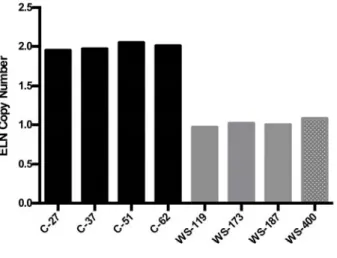
Recruitment of cases and controls was performed as part of institu- tional review board–approved studies (WS cases through Washington University School of Medicine [WUSM] and the Massachusetts General Hospital, adult controls through WUSM, and pediatric controls through Semmelweis University). Each participant (adult controls) or their parent/caregiver (pediatric controls and all WS cases) consented to participation in the study. Procedures followed were in accordance with institutional guidelines. Historical, medical, and surgical data were obtained by the use of a questionnaire. Medical records, including echocardiogram, were reviewed to validate questionnaire data when possible. Participants with WS underwent cardiovascular physical examination, and WS phenotype was verified on physical examination by experienced clinical geneticists. Molecular confirmation of WS was achieved either by review of clinical testing results (fluorescent in situ hybridization or microarray) or by research quantification of ELN CN (Figure S1 in the online-only Data Supplement).
Pulse Wave Velocity
Height and weight were formally measured, and individuals were then placed in a supine position and allowed to rest quietly. Blood pressure was manually assessed in both arms. PWV was determined from contour analysis of arterial waveforms recorded by applanation tonometry using a highly reproducible technique.7,13,14
NCF1 Gene and Pseudogene CN Determination
The WS critical region on chromosome 7 is flanked by 3 regions of low CN repeats,8,15 each containing the NCF1 gene or 1 of its 2 NCF1 pseu- dogenes (NCF1B and NCF1C; Figure S2). The absence of 2 base pairs (ΔGT) at the beginning of exon 2 in the pseudogenes distinguishes them from the NCF1 gene.16 To calculate relative CN for the NCF1 gene to its pseudogenes, both the genes and pseudogenes were amplified together using polymerase chain reaction primers surrounding the ΔGT region.
After polymerase chain reaction amplification, the product was gel purified and Sanger sequenced. At the ΔGT, the gene and pseudogene sequences diverge, and the relative peak heights of the next 27 bases are determined from the tracings as previously described.17 NCF1 gene and pseudogene CN were assigned using the ratio table in Table S1, and NCF1 gene number was used in subsequent statistical analysis.
Statistical Analyses
PWV in WS Versus Matched Controls
Because PWV normally increases with age,7,14,18 our initial analysis evaluated the pediatric (n=36) and adult (n=41) cohorts separately.
Cases and controls were matched, and PWV readings were com- pared using results from paired t tests. To determine whether PWV increases with age at the same rate in participants with WS versus controls, we also performed regression in the full-matched data set (n=77) with age as the covariate.
Covariate Analysis (PWV)
To identify additional features associated with higher PWV in the WS population, we performed regression on PWV data from participants with WS using diabetes mellitus, hypertension, antihypertensive
medication, or NCF1 CN status as the covariate. For the NCF1 CN analysis, because of the observed protective effect of antihyperten- sives on PWV in WS (Figure 2B) and others,19 individuals known to use these medications were excluded from this analysis.
Covariate Analysis (Hypertension)
The Fisher exact test was used to compare the prevalence of hyper- tension in WS individuals with NCF1 gene CN of 1 versus ≥2.
Hypertension was defined as any participant who had received a diag- nosis of hypertension (treated or untreated). For each analysis, only participants with the necessary data components were analyzed, and n for each study is reported. All statistical analyses were performed using Prism statistical software.
Results
Study PopulationsWS Cases
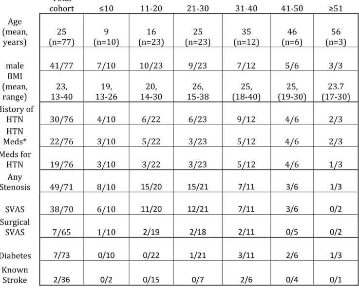
A total of 103 individuals with WS (aged 7–62 years) were con- sented for the WS-SAVE study. Quality PWV measurements were obtained in 77 of the 103 (74.8%) participants with WS.
Rates of vascular disease in those with successful versus unsuc- cessful PWV measurements were similar for most phenotypes assessed (see Table S2 for P values), with the unsuccessful cohort having a higher percentage of women, individuals with elevated body mass index (>35), and SVAS repair. No partici- pants were fully excluded from the study, but individuals were excluded from portions of the analysis in which relevant data were not available. For example, history of hypertension was assessed in 101 of 103 participants. Similarly, DNA was of suf- ficient quality to calculate NCF1 CN in 101 of 103 individuals.
Controls Adult Controls
Adult controls (n=41; aged 21.5–62.4 years) were ambula- tory subjects selected from participants of institutional review board–approved cardiovascular studies at WUSM. Subjects were excluded for any of the following: left ventricular sys- tolic dysfunction (ejection fraction <55%), reported or sus- pected coronary artery disease, pulmonary hypertension, stage C or worse chronic kidney disease, reported infection by the HIV, and reported sickle cell disease. Adults were matched to participants with WS, in aggregate, for body mass index, hypertension diagnosis, use of antihypertensives, and diabetes mellitus, in addition to age and sex (paired t tests revealed nonsignificant results for these variables; Table S3).
Pediatric Controls
Pediatric controls (n=36; aged 7.6–21.2 years) were enrolled as part of an international study establishing reference values of PWV in children and adolescents.18 Children with known history of hypertension or diabetes mellitus were excluded. Children were matched by age (mean difference±SD [WS-C], 0.04±0.19 years), sex, and height (mean difference [WS-C] −6±7 cm) to WS youth.
Clinical Characteristics of the WS-SAVE Cohort To determine whether the increased arterial stiffness noted in elastin-insufficient mice20–22 extrapolates to humans with elas- tin insufficiency, we attempted PWV in a cohort of 103 individ- uals with WS. Clinical features of the 77 subjects successfully completing the PWV portion of the study are included in Table S4. Sixty-nine percent of those with successful PWV mea- surements have a history of vascular stenosis (any location).
at Semmelweis University (Egyetem) on January 13, 2015 http://hyper.ahajournals.org/
Downloaded from
76 Hypertension January 2014
Fifty-four percent had SVAS, but only 10% had SVAS requir- ing surgical intervention. Forty percent of the cohort reported a history of hypertension, but only 25% had hypertension that was treated with antihypertensive medication. Ten percent reported a diagnosis of diabetes mellitus. Stroke (any type) was reported in 6% (3/48) of all individuals asked about this pheno- type; among the subset with a successful PWV measurement, 2 of 36 (6%) reported a history of stroke.
Children and Adults With WS Show Stiffer Vessels Than Matched Controls
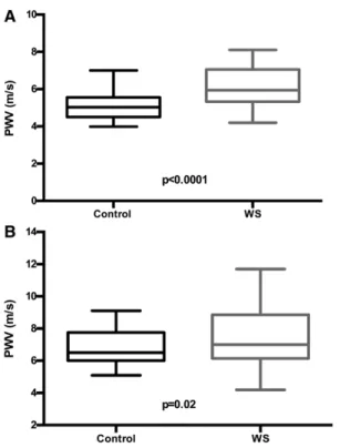
PWVs from the 77 individuals with WS were compared with pediatric and adult controls. When the pediatric subset was compared in paired t tests, those with WS were found, on aver- age, to have significantly higher PWV (mean±SD, 6.1±1.0 [WS] versus 5.1±0.8 m/s [control]; P<0.0001; Figure 1A).
Increased PWV was obvious in even the youngest participants with WS (see Figure S3A for raw WS data plotted against healthy population control means).14,18 In adults, paired t tests again showed higher PWV in WS adults (7.5±1.8 m/s, WS) versus controls (6.9±1.1 m/s, controls; P=0.02; Figure 1B).
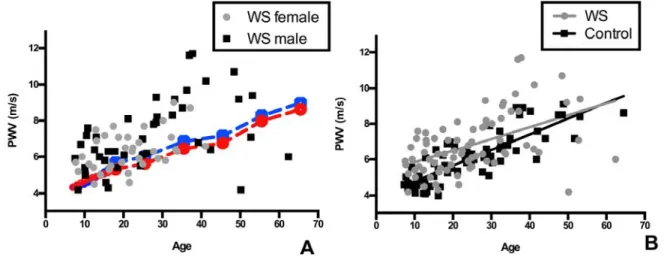
PWV normally increases with age.7,18 To evaluate for pos- sible differential effects of aging on PWV in WS, regression was performed using the full WS data set and matched controls (n=77 each). This analysis showed higher PWVs in participants
with WS across the whole age distribution (P<0.0001 for eleva- tion), with no statistical difference in the rate of PWV increase with age compared with controls (P=NS for slope; Figure S3B).
PWV: WS Clinical Phenotypes Associated With Vascular Stiffness
We sought to determine whether hypertension and diabetes mellitus, previously linked to increased vascular stiffness in non-Williams cohorts,7,23,24 were associated with higher PWV in WS. Regression analyses showed no significant relation- ship between PWV and hypertension (treated and untreated;
Figure 2A) or diabetes mellitus (Figure S4). We did, however, see relative protection from increased PWV in WS individuals taking antihypertensives (P=0.001; Figure 2B). Of note, 58%
(21/36) of those with PWV >1 m/s higher than the popula- tion mean for age and sex14,18 had no history of hypertension, whereas 36% (4/11) of those with reported untreated hyper- tension did not show elevated PWV.
Decreased NCF1 Gene CN Protects From Vascular Stiffness in WS
We investigated whether NCF1 CN was also associated with changes in vascular stiffness, as predicted by quantitative trait
Figure 1. Pediatric cases and adults with Williams syndrome (WS) have higher pulse wave velocity (PWV) than matched controls.
PWV was measured in WS pediatric cases (n=36; aged 7.6–21.2 years) and adults (n=41; aged 21.5–62.4 years). When compared in paired t tests, youth with WS were found, on average, to have significantly higher PWV (A). The WS adults were compared with those with a similar compilation of hypertension, diabetes mellitus, antihypertensive medication use, and body mass index, in addition to matched age and sex, and also had higher PWV (B). Controls are black, and participants with WS are gray. The box shows the SD with the mean PWV marked inside the box. The whiskers on the boxes depict the extreme reach of the PWVs measured.
Figure 2. Use of antihypertensives, but not hypertension, influences the severity of vascular stiffness in Williams syndrome (WS).
Regression analyses were performed by plotting WS participant data points by age and pulse wave velocity (PWV) and dividing the cohort by history of hypertension (A; n=76, 30 with hypertension [HTN] and 46 without HTN) or use of antihypertensive medications (B; n=76, 22 using medication [meds] and 54 not using medication).
Data from participants without the described feature are gray, and those with the phenotype are black. Regression lines are shown.
The slope of the regression line was significantly different only in B, in which the use of antihypertensive medication seems to be protective from increases in PWV (P=0.001).
at Semmelweis University (Egyetem) on January 13, 2015 http://hyper.ahajournals.org/
Downloaded from
Kozel et al Vascular Stiffness in Williams Syndrome 77
analysis in the Eln+/− mouse.25 Regression analysis showed higher PWVs in participants with WS who had ≥2 copies of NCF1 rather than 1 (P=0.05 for elevation; Figure 3). There was no statistical difference in the rate of PWV increase with age (P=NS for slope) in individuals with CN=1 or 2 in this analysis. Interestingly, removal of the single participant with the highest PWV (denoted by #) increases the differ- ence in PWV observed between CN=1 and CN=2 individuals (P=0.005 for elevation), with a trend toward better protection from increasing PWV with older age (P for slope improves from 0.6 to 0.1; Figure S5).
NCF1 Gene CN and Hypertension in WS
To determine whether the deletion of NCF1 affected hyperten- sion in our cohort, we compared the frequency of hyperten- sion between WS individuals with 1 versus ≥2 NCF1 genes.
In our total cohort of 99 individuals with WS in whom both NCF1 CN and hypertension status were known, a single copy of NCF1 was associated with protection from hypertension (P=0.03; Figure S6). This association persisted even when restricting the analysis to those aged <18 years (P=0.04; data not shown) in which the WS-related component of hyperten- sion may be more dominant. These results confirm the pre- viously reported protective association of reduced NCF1 CN and risk of hypertension in WS.8
Discussion
In a developing blood vessel, collagen is responsible for ten- sile strength, whereas elastin provides recoil capability. When the elastin gene is mutated or deleted, the resulting elastin insufficiency leads to multiple cardiovascular abnormalities.
Most consistently described in these populations are focal arterial stenoses and hypertension.5,6,26,27 However, the sever- ity of the vascular features in WS is variable. The frequency of WS-associated vascular symptoms in WS-SAVE is simi- lar to previous reports,6,28 with history of SVAS in 55% and hypertension in 40%. Importantly, only 25% of the cohort reported using antihypertensive medication for blood pres- sure control. Decision to treat was made by each individual’s
primary medical team and may reflect overall population undertreatment or possible hesitancy to treat hypertension in syndromic individuals in whom high blood pressure measure- ments are often felt to represent anxiety in the medical setting.
Interestingly, studies on this question have shown a paucity of true white coat hypertension in this population.28
Previous investigations of vascular stiffness in WS yielded contradictory results, ranging from increased arterial stiff- ness29,30 to normal or even paradoxically reduced values31,32 in small studies consisting of 3 to 29 participants. Consistent with these reports and with other cardiovascular features in WS, our larger analysis found considerable interpersonal PWV vari- ability. However, even with this variability, individuals with WS had significantly stiffer vessels than matched controls. As in the Eln+/− mouse, this stiffness is apparent from the earliest ages studied20,33 and progresses with increasing age at the same rate as controls, suggesting that elastin insufficiency causes early-onset and possibly congenital arterial stiffness. Eln+/−
mice generally become hypertensive in the neonatal period, a process suggested to be a physiological response to the altered vascular mechanics brought on by elastin insufficiency.21 In many of our cases, vascular stiffness was present without hypertension, and conversely, in some cases, hypertension was present without stiffness, suggesting that the 2 features may be related but independent effects of elastin insufficiency. For some of the studied individuals, increased PWV is the only known cardiovascular manifestation of the disorder. The devel- opment of hypertension in WS is likely multifactorial with influences due to vascular stiffness, complex effects of genetic background, and other features of WS such as renal artery ste- nosis and small-for-gestational-age birth.28
Our data show that vessels of pediatric participants with WS are more uniformly stiff than matched controls. Greater vari- ability is seen in older participants with WS, with evidence of protection in those on antihypertensive medications. The mean PWV observed in pediatric participants with WS is 6.1 m/s, approaching that seen in predialysis youth with end-stage renal disease (6.6 m/s)11 and youth with type 2 diabetes mellitus (6.4 m/s12, both with similar age demographics). Interestingly, in the end stage renal disease study, no improvement resulted from hemodialysis, suggesting a final common pathway to these conditions ending in destruction of elastic matrices and lasting alteration of the biomechanical properties of the vessel wall.
The fact that we did not see a primary hypertension-by- PWV effect in regression analysis may result from an admix- ture of untreated hypertensive individuals (who generally have higher PWVs) with treated hypertensive WS participants (in whom our data show lower PWVs). This mixing may ulti- mately act to normalize the PWV for the hypertensive group, causing it to appear similar to the nonhypertensive subset.
Further subgroup analysis, however, was underpowered. The use of antihypertensive medication showed greater protection in older individuals with WS, possibly suggesting cumulative effects of longer treatment duration; however, longitudinal evaluation is needed to confirm this.
Because vascular stiffness is strongly associated with negative cardiovascular consequences, such as myocardial infarction and stroke,1 further investigation into the predic- tive power of PWV for clinical outcome in individuals with Figure 3. NCF1 copy number (CN) modifies vascular disease
severity in Williams syndrome. Regression analysis is used to assess the effect of NCF1 CN on pulse wave velocity (PWV;
n=55). Those with NCF1 CN ≥2 (black) have higher PWV than those with CN=1 (gray; P=0.05 for elevation), but there is no difference in the increase in stiffness over time in this cohort (P=NS for slope). # denotes the single participant with CN=1 outlier.
at Semmelweis University (Egyetem) on January 13, 2015 http://hyper.ahajournals.org/
Downloaded from
78 Hypertension January 2014
WS is warranted. In our study, 48 participants were surveyed about their stroke history, and 3 of 48 (6%), aged 22, 32, and 32 years, reported a history of stroke. The prevalence of age- adjusted stroke for control individuals aged 18 to 45 years is 0.6% to 0.7%34 compared with 12% (3/25) when we consider only the subset of participants with WS in this demographics.
In 2 of 3 WS participants with stroke, we obtained successful PWV measurements, and both were elevated (+0.6 and +1.4 m/s relative to age- or sex-matched controls), although both participants were receiving antihypertensive medications at the time of PWV measurement. Currently, the best estimate of risk of sudden death in WS is 25- to 100-fold increased rela- tive to the general population.10 It is likely that this risk does not apply equally to all individuals with WS, and the use of PWV may enable individualized risk assessment. Full details about the nature of strokes in these participants are not avail- able, and further investigation into this area is warranted.
Because of its status as a variably deleted gene in WS, NCF1 CN was investigated and found to be a significant modifier of arterial stiffness in WS. Data from the current cohort also con- firm the reduced risk of hypertension associated with NCF1 CN=1 originally described by Del Campo et al.8 Although our study found generally lower PWVs in WS participants with only 1 copy of NCF1 relative to those with ≥2, initial analysis revealed no difference in the rate of age-associated increase in PWV between the 2 groups. However, when a single outlier is removed, a trend toward progressive protection with advancing age is identified in those with CN=1. Because NCF1 is a mem- ber of the NADPH oxidase family and is involved in the genera- tion of reactive oxygen species in tissues,35 its association with severity of vascular stiffness suggests a role for chronic oxidative stress in the pathology of vascular stiffness in WS. Additional studies with focused recruitment of older individuals with WS, although difficult, may help clarify this intriguing finding.
Limitations of our study include the possibility that because of the topic of the study, parents of individuals with signifi- cant vascular disease were more interested in participating.
However, our study includes many subjects with no previ- ously known vascular features of WS (who, in fact, have high PWV). Second, challenges inherent to WS limited the number of successful PWV studies we were able to obtain. Individuals with WS have an average intelligence quotient of 55 and are often anxious, making it difficult to obtain resting measure- ments in younger children with WS. High body mass index, in combination with these features, additionally complicated PWV acquisition in some adults. Reassuringly, phenotypes of participants on whom we failed to obtain PWV trended toward more severe vascular disease with higher rates of hypertension and stenosis seen in this group. These findings suggest that it is unlikely that the data loss excluded a milder cohort that might skew the PWV results. Finally, our studies are merely a single snapshot in time. To investigate the interaction between vascular stiffness and hypertension in elastin insufficiency, additional larger prospective studies are needed to document the natural evolution of these phenotypes.
Perspectives
This study successfully evaluated vascular stiffness in the largest cohort of individuals with WS reported to date and
demonstrates that individuals with WS, regardless of their blood pressure, are at risk for increased aortic stiffness, a well- established surrogate for adverse cardiovascular outcomes in many disease processes. This increase in vascular stiffness is caused by elastin insufficiency, as evidenced by Eln+/−mouse studies20–22 and, importantly, is modified by antihypertensive use and NCF1 CN. PWV differences are detectable at the ear- liest ages studied and continue into adulthood, making WS and elastin insufficiency useful models in which to study the long-term effects of chronic and early-onset vascular stiffness.
Overall, this study suggests that (1) monitoring of vascular stiff- ness is warranted in WS; (2) antihypertensive treatment might protect against vascular stiffness and consequent adverse car- diovascular events in individuals with WS, even without overt hypertension. Consequently, at a minimum, antihypertensives should be considered in all WS individuals with hypertension;
and finally, (3) novel medications, aimed at the NADPH oxi- dase pathway, should be investigated as potential modulators of vascular disease severity in this condition with use poten- tially determined by NCF1 CN. To generate formal treatment guidelines, larger prospective studies are needed to quantify the effects of antihypertensive drug choice and duration in treating arterial stiffness in WS. In addition, more work is needed to bet- ter understand the prognostic relevance of early-onset vascular stiffness in those with WS, with special attention given to organ systems known to be adversely affected in aging adults with vascular stiffness, such as the kidneys, heart, and brain. Finally, medication by NCF1 CN studies are needed to determine which patients may most benefit from pharmacological intervention.
Acknowledgments
We thank the Williams Syndrome Association and the families who participated in the WS-SAVE study. We gratefully acknowledge the assistance of Dr Chengsheng Zhang in clarifying the copy num- ber status of genes within the Williams region for select patients, Dr Mark Johnson for review of Williams echocardiograms, and Dr Richard Feinn for recommendations on statistical tests. We thank Dr Carmel McEniery for sharing updated normative data from the Anglo-Cardiff Collaborative Trial14 and Dr Mark Levin for his helpful review of the article.
Sources of Funding
Funding was provided to Dr Kozel by the Children’s Discovery Institute of Washington University and St. Louis Children’s Hospital.
In addition, Dr Kozel received funding for the study through her appointment as a scholar of the Child Health Research Center in Developmental Biology (National Institutes of Health [NIH] K12- HD01487) and the Genetic Basis of Inflammatory Airway Disease (NIH K12-HL089968). Original pediatric control studies were sup- ported by the Hungarian National Research Fund, OTKA 100909.
Disclosures
None.
References
1. Franklin SS. Beyond blood pressure: arterial stiffness as a new biomarker of cardiovascular disease. J Am Soc Hypertens. 2008;2:140–151.
2. Kozel BA, Mecham RP, Rosenbloom J. Elastin. In: Mecham RP, ed. The Extracellular Matrix, an Overview. Verlag, Belin, Heidelberg: Springer;
2011:267–302.
3. Curran ME, Atkinson DL, Ewart AK, Morris CA, Leppert MF, Keating MT. The elastin gene is disrupted by a translocation associated with supra- valvular aortic stenosis. Cell. 1993;73:159–168.
4. Metcalfe K, Rucka AK, Smoot L, et al. Elastin: mutational spectrum in supravalvular aortic stenosis. Eur J Hum Genet. 2000;8:955–963.
at Semmelweis University (Egyetem) on January 13, 2015 http://hyper.ahajournals.org/
Downloaded from
Kozel et al Vascular Stiffness in Williams Syndrome 79
5. Urbán Z, Michels VV, Thibodeau SN, Davis EC, Bonnefont JP, Munnich A, Eyskens B, Gewillig M, Devriendt K, Boyd CD. Isolated supravalvular aortic stenosis: functional haploinsufficiency of the elastin gene as a result of nonsense-mediated decay. Hum Genet. 2000;106:577–588.
6. Pober BR, Johnson M, Urban Z. Mechanisms and treatment of car- diovascular disease in Williams-Beuren syndrome. J Clin Invest.
2008;118:1606–1615.
7. Boutouyrie P, Vermeersch SJ. Determinants of pulse wave velocity in healthy people and in the presence of cardiovascular risk factors: “estab- lishing normal and reference values”. Eur Heart J. 2010;31:2338–2350.
8. Del Campo M, Antonell A, Magano LF, Muñoz FJ, Flores R, Bayés M, Pérez Jurado LA. Hemizygosity at the NCF1 gene in patients with Williams-Beuren syndrome decreases their risk of hypertension. Am J Hum Genet. 2006;78:533–542.
9. Bird LM, Billman GF, Lacro RV, Spicer RL, Jariwala LK, Hoyme HE, Zamora-Salinas R, Morris C, Viskochil D, Frikke MJ, Jones MC.
Sudden death in Williams syndrome: report of ten cases. J Pediatr.
1996;129:926–931.
10. Wessel A, Gravenhorst V, Buchhorn R, Gosch A, Partsch CJ, Pankau R.
Risk of sudden death in the Williams-Beuren syndrome. Am J Med. Genet A. 2004;127A:234–237.
11. Covic A, Mardare N, Gusbeth-Tatomir P, Brumaru O, Gavrilovici C, Munteanu M, Prisada O, Goldsmith DJ. Increased arterial stiffness in children on haemodialysis. Nephrol Dial Transplant. 2006;21:729–735.
12. Wadwa RP, Urbina EM, Anderson AM, Hamman RF, Dolan LM, Rodriguez BL, Daniels SR, Dabelea D; SEARCH Study Group. Measures of arterial stiffness in youth with type 1 and type 2 diabetes: the SEARCH for diabetes in youth study. Diabetes Care. 2010;33:881–886.
13. Kis E, Cseprekál O, Kerti A, Salvi P, Benetos A, Tisler A, Szabó A, Tulassay T, Reusz GS. Measurement of pulse wave velocity in children and young adults: a comparative study using three different devices.
Hypertens Res. 2011;34:1197–1202.
14. McEniery CM, Yasmin, Hall IR, Qasem A, Wilkinson IB, Cockcroft JR;
ACCT Investigators. Normal vascular aging: differential effects on wave reflection and aortic pulse wave velocity: the Anglo-Cardiff Collaborative Trial (ACCT). J Am Coll Cardiol. 2005;46:1753–1760.
15. Pober BR. Williams-Beuren syndrome. N Engl J Med. 2010;362:239–252.
16. Roesler J, Curnutte JT, Rae J, Barrett D, Patino P, Chanock SJ, Goerlach A. Recombination events between the p47-phox gene and its highly homologous pseudogenes are the main cause of autosomal recessive chronic granulomatous disease. Blood. 2000;95:2150–2156.
17. Heyworth PG, Noack D, Cross AR. Identification of a novel NCF-1 (p47- phox) pseudogene not containing the signature GT deletion: significance for A47 degrees chronic granulomatous disease carrier detection. Blood.
2002;100:1845–1851.
18. Reusz GS, Cseprekal O, Temmar M, Kis E, Cherif AB, Thaleb A, Fekete A, Szabó AJ, Benetos A, Salvi P. Reference values of pulse wave velocity in healthy children and teenagers. Hypertension. 2010;56:217–224.
19. Shahin Y, Khan JA, Chetter I. Angiotensin converting enzyme inhibitors effect on arterial stiffness and wave reflections: a meta-analysis and meta- regression of randomised controlled trials. Atherosclerosis. 2012;221:18–33.
20. Wagenseil JE, Ciliberto CH, Knutsen RH, Levy MA, Kovacs A, Mecham RP. Reduced vessel elasticity alters cardiovascular structure and function in newborn mice. Circ Res. 2009;104:1217–1224.
21. Faury G, Pezet M, Knutsen RH, Boyle WA, Heximer SP, McLean SE, Minkes RK, Blumer KJ, Kovacs A, Kelly DP, Li DY, Starcher B, Mecham RP. Developmental adaptation of the mouse cardiovascular system to elas- tin haploinsufficiency. J Clin Invest. 2003;112:1419–1428.
22. Pezet M, Jacob MP, Escoubet B, Gheduzzi D, Tillet E, Perret P, Huber P, Quaglino D, Vranckx R, Li DY, Starcher B, Boyle WA, Mecham RP, Faury G. Elastin haploinsufficiency induces alternative aging processes in the aorta. Rejuvenation Res. 2008;11:97–112.
23. Stehouwer CD, Henry RM, Ferreira I. Arterial stiffness in diabetes and the metabolic syndrome: a pathway to cardiovascular disease. Diabetologia.
2008;51:527–539.
24. Kaess BM, Rong J, Larson MG, Hamburg NM, Vita JA, Levy D, Benjamin EJ, Vasan RS, Mitchell GF. Aortic stiffness, blood pressure progression, and incident hypertension. JAMA. 2012;308:875–881.
25. Kozel BA, Knutsen RH, Ye L, Ciliberto CH, Broekelmann TJ, Mecham RP. Genetic modifiers of cardiovascular phenotype caused by elastin haploinsufficiency act by extrinsic noncomplementation. J Biol Chem.
2011;286:44926–44936.
26. Eronen M, Peippo M, Hiippala A, Raatikka M, Arvio M, Johansson R, Kähkönen M. Cardiovascular manifestations in 75 patients with Williams syndrome. J Med Genet. 2002;39:554–558.
27. Park S, Seo EJ, Yoo HW, Kim Y. Novel mutations in the human elastin gene (ELN) causing isolated supravalvular aortic stenosis. Int J Mol Med.
2006;18:329–332.
28. Broder K, Reinhardt E, Ahern J, Lifton R, Tamborlane W, Pober B.
Elevated ambulatory blood pressure in 20 subjects with Williams syn- drome. Am J Med. Genet. 1999;83:356–360.
29. Bassareo PP, Mercuro G. Increased arterial stiffness in children with Williams syndrome and normal blood pressure. Blood Press Monit.
2010;15:257–261.
30. Salaymeh KJ, Banerjee A. Evaluation of arterial stiffness in children with Williams syndrome: does it play a role in evolving hypertension? Am Heart J. 2001;142:549–555.
31. Aggoun Y, Sidi D, Levy BI, Lyonnet S, Kachaner J, Bonnet D. Mechanical properties of the common carotid artery in Williams syndrome. Heart.
2000;84:290–293.
32. Lacolley P, Boutouyrie P, Glukhova M, Daniel Lamaziere JM, Plouin PF, Bruneval P, Vuong P, Corvol P, Laurent S. Disruption of the elastin gene in adult Williams syndrome is accompanied by a paradoxical reduction in arterial stiffness. Clin Sci (Lond). 2002;103:21–29.
33. Le VP, Knutsen RH, Mecham RP, Wagenseil JE. Decreased aortic diam- eter and compliance precedes blood pressure increases in postnatal devel- opment of elastin-insufficient mice. Am J Physiol Heart Circ Physiol.
2011;301:H221–H229.
34. Fang J, Shaw K, George M. Prevalence of stroke—United States, 2006–
2010. MMWR Morb Mortal Wkly Rep. 2012;61:379–382.
35. Lassègue B, Griendling KK. NADPH oxidases: functions and pathologies in the vasculature. Arterioscler Thromb Vasc Biol. 2010;30:653–661.
What Is New?
• The Williams Syndrome (WS)-Skin And Vessel Elasticity study is the larg- est multi-institutional study of vascular stiffness in Williams syndrome to date, revealing increased pulse wave velocity in individuals with this rare condition.
What Is Relevant?
• Increased pulse wave velocity was identified in even the youngest Wil- liams participants, suggesting that vascular stiffness is of early, if not congenital, onset. Stiffness seems to be independent of hypertension in WS, making vascular stiffness a new primary WS phenotype. Protection against increasing stiffness is afforded by antihypertensive medication
and also by deletion of NCF1, an NADPH oxidase component and gene variably deleted in WS.
Summary
Elastin insufficiency causes increased arterial stiffness, a pheno- type associated with multiple negative cardiovascular outcomes complicating WS. Elevated pulse wave velocity is seen less often in those with WS deletions also removing the NCF1 gene and in those on antihypertensive therapy. Our findings suggest the need for en- hanced evaluation and anticipatory treatment of vascular disease in Williams syndrome.
Novelty and Significance
at Semmelweis University (Egyetem) on January 13, 2015 http://hyper.ahajournals.org/
Downloaded from
WILLIAMS SYNDROME PREDISPOSES TO VASCULAR STIFFNESS MODIFIED BY ANTI‐
HYPERTENSIVE USE AND COPY NUMBER CHANGES IN NCF1
Beth A. Kozel*, Joshua Danback*, Jessica Waxler
+, Russell H. Knutsen
%, Lisa de las Fuentes
~, Gyorgy S. Reusz
&, Eva Kis
&, Ami Bhatt
^, and Barbara R Pober
#+@.
*Dept. of Pediatrics, Washington University School of Medicine.
#Dept. of Medical Sciences, Netter School of Medicine, Quinnipiac University.
+Dept. of Pediatrics, Massachusetts General Hospital.
@Dept. of Pediatrics, Harvard Medical School.
%
Washington University School of Medicine, Dept. of Cell Biology and Physiology,
~
Cardiovascular Imaging and Clinical Research Core Laboratory, Cardiovascular Div., Dept. of Internal Medicine, Washington University School of Medicine.
&1
stDepartment of Pediatrics, Semmelweis University,
^Division of Cardiology, Brigham & Women's Hospital, Boston, MA.
+Dept. of Pediatrics, Harvard Medical School.
Supplemental Methods:
Study examination and historical data acquisition
Each individual was examined by a clinical geneticist experienced in the diagnosis of WS (BP and BK) and was felt to possess physical features consistent with WS. In addition to examination of facial features, individuals underwent cardiovascular examination including cardiac auscultation, assessment of peripheral pulses by palpation and auscultation for abdominal bruits. Parents/caregivers answered questions about cardiovascular history including presence of stenosis, hypertension, and diabetes. A subgroup was also asked about stroke history. Release of records was obtained for the most recent echocardiogram.
Historical information obtained by questionnaire was validated in medical records when possible.
DNA collection and Confirmation of WS Diagnosis:
Molecular confirmation of the WS diagnosis was sought through review of clinical testing results (ELN FISH or chromosomal microarray). If clinical testing was not available, elastin deletion was confirmed by qPCR (research).
Saliva was collected in Oragene saliva kits (DNA Genotek, Kanata, Ontario, Canada) at the time of in‐person assessment. DNA was prepared from the saliva sample according to manufacturer’s instructions. We performed ELN copy number analysis on a Viia7
TMReal‐
Time PCR System (Applied Biosystems, (ABI), Foster City, CA) using a TaqMan Copy
Number Assay. Probes (Elastin (ABI, #4400291, labeled FAM) and RNase P, (the reference gene, ABI, #4403326, labeled VIC)) were used in the reaction with 25ng of genomic DNA and TaqMan Genotyping Master Mix. PCR parameters were as specified in the
manufacturer’s Copy Caller protocol (ABI). DNAs from four unaffected individuals (CN= 2) and one WS individual with a known positive ELN FISH (CN=1) were used as controls. A no DNA control failed to amplify. Copy Caller software (Applied Biosystems) was used to analyze the data and assign copy number. All consented individuals have phenotypically and molecularly confirmed WS (See Figure S1 for representative data.)
NCF1 gene and pseudogene copy number determination
Saliva was collected and DNA purified as above. To calculate the NCF1 gene copy number, both the genes and pseudogenes (which differ by two bp, pseudogenes have ∆GT at the beginning of exon 2) were amplified together using PCR primers surrounding the divergent region. Amplification primers used were NCF1F 2LB2 (5’‐‐GTGCACACAGCAAAGCCTCT‐‐3’) and NCF1R 2RB2 (5’‐‐CTAAGGTCCTTCCCAAAGGGT—3’). Following PCR amplification, the product was gel purified and Sanger sequenced. At the ∆GT, the gene and pseudogene sequences diverge and the peak heights (gene and pseudogene) are determined from the tracings for the next 27 bases using Applied Biosystems Sequence Scanner 1.0 software.
Pseudogene:Gene (P:G) peak height ratios are calculated for each base and averaged. From the raw NCF1 P:G ratio, pseudogene and gene copy number were assigned using the ratio table in Table S1.
Using the relative peak height method, the most commonly observed pattern in
those without WS is 4 pseduogenes:2 genes, while individuals with typical WS deletions
(1.5 or 1.8 MB) display the absence of one or two pseudogenes and/or genes, respectively, perturbing the 4:2 ratio. Of note, 7 of 103 WS participants were found to have more than 2 copies of NCF1, but fewer copies of the pseudogenes, a finding reported previously in WS
1and control populations
2and thought to be due to inherited gene conversion between the gene and pseudogenes. Copy number frequencies are reported for individuals as either one (CN=1) or two or more (CN≥2) functional NCF1 copies.
Vascular Phenotype assessment (Hypertension and PWV)
Hypertension: History of hypertension was ascertained in 101/103 participants. Survey responses indicated inadequate information existed to determine hypertension status in 10/103 WS participants. Eight of these did not report use of anti‐hypertensive in their medication list and their blood pressure was normal in both arms on the day of in‐person evaluation (<95%ile for age, gender and height in pediatric patients or<140/90 in adults);
these individuals were treated as not having hypertension for the duration of the analysis.
The last two remaining individuals had insufficient information to determine their blood pressure status and were excluded from analyses that required this parameter.
PWV: Applanation tonometry was used to calculate PWV in each participant. To do so, waveforms were recorded from the carotid and femoral arteries while EKG was recorded.
PWV was calculated from these waveforms and measurements from standard landmarks ((sternal notch to arterial femoral site distance)‐(sternal notch to carotid site distance)).
PWV was determined using a SphygmoCor device (AtCor Medical, Sydney) for the WS‐SAVE participants and the WUSM adult control groups and with the Pulse Pen (DiaTecne, Milan) in the Semmelweis pediatric control group. Published studies show high agreement for PWV between the SphygmoCor and the Pulse Pen
3.
References:
1. Del Campo M, Antonell A, Magano LF, Munoz FJ, Flores R, Bayes M, Perez Jurado LA.
Hemizygosity at the ncf1 gene in patients with williams‐beuren syndrome decreases their risk of hypertension. Am J Hum Genet. 2006;78:533‐542.
2. Heyworth PG, Noack D, Cross AR. Identification of a novel ncf‐1 (p47‐phox) pseudogene not containing the signature gt deletion: Significance for a47 degrees chronic granulomatous disease carrier detection. Blood. 2002;100:1845‐1851.
3. Kis E, Cseprekal O, Kerti A, Salvi P, Benetos A, Tisler A, Szabo A, Tulassay T, Reusz GS. Measurement of pulse wave velocity in children and young adults: A
comparative study using three different devices. Hypertens Res. 2011;34:1197‐1202.
4. Pober BR. Williams‐beuren syndrome. N Engl J Med. 2010;362:239‐252.
5. Reusz GS, Cseprekal O, Temmar M, Kis E, Cherif AB, Thaleb A, Fekete A, Szabo AJ, Benetos A, Salvi P. Reference values of pulse wave velocity in healthy children and teenagers. Hypertension. 2010;56:217‐224.
6. McEniery CM, Yasmin, Hall IR, Qasem A, Wilkinson IB, Cockcroft JR. Normal vascular aging: Differential effects on wave reflection and aortic pulse wave velocity: The anglo‐cardiff collaborative trial (acct). J Am Coll Cardiol. 2005;46:1753‐1760.
Supplemental Tables and Figures:
Table S1: NCF1 genotyping. NCF1 gene number was determined after assignment to a genotype bin based on the ratio of NCF1 pseudogenes:gene peaks (P height/G height for each base, averaged over 27 bases). Nine non‐WS controls were used. All controls have the expected 6 total NCF alleles. Of note, 7 of 103 WS participants and two of nine controls were noted to have more than 2 copies of NCF1, but fewer copies of the pseudogenes. This finding was reported previously in WS
1and in control populations
2and is thought to be due to inherited gene conversion between the gene and pseudogenes. Establishing a breakpoint between the 3:1 and 4:1 P:G bins requires knowledge of the deletion size (1.5 MB (5 total alleles) vs 1.8 MB (4 total alleles)). This analysis was not performed because both groups have an NCF1 gene copy number of 1 and differentiation between the two would not affect further statistical analysis. DNAs from two individuals were not of sufficient quality to provide copy number information.
NCF1 Genotype Ratio range Number of Participants
Pseudogene:Gene WS Control
2:3 0.68‐0.78 5 0
3:3 or 2:2 1.04‐1.07 2 2
3:2 1.35‐1.95 47 0
4:2/2:1 1.96‐2.15 0 7
4:1/3:1 2.4‐4.19 47 0
Table S2: Characteristics of WS participants on which PWV was not achievable. PWV was attempted on 103 participants and 77 had successful attempts. The successful and unsuccessful groups of participants were compared for differences in age and body mass index (BMI) (t test) and percent history of hypertension, use of anti‐hypertension
medication, stenosis (any type), supravalvar aortic stenosis (SVAS), surgical SVAS and diabetes (Fisher’s exact test). The unsuccessful group had a higher proportion of females (p=0.02) and had a higher BMI (p=0.002) than those in which PWV was achieved. In addition, the unsuccessful cohort had a higher rate of surgery for stenosis (p=0.02). With each of the remaining features evaluated, the unsuccessful cohort trended toward having more severe vascular disease but none of these comparisons reached statistical
significance. These results suggest that the unsuccessful cohort is unlikely to have milder features that would unfairly bias results coming from the successful group. *denotes statistically significant results.
Table S2:
Failed PWV Participants
N=26
Successful PWV Paricipants
N=77
p value
Average (range)
Average
(range) Age (years) 24.1 (7‐54) 24.6 (7‐62) 0.85 BMI 28.2 (10‐48) 22.9 (13‐40) 0.002*
% (feature present/
total)
% (feature present/
total) Male (%) 27%
(7/26)
53%
(41/77) 0.02*
Hx of
Hypertension
52%
(13/25)
40%
(30/76) 0.35 Hypertension
Medication (any
indication)
40%
(10/25)
29%
(22/76) 0.32
Hypertension Medication for HTN Diagnosis
35%
(9/25)
25%
(19/76) 0.31
Any Stenosis (%)
83%
(20/24)
69%
(49/71) 0.20 SVAS (%) 67%
(16/24)
54%
(38/70) 0.34 Surgical SVAS
(%)
33%
(8/24)
11%
(7/65) 0.02*
Diabetes (%) 21%
(5/24)
10%
(7/73) 0.16
Table S3. Phenotypes of matched adult WS and controls. Mean age and BMI, as well as percent of participants with hypertension (HTN), diabetes, or anti‐hypertensive use are shown for both the WS cohort and the matched adult control group. P values for the t test (age and BMI) or Fisher’s exact test (HTN, HTN meds, and diabetes) are shown.
Age BMI Hx of HTN (%) Use of HTN Meds (%) Diabetes (%)
Control 33.9 27.4 44 29 13
WS 33.7 25.7 49 34 18
p value 0.91 0.24 0.82 0.81 0.76
Table S4: Demographic data for the 77 WSSAVE participants with quality PWV tracings. Data shown include the frequency of various phenotypes in the total cohort or in individuals of decade binned groups (≤10 years, 11‐20 years, etc). BMI=Body Mass Index, HTN=Hypertension, SVAS=Supravalvar Aortic Stenosis. * Anti‐hypertensive medications used include (beta blockers (8), ACE inhibitors/Angiotensin II receptor blockers (14), calcium channel blockers (3), diuretics (2), and alpha adrenergic blockers(1)). Five individuals used more than one anti‐hypertensive (in each case, an ACE inhibitor or ARB were used in addition to a second medication (4 individuals) or two additional medications (1 individual). Anti‐hypertensive medications were prescribed for blood pressure control in all but three individuals (1 used only an alpha adrenergic receptor blocker to treat attention deficit disorder, one received a beta blocker for anxiolysis, and one received an ACE inhibitor for afterload reduction for mitral valve prolapse with regurgitation).
Table S4
Total
cohort ≤10 11‐20 21‐30 31‐40 41‐50 ≥51
Age (mean,
years) 25
(n=77) 9
(n=10) 16
(n=23) 25
(n=23) 35
(n=12) 46
(n=6) 56 (n=3)
male 41/77 7/10 10/23 9/23 7/12 5/6 3/3
BMI (mean,
range) 23,
13‐40 19,
13‐26 20,
14‐30 26,
15‐38 25,
(18‐40) 25,
(19‐30) 23.7 (17‐30) History of
HTN 30/76 4/10 6/22 6/23 9/12 4/6 2/3
HTN
Meds* 22/76 3/10 5/22 3/23 5/12 4/6 2/3
Meds for
HTN 19/76 3/10 3/22 3/23 5/12 4/6 1/3
Any
Stenosis 49/71 8/10 15/20 15/21 7/11 3/6 1/3
SVAS 38/70 6/10 11/20 12/21 7/11 3/6 0/2
Surgical
SVAS 7/65 1/10 2/19 2/18 2/11 0/5 0/2
Diabetes 7/73 0/10 0/22 1/21 3/11 2/6 1/3
Known
Stroke 2/36 0/2 0/15 0/7 2/6 0/4 0/1
Figure S1: Confirmation of WS diagnosis by ELN copy number. We performed ELN copy number analysis to confirm the WS diagnosis. DNA from four unaffected individuals (black bars) and one WS individual with a known positive ELN FISH (WS‐400, CN=1, gray bar with white dots) were used as controls. Representative WS participants are shown as gray bars. A no DNA control failed to amplify. All consented WS individuals were shown to have molecularly confirmed WS.
Figure S2: Depiction of the Williams deletion region. The WS critical region (blue) on chromosome 7 is flanked by three low copy number repeat regions, most often consisting of NCF1 (purple) and one of its pseudogenes (NCF1B, green; NCF1C, red)
1, 4. The majority of non‐WS individuals in the general population have a total of 6 NCF1 pseudogenes (P) + genes (G) in a P:G ratio of 4:2. The ratio is altered in WS, in both the 1.5 MB and less frequent 1.8 MB deletion. In individuals with the 1.5 MB deletion, either the NCF1 gene (purple) or the NCF1B pseudogene (green) is deleted; this leads to a total of 5 P+G, with ratios of P:G 4:1 or 3:2. In individuals with the 1.8 MB deletion, the recombination occurs between the two pseudogene regions (green and red), leading to the deletion of the NCF1 gene (purple) AND deletion of either the NCF1B (green) or the NCF1C (red) pseudogene;
deletion of green and purple shown as example). This result in 4 total P + G with a P:G ratio of 3:1. Some individuals (WS and control) have more than 2 copies of NCF1, but fewer copies of the pseudogenes. This finding is thought to be due to inherited gene conversion between the gene and pseudogenes (not shown in the figure).
Figure S
2
Figure S3: Individuals with WS have stiffer blood vessels than normal population controls. In panel A, raw PWV from the 77 participants with quality PWV studies are plotted against the age of the patient. Male WS participants are denoted as black squares.
Female WS participants are grey circles. Normative data for pediatric patients
5are noted by solid lines while adult normative data (derived from
6) are shown by hatched lines. In both control groups, males are indicated by a blue line and females, by a red line. Most, but not all, individuals with WS have higher PWV than age/gender matched peers. To evaluate for possible differential effects of aging on PWV in WS relative to controls, regression was performed using the full data PWV set (n=77 adults and children, Panel B). This analysis showed higher PWVs in WS participants across the whole age distribution (p < 0.0001 for elevation) with no statistical difference in the rate of PWV increase with age compared to controls (p=NS).
Figure S4: Lack of influence of diabetes on the severity of vascular stiffness in WS.
Regression analysis was performed by plotting WS participant data points by age and PWV in those with, versus those without, diabetes (N=74, 7 with diabetes (black) and 67 without (gray)). Regression lines are shown. Neither the elevation nor the slope of the regression lines were significantly different (p=NS for both) in those with or without diabetes.
Figure S5. NCF1 copy number modifies vascular disease severity in WS. Regression analysis is used to assess the effect of NCF1 copy number on PWV (n=55). Removal of the single participant with the highest PWV (denoted # in Figure 3) increases the difference in PWV observed between CN=1 and CN=2 individuals, with those with NCF1 CN≥2 (black) having higher PWV than those with CN=1 (gray) (p< 0.005 for elevation). In addition, there is a trend toward better protection from increasing PWV with older age (p value for slope improves from 0.6 (in Figure 3) to 0.1 (below).
Figure S6. NCF1 copy number modifies hypertension risk in WS. Fisher’s exact test was performed to evaluate the prevalence of hypertension in WS participants with 1 vs ≥2 copies of NCF1. (N= 99 WS individuals for whom hypertension status and NCF1 copy number were known). Gray bars show those with hypertension and black bars show those without. Prevalence of hypertension is reduced in those with NCF1 CN=1 (p =0.03).
Gyorgy S. Reusz, Eva Kis, Ami B. Bhatt and Barbara R. Pober
Beth A. Kozel, Joshua R. Danback, Jessica L. Waxler, Russell H. Knutsen, Lisa de las Fuentes, NCF1
and Copy Number Changes in
Williams Syndrome Predisposes to Vascular Stiffness Modified by Antihypertensive Use
Print ISSN: 0194-911X. Online ISSN: 1524-4563
Copyright © 2013 American Heart Association, Inc. All rights reserved.
is published by the American Heart Association, 7272 Greenville Avenue, Dallas, TX 75231 Hypertension
doi: 10.1161/HYPERTENSIONAHA.113.02087
2014;63:74-79; originally published online October 14, 2013;
Hypertension.
http://hyper.ahajournals.org/content/63/1/74
World Wide Web at:
The online version of this article, along with updated information and services, is located on the
http://hyper.ahajournals.org/content/suppl/2013/10/14/HYPERTENSIONAHA.113.02087.DC1.html
Data Supplement (unedited) at:
http://hyper.ahajournals.org//subscriptions/
is online at:
Hypertension Information about subscribing to
Subscriptions:
http://www.lww.com/reprints
Information about reprints can be found online at:
Reprints:
document.
Permissions and Rights Question and Answer this process is available in the
click Request Permissions in the middle column of the Web page under Services. Further information about Office. Once the online version of the published article for which permission is being requested is located,
can be obtained via RightsLink, a service of the Copyright Clearance Center, not the Editorial Hypertension
in
Requests for permissions to reproduce figures, tables, or portions of articles originally published Permissions:
at Semmelweis University (Egyetem) on January 13, 2015 http://hyper.ahajournals.org/
Downloaded from