https://doi.org/10.1177/1756286419850433 https://doi.org/10.1177/1756286419850433
Therapeutic Advances in Neurological Disorders
journals.sagepub.com/home/tan 1 Ther Adv Neurol Disord 2019, Vol. 12: 1–7 DOI: 10.1177/
1756286419850433
© The Author(s), 2019.
Article reuse guidelines:
sagepub.com/journals- permissions
Creative Commons Non Commercial CC BY-NC: This article is distributed under the terms of the Creative Commons Attribution-NonCommercial 4.0 License
Introduction
The limb–girdle muscular dystrophies (LGMDs) are a group of genetically heterogeneous disorders characterized by predominant proximal muscle weakness with histopathological signs of progres- sive degeneration in muscle.1 The estimated inci- dence for all forms of LGMDs is 1:100,000, the clinical spectrum of LGMDs can vary from severe childhood forms with early loss of independent ambulation to milder adult-onset slowly progres- sive subtypes.2 They are divided into two major groups according to the hereditary trait: autoso- mal dominant (AD) forms that are indicated as LGMD type D, autosomal recessive (AR) forms that are classified as LGMD type R.1
One subtype of dominant LGMD is due to a mutation of TNPO3 gene (LGMD D2). This rare disorder was first identified in a large Italo-Spanish family3 in which patients presented proximal limb–
girdle muscles weakness with variable onset rang- ing from 1 to 58 years.4 In this family, the disease locus has been mapped to chromosome 7q32.1- 32.2.4 In 2013, using the next-generation sequenc- ing (NGS) technique we identified a heterozygous
frame-shift variant in the Transportin-3 (TNPO3) gene, that encodes Transportin-3.5,6 Transportin-3 is a nuclear protein member of the importin beta superfamily that imports serine–arginine-rich pro- teins into the nucleus, which is important for mRNA splicing. The clinical phenotype of LGMD D2 is characterized by severe weakness occurring first in the pelvic girdle muscle, then the shoulder girdle. In severe clinical cases, distal muscle weak- ness, winging scapulae, scoliosis and occasional facial weakness are present. No cognitive impair- ment or cardiac involvement has been observed (Table 1). Only in the juvenile-onset severe pheno- type, respiratory involvement has been reported.7 We present a family with a congenital myopathy- LGMD phenotype in a child and his mother of Hungarian origin. A written informed consent was obtained from the mother (Patient 1) to pub- lish the medical data and images of both her and her son (Patient 2).
Molecular analysis, magnetic resonance imaging (MRI), histopathological study, and a assessment of quality of life were performed in this new
A new family with transportinopathy:
increased clinical heterogeneity
Corrado Angelini , Roberta Marozzo, Elena Pinzan, Valentina Pegoraro, Maria Judit Molnar, Annalaura Torella and Vincenzo Nigro
Abstract: We describe a family with a novel TNPO3 mutation of limb–girdle muscular dystrophy D2 (or LGMD 1F), a rare muscle disorder with autosomal dominant inheritance, first identified in an Italo-Spanish family where the causative defect has been found to be due to TNPO3 gene mutation, encoding transportin-3 protein (TNPO3). We present the clinical, histopathological and muscle magnetic resonance imaging (MRI) features in two patients, mother and son Hungarian origin, affected by LGMD D2 and correlate their clinical, MRI and histopathological data found in this condition. The affected son presented early pelvic girdle muscle weakness and thin muscles similar to a congenital myopathy; the mother was less compromised and had an LGMD phenotype. Muscle MRI showed a very pronounced lower limb muscle atrophy in both patients. The most relevant change obtained in the child muscle biopsy was a generalized type 1 fibre atrophy. The two patients presented the same mutation, but a different phenotype has been observed in mother and son.
Keywords: LGMD, TNPO3, transportinopathy
Received: 10 September 2018; revised manuscript accepted: 21 April 2019
Correspondence to:
Corrado Angelini IRCCS San Camillo Hospital, Via Alberoni 70, Venice, 30126, Italy corrado.angelini@
ospedalesancamillo.net Roberta Marozzo Elena Pinzan Valentina Pegoraro IRCCS San Camillo Hospital, Venice, Italy Maria Judit Molnar Institute of Genomic Medicine and Rare Disorders, Budapest, Hungary
Annalaura Torella Vincenzo Nigro TIGEM (Telethon Institute of Genetics and Medicine), University at Campania, Naples, Italy
Case Report
family of LGMD D2 and compared with previous findings in the first reported family.8,9
Case report Patient 1
Patient 1, a 43-year-old woman of Hungarian ori- gin, had two sons, the first, Patient 2, affected by LGMD D2, whereas the second, 10 years old, is healthy. Her parents were reported to be unaf- fected, but refused further DNA investigation.
The woman presented the first symptoms during her childhood: she started walking at 15 months and was always weak compared with peers.
Electromyography (EMG) was performed at age 6 and myopathic alterations were found. During her second pregnancy at 30 years, she underwent a C-section, presented difficulty breathing and felt ‘paralyzed’. At age 41, she had elevated cre-
muscle strength in the forearm and finger exten- sors and in the back muscles. She had also a prox- imal weakness primarily affecting proximal lower limb muscles.
On the last exam at age 43, she was able to walk, but had difficulty running. She complained of fre- quent falls, had difficulty keeping legs elevated or raising from the floor (Gower’s manoeuvre) and had to climb stairs using the handrail. She has reduced forced vital capacity (FVC) on spirome- try (57%) and presented difficulty in her speech and swallowing. She had no cardiac involvement or cognitive impairment.
On last neuromuscular evaluation (IRCCS San Camillo Hospital, March 2018), specific clinical myopathic signs were observed such as pigeon toe–feet and kyphoscoliosis.
Muscle strength graded by Medical Research Table 1. The genetic and clinical data in limb–girdle muscular dystrophy type D2 (LGMD D2).
LGMD D2
Disease symbol LGMD D2 or 1F
Gene symbol TPNO3
Protein Transportin-3
Chromosome locus 7q32.1-q32.2
Inheritance Autosomal dominant
Principal clinical features
Early onset Late onset
Age 5–15 years 20–30 years
Phenotype Similar to congenital myopathy LGMD phenotype
Scapular winging 30% -
Dysphagia 30% -
Respiratory involvement 30% No
Finger contracture Yes No
Wheelchair bound age 30 years Over 30 years
Creatine kinase level Mildly elevated Slightly elevated
Muscle biopsy Central nuclei, muscle fibres atrophy Fibre degeneration atrophy, increased acid phosphatase
Quality of life Mildly impaired Slightly impaired
weakness of triceps and deltoid (2/5), neck flexors 3/5, neck extensors 4/5, biceps 4/5, wrist exten- sors 4/5 and pectoralis 3/5. In lower limbs, quadri- ceps and ileo-psoas had 3/5 score, semitendinous and semimembranous 4/5 and tibial anterior 3/5.
Patient 2
Patient 2, a 12-year-old child, presented weak- ness since the first year of life. He walked at 18 months but could not raise easily from the floor.
At 10 years, he walked with a slow gait up to about 3 km, but then he felt fatigued and fell fre- quently. At 12 years, he walked only 50–100 m because he had become weaker in recent years.
He could not rise from a laying position without grasping his knees and presented a weak grip. He had difficulty swallowing with moderate dyspha- gia. CK was normal. He did not present any res- piratory or cardiac problem. He goes to school by car and has no learning difficulties.
On the last examination at IRCCS San Camillo Hospital in March 2018, he had a waddling gait, Gowers sign and climbed stairs using the hand- rail. He presented a myopathic face and moderate scapular winging. He was not able to stand from the floor and was unable to lift legs from the bed.
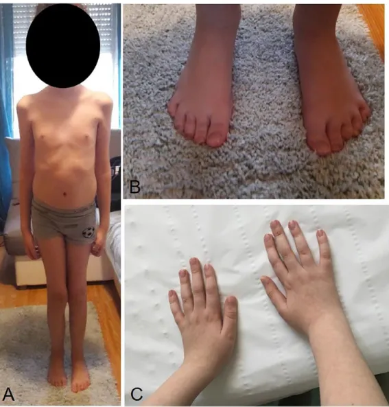
He appeared thin, frail and weak (Figure 1A–C).
Deep tendon reflexes were absent. Muscle strength assessed by MRC scale showed a pro- nounced weakness of triceps (2/5), deltoid (3/5), wrist extensor (3/5) and neck flexor muscles (3/5).
In lower limbs, he had low MRC score in quadri- ceps, iliopsoas, semitendinous and semimembra- nous muscles (3/5).
Quality of life
Quality of life (QoL) was assessed in patient 1 using an Individualized Neuromuscular Quality of Life (INQoL) test, validated in Italian11 and UK popu- lations12 in a variety of muscular diseases. Patient 1 self-evaluation on the impact of muscle disease on her physical, psychological and social functioning in INQoL showed a mild impairment.
Laboratory exams
Muscle biopsy histopathology was performed in both patients according to standard procedures.
A biopsy was performed in the child at 5 years of age and histochemistry and electron microscopy
were diagnosed as compatible with congenital myopathy. The mother underwent three muscle biopsies at different ages (24, 36 and 38 years), that showed aspecific myopathic signs such as type 1 fibre predominance and changes in nicoti- namide adenine dinucleotide tetrazolium reduc- tase (NADH-TR) stain similar to ‘cores’.
In March 2018 mother and son came for diagnos- tic purposes to the IRCCS San Camillo Hospital because transportinopathy was suspected and where clinical, biomedical, molecular, muscle MRI and a skin biopsy were performed. Muscle MRI was evaluated according to Mercuri scale. A skin biopsy was performed in patient 1 and fibro- blast collected and cultured; the pellet of fibro- blasts was sent to the Telethon Institute of Genetics and Medicine (TIGEM) for genetic analysis.
Histopathological studies
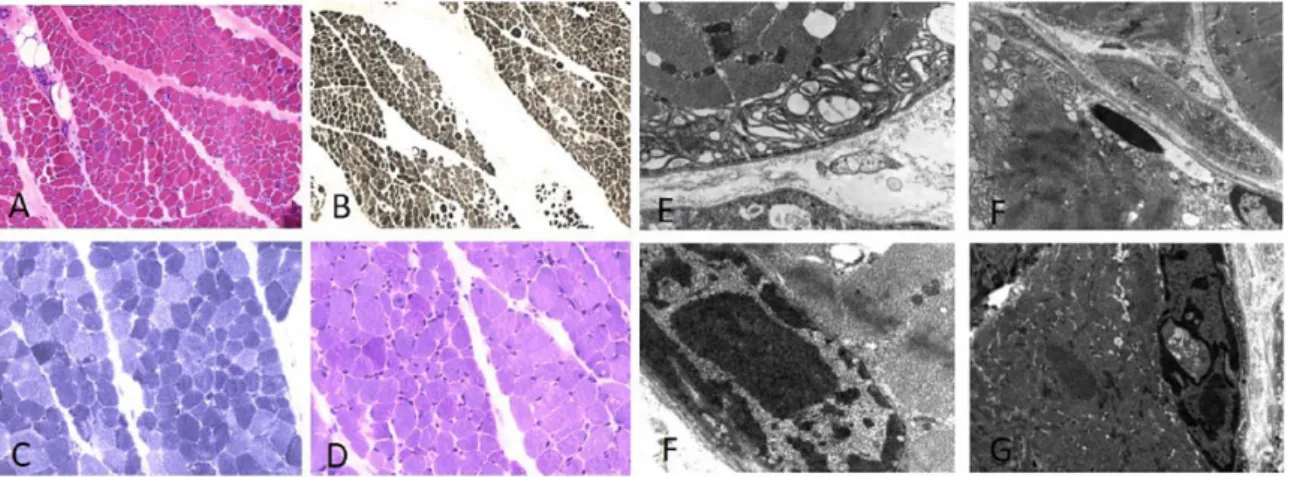
In the child muscle biopsy, morphologic findings by haematoxylin–eosin (HE) staining showed prominent fibre atrophy with acid ATPase stain (pH 4) with a 92% type 1 fibre predominance (Figure 2A and B) was observed. NADH-TR showed the abnormal presence of mitochondrial rims and periodic acid–Schiff (PAS) reaction detected focal glycogen accumulation in some fibres (Figure 2C and D). Ultrastructural analysis showed abnormal mitochondrial accumulation, myelinoid bodies and occasional cytoplasmic bodies; several abnormal nuclei had a fenestrated appearance (Figure 2E–G).
Muscle MRI
Muscle MRI was performed in both patients using a 1.5-T MRI scanner in T1-weighted sequences; muscle pattern was ranked according to Mercuri scale.13
Muscle MRI of patient 1 revealed marked atro- phy, fatty tissue replacement in the pelvic girdle and anterior thigh muscles, especially in vastus lateralis, vastus medialis and intermedius, whereas in posterior thigh semitendinous and semimem- branous muscles were relatively spared (Figure 3A). Posterior leg muscles were characterized by marked atrophy, in particular, soleus and gastroc- nemius muscles were completely replaced by fatty infiltration (Figure 3B).
In patient 2, muscle MRI showed a pronounced hypotrophy of anterior thigh muscles, whereas leg muscles were preserved (Figure 3C and D).
Mutation analysis
Genomic DNA was extracted from cultured fibroblasts of patient 1, analysed by whole exome sequencing (WES) and confirmed by Sanger sequencing. A heterozygous frameshift single base pair deletion c.2767delC (p.Arg923Aspfs*17) in exon 23 of TNPO3 gene was identified in both the mother and son genomic DNA. The number associated to the reference sequence is NM_012470.3: c.2767del;
NP_036602.1: p.(Arg923Aspfs*17). This vari- ant has not previously been reported and in silico predicted to be damaging (PoliPhen score 0.992; sensitivity 0.49; specificity 0.95).
The mutation identified is very similar and close to the first described mutation5 because it always involves the C-terminal therefore the final effect might be the same. The deletion of the base is in the codon immediately preceding to the other family variant (base position chr7:
128,597,310; NM_012470.3: c.2771delA, NP_036602.1p. (Ter924Cys)) and could be resulting in a longer protein (15 aa longer) than the wild type.
Figure 1. The child, 12 years old, was thin and weak and presented flat feet and short fingers with weak extensor indicis and carpi ulnaris muscles.
Discussion
Clinical, molecular, histopathological and radio- logical data, along with electron microscopy find- ings have been described so far only in a TNPO3 family.9,6
This dominant LGMD is characterized by clini- cal heterogeneity. The disorder has onset in child- hood or adolescence, but also frequently in adulthood; in adults, it has a benign course com- patible with a normal life. In the Italo-Spanish Figure 2. Muscle biopsy of patient 2 shows diffuse fibre atrophy and some central nuclei with haematoxylin–
eosin stain (A). Acid ATPase (pH 4) shows 92% type 1 fibre predominance (B). Nicotinamide adenine dinucleotide tetrazolium reductase (NADH-TR) showed abnormal nuclei and mitochondrial accumulation (C), periodic acid–Schiff (PAS) stain shows deposit of glycogen localized in some fibres (D). Ultrastructural analysis shows atrophic fibres with myelinoid body in subsarcolemmal position (E), cytoplasmatic body with mitochondrial alterations and vacuoles in nuclei (F). Abnormal dense nuclei with central vacuoles can be seen in (F, G).
Figure 3. Evaluation of muscle magnetic resonance imaging (MRI) in lower limbs in patients 1 and 2 according to the Mercuri score. (A) Slight atrophy was found in both legs and thighs of the female patient (mother of patient 2). Vastus lateralis, medialis and intermedius presented a 4 Mercuri score; semitendinous and semimembranous muscles were spared with Mercuri 1 score. (B) Advanced atrophy was present, in posterior compartment where soleus and gastrocnemius muscles were completely substituted with fatty infiltration.
(C) Muscles of patient 1 were characterized by hypotrophy in the anterior thigh compartment. (D) Leg muscle mass was less compromised.
family, the main characteristic features and pos- sible clues for diagnosis observed were abnor- mally long finger (arachnodactyly), pes cavus, Achilles retraction, and kyphoscoliosis (Table 1).
On the other hand, the main clinical signs observed in Hungarian patients were the diffi- culty in lifting arms over the head, due to scapular winging, and inability to lift legs from the bed.
Respiratory insufficiency might occur around 20–
30 years in early onset cases:3 the mother had severe impairment of the respiratory function, while the son presented normal spirometry.
Gamez et al.3 in the biopsy of the Italo-Spanish LGMD patients observed diffuse atrophic fibres, basophilic cytoplasmic regions with spots of cyto- plasmic lysosomal acid phosphatase reaction. By an ultrastructural study, Cenacchi et al.8 showed fibre atrophy, abnormal mitochondria accumulations with rare paracrystalline-like inclusions and autophagosomal vacuoles containing cytoplasmic debris and myeloid bodies. Similar histopathologi- cal and ultrastructural changes were observed in the biopsy of the son such as fibre atrophy, mitochon- drial accumulation, myelinoid bodies, autophago- somal vacuoles and central fenestrated nuclei.
In the Italo-Spanish family, the mutation was identified as a single nucleotide deletion (c.2771delA, p.X924C, exon 22) in the TNPO3 gene not present in databases, which determines a nonstop mutation and consequently a 15-ami- noacid extension of the C-terminus of the pro- tein.5,6 Molecular analysis showed that the causative mutation of LGMD D2 in the Hungarian patients is due to a single nucleotide deletion in the termination codon of transportin 3 in exon 23 and might result in TNPO3 reduction.
In this infantile-onset case, there was a congenital myopathy phenotype. The neurological examina- tion highlighted this peculiar aspect: the child presented a severe phenotype with hypotonia and had thin and weak muscles.
Data derived from MRI study in our cases of this new family confirm a pattern of early onset and a late onset with proximal muscle involvement.
The MRI changes in the mother at age 43 were more marked than in her son and were compati- ble with an LGMD phenotype.9
This AD disorder has been previously demon- strated to have incomplete penetrance in the orig- inal family.14 The lack of clinical signs in the
parent(s) of patient 1 could be due to an incom- plete penetrance rather than being a spontaneous mutation.
Transportin 3 orchestrates nuclear import of splicing factors and it is implicated in HIV resist- ance.15 The role of TNPO3 protein in muscle is unknown, but its involvement in nuclear import of splicing factors and protein involved in the RNA metabolism leads to a hypothesis about how TNPO3 mutation can cause LGMD D2.
The mutations abolish the stop codon, therefore the synthesis of a limited amount of TNPO3 pro- tein with increased molecular weight is expected to occur in association with the mutation found in this family: a perturbed nuclear–myofibrillar interaction could result in myofibrillar disarray.
Regarding INQoL in our LGMD case, the results are consistent with those published by Peric et al.16 in 46 patients with LGMD. In both cases, the scores were similar, showing worse self- reported evaluation for weakness and fatigue and better results for social relationships and emo- tions. There was no effect on independent life.
The clinical features in these two patients of Hungarian family add new data to the spectrum of the clinical phenotypes so far described in LGMD D2. In particular, the clinical features appear severe in this family because the onset of symptoms was in the first year of life, including distal, axial, facial and bulbar weakness. However, onset in the first years of childhood,3,4,6,9 delayed motor skills,6 distal leg weakness,3,6,9 axial weak- ness,9 facial weakness3,9 and bulbar signs9 are also a feature of previously reported cases, and epige- netic and genetic modifiers are probably impor- tant in determining the onset, the characteristics and the progression of this disorder.
Funding
This research received grants from Conquistando Escalones Association, Telethon GGP14066, AFM, Eurobiobank and BBMRNR.
Conflict of interest statement
The authors declare that there are no conflicts of interest.
ORCID iD
Corrado Angelini https://orcid.org/0000-0002 -9554-8794
References
1. Straub V, Murphy A and Udd B; LGMD Workshop Study Group. 229th ENMC international
workshop: limb girdle muscular dystrophies – nomenclature and reformed classification, Naarden, the Netherlands, 17-19 March 2017.
Neuromuscul Disord 2018; 28: 702–710.
2. Magri F, Nigro V, Angelini C, et al. The Italian limb girdle muscular dystrophy registry: relative frequency, clinical features, and differential diagnosis. Muscle Nerve 2017; 55: 55–68.
3. Gamez J, Navarro C, Andreu AL, et al.
Autosomal dominant limb-girdle muscular dystrophy: a large kindred with evidence for anticipation. Neurology 2001; 56: 450–454.
4. Palenzuela L, Andreu AL, Gàmez J, et al. A novel autosomal dominant limb-girdle muscular dystrophy (LGMD 1F) maps to 7q32.1–32.2.
Neurology 2003; 61: 404–406.
5. Torella A, Fanin M, Mutarelli M, et al. Next- generation sequencing identifies transportin 3 as the causative gene for LGMD1F. PLoS One 2013; 8: e63536.
6. Melià MJ, Kubota A, Ortolano S, et al. Limb- girdle muscular dystrophy 1F is caused by a microdeletion in the transportin 3 gene. Brain 2013; 136: 1508–1517.
7. Angelini C. Genetic Neuromuscular Disorders: A Case-based Approach. Berlin: Springer, 2018.
8. Cenacchi G, Peterle E, Fanin M, et al.
Ultrastructural changes in LGMD1F.
Neuropathology 2013; 33: 276–280.
9. Peterle E, Fanin M, Semplicini C, et al.
Clinical phenotype, muscle MRI and muscle pathology of LGMD1F. J Neurol 2013; 260:
2033–2041.
10. Vlak M, Van Der Kooi E and Angelini C.
Correlation of clinical function and muscle CT scan images in limb-girdle muscular dystrophy.
Neurol Sci 2000; 21(5 Suppl.): S975–S977.
11. Sansone VA, Panzeri M, Montanari M, et al.
Italian validation of INQoL, a quality of life questionnaire for adults with muscle diseases. Eur J Neurol 2010; 17: 1178–1187.
12. Vincent K, Vincent K, Carr J, et al. Construction and validation of a quality of life questionnaire for neuromuscular disease (INQoL). Neurology 2007;
68: 1057.
13. Mercuri E, Pichiecchio A, Counsell S, et al. A short protocol for muscle MRI in children with muscular dystrophies. Eur J Paediatr Neurol 2002;
6: 305–307.
14. Fanin M, Peterle E, Fritegotto C, et al.
Incomplete penetrance in limb-girdle muscular dystrophy type 1F. Muscle Nerve 2015; 52:
305–306.
15. Maertens GN, Cook NJ, Wang W, et al.
Structural basis for nuclear import of splicing factors by human Transportin 3. Proc Natl Acad Sci USA 2014; 111: 2728–2733.
16. Peric M, Peric S, Stevanovic J, et al. Quality of life in adult patients with limb-girdle muscular dystrophies. Acta Neurol Belg. 2018; 118(2):
243–250.
Visit SAGE journals online journals.sagepub.com/
home/tan
SAGE journals