III./10.3. Clinical characteristics and symptoms
After studying this chapter, you should be able to recognize the symptoms of the most important muscle diseases, and to classify muscle disorders.
Introduction
The majority of patients suffering from muscle diseases visit the doctor with the following complaints: muscle weakness, muscle atrophy, muscle pain and muscle cramps. Detailed medical history and thorough physical examination may already give a tentative diagnosis and they are the basis for further examinations, such as special laboratory tests, histopathology, electrophysiological examination, and medical imaging techniques. In this chapter, you will learn about the clinical characteristics of the diseases described in the first and second sections of this chapter.
Keywords: muscle weakness, muscle pain, muscle cramps, Duchenne muscular dystrophy, limb girdle muscular dystrophy (LGMD), myalgia, facioscapulohumeral muscular dystrophy (FSHD), myotonic dystrophy, mitochondrial myopathy, polymyositis, dermatomyositis
Structure of the chapter
A.) Most common clinical symptoms of muscle diseases B.) Symptoms of individual diseases
B.)1. Hereditary disorders B.)2. Immune mediated disorders B.)3. Toxic and iatrogenic myopathies C.) Summary
A.) The most common clinical symptoms of muscle diseases
Muscle weakness
Muscle weakness (paresis) should be differentiated from fatigue. Muscle weakness may be present in various muscle groups in varying degree. When hip flexors and lower limb muscles are weak, the patient reports of difficulty of standing up and climbing stairs. If the lower leg muscles are weak, „tap dancing“ type of gait (i.e. weakness of the anterior tibial muscle group) or shambling or wobbling (i.e. weakness of the triceps surae muscle group) are seen. Raising the arms above the patient’s head (for instance to take
something from the shelf) is difficult if the proximal muscles of the upper limbs are weak. Opening of a can or turning the water tap is difficult if the distal muscles of the arm are weak. Patients with weak neck muscles complain of the difficulty of lifting their head from the pillow. If the muscles of facial expression are affected, so called „facies myopathica“ is seen, which is often associated with the drooping of the eyelids (ptosis).
The distribution of weakness and muscle atrophy is often characteristic for a particular muscle disorder, thus a precise physical examination may facilitate differential
diagnosis.
Fig. 1.: The distribution of weakness relevant for the clinical classification of muscle diseases. A:
Duchenne/Becker. B: Emery Dreifuss. C: limb girdle muscular dystrophy (LGMD), polymyositis, dermatomyositis. D: facioscapulohumeral muscular dystrophy (FSHD). E: distal muscle dystrophy. F:
oculopharyngeal muscle dystrophy. C+E: inclusion body myositis
In children, the most common muscular dystrophy is the Duchenne/Becker type of muscular dystrophy (DMD/BMD). In the adult population, myotonic dystrophy (DM), facioscapulohumeral muscular dystrophy (FSHD) and limb girdle muscular dystrophies (LGMD) are the most common forms. The traditional classification of muscle disorders was based on the distribution of muscle atrophy and weakness. In DMD/BMD, proximal (limb girdle) muscles, lower leg muscles, muscles of the neck and back are most often affected. Furthermore, cardiac muscle involvement is also not uncommon in
DMD/BMD. In LGMDs, the distribution of muscle weakness is similar, but the
quadriceps muscle is less affected. In Emery Dreifuss dystrophy, weakness of the upper limbs and cardiac muscle involvement are prominent. In FSHD, facial muscles,
periorbital, perioral and buccal muscles are weak, and shoulder girdle muscles and the anterior tibial muscle group are also affected.
In myotonic dystrophy, muscles of the neck and face and distal limb muscles are most commonly affected. In myositis, proximal weakness of the limbs is seen.
Fig. 2 and 3: Paresis and muscle atrophy in polymyositis.]]
Fatigue
Unlike muscle weakness which is persistent, fatigue usually shows a daily fluctuation and depends on muscle exertion. The most common cause of fatigue is the disturbance of neuromuscular transmission, i.e. in myasthenia gravis. Myasthenia often presents with ocular symptoms (ptosis, weakness of extraocular eye muscles, double vision). This type is called as ocular myasthenia. In the generalized form of myasthenia, fatigue and weakness are present in skeletal muscles as well.
Muscle pain (myalgia)
Muscle pain may be present in rest or appear after physical activity, resembling normal muscle pain felt after exertion. Most patients suffering from dermatomyositis complain
of muscle pain, but it is also common in metabolic myopathies, and may take the form of exercise intolerance in mitochondrial myopathies or in disorders of glycogenolysis (e.g. McArdle’s disease).
Muscle cramps
Muscle cramps are not a common sign of primary muscle diseases, except for some of the metabolic disorders (i.e. disturbed energy supply of the muscles).
Muscle tone
In myopathies, muscle tone may be normal or decreased.
In certain muscle diseases, relaxation of skeletal muscles is disturbed, which is called myotonia. In myotonia, tapping of the muscle during physical examination causes a localized bump or deformity, which disappears only after seconds or even minutes, due to the slow relaxation of the muscle.
Video 1.: Percussion phenomenon in the muscles of the right thenar eminence.]]
Video 2.: Percussion phenomenon in the muscles of the tongue.]]
Video 3.: Myotonic phenomenon after handshake.]]
Deep tendon reflexes
Tendon reflexes in muscle diseases are depressed or absent. In case of myasthenia gravis, muscle reflexes are often preserved or moderately depressed, in spite of decreased muscle power.
B.) Symptoms of individual muscle disorders
B.)1. Hereditary disorders
Duchenne muscular dystrophy (DMD)
DMD/BMD is a dystrophinopathy, showing an X-linked recessive inheritance. Its reported incidence is 1 in 3500 newborn boys, and its reported prevalence is
2.7/100,000. The first symptoms of Duchenne muscular dystrophy appear at the age of 3-5 years as a difficulty of standing up from squatting, climbing stairs, and running. The disease progresses quickly. Affected children usually need assistance with walking by the age of 8-9 years, and ambulation is lost in most patients by the age of 10-11 years.
They usually die in their twenties due to respiratory insufficiency. After patiensts have become wheelchair-bound, deformities of the extremities and the spine develop fast.
Spinal deformities exert a further adverse effect on respiratory function. Becker dystrophy has a later onset with first symptoms appearing during puberty, and shows a slower progression. Boys with Becker dystrophy usually loose ambulation between the age of 20 and 30 years.
Limb girdle muscular dystrophy (LGMD)
LGMD is a heterogeneous group of muscular dystrophies, in which the atrophy and weakness of limb girdles muscles are the most specific symptoms. The disease can start at any age between 4 and 40 years. The earlier it starts, the faster it progresses, and similarly to BMD, patients usually lose their ambulation in young adulthood. Early Achilles tendon contractures are typical, and lumbar lordosis increases due to the weakness of the long back muscles.
Facioscapulohumeral muscular dystrophy (FSHD)
The reported prevalence of FSHD is 1-5/100,000. Weakness first appears in the facial, shoulder girdle, and periscapular muscles. Patients are unable to purse the lips and to whistle, their eyes are often open during sleep, and have a transverse smile and drooping lips. Weakness of the latissimus dorsi, trapezius and rhomboid muscles results in
winging of the scapula, which is called scapula alata. As a consequence, patients are unable to abduct their arms. Dorsiflexors of the legs are also affected later on. Muscle power of the bulbar, extraocular, deltoid, and respiratory muscles is preserved.
Myotonic dystrophy (Type 1)
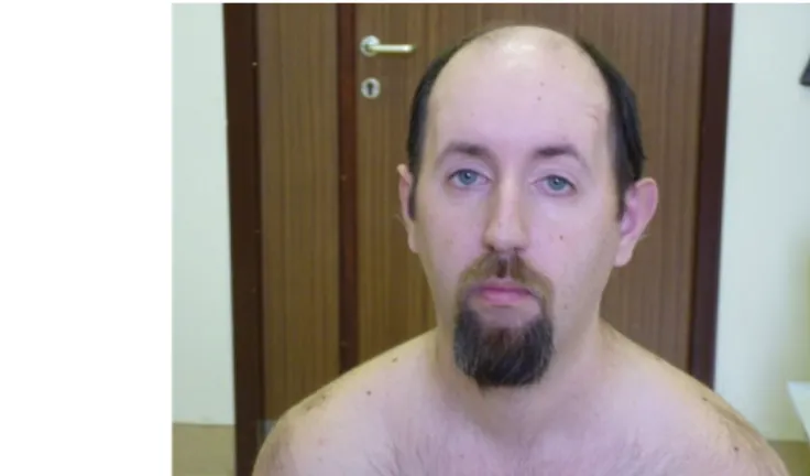
This is the most common muscle dystrophy in adulthood. Its reported prevalence is 13.5/100,000 newborns. The onset of the disease is variable, it may start at any time from infancy into older age. In cases with childhood onset, mental retardation is more commonly present and thus motor development is also delayed. The most specific symptom of myotonic dystrophy is myotonia, the disturbance of muscle relaxation. For example, patients are unable to release the water tap after turning it strongly. Patients have a typical face (ptosis, atrophy of masseter muscles), and speech is characteristic due to the weakness of tongue and the gothic soft palate. Ophthalmoplegia is not typical for this disease. Severe atrophy and weakness of skeletal muscles can develop. Distal muscles of the leg are typically affected, frequently causing sprained ankles. Muscle power of the pelvic girdle muscles, extensor muscles of the thigh, soleus muscle and the gastrocnemius muscles is usually preserved. Myotonic dystrophy is a multisystemic disease, with associated symptoms of cataracta, gynecomastia, endocrine dysfunction and cardiomyopathy. EMG is essential in the diagnosis of the disease, showing typical myotonic discharges in the muscles.
Fig. 4: The typical face of patients suffering from myotonic dystrophy–weakness of facial muscles is seen.
Video 3.: Myotonic phenomenon after handshake.
Myotonic dystrophy (Type 2, PROMM: PROximal Myotonic Myopathy)
In this type of myotonic dystrophy, the quadriceps muscle of the thigh and the triceps brachii muscle of the upper arm are preferentially affected. Lower legs are often hypertrophic, and patients complain of muscle pain and muscle cramps.
Neuropsychologic symptoms and histopathologic findings are similar to those in myotonic dystrophy Type 1.
Mitochondrial myopathies
In mitochondrial encephalomyopathies, isolated skeletal muscle involvement is rare. It is more typical that mitochondrial diseases show multisystemic manifestations, including skeletal muscle involvement. In the following part, the most common forms of
mitochondrial diseases are discussed:
Kearn-Sayers-syndrome (KSS) is caused by deletion of mtDNA. Diagnostic criteria:
onset at a young age, ptosis, external ophthalmoplegia, conduction defects in the heart, and retinitis pigmentosa. These core symptoms are often associated with diabetes mellitus, cerebellar ataxia and hearing impairment.
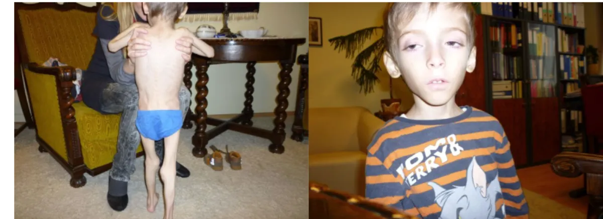
Fig. 5.: Severe generalized mitochondrial myopathy. Fig. 6.: Facies myopathica and bilateral partial ptosis in generalized mitochondrial myopathy.
Chronic Progressive External Ophthalmoplegia (CPEO) may start in adolescence or adulthood. In addition to the involvement of external and internal ocular muscles, other tissues may be affected as well to some extent. Associated symptoms mentioned at KSS may be present, but the diagnostic criteria are not as strict as for KSS.
In patients suffering from MELAS (Mitochondrial Encephalomyopathy, Lactic Acidosis, and Stroke-like episodes), first symptoms usually occur before the age of 20 years. In addition to the repeated ischemic stroke-like symptoms in adolescence, patients often complain of migraine, and epileptic seizures are also not unusual. Other symptoms may include diabetes mellitus, myopathy, and cardiomyopathy.
MERRF (Myoclonic Epilepsy with Ragged Red Fibers) syndrome may begin at any age.
Typical symptoms include myoclonic epilepsy and cerebellar ataxia, which may be associated with dementia, polyneuropathy, optic nerve atrophy, deafness, dorsal lipomatosis, and cardiomyopathy.
In MNGIE (Mitochondrial NeuroGastroIntestinal Encephalomyopathy) syndrome, characteristic symptoms include external ophthalmoplegia, dementia, progressive leukodystrophy, and involvement of the gastrointestinal tract.
Glycogenoses
The most typical symptom of McArdle’s disease is exercise intolerance. Immediately after physical exercise, patients complain of myalgia, muscle cramps, and even muscle contractures may appear after a prolonged exercise. Physical activity during tooth brushing may often cause unbearable pain for patients.
Pompe disease (alpha glucosidase deficiency) may begin at any time from infancy to adulthood. In infants, it presents with severe hypotonia and severe cardiomyopathy. In the adult type of the disease, limb girdle type muscular dystrophy is typical.
Video 4: Pompe disease. Abnormal gait as a consequence of lower limb girdle weakness.
Disorders of the Beta oxidation
In carnitine deficiency myopathy, proximal muscle groups are affected and weakness progresses slowly, but may also show a relapsing-remitting course. Cranial muscles may also be involved.
Carnitine-palmytoil-transferase II deficiency causes exercise intolerance, and subsequent myoglobinuria similar to McArdle’s disease. Symptoms are elicited – or enhanced – by fasting, stress or infections.
B.)2. Immune related muscle disorders
Dermatomyositis (DM)
Dermatomyositis is responsible for approximately 50%t of idiopathic myositis cases.
Clinical symptoms can start any time between 20 and 80 years of age. In typical cases, patients complain of weakness and pain in the proximal limb muscles. It should also be noted that dysphagia is not an uncommon symptom. Cranial and extraocular muscles are never affected. In most cases, abnormal skin symptoms are also present, such as
heliotrope discoloration of the eyelids, erythema of the face and the upper chest, bluish skin discoloration above the elbow and knee. The so called Gottron papules on the skin are specific signs of dermatomyositis.
Polymyositis (PM)
First symptoms usually appear around the age of 30 to 60 years, and progress slowly.
The chronic form is difficult to differentiate from metabolic myopathies and muscle dystrophies. The most specific sign is weakness and atrophy of the limb girdle and proximal muscles. The involvement of respiratory muscles and dysphagia is less
frequent compared to dermatomyositis. As opposed to dermatomyositis, muscles are not sensitive to pressure and joint complaints are also less frequent. Severe residual
symptoms develop if the disease is not treated properly.
Inclusion body myositis (IBM)
Symptoms of inclusion body myositis start in the sixth and seventh decade. Inclusion body myositis is more common in males. The onset of the disease is usually difficult to recognize, and it progresses slowly. Muscle weakness is present both in proximal and distal muscles. In the classic form of IBM, atrophy and weakness of the quadriceps muscles are typical. With progression of the disease, patients may lose ambulation. In about half of the cases, dysphagia is also associated with inclusion body myositis.
Myasthenia gravis
Dysfunction of the neuromuscular junction is called myasthenia gravis (MG). The specific clinical sign of myasthenia gravis fluctuating muscle weakness in the
extraocular, bulbar, respiratory, and limb muscles. The most common pathomechanism of MG is an autoimmune reaction against the postsynaptic membrane of the
neuromuscular junction. This type is called myasthenia gravis paralytica with a reported prevalence of approximately 2-10/100,000. In most of the cases, this form appears in females in their twenties and in males older than 60 years. The typical symptom of MG is an exercise-dependent muscle weakness, which increases during the course of the day.
Weakness may be present in the ocular, bulbar, postural, and proximal limb muscles.
The first clinical signs often occur at a young age.
B.)3. Toxic and iatrogenic myopathies
The most common forms of toxic and iatrogenic myopathies are the following:
1. Acute necrotizing myopathy with or without myoglobinuria. Possible agents:
ethanol, Epsilon Amino-Caproic Acid (EACA), cocaine, carbon monoxide 2. Subacute painful myopathy. Possible agents: clofibrate, aldose reductase inhibitors, EACA, and emetine.
3. Chronic proximal myopathy. Possible agents: chloroquine, corticosteroid, drugs and other factors causing hypokalemia.
4. Inflammatory myopathy. Possible agents: penicillamine.
5. Focal necrosis, or fibrosis at the site of intramuscular (i.m.) injection. Possible agents: opiates, paraldehyde.
Acute Quadriplegic Myopathy (AQM; myosin depletion syndrome):
Patients with AQM are usually treated in intensive care units with status asthmaticus who have received neuromuscular blocking agents to facilitate mechanical ventilation and corticosteroids in high-dose. After a couple of days, asthmatic symptoms improve and the administration of neuromuscular blocking agents muscle relaxant drug is discontinued, but respiratory insufficiency and quadriplegia are observed. Serum creatine kinase (CK) levels are slightly elevated. AQM may completely resolve, with recovery lasting for several weeks or months.
Chronic steroid myopathy: After chronic steroid administration, myopathy similar to Cushing syndrome may develop. Typical clinical sign is the weakness of the pelvic girdle, and proximal leg muscles. The quadriceps muscles and the psoas major muscles are preferentially affected. If steroids are administered to treat other muscle disorders, the development of steroid myopathy may exacerbate weakness due to the underlying disease. Symptoms improve with the reduction of the steroid dose. The pathomechanism of chronic steroid myopathy is not fully understood. Presumably, advanced age, lack of exercise and low-protein diet are predisposing factors of this disease.
Summary
The clinical presentation of muscle diseases is similar. The typical symptom is muscle atrophy and weakness of proximal limb muscles. In certain forms, symptoms such as involvement of other muscle groups (for example facial muscles, quadriceps muscles), or muscle contractures, cardiac muscle involvement or erythema, etc. may help in the differential diagnosis of muscle disorders.
![Fig. 2 and 3: Paresis and muscle atrophy in polymyositis.]]](https://thumb-eu.123doks.com/thumbv2/9dokorg/1160883.84116/2.892.208.808.635.862/fig-paresis-muscle-atrophy-polymyositis.webp)