Lesson 6 - The biomechanics of skeletal muscles
This curriculum has been made at the University of Szeged, and supported by the European Union. Project identity number: EFOP-3.4.3-16-2016-00014.
This lesson contains 10 screens teaching text, 4 zoomable figures and 7 videos. This lesson requires approximately 2 - 4 hours of study but can vary depending on the student.
Function of the skeletal muscles
Skeletal muscleconstitutes of 40% of the body weight, itforms the large majority of our muscle tissue. Skeletal muscles are the key componentof the locomotor system due to their ability to convert the chemical energy into mechanical energy (13). The skeletal muscles serve as stabilizer and mobilizer organs, provide strength, protection and shock absorption to the skeleton. The dynamic work of the skeletal muscles enable the bones to move at the joints and the static work of muscles maintains the body posture by stabilizing the joints. Muscles also protect the bones and organs by absorbing shock and reducing friction in the joints (10).
Structure and composition of skeletal muscle
Gross structure
A skeletal muscle consists of numerous muscle cells called muscle fibers which are linked together by three layers of collagenous connective tissue. These connective tissues associated with muscles follow (10):
The endomysium is the connective tissue that encompasses each muscle fiber.
The perimysium encases the group of muscle fibers, forming a fascicle.
The epimysium encircles all the fascicles to form a complete muscle.
Please watch the video below about the connective tissue layers of muscle:
https://www.youtube.com/watch?v=SCznFaTwTPE&pbjreload=10
Skeletal muscle fibers can be quite large for human cells, with diameters up to 100 μm and lengths up to 30 cm in the Sartorius muscle. The plasma membrane of muscle fibers is called the sarcolemma, the cytoplasm is referred to as sarcoplasm, and the specialized smooth endoplasmic reticulum is called the sarcoplasmic reticulum.
Muscle fibers (cells) are composed of a large number of contractile myofibrils. Each myofibril is composed of fibrous filaments of two types: the thin actin filaments and the thick myosin filaments.
The functional contractile unit of the muscle fiber is the sarcomere. The sarcomere itself is bundled within the myofibril that runs the entire length of the muscle fiber and attaches to the sarcolemma at its end (2).
When signaled by a motor neuron, a skeletal muscle fiber contracts. During this process, the thin filaments are pulled and then slide past the thick filaments within the fiber’s sarcomeres.
The mechanism can be explained with the sliding filament model. According to this model, shortening of the sarcomere results from the relative movement of actin and myosin filaments (2, 10).
Please watch this video below and brush up your physiological knowledge about the structure and contraction of muscle:
https://www.youtube.com/watch?v=E2RFiNhHHaA
Motor unit – the functional unit of skeletal muscle
In anatomical terms, the motor unit consists of a single motor neuron, its axon, and all the muscle fibers innervated by that axon and its branches. A single motor neuron and its associated muscle fibers together constitute the smallest unit of force that can be activated to produce movement. The function of the muscle and the number of muscle fibers forming a motor unit are closely related to each other (10). The activation of the motor units is gradually according
to their size and the needed muscle force. This means thatsmall motor units are recruited before larger ones. The motor unit recruitment happens automatically since small neurons are more easily depolarized and means that movements requiring minimal force can be more finely graded (7).
Please watch this video about the activation of a muscle fiber via neuromuscular junction:
https://www.youtube.com/watch?v=e7BNM-aRVZg
Types of muscle fibers
Muscle fibers can be classified in terms of contractile and metabolic properties.
Human muscular system basically has three different types of skeletal muscle fibers.
Slow-twitch (Type I) fibers are identified by a slow contraction time and a high resistance to fatigue. Structurally, they have a small motor neuron and fiber diameter, a high mitochondrial and capillary density, and a high myoglobin content. Energetically, they contain many of the enzymes involved in the oxidative pathways. Functionally, slow-twitch fibers are used for aerobic activities requiring low-level force production, such as walking and maintaining posture.
Fast-twitch (Type II) fibers are identified by a quick contraction time and a low resistance to fatigue. Fast-twitch fibers are further divided into fast-twitch A (Type IIA) and fast- twitch B (Type IIB) fibers.
Fast-twitch A fibers have a moderate resistance to fatigue. Structurally, they have a large motor neuron and fiber diameter, a high mitochondrial density, a medium capillary density, and a medium myoglobin content. They have both a high glycolytic and oxidative enzyme activity.
Functionally, they are used for prolonged anaerobic activities with a relatively high force output.
Fast-twitch B fibers are very sensitive to fatigue and are used for short anaerobic, high force production activities. They have a large motor neuron and fiber diameter, but a low mitochondrial and capillary density and myoglobin content. They contain many glycolytic enzymes but few oxidative enzymes (8).
Types of muscle contraction
Muscle contractions can be divided into two basic categories. The dynamic muscular work, which performs a movement, includes concentric and eccentric types of contraction. During concentric dynamic contraction, the muscle products force concurrently with the shortening.
During eccentric dynamic contraction, the muscle products force concurrently with the lengthening.
The static muscular work, which means generated force without changing the length of the muscle, includes the isometric type of contraction. During isometric contraction,the muscle- tendon unit as a whole is under tension but neither shortens nor lengthens, the contractile components are shortened, but the elastic components are lengthened concurrently. The function of the isometric contraction is to provide a stabilizing effect (4, 12).
Please watch these short videos about muscle contraction types:
https://www.youtube.com/watch?v=fkqg3MMUXFw https://www.youtube.com/watch?v=4LRBnAW5EzE
Mechanical properties of skeletal muscles Muscle architecture
Three-element model
The mechanical behavior of the sarcomere, and the whole musculotendinous unit, can be described by a three-element model. In this model, the PE represents the parallel elastic elements, SE symbolizes the series elastic elements, and the CE means the contractile elements (Fig. 1).
Fig. 1 The mechanical model of a muscle – the three element model
The parallel elastic elements (muscle membranes e.g. sarcolemma, endomysium, epimysium, perimysium) supplies resistance and provides protective function, when the muscle is passively lengthened and stretched. The series elastic element (e.g. tendon, fascia) can store the elastic energy when the tensed muscle is stretched. The elasticity of skeletal muscles is to be primarily to the series elastic element (2, 4).
Fiber architecture
The force that muscle excerpts depends on not only the mechanical properties of its contractile elements but also on the physical arrangements of these contractile elements. Two main categories of muscle fiber arrangement are named parallel and pennate, which means different fiber orientation in relation to the longitudinal axis of the muscle (4). Muscles such as the sartorius and the rectus abdominis are considered to have a parallel muscle fiber arrangement.
Most muscle fibers in parallel arrangement are oriented parallel to the muscle’s longitudinal axis. Muscles with parallel arranged fibers can make larger joint motions due to greater shortening of the muscle, than comparably sized pennate-fibered muscles. The pennate fiber arrangement is a category in which the fibers run at an angle (α) to the muscle’s longitudinal axis. Usually, short fibers attach to one or more tendons within the pennate muscle. The pennate muscles contain more fibers than longitudinally arranged muscles in an equal unit of volume and it can generate more force than parallel muscles of the same size, due to this difference.
When the fibers of the pennate muscle contract and the muscle shorten, the volume of the muscle remains relatively constant but the angle of pennation (α) increases concurrently. As demonstrated in Fig 2. the greater angle of pennation lead to decreased effective force transmitted to the tendon to move the attached bones, and to generate articulate motion (4).
Fig 2. Effect of the pennate fiber arrangement on the effective force vector
Muscle mechanics
The muscle mechanics concerns the external mechanical variables of muscle (e.g., force, length, power) in relation to the internal contractile state of muscle. (2)
Muscle length
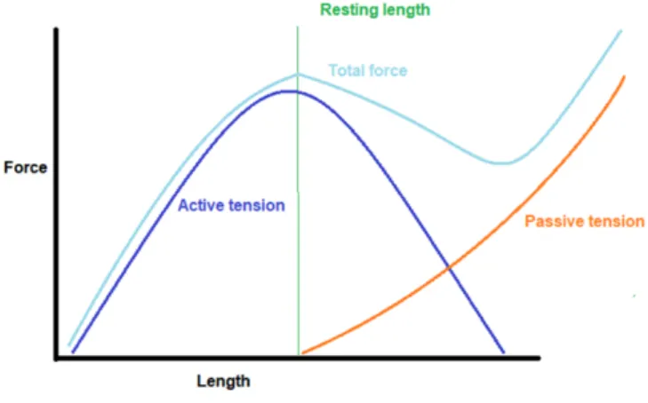
When the muscle contracts dynamically, the length of the muscle and sarcomere changes during the filaments sliding and the number of the actin filament binding sites available for the cross- bridges changes. The amount of overlap, as the possibility of interaction between myosin cross- bridges between actin and myosin, varies with the length of the muscle. The tension of muscle varies with the amount of overlap.
This principle demonstrates the active length-tension relationship.
When the sarcomere is overly contracted the overlap between the thin and thick filaments is high and muscle contraction causes actin filaments to slide over one another and the ends of myosin filaments. Tension decreases due to this pause in cross-bridge cycling and formation.
If the resting muscle length increases, more cross-bridges cycling and increased tension occurs when muscles are stimulated to contract. Maximum tension is produced when sarcomeres are about 2.1 to 2.2 μm long. This is the optimal resting length of the sarcomere for producing the maximal tension.
By increasing the muscle length beyond the optimum, the actin filaments become pulled away from the myosin filaments and from each other. In this case, there is little interaction between the filaments and less tension is produced because very few cross-bridges can form (2, 10).
Furthermore, the muscle force is not dependent only on the active mechanism, because the muscle includes a considerable amount of connective tissue. These passive components exert an additional force during muscle activity, which combines with the effect of the active components results in a combined force which characterize the biomechanical properties of muscle. Due to the elastic properties of muscle, resulting from the tendons and surrounding connective tissue as muscle length increases, passive tension also increases. The force-length curve of a muscle describes the contributions of the active and passive components to the total
muscle force as muscle length varies from the contraction length to the maximum stretched length (Fig.3).
Fig 3. The force-length curve of a muscle
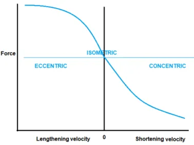
In concentric contractions, the force generated by the muscle is always less than the muscle's maximum. As the load the muscle is required to lift decreases, contraction velocity increases.
This occurs until the muscle finally reaches its maximum contraction velocity. By performing a series of constant velocity shortening contractions, a force-velocity relationship can be determined (2).
The force-velocity relationship of the muscle describes the force generated by a muscle as a function of velocity under conditions of constant load. In other words, the velocity of muscle contraction depends on the force resisting to muscle.
The force generated by a muscle depends on the total number of cross-bridges attached.
Because it takes a finite amount of time for cross-bridges to attach, as filaments slide past one another faster and faster, as the muscle shortens with increasing velocity, force decreases due to the lower number of cross-bridges attached. Conversely, as the relative filament velocity decreases, as muscle velocity decreases, more cross-bridges have time to attach and to generate force, and thus force increases (2) (Fig.4).
Fig. 4 The force-velocity relationship of the muscle
You can understand the difference of the force-velocity relationship for the concentric and eccentric contraction with the following video:
https://www.youtube.com/watch?v=ERWQXiuwCFc
Please watch the following video and practice your knowledge about muscle mechanics.
https://www.youtube.com/watch?v=eTs9EnlbFew&t=13s
Effect of immobilization and physical training
Muscle atrophy is defined as a decrease in the mass of the muscle mainly caused by immobilization.
Immobilization causes a rapid loss of muscle mass and strength. A substantial amount of skeletal muscle mass and strength is lost during joint surgery‐related immobilization, unloading space flight, bed rest, myopathies, and aging (5). Works from animal studies conclude that muscle protein synthesis is reduced and muscle protein breakdown is increased following a short, 6 and 24 h of cast immobilization (1, 9). Human muscle biopsy researches demonstrated that mainly the type I. fibers atrophy with immobilization. Several studies suggest that an intermediate or early movement program may prevent muscle atrophy due to disusing. The
physical training increases the cross-sectional area of all muscle fibers, the volume, and the strength of the muscle (4, 12).
Effect of aging
The aging markedly affects muscle mechanical function, furthermore, elderly individuals are frequently exposed to periods of muscle disuse caused by disease or injury (6). Skeletal muscle cross-sectional area decreases with age due to the reduction in muscle fiber size, and number.
Another fact that effects on the muscle function that the number of α-motoneurons decreases with age as well (11).
Study questions:
TRUE/FALSE questions
Read each statement below carefully. Choose T if you think a statement is TRUE.
Choose F if you think the statement is FALSE.
1. Immobilization causes a slow loss of muscle mass and strength. T or F 2. The pennate muscles contain less fibers than longitudinally arranged muscles in an equal
unit of volume. T or F 3. Muscle atrophy is defined as an increase in the mass of the muscle mainly caused by
immobilization. T or F 4. The endomysium is the connective tissue that encompasses the fascicle. T or F
5. The motor unit consists of a single motor neuron, its axon and all the muscle fibers innervated by that axon and its branches. T or F 6. Skeletal muscle cross-sectional area decreases with age due to the reduction in muscle
fiber size and number. T or F 7. Each myofibril is composed of fibrous filaments of two types: the thick actin filaments
and the thin myosin filaments T or F 8. Fast-twitch (Type II) fibers are identified by a slow contraction time and a high
resistance to fatigue. T or F
Matching questions
In this exercise, you have to match each word with a definition.
1. The force-length curve of a muscle
2. Eccentric dynamic contraction
3. Isometric contraction 4. The force-velocity
relationship of the muscle
5. Concentric dynamic contraction
A. The muscle products force against an external load concurrently with the shortening.
B. The muscle-tendon unit as a whole is under tension but neither shortens nor lengthens.
C. It describes the force generated by a muscle as a function of velocity under conditions of constant load.
D. It describes the contributions of the active and passive components to the total muscle force as muscle length varies from the contraction length to the maximum stretched length.
E. The muscle products force concurrently with the lengthening.
References
1. Booth FW, Seider MJ (1979): Early change in skeletal muscle protein synthesis after limb immobilization of rats. J Appl Physiol 47: 974-977
2. Enoka RM: Neuromechanical Basis of Kinesiology. 1994. Human Kinetics
3. Williams GN, Higgins MJ, Lewek MD (2002): Aging Skeletal Muscle: Physiologic Changes and the Effects of Training, Physical Therapy, 82:1 62–68 https://doi.org/10.1093/ptj/82.1.62
4. Hall SJ: Basic Biomechanics 2002. McGrawHill Coll; 4th Edition
5. Hortobágyi T, Dempsey L, Fraser D, Zheng D, Hamilton G, Lambert J, Dohm L (2000):
Changes in muscle strength, muscle fibre size and myofibrillar gene expression after immobilization and retraining in humans The Journal of Physiology 524:1 293-304
6. Hvid L, Aagaard P, Justesen L, Bayer ML, Andersen JL, Ortenblad N, Kjaer M, Suetta C (2010): Effects of aging on muscle mechanical function and muscle fiber morphology during short-term immobilization and subsequent retraining. J Appl Physiol. 109: 1628- 1634. 10.1152/japplphysiol.00637.2010
7. Johns P, Chapter 4 - Sensory and motor pathways. Editor(s): Paul Johns, Clinical Neuroscience,Churchill Livingstone,2014,Pages 49-59,ISBN 9780443103216,
8. Karp, J. R., 2001. Muscle Fiber Types and Training. Track Coach. 155:4943-4946
9. Kobayashi H , Kato H , Hirabayashi Y, Murakami H, Suzuki H (2006): Modulations of muscle protein metabolism by branched-chain amino acids in normal and muscle- atrophying rats. J Nutr 136: 234-236
10. Martini Frederic Martini (Author), William C. Ober (Author), Claire W. Garrison (Author), Kathleen Welch (Author), Ralph T. Hutchings (Author) Fundamentals of Anatomy & Physiology, 5th edition, hc, 2001 Prentice Hall College Div; 5th edition (January 2001)
11. Mittal KR, Logmani FH (1987): Age-related reduction in 8th cervical ventral nerve root myelinated fiber diameters and numbers in man. J Gerontol 42:8–10
12. Nordin M, Frankel VH: Basic Biomechanics of the Musculoskeletal System. 2014.
Wolters Kluwer Health; 4th Edition
13. Trovato FM, Imbesi R, Conway N, Castrogiovanni P (2016): Morphological and Functional Aspects of Human Skeletal Muscle J. Funct. Morphol. Kinesiol. 1(3): 289- 302