Diseases Caused by Hyphomycetous Fungi
M. F. MADELIN
Department of Botany, The University, Bristol, England
I. Introduction 233 II. T h e Pathogens 234 III. Modes of Parasitism 236
IV. Infection 237 A. T h e Infective U n i t 237
B. Sites and Modes of Infection 240 C. Factors T h a t Influence Infection 243 V. Colonization of the Infected Insect 247
VI. Symptom Production 250 VII. Death of the Diseased Insect 252 VIII. Postmortem Behavior of the Fungus 255
A. Saprophytic Colonization of the Host 255 B. Emergence of the Fungus through the Integument . . 255
C. Sporulation of the Fungus 256 IX. Natural Life Cycle of the Pathogen 256
X . Opposition to Fungal Invasion 259 X L Host Specificity of the Pathogen 260 X I I . Physiological Characteristics of the Pathogen 261
XIII. R o l e of Stress in the Incidence of Mycoses 263
XIV. Conclusion 263 References 264
I. INTRODUCTION
T h e aim of this chapter is to treat comparatively the diseases of in
sects caused by hyphomycetous Fungi Imperfecti (Deuteromycetes) not usually associated in n a t u r e with a perfect stage. Hyphomycetes are mycelial fungi reproducing by means of conidia which are generally produced on free or aggregated conidiophores on the surface of their
233
234 Μ. F. MADELIN
substratum. T h e so-called muscardine diseases, all of which are caused by Hyphomycetes, d o not as a g r o u p differ fundamentally from most other mycoses caused by Hyphomycetes.
II. T H E PATHOGENS
T h e r e are many hyphomycetous species which grow on insects (see, e.g., Charles, 1941; Kobayasi, 1941; Petch, 1948), b u t for a large n u m b e r it remains to be proved whether they are parasitic or saprophytic. T h e literature on the bionomics of insect species, however, reveals that there is only a rather small group of species which are very common, wide
spread, and important in n a t u r e as biotic factors influencing the size of insect populations. Those most widely encountered are members of the genera Beauveria, Metarrhizium, and Isaria (or Spicaria), and to a lesser extent Aspergillus, Cephalosporium, Sorosporella, Aegerita, Hirsu- tella, and Acrostalagmus.
T h e genus Beauveria has recently been thoroughly studied by Mac
Leod (1954b), who concluded that 14 species h a d been named which were characteristic of the genus, b u t that only two were tenable. H e concluded that B. stephanoderis (Bally) Petch, B. laxa Petch, B. globu- lifera (Spegazzini) Picard, Β. effusa (Beauverie) Vuillemin, B. doryphorae Poisson and Patay, B. delacroixii (Saccardo) Petch, Isaria vexans Pettit, and Botrytis acridiorum Brongniart and Delacroix are strains of Beau
veria bassiana (Balsamo) Vuillemin (Fig. 1, A), in which globose and oval spores occur in about equal proportions; a n d B. densa (Link) Picard, Β. brongniartii (Saccardo) Petch, B. shiotae (Kuru) Langeron (which was not isolated from an insect), a n d Botrytis melolonthae Sac
cardo are strains of the almost entirely oval-spored species, Beauveria tenella (Delacroix) Siemaszko. I n subsequent references to species of Beauveria, the specific epithets employed by the workers concerned will, in general, be used in order to preserve indications of the strains in
volved.
T h e commonest species of Metarrhizium is Metarrhizium anisopliae (Metchnikoff) Sorokin, the cause of the green muscardine disease of divers insects (Fig. 1, C). Its synonyms include Oospora destructor Metch
nikoff, Entomophthora anisopliae Metchnikoff, and Penicillium anisop
liae (Metchnikoff) Vuillemin. Johnston (1915) described two forms, major and minor, occurring on different hosts, and Friederichs (1920) similarly recognized long- and short-spored forms. R a d h a et al. (1956) are inclined to accept Johnston's division of the species, with 10.6 to 12 μ and 3.5 to 8.2 μ as the two characteristic ranges of spore lengths. A vari
ety americana has been described by Pettit (1895), and two other entomog
enous species, album and brunneum, have been described by Petch (1931, 1935).
Isaria is a form genus whose members form conidiophores in fascicles called coremia. These are often more than a centimeter tall. Because Vuillemin (1911) considered that the method of spore production was more i m p o r t a n t for classification than the presence or absence of core
mia, he transferred the common insect parasite, Isaria farinosa Fries to
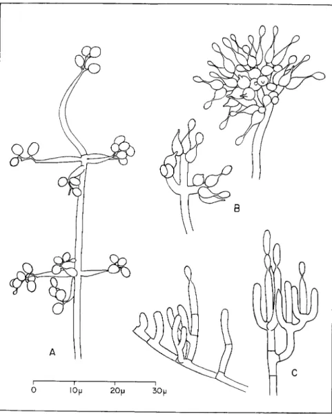
FIG. 1. Individual conidiophores of three Hyphomycetes parasitic o n insects.
(A) Beauveria bassiana (Balsamo) V u i l l e m i n , from a slide culture o n malt agar.
(B) Spicaria farinosa (Fries) Vuillemin, from coremia on naturally infected insects.
(C) Metarrhizium anisopliae (Metchnikoff) Sorokin, from a naturally infected sugar
cane froghopper, Tomaspis saccharina Distant.
236 Μ. F. MADELIN
the genus Spicaria. More recently Brown and Smith (1957) have trans
ferred it to Paecilomyces. T h e r e are allied organisms which parasitize insects, including Spicaria fumoso-rosea (Wize) Vassiljevski and S. fari- nosa verticillo'xdes Fron. However, Picard (1914) and Brown and Smith consider that the latter is synonymous with S. farinosa (Fries) Vuillemin (Fig. 1, B).
T h e characteristics of many pathogenic fungi have recently been dis
cussed in a Japanese text by K. Aoki (1957). T h i s present chapter deals only with Hyphomycetes not usually associated in n a t u r e with a perfect stage, those Ascomycetes in which a hyphomycetous asexual state ac
companies the perithecial state being specifically excluded, as, for ex
ample, certain species of Cordyceps, Torrubiella, Sphaerostilbe, and Nectria. Perithecial stages have nevertheless been recorded for certain species considered here. Schaerffenberg (1955, 1957a, 1959) reported find
ing perithecia of B. bassiana, B. densa, and M. anisopliae. These peri
thecial stages have not been reported by other workers and their natural role may not be great. N o perfect stage has been described for S. farinosa, although it was for a long time erroneously believed to be Cordyceps militaris (Linnaeus) Link (Petch, 1936).
I I I . MODES OF PARASITISM
As a group, the hyphomycetous parasites are facultative. Virtually all grow readily in artificial culture, and with few exceptions, such as certain Hirsutella species (MacLeod, 1954a, 1960), they are rather unexacting nutritionally. A n u m b e r have been observed to grow in na
ture on dead substrates other than insect bodies, and for some, notably species of Aspergillus, a saprophytic existence may be the d o m i n a n t life habit (Lepesme, 1938).
Although most Hyphomycetes that attack insects are internal patho
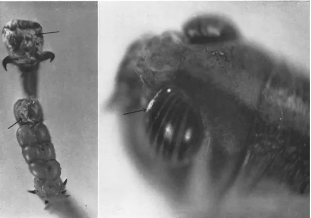
gens, the superficial parasitic habit is represented, notably by Fusarium acridiorum (Trabut) Brongniart and Delacroix. T h i s was observed first by Kunckel d'Herculais and Langlois in Algeria in 1891, attacking desert locusts (Schistocerca gregaria (Forskäl)). It forms white powdery masses or a grayish down on the surface of parts of the head, thorax, abdomen, and hind feet (Giard, 1891b) (see Fig. 2). It is entirely superficial u p o n the cuticle, which it darkens (Trabut, 1891; Brongniart, 1891). Neverthe
less, occasionally it deforms segments of the body to the extent of im
peding egg-laying (Kunckel d'Herculais and Langlois, 1891). Although Kunckel d'Herculais and Langlois reported that it attacks locusts only at the end of their life cycles, Brongniart (1891) observed that in a con
siderable epizootic most of the many females killed died without laying eggs. T h e disease is slow to develop and not very contagious. Giard (1892b) states that it becomes dangerous to the insect only when the fungus
invades the tracheae and causes asphyxiation. H e observed that it had not been shown that it could also attack locust eggs. Although Marchi- onatto (1933) recorded a species of Fusarium allied to F. acridiorum which covered eggs of Schistocerca paranensis Burmeister with a white cottony growth, and Akbar et al. (1958) observed a similar growth on eggs of S. gregaria which compared favorably with a description of F.
acridiorum, there appears to be no proof that the actual organism which superficially parasitizes adult locusts also attacks the eggs. Although the fungus has been mentioned in literature several times since 1892, few new facts have been revealed. T h i s superficial parasite differs from most endoparasitic fungi principally in the slowness with which it progresses and in its usual sporulation on the surface of the still-living host.
T h e internally parasitic fungus may be envisaged as one which through possessing special attributes is able to exploit more fully the live insect as a nutritive environment than is the superficial parasite.
T h e first obstacle which it successfully overcomes is penetration of the host's boundary layers. Once inside, the fungus proceeds to colonize the body, exercizing in the process an influence which evokes the condition we call disease. Generally the diseased insect sooner or later succumbs, whereupon the fungus proceeds to live as a saprophyte on the dead tissues. Usually when colonization is more or less complete, and condi
tions are suitable, the fungus sporulates. These different stages will be discussed separately.
IV. INFECTION
A. The Infective Unit
Internally parasitic Hyphomycetes can enter intact insects only by the activity of hyphal tips arising from infective units in the insects' im
mediate environment. T h e infective unit of the hyphomycetous patho
gen in an exposed situation is usually the conidium. I n a more sheltered habitat, such as in leaf litter and soil, the situation is less easily observed, but it is likely that there too the conidium can infect directly. Many workers have infected soil-inhabiting insects by adding spores to the soil, but it has not been shown whether infection proceeds directly from these or from an intervening soil-inhabiting mycelial phase. Infection by vege
tative hyphae certainly may occur in subaerial environments. H y p h a e may spread from larvae of Dialeurodes citri (Ashmead) killed by Aegerita webberi Fawcett on one citrus leaf to another leaf where they infect fresh hosts (Morrill and Back, 1912). Similarly Williams (1915) reported an unidentified fungus on pea thrips, Kakothrips robustus (Uzel), whose hyphae radiated out from attacked larvae and infected nearby healthy ones even in dry conditions.
T h e r e is evidence that mycoses can arise from d o r m a n t infections.
FIG. 2. Fusarium acridiorum (Trabut) Brongniart and Delacroix spontaneously infecting adult of Nomadacris septemfasciata Serville reared in laboratory. Superficial growth of sporulating mycelium on foot and c o m p o u n d eye.
240 Μ. F. MADELIN
Lefebvre (1934) found that larvae of Pyrausta nubilalis (Hübner) from Manchuria carried a d o r m a n t B. bassiana infection which became viru
lent when the temperature was raised from 10-14°C to room heat.
Pospelov (1938, 1940) similarly concluded that M. anisopliae could ap
parently exist as a latent infection within the organs of larvae of Cleonus punctiventris (Germar) in an unrecognized form.
B. Sites and Modes of Infection
It is necessary to consider briefly the nature of the insect's b o u n d a r y with its environment, for it is there in particular that important factors operate which differentiate between pathogenic and nonpathogenic fungi and between resistant and susceptible hosts.
T h e insect's outer integument is composed of a layer of epidermal cells overlain with the procuticle. T h i s consists of protein and chitin, which is a polymer of N-acetyl glucosamine. Over this lies the epicuticle, which among other materials typically contains wax. T h e cuticle is con
tinuous with the much thinner layers which line the tracheae and the foregut and hindgut. T h e midgut epithelium is commonly protected by a chitin-containing peritrophic m e m b r a n e which forms a sheath sepa
rating the contents of the intestine from the gut wall.
T h e exact site of infection depends on the fungus, the insect, the conditions, and the opportunity. T h e four m a i n routes of infection—
through outer integument, digestive tract, tracheae, and wounds—will be considered separately.
T h e passage of germ tubes of B. bassiana through the outer integu
ment has been directly observed in larvae of Pyrausta nubilalis (Lefebvre, 1934; Metalnikov and Toumanoff, 1928), Bombyx mori (Lin
naeus) (Conte and Levrat, 1907; Paillot, 1930), Leptinotarsa decemline- ata (Say) (Schaerffenberg, 1957a), and Loxostege sticticalis (Linnaeus) (Pilat, 1938). Larvae of Phthorimaea operculella Zeller are probably in
fected the same way by B. bassiana, B. densa, and B. globulifera (Picard, 1913), as also are adults of L. decemlineata by B. effusa (Dieuzeide, 1925).
Metarrhizium anisopliae has been seen to penetrate the integuments of house flies (Musca domestica Linnaeus), larvae of L. decemlineata (Schaerffenberg, 1959), hibernating larvae of P. nubilalis (Wallengren and Johansson, 1929) and a n u m b e r of other susceptible insects (Notini and Mathlein, 1944). Externally applied spores successfully infected larvae of B. mori (Glaser, 1926) and Melolontha melolontha (Linnaeus) ( H u r p i n and Vago, 1958). T h e r e are many other records of insects be
coming diseased from conidia of M . anisopliae applied to their skins, b u t it is not always certain that the spores did not also enter the tracheae or digestive tract.
Spicaria farinosa can infect larvae of M. melolontha and P. nubilalis through the cuticle (Toumanoff, 1933; H u r p i n and Vago, 1958), b u t Aspergillus flavus Link appears to behave differently on different hosts.
Its germ tubes have been seen to penetrate the integument of P. nubilalis (Toumanoff, 1933) and of Platysamia cecropia Linnaeus (Sussman, 1952a), b u t though pathogenic by other infection routes they were un
able to infect in this way either adults of Schistocerca gregaria (Lepesme, 1938) or honey bees (Burnside, 1930). Speare (1920) believes that Soro
sporella uvella (Krassilstschik) Giard, the red muscardine fungus, can infect noctuid larvae through the integument as well as through the intestinal wall. Cephalosporium lecanii Zimmerman apparently invades coccids through the integument (Ganhäo, 1956).
Infection through the digestive tract is difficult to demonstrate with certainty. It is difficult to introduce conidia into the gut without con
taminating the skin at the same time. T h e only real proof is actually to observe penetration by histological study, b u t the paucity of positive results by this method suggests its difficulty or the rarity of the event.
Much of the evidence for infection via the gut wall is thus rather indi
rect.
Infection through the digestive tract has been observed histologically for B. bassiana in larvae of Loxostege sticticalis (Pilat, 1938). Lefebvre (1934) considers that there is good evidence for this in larvae of P. nu
bilalis. H u r p i n and Vago (1958) reported that a n u m b e r of strains of Beauveria could infect larvae of M. melolontha which ingested their spores, while Schaerffenberg (1957b) reported the same for B. bassiana in larvae of Leptinotarsa decemlineata. However, Dieuzeide (1925) was unable to infect the latter in this way with spores of B. effusa (— B.
bassiana, fide MacLeod, 1954b). Similarly adults of Phthorimaea opercu- lella fed with spores of the allied B. globulifera suffered no ill effects (Picard, 1913). Comparably diverse findings have been reported for Metarrhizium anisopliae. Spores fed to larvae of Melolontha melolontha caused infection ( H u r p i n and Vago, 1958) b u t fed to honey bees killed fewer than 10 percent (Burnside, 1930) and fed to silkworms killed none (Glaser, 1926). O n the other hand, Masera (1957) could infect silkworms with this fungus only by feeding spores to them or by direct injection;
external application failed.
Gabriel (1959) introduced spores directly into the midgut of test insects with a microinjector. W i t h B. bassiana, 36 out of 86 died, and with M. anisopliae 18 out of 86, as compared with more than 82 out of 86 when externally contaminated with these fungi. It was suggested that the bacterial infections which occurred in about a quarter of the micro- fed insects might have resulted from accidental injury to the gut caused
242 Μ. F. MADELIN
by the microfeeder. T h e possibility that wounding may similarly have favored infection by the introduced fungus spores cannot be ignored.
Burnside (1930) reported that spores of pathogenic Aspergillus spe
cies ingested by honey bees produced an extensive mycelium in the gut lumen from which the gut wall was soon invaded, although commonly, when the diseased bee was capable of b u t feeble movements, there were only a few scattered hyphae in the alimentary canal. Toumanoff (1931) indeed found that of 150 bees which died after ingesting spores of A. flavus, in only two was there mycelium in the general body cavity at the time of death. It therefore appears that death from ingestion of spores of aspergilli sometimes does not involve a true infective phase at all. Death is instead apparently caused by toxins (see Section VII), and penetration of the gut wall is often entirely post mortem. However, germ tubes of A. flavus have been seen to penetrate the chitinous lining of the hindgut of living larvae of Platysamia cecropia (Sussman, 1952a).
Introduction of spores through the stigmata, particularly of the thorax, was the only way desert locusts (5. gregaria) could be infected with A. flavus (Lepesme, 1938), and the most successful way by which certain other acridids could be infected by Aspergillus parasiticus Speare (Ogloblin and Jauch, 1943). Larvae of L. decemlineata can be infected via the tracheae by B. bassiana (Schaerffenberg, 1957b). O n the other hand, the spores of Aspergillus species appear unable to germinate in the tracheae of honey bees owing to the dryness there resulting from aeration (Burnside, 1930).
T h e problem of penetrating the insect's boundaries is solved for the pathogen where it is directly introduced into a fresh or recent deep wound. T h i s method of infection applied artificially proves highly suc
cessful b u t its natural importance has not been widely established. Vou- kassovitch (1925) believes that hymenopterous parasites implant spores of S. farinosa verticilloides into p u p a e of Polychrosis botrana Schiffer
müller during oviposition. Further, some Aspergillus and Fusarium spe
cies depend on wounds for their ability to infect M . melolontha larvae ( H u r p i n and Vago, 1958), as does Trichothecium roseum (Persoon) Link to attack silkworms (Jolly, 1959) and Penicillium brevi-compactum Dierckx to attack larvae of Malacosoma neustria (Linnaeus) (Müller- Kögler and Huger, 1960).
Most data relating to the actual process by which penetration is effected concern infection through the integument under laboratory conditions. Giard (1892a), who studied the parasitism of B. densa on cockchafers, appears to have been the first to have inferred that the cuti
cle is penetrated with the aid of an enzyme secreted at the apex of the penetrant hypha. A n u m b e r of later workers have arrived at similar con-
elusions with B. bassiana (Conte and Levrat, 1907; Paillot, 1930; Le- febvre, 1934; Pilat, 1938) a n d Metarrhizium anisopliae (Wallengren and Johansson, 1929; Notini and Mathlein, 1944). A clear zone surrounding the penetrant germ tube has been interpreted as a zone of chemical al
teration of the chitinous cuticle by secreted enzymes. T h e fact that the clear zone appears to be uniformly thick along the length of the pene
trant hypha suggests that the agent responsible emanates solely from its tip.
Although most workers have attributed the chemical aspects of the process of penetration to secretion of a chitinase, and indeed H u b e r (1958) has shown that B. bassiana, M. anisopliae, and A. flavus all can hydrolyze chitin in vitro, it must not be overlooked that chitin itself is generally not the major component of the procuticle, nor is it responsi
ble for its hardness. T h e r e is usually a greater percentage of protein than of chitin (Richards, 1953). T h e ability of at least the more common pathogenic fungi to digest certain proteins has, however, been demon
strated.
T h a t a mechanical action is also involved has been suggested by Wallengren and Johansson (1929) for M. anisopliae, and by Lefebvre (1934) for B. bassiana, because the layers of chitin are often somewhat indented in advance of the penetrating hyphae.
T h e way in which the epicuticle is penetrated has not been estab
lished. T h e similarly waxy cuticle of plant surfaces, as far as is known, is penetrated by pathogenic fungi solely by mechanical means.
Wallengren and Johansson (1929) observed that the conidia of M.
anisopliae adhering to the surface of fifth-stage larvae of P. nubilalis cause the adjacent cuticle to yellow before there is any penetration and indeed before the spores have produced germ tubes. T h e cuticle becomes granular in appearance. T h e pigment bodies in the outer cuticle disin
tegrate, and there the germ tubes eventually penetrate. These observa
tions suggest that a substance is produced by the spores which acts on the adjacent cuticle in advance of penetration by the germ tube. Rele
vant to this is the observation by Dresner (1950) that germinating spores of B. bassiana produced a toxin capable of rapidly paralyzing and killing certain species of insects. However, Steinhaus and Bell (1953) in similar experiments obtained at best b u t slight and inconsistent toxic reactions.
C. Factors That Influence Infection
W h e r e spores are the infective units, a prerequisite of infection is that they should germinate. A h u m i d atmosphere usually in excess of 80 percent relative humidity (R.H.) is essential. It is, therefore, not sur
prising that in many instances natural or artificially induced outbreaks
244 Μ. F. Μ ADELIN
of fungus disease in insect populations have been correlated with the existence of h u m i d conditions. T h u s of eight experiments conducted by Feytaud (1913) with larvae of P. botrana, Spicaria farinosa verticilloides infected a large proportion in solely that one in which the atmospheric humidity was deliberately made high. Doane (1959) found that beneath the bark of elms growing in a d a m p habitat, 92 percent of the overwin
tering larvae of Scolytus multistriatus (Marsham) were infected with B.
bassiana, whereas in a dry habitat only 4 percent were. Further, Michel- bacher et al. (1950) found that because of the moister conditions, more bark-inhabiting larvae succumbed to mycoses on the n o r t h side of trees (in the N o r t h e r n Hemisphere) than on the south.
I n the soil, too, the moisture level is a potent environmental factor.
Spicaria farinosa was observed on M. melolontha by H u r p i n and Vago (1958) only when the soil containing the insects was very moist. Schaerf
fenberg (1952) found that the level of infection of Melolontha sp. with B. densa in trials in field plots with sandy soil was very dependent on the a m o u n t of rainfall, whereas in humus-rich soil it remained uniformly high despite rainfall variation. By contrast, Pospelov (1913) reported that excessive drought provoked the outbreak of Sorosporella uvella in Cleo- nus punctiventris in Russia.
Soil p H can affect the behavior of pathogenic fungi. Pospelov (1940) recorded that M. anisopliae infected proportionally more individuals of C. punctiventris in acid than in other soils, while Pyatnitzkii (1940) re
ported that the mortality of immature stages of this weevil is high in soils poor in humus, due to M. anisopliae where the soils are acid and to S. uvella where they are more alkaline. Further, the use of mineral fer
tilizers that increase the acidity of the soil increased the rate of infection by M. anisopliae and decreased that by S. uvella whereas those that ren
dered the soil more alkaline h a d the reverse effect.
Although it is unlikely that the conidia of any insect-parasitizing hyphomycete can germinate below 80 percent R.H., there are reports of insects becoming infected at m u c h lower humidities, even as low as 46 percent R . H . (Metalnikov and Toumanoff, 1928). For example, larvae of P. nubilalis can be infected at low humidities by B. bassiana, B. globu- lifera, Spicaria farinosa, A. flavus, and Sterigmatocystis nigra van T i e g h e m (Toumanoff, 1928, 1933; Metalnikov and Toumanoff, 1928), and those of Carpocapsa pomonella (Linnaeus) by B. bassiana (Jaynes and Marucci,
1947). Probably the skin of P. nubilalis larvae is kept moist enough for the spores to germinate by cuticular secretions or transpiration (Tou
manoff, 1928, 1933). Similar microclimatic effects might prove to be im
portant in the soil, for Fidler (1936) showed that larvae of melolonthids are able slightly to raise by evaporation the humidity of the air in the cells in which they live.
T e m p e r a t u r e affects the numbers of insects killed by a fungus patho
gen, b u t it is not always clear whether the effect is on germination, pene
tration, or the pathogenic processes that ensue. Against larvae of L. de- cemlineata, B. bassiana declined in infectivity below 6°C, B. densa below 10°C, and M . anisopliae below 15°C (Schaerffenberg, 1957b). Müller- Kögler (1942) similarly found that B. bassiana failed to infect larvae of Bupalus piniarius (Linnaeus) at 0.5°C whereas it succeeded, though slowly, at 8°C. S. farinosa verticilloides infected at all temperatures from 0.5°C to 25°C. By contrast Boyce and Fawcett (1947) found that an Aspergillus species near A. parasiticus was unable to kill mealybugs at
17CC and below although it was pathogenic at higher temperatures.
Besides the major factors which influence germination, certain special factors have been reported. Notini and Mathlein (1944) detected a stim
ulus to the germination of conidia of M. anisopliae which emanated from the lipoid layer of the chitinous integument of certain insects.
Conidia germinated slowly on portions of the cuticle of Cossus cossus (Linnaeus) treated with a fat solvent, b u t rapidly on untreated cuticle.
Further, Nolla (1929) found that the juices of aphids stimulated germina
tion of spores of Acrostalagmus aphidum O u d e m a n s to a degree depend
ing on the species.
T h e n a t u r e of the stimulus which causes a germ tube growing on the surface of the integument to change its direction of growth and to pierce the cuticle remains to be established. Giard's (1892a) view that hyphae of B. densa are chemo tropically attracted through the chitinous integument by an acid in the blood is not based on strong evidence.
T h e epicuticles of some insects contain potent resistance factors which can be modified by suitable treatments. P u p a e of Platysamia cecro- pia dewaxed with ether lose their resistance to infection through the integument by Aspergillus flavus (Sussman, 1951a). Larvae of Chilo sim
plex (Butler) similarly become highly susceptible to this and other fungi when their epicuticular lipids are removed (Koidsumi, 1957). Vago (1959) found that the usually innocuous fungus, Fusarium poae (Peck) Wollenweber, sometimes grew where the integument of larvae of M.
melolontha was naturally abraded, and from thence penetrated into the body cavity.
Since chitin digestion is at least a part of the process of penetrating the procuticle, Huber's (1958) discovery that in vitro breakdown of chitin by B. bassiana, Metarrhizium anisopliae and A. flavus was suppressed by moderately high levels of alternative nutrients suggests that the invasive capacity of hyphae in n a t u r e might be influenced by the general nutrient status of the environment.
T h e texture of the cuticle, which varies in different species and in different places on the one insect, can affect the behavior of pathogens.
246 Μ. F. MADELIN
Lefebvre (1934) saw that 22. bassiana could penetrate only the thinner in
tersegmental regions of the integument of p u p a e of Pyrausta iiubilalis;
he never saw hyphae in the thicker portions of the integument. Thick
ness per se, however, is not a factor to which germ tubes could be di
rectly sensitive. Differences in thickness might be associated with critical differences in composition. J u d g i n g from the distribution of hyphae of B. bassiana in the cuticle of diseased silkworms, the different layers differ in hardness (Ito, 1951). T h e r e might also be differences in permeability so that certain sites on insects' integuments are moister and thereby more prone to infection (see Toumanoff, 1933). Permeability of the integument to water is controlled primarily by the epicuticle (Richards, 1953).
W o u n d i n g can predispose insects to infection or itself be the agency of penetration. T h u s hemorrhagic wounds in silkworm larvae make entry more easy for fungi normally capable of penetrating the intact cuticle, so that greater mortalities ensue, and allow weakly pathogenic or non
pathogenic fungi like Penicillium granulatum Bainier and Fusarium co- eruleum (Libert) Saccardo to assume a certain virulence (Vago, 1959).
Snow (1896) observed that injured or weakened chinch bugs (Blissus leu- copterus (Say)) fall victim to Beauveria globulifera more easily t h a n healthy ones. It is seldom clear whether wounding aids infection by lowering the resistance of the insect generally or by providing an easy access to the body cavity.
T h e quantity of inoculum affects certainly the incidence of disease produced in a population and probably also the rate of progress in the individual. However, unless the inoculum is applied directly to the in
sect, interpretation of the results is difficult. Unfortunately most pub
lished results relate to addition of the inoculum to the insects' surround
ings. Rock wood (1951) found that a sixfold increase in the a m o u n t of M . anisopliae added to sand containing wireworms increased their mor
tality from 32 to 57 percent, clearly not a proportional increase. Fox and Jaques (1958) in similar experiments found that the quantity of spores added h a d little effect by comparison with that of the environment. T h e size of the inoculum of Beauveria globulifera and B. bassiana added to the surroundings affected the rate at which larvae of Tenebrio molitor Linnaeus were killed more than the final mortality figures (Masera,
1936).
A n existing microbial infection may predispose an insect to more severe infection by other microorganisms. Different stages of Schizonycha profuga Peringuey deformed by the effect at molting of a slowly develop
ing species of Torrubiella, an ascomycete, soon succumb in the field to B. bassiana and M . anisopliae (Bünzli and Büttiker, 1959), whereas fun-
gal infection predisposes larvae of several insects to septicemias caused by Micrococcus sp. (Vago, 1959). Mixed inocula may prove less virulent than simple ones. I n the laboratory, B. bassiana killed fewer larvae of Tenebrio molitor when combined with the bacterium Serratia marces
cens Bizio than when used alone (Masera, 1936).
Infection via the gut depends on conditions rather different from those affecting infection by way of the integument. T h e spore must re
m a i n viable in the presence of the digestive juices of the insect; it must be able to germinate u n d e r the physical and chemical conditions prevail
ing in at least some part of the intestine; and its germ tube must be able to penetrate the gut wall. Conditions vary in different parts of the gut (Day and Waterhouse, 1953) so that one might expect fungi to be rather specific in their sites of infection, b u t little information is available.
Because some honey bees to which spores of A. flavus were fed de
veloped no mycelium in the gut and remained healthy, Toumanoff (1931) suspected that conditions there were not always favorable for germination. Lepesme (1938), who was unable to infect Schistocerca gre
garia per os with A. flavus was led to suppose that this was because the gut contents were nutritionally unsuitable for the fungus. It is significant that A. flavus appears to act in the gut of bees by producing a toxin (Toumanoff, 1930) for which a nourishing substrate might be necessary.
Infection through the foregut or h i n d g u t necessitates penetration of thin chitinous membranes, and would, in many insects, also necessitate this if proceeding through the midgut. Virtually n o t h i n g is known of fungal penetration of these membranes, b u t presumably the process cannot be instantaneous. T h e germ tube must remain in undisturbed contact with one particular place for some time. O n e would therefore expect the feeding activity of the insect to affect the readiness with which it becomes infected. Gabriel (1959) found that starved silkworms micro-fed with spores of B. bassiana died earlier than larvae which were fed throughout the experiment, and Rozsypal (1930) claimed that beet weevils (C. punctiventris) which h a d suspended feeding on cold and rainy days easily succumbed to infection by B. bassiana, partly because the inactivity of the intestinal tract was specially favorable to the development of the spores. Insofar as A. flavus, B. bassiana, and Spicaria farinosa can all kill bees solely by toxin production within the gut (Toumanoff, 1931), it is possible that a slowing of peristalsis by the action of toxins liberated by germinating spores precedes normal infection.
V. COLONIZATION OF THE INFECTED INSECT
T h e hyphae which have penetrated the cuticle arrive in the hypo- dermal region where usually they proliferate. T h e r e , Beauveria bassiana
248 Μ. F. MADELIN
and Metarrhizium anisopliae form stellate or padlike colonies of limited extent (Schaerffenberg, 1957a, 1959). W i t h Spicaria farinosa and S.
fumoso-rosea in Melolontha melolontha, these hypodermal colonies re
main localized for some time ( H u r p i n and Vago, 1958). Hemocytes characteristically aggregate about the invading mycelium. I n silkworms attacked by Metarrhizium anisopliae they accumulate to form a stratified pseudotissue within which the hyphae follow the layers of cells (Vago, 1959), and in larvae of Galleria mellonella (Linnaeus) they form a sort of abscess (Boczkowska, 1935). At this stage the insect is generally still fully active, though possibly showing local symptoms associated with penetration, such as discoloring. Baird (1954), however, recorded that a Cephalosporium species killed larvae of Pyrausta nubilalis even before it h a d penetrated beyond the hypodermis.
After the hypodermal growth phase, the fungus invades deeper tis
sues either by abstricting free cells or hyphal fragments or by growing continuously as filamentous hyphae. Both methods find their analogues in mycoses of higher animals including man. Free cells and free fila
ments probably generally represent different stages of development of the same structures, for in general the later stages of disease are associ
ated with the gradual production of longer filaments. Free cells can originate in various ways: by abstriction from the original hypodermal hyphae; by b u d d i n g from preexisting free cells; by separation of the cells comprising free-floating filaments, themselves developed from free cells; a n d by abstriction from terminal or lateral pegs on cells of free filaments.
Isolated fungal cells in the blood have been recorded for B. bassiana, B. effusa, and B. globulifera in different insects (Picard, 1913; Rock- wood, 1916; Dieuzeide, 1925; Paillot, 1930; Schaerffenberg, 1957a). I n these species, the free-cell stage gives way to or becomes associated with one with filaments of varying length. Observed differences in the pro
portions of free cells to free filaments may in part be attributable to differences in the stage to which the disease had progressed at the time.
T h e free cells in B. bassiana appear to originate by being abstricted in little clusters from the tips of hypodermal hyphae as well as by the sub
sequent multiplication of these cells by sprouting or cell division (Schaerffenberg, 1957a). I n B. effusa it appears from Dieuzeide's (1925) illustrations that they are also produced on lateral pegs on the fila
ments. It therefore seems likely that Beauveria species can produce them in all the ways listed above.
Free cells have also been recorded for M. anisopliae in silkworms where, as in Beauveria, there is a later transition to short filaments (Glaser, 1926). I n Sorosporella uvella the tips of penetrant germ tubes abstrict cells which multiply like yeasts (Speare, 1920). Spicaria heliothis
Charles produces similar cells (Charles, 1938). By contrast there seems to be no record of free cells playing a role in an Aspergillus infection.
O n the other hand, a b u n d a n t filamentous mycelium has been observed in the tissues of several species of insect before death from aspergilloses (Toumanoff, 1928; Sussman, 1952a; Madelin, 1960).
Different tissues and organs of the body are not attacked simulta
neously; certain of them are spared until after death. I n certain insects Beauveria species and M. anisopliae are restricted to the blood for as long as the insect remains alive (Picard, 1913; Glaser, 1926; Paillot, 1930). O n the other h a n d all tissues of larvae of P. nubilalis are reported to be susceptible to the attack of B. bassiana, as indeed of A. flavus and S. farinosa (Toumanoff, 1933). Lefebvre (1934) observed that B. bassiana attacked first the fat body of P. nubilalis, then the silk glands and Malpighian tubes, then the chitinous lining of the foregut and hind
gut, and finally the muscles, nervous system, and gonads. Notini and Mathlein (1944) found that M. anisopliae too h a d an affinity for the fatty tissue in the insects they studied.
W h y in one insect do these pathogens remain confined to the blood and in another attack the internal organs, often in a definite sequence?
It is possible that penetration of an internal organ is beyond the capa
bility of isolated free cells which, bathed in the blood, are unable to digest enzymatically or thrust mechanically a way into a discrete organ or tissue. It is likely that the initial phase of penetration involves mechanical pressure which only an anchored filament could apply, in which case only when conditions in the insect allow the fungus to grow as filaments will it attack the internal organs. Evidence that this might be so can be adduced from the work of Schaerffenberg (1957a).
In larvae of L. decemlineata and cockchafers, B. bassiana initially formed stellate colonies of limited size at the expense of the fat body and musculature. Free cells were then produced which multiplied in the blood. At death, the musculature, the nervous system, and the bulk of the fat body were still unattacked, save at the site of infection. A few hours after death, the free cells in the blood produced hyphae which then permeated all the organs with the exception of the gut. T h u s the hyphae at the site of infection and those produced after death penetrated internal organs, while the free cells did not. Speare (1920), too, found that the yeast-like cells of S. uvella which multiply in the blood never intrude into the organs of the host. Courses of colonization might there
fore differ because of differences in the sensitivity of fungal pathogens to factors in the hemolymph which affect their growth form, and dif
ferences in these factors according to the species and condition of the host insect.
T h e sequence in which Aspergillus species attack organs tends to be
250 Μ. F. MADELIN
rather different. Α. flavus invades first the thoracic muscles of Schistocerca gregaria, but never any other tissue while the locust lives. Excised fat body is positively unfavorable for its growth (Lepesme, 1938). Aspergillus species attack all the softer tissues of bees b u t especially the thoracic musculature (Burnside, 1930). All internal tissues of larvae of Pyrausta nubilalis and Platysamia cecropia were susceptible to A. flavus (Tou
manoff, 1933; Sussman, 1952a). T h i s ability of aspergilli readily to attack discrete live tissues might be related to their entirely hyphal mode of growth.
Colonization of the blood entails more than mere multiplication of cells in a liquid medium, for there is strong interaction between the hemocytes and the fungus. Cells of M. anisopliae in the blood of silk
worms are too large to be ingested by the phagocytes which aggregate about them (Glaser, 1926). Those of B. effusa are ingested, b u t the phagocytes generally then die, few remaining by the eighth day (Dieuzeide, 1925). A similar situation arises in noctuid larvae infected by S. uvella (Speare, 1920).
T o o little is known of the course which infection follows after inva
sion via the intestine, as compared with through the integument, to war
rant generalizing about the differences. Lefebvre (1934) did, however, find that while B. bassiana attacked the glandular tissues of larvae of P. nubilalis last when infected by way of the skin, there was some evi
dence that these were attacked earlier after spores were injected into the alimentary tract.
While the diseased insect lives, the pathogenic hyphomycete generally produces no typical spores. However, Boyce and Fawcett (1947) found that mealybugs infected with a fungus close to Aspergillus parasiticus occasionally bore several conidiophores on their bodies shortly before death, while A. parasiticus sometimes sporulated within the thoracic air sacs of live acridids (Ogloblin and Jauch, 1943). H y p h a e of A. flavus, which might have become conidiophores, were observed by Madelin (1960) to emerge from Schistocerca gregaria while it lived. Occasionally the red resting spores of Sorosporella uvella begin to form within infected cutworms before they are dead (Speare, 1920). It is significant that most records of premortem emergence or sporulation by Hyphomycetes con
cerns species of Aspergillus. T h e r e appear to be none for Beauveria and Metarrhizium, a fact which might be correlated with their mode of growth in their parasitic phase.
V I . SYMPTOM PRODUCTION
T h e attack of the fungus results in the production of a n u m b e r of signs and symptoms, of which some may be characteristic of the particular
host-parasite combination though many are rather general. For con
venience they may be arranged in four groups: behavioral, external, internal, and physiological.
Loss of appetite is often an early behavioral symptom (Glaser, 1926;
Nirula, 1957; Jolly, 1959). So too is restlessness. H o n e y bees infected with aspergilli continuously strive to escape from clusters of healthy bees and remain active when the healthy bees are still (Burnside, 1930).
Commonly the diseased insect climbs u p adjacent supports to a high position (Rorer, 1910; Marchionatto, 1934), or if buried in the soil may ultimately emerge on the surface to die (Dieuzeide, 1925; Nirula, 1957).
General sluggishness, weakness, and decreased irritability are usual later symptoms of attack by Beauveria, Metarrhizium, Spicaria, and Aspergil
lus species (Glaser, 1926; Toumanoff, 1928; Wallengren a n d Johansson, 1929; Lefebvre, 1934; Boyce and Fawcett, 1947; Sussman, 1952a; Nirula, 1957). Inability of the insect to right itself is one aspect of this syndrome (Wallengren a n d Johansson, 1929; Burnside, 1930). As the disease pro
gresses there is a gradual loss of function resulting in general or partial paralysis (Burnside, 1930; Lepesme, 1938). T h e gradual loss of function in honey bees fed large numbers of spores of A. flavus a n d B. bassiana, which Toumanoff (1931) found could kill even without penetrating the gut wall, was reproduced by feeding healthy bees on filter-sterilized extracts of colonies of these fungi. Fungal infection also can reduce the capacity of female insects to lay eggs (Karpinski, 1937; Pascalet, 1939).
I n Russia, a species of Isaria sometimes infects the ovaries of female adults of Loxostege sticticalis a n d causes sterility (Korab, 1927; KrishtaF and Petrukha, 1930; Pliginskii, 1930).
External signs often comprise color changes. At the site of infection there is often blackening, b u t melanic reactions at later stages of pathogenesis are not u n c o m m o n (Wallengren and Johansson, 1929;
Ogloblin and Jauch, 1943; Nirula, 1957). Similar melanic reactions may occur internally, as in the blood of Platysamia cecropia larvae and in the thorax of Schistocerca gregaria, both infected with A. flavus (Lepesme, 1938; Sussman, 1952c). Melanic patches on the cuticle can, however, be the signs of existing wounds through which weak fungal pathogens have established themselves (Vago, 1958). Besides blackening of the body, there are often changes of color, generally at late stages in the course of the disease, to more or less bright hues. These changes are sometimes post mortem. Color changes to yellow, pink, brown, red, and p u r p l e have been described. T h e causes of the color changes are not always known. In Beauveria there is not only variation among strains in respect of color production in infected larvae b u t even dif
ferences in the ways spores from a single source may color larvae of
252 Μ. F. MADELIN
the same insect species (MacLeod, 1954b). In artificial culture at least, red color is produced by Beauveria species only in the light (Dieuzeide, 1925; MacLeod, 1954b). Although it has been claimed that coloration in Beauveria mycoses is due to chromogenic bacteria associated with the pathogen (Perroncito, 1886; Siemaszko and Jaworski, 1939), MacLeod (1954b) has concluded from his experiments that the fungus is respon
sible. T h e very characteristic ochre coloring of the thorax of S. gregaria attacked by A. flavus is caused by a yellow pigment secreted by the fungus (Lepesme, 1938).
Eggs, too, when attacked may change color depending on the fungus involved. Those of Melolontha melolontha attacked by Metarrhizium anisopliae become brownish ( H u r p i n and Vago, 1958), while those attacked by Beauveria densa turn pink or reddish violet (Blunck, 1939).
External anatomical deformations are not common, presumably be
cause the insect commonly has a firm exoskeleton. However, in larvae of P. botrana infected with Spicaria farinosa verticilloides protuberances appear, chiefly near the pseudopods and anus (Voukassovitch, 1925); and in larvae of Leptinotarsa decemlineata infected by M. anisopliae, hypo- dermal lesions may enlarge so m u c h that they r u p t u r e the cuticle (Schaerffenberg, 1959).
Internal signs are various and some have been mentioned. T o these may be added the observation of Paillot (1930) that in silkworms the cells in those regions of the hypodermis near to, b u t not touched by, hyphae of B. bassiana develop enormous vacuoles at their outermost ends and the overlying newly formed cuticle largely lacks its usual stain
ing reaction.
Concerning physiological symptoms, Sussman (1952b) showed that p u p a e of Platysamia cecropia attacked by A. flavus lost weight (by spiracular water loss) about seven times faster than healthy ones. Kodaira (1956) found that the organic acid content of the blood of silkworms changed as disease due to Beauveria progressed. T h e total blood acidity also altered.
VII. DEATH OF THE DISEASED INSECT
T h e time that it takes a fungal pathogen to kill its host insect de
pends on a n u m b e r of factors, of which the more important are the identity of the two organisms, their respective virulence and resistance, the intensity of infection, and the environmental conditions. Death commonly occurs between 2 days and 2 weeks after infection, b u t may be in even less than 24 hours (Katsura, 1938; Bonnemaison, 1952) or as m u c h as 5 weeks (Nirula et al., 1955). T h e few records which indicate
that d o r m a n t infections can occur (Section IV, A) suggest that longer infection periods are possible. Because the members of a test population do not succumb simultaneously, some workers have adopted the time it takes for 50 percent of the population to be killed or for half of the ultimate mortality to be attained as convenient expressions of mortality (e.g., Kerner, 1959; Getzin, 1961).
T h e time taken to kill is influenced by the mode of inoculation.
Direct injection of spores kills larvae of G. mellonella faster than does inoculation by external application (Boczkowska, 1935), while vege
tative hyphae of S. farinosa verticilloides infect and kill chrysalids of P. botrana sooner than do spores (Voukassovitch, 1925). T h e size of the inoculum is important, whether it is applied superficially or injected (Janisch, 1938; Baird, 1954), as also is the stage of the insect, earlier instars commonly dying quicker than later ones (Bartlet and Lefebvre, 1934; Schaerffenberg, 1957b). Progressively increased temperature within limits serves generally to reduce the time taken for disease to kill the insect. For some Aspergillus species the m i n i m u m temperature for dis
ease production is unusually high. For a species near A. parasiticus, parasitic on mealybugs, this was 17°C (Boyce and Fawcett, 1947), while for A. flavus parasitic on larvae of P. cecropia it was between 10 and 15°C (Sussman, 1952b).
It is generally unlikely that any single activity of the fungal parasite is alone responsible for death of the infected insect. A n u m b e r of potentially lethal activities may be recognized, to which different workers have attributed important roles. T h e damaging effect of the physical presence of the mycelium of the pathogen is considered by Burnside (1930) to be important in aspergilloses of honey bees, where the advanc
ing hyphae force apart the muscle fibers, and by Lepesme (1938) in A. flavus infections of Schistocerca gregaria, where there is also a his- tolytic action. A n important role is also attributed to histolytic action by Speare (1920) in the killing of cutworms by Sorosporella uvella. A special aspect of mechanical action is that in which free cells of the fungus multiply so greatly in the blood that it becomes viscous and stops circulating. T h e killing of certain insects by B. bassiana and M. an
isopliae is partly attributed to this by Schaerffenberg (1957a, 1959).
T h e r e is also progressive destruction of blood cells, a condition recorded in many other instances.
Mechanical blockage of the gut by a mass of mycelium is believed by Vincens (1923) to be responsible for the death of honey bees by a fungus near A. flavus. However, he noted that in some bees the mycelium was sparse and closely applied to the intestinal wall, when it could have
254 Μ. F. Μ ADELIN
caused the gut to remain full of unevacuated matter only by having paralyzed it.
T o x i n production has been implicated by several workers in the lethal action of divers fungi. H u r p i n and Vago (1958) concluded that, in aspergilloses of Melolontha melolontha in which the pathogens re
mained localized, the hypothesis of killing by a toxic effect was the one most compatible with the observed phenomena. Similarly, Notini and Mathlein (1944) and Schaerffenberg (1959) believe that toxin produc
tion is a way by which Metarrhizium anisopliae kills. Toumanoff (1931) found that sterile extracts of colonies of A. flavus and B. bassiana were very toxic to bees to which they were fed and is of the opinion that toxins produced in the gut are also the cause of death when spores have been ingested. Burnside (1930) suspected that toxins played a role in the pathogenic action of Aspergillus mycelia actually within the tissues of bees; he obtained an ether-soluble toxic substance from n u t r i e n t media on which A. flavus had grown. Lepesme (1938) suspected the same in S. gregaria attacked by A. flavus, b u t was unable to demonstrate a toxin in culture filtrates. Schaerffenberg (1957a) extracted from cock
chafer larvae, shortly before they would have died from B. bassiana mycosis, a substance which, if applied in sufficient concentration to potato leaves on which larvae of Leptinotarsa decemlineata fed, killed them. Dresner's (1950) report of a toxin produced by spores of B.
bassiana has been mentioned in Section IV, B.
T h e r e is, then, evidence that materials may be present in pathogenic Hyphomycetes, their culture media or their diseased hosts, which are toxic to live insects. However, it is difficult to prove that toxins actually play a role in the normal pathogenic processes. It is highly likely that some of the metabolic products of fungal growth are harmful to in
sects, b u t the extent to which insect-parasitizing fungi are specifically and excessively endowed with toxin-producing powers remains to be demonstrated. T h e fact that normally innocuous species are sometimes highly pathogenic when they enter the body cavity directly (Burnside, 1930; Boczkowska, 1935; Jolly, 1959) suggests that virulence toward in
sects is not necessarily related to a special ability to produce toxins.
Sussman (1952b) suggested that one of the ways by which A. flavus kills p u p a e of P. cecropia is by interfering with the animals' tracheoles and spiracles, either directly, or indirectly by destroying the nervous sys
tem. T h e tracheoles and ganglionic tissue are indeed attacked by this fungus (Sussman, 1952a). H e also suggested that the polyphenols which accumulate in the blood d u r i n g infection possibly contribute to the insects' death (Sussman, 1952c).
V I I I . POSTMORTEM BEHAVIOR OF THE FUNGUS
A. Saprophytic Colonization of the Host
W h e n the host dies, the pathogen usually proceeds to grow and spread in a filamentous manner, even if hitherto it has existed in the form of free cells (Glaser, 1926; Schaerffenberg, 1957a). Sorosporella uvella, however, behaves rather differently. Instead of forming hyphae, the free cells form coherent colonies of cells which replace the disin
tegrating host tissues a n d become directly metamorphosed into resting spores without an intervening phase of mycelial growth (Speare, 1920).
Hyphomycetes generally colonize the whole body, b u t for reasons un
known they sometimes spare the gut (Poisson and Patay, 1935; Charles, 1938; Schaerffenberg, 1957a). Often d u r i n g this phase, the color of the cadaver changes in ways like those already described (Section VI).
T h e rapid spread of the fungus through virtually all tissues after the host dies may be because the tissues have lost their vital resistance, b u t there may be another explanation. If conditions in the live host permit the fungus to form only free cells, and if, as it seems, these can
not penetrate intact organs, then only when conditions change at death a n d hyphal growth is resumed will the fungus invade these structures.
W h e n an insect is killed by a fungal pathogen, it sometimes then is attacked by unspecialized saprophytes, b o t h fungal and bacterial (Burn
side, 1930; Boyce and Fawcett, 1947; Dresner, 1949). T h i s event is, how
ever, rare, perhaps because of the advantageous position of the primary pathogen, perhaps because of antibiosis. Residues of tissues of insects killed by the ascomycete Cordyceps militaris are resistant to decay, apparently because of an antibiotic, cordycepin, produced by the fungus (Cunningham et al, 1951). A n u m b e r of Aspergillus species for which pathogenic strains have been recorded also produce antibiotics, b u t there appears to be n o record for Beauveria and Metarrhizium (see Brian,
1951).
B. Emergence of the Fungus through the Integument
Colonization of the dead insect culminates in the formation of a compact, often hard, mass of mycelium within the more or less intact integument. T h i s is often termed a Sclerotium. It can lie d o r m a n t for fairly long periods, though records of longevity are few. W h e t h e r or not it becomes d o r m a n t depends almost entirely on external conditions.
If these are moist and suitably warm, generally in 1 to 4 days after the insect's death hyphae emerge, usually through weaker parts of the integument such as articulating membranes (e.g., N i r u l a et al, 1955;
Schaerffenberg, 1959) (see Fig. 3), spiracles (Lepesme, 1938), and wax
256 Μ. F. MADELIN
glands (Speare, 1912). At least part of the process of emergence appears to be effected by digestion of the cuticle, for hyphae of B. bassiana can develop within the cuticle itself (Arnaud, 1927). However because the penetrant hyphae are especially slender, Lefebvre (1934) concluded that a great deal of mechanical pressure is involved. T h e integument may also rupture, perhaps aided by pressure from the hyphal mass beneath.
C. Sporulation of the Fungus
T h e more common hyphomycetous pathogens produce spores abun
dantly and release them in situations conducive to efficient dispersal.
Speare (1912) calculated that one adult of Rhabdocnemis obscura (Bois- duval) killed by M. anisopliae gives rise to 66.4 million spores. Frequently diseased insects die in elevated or exposed positions. Further, particu
larly in insects killed on or just in the soil, the spores are sometimes formed on coremia, of which one insect may give rise to several (Siemaszko, 1937). T h e fertile tips of these coremia are raised into ex
posed positions as a result of their growth toward the light (Boczkowska, 1934). Coremia are sometimes encountered in species not known charac
teristically to form them, such as B. densa and B. bassiana (Petch, 1930, 1932). Schaerffenberg (1957a, 1959) has recently claimed that ascospores of B. bassiana, B. densa, and M. anisopliae, discharged from perithecia 2 to 3 weeks after the death of the insect, germinated on the corpse to form conidiophores which aggregated as coremia. These interesting ob
servations, however, remain to be confirmed.
A high humidity is essential for spore production on the mummified insect. It affects both the rate and density of development of the sporu- lating mycelium (Toumanoff, 1933). Burnside (1930) found that if bees killed by aspergilli were kept in too dry an atmosphere, the fungus sporulated within the exoskeleton instead of on its surface.
T h e reproductive structures of the pathogen itself may become as
sociated with or attacked by secondary fungi (e.g., see Morrill and Back, 1912; Blunck, 1939; Aoki, 1957). Petch (1931) warns that it cannot be concluded that all the fungi found on an insect at the same time are stages of the same fungus, nor that the most obvious fungus on an insect is the one that killed it.
IX. NATURAL L I F E CYCLE OF THE PATHOGEN
Since similar conditions favor infection and sporulation, simple in
sect-to-insect infection cycles can flourish when these conditions prevail.
However, at the end of an outbreak of disease the surviving insects may be few and widely dispersed. H o w does the pathogenic fungus then survive? Several theoretically possible ways exist. T h e fungus may persist
in the insect p o p u l a t i o n at a very low incidence; it may successively in
fect different susceptible species; it may possess durable resting stages;
it may persist as d o r m a n t infections; or it may enter a saprophytic phase, e.g., in the soil. Since comparatively little systematic study has been made of natural life cycles, those characteristics which underlie survival by the above methods will be reviewed. T h e y are the degree of host specificity, the possession of resting stages, d u r a t i o n of viability of different stages, ability to form d o r m a n t infections, and capacity to live saprophytically.
Species of Beauveria, Metarrhizium, Spicaria, and Aspergillus are generally able to attack many different host species. It is possible though that specialized races exist within the different fungal species. Only a particular strain of M. anisopliae attacks larvae of Oryctes rhinoceros (Linnaeus) ( R a d h a et al., 1956), while this species isolated from the cara- bid Amara obesa (Say) h a d little virulence for wireworms, unlike a strain isolated from an elaterid (Rockwood, 1951). Similarly B. bassiana iso
lated from Loxostege sticticalis was more virulent toward this species than a strain from Agrotis segetum Schiffermüller (Volkoff, 1938). If host specialization did not exist, one would expect epizootics of mycoses to involve more than the one insect species in the same environment, b u t there are virtually no records of this happening.
Although conidia are not characteristically long-lived structures, they sometimes survive for fairly long periods, depending on conditions.
Unfortunately reports of d u r a t i o n of viability have not always specified these conditions. I n fungi generally, low temperatures and low humidi
ties are most conducive to long survival of spores. At 4°C, dry spores of B. bassiana survived u p to nearly two and a half years, whereas at 23°C they survived no more than 12 weeks (Steinhaus, 1960a). M. anisopliae conidia have been reported as surviving from more than one year to even three years (Glaser, 1926; Vouk and Klas, 1931; Boczkowska, 1935) although Masera (1957) found that they usually lost their virulence in the laboratory after 4 months. Spores of S. farinosa verticillotdes and Aspergillus ochraceus Wilhelm can survive about a year (Voukassovitch,
1925; Burnside, 1930). It would be rare, however, for a spore to en
counter conditions ideal for long survival in nature. Dampness, solar radiation, and climatic extremes take their toll. As little as 3 hours' di
rect exposure to the sun destroys the infectivity of spores of B. bassiana and B. globulifera (Toumanoff, 1933). Besides conidia, certain Hypho
mycetes such as Verticillium cinnamomeum Petch, a pathogen of citrus whitefly (Dialeurodes citri), and Sorosporella uvella form special spores suited to surviving adverse conditions (Morrill and Back, 1912; Speare,
1920).
Probably the most important resting stage in the natural life cycle
258 Μ. F. MADELIN
of most pathogenic Hyphomycetes is the S c l e r o t i u m within the m u m m i fied insect. T h e r e appear to be n o records of their m a x i m u m periods of survival. Reports of survival for several months are surely m u c h below the m a x i m u m (Harrar and McKelvey, 1942; Jaynes a n d Marucci, 1947).
Spicaria farinosa verticilloides in mummified chrysalids of P. botrana can survive for more t h a n a year in dry places a n d can in moist con
ditions produce several successive lots of conidiophores before it exhausts its nutritive reserves (Voukassovitch, 1925).
Reports of d o r m a n t infections have been noted above (Section IV, A), b u t how widespread these are and what conditions lead to their estab
lishment are not known.
Most insect-pathogenic Hyphomycetes readily grow saprophytically on artificial media, b u t they may do so less readily in nature. T h e vege
tative mycelium of some species, notably of Beauveria, spreads exten
sively from buried insect cadavers into the surrounding soil (Giard, 1892a; Rockwood, 1916; T i m o n i n , 1939; H u r p i n and Vago, 1958).
Blunck (1939) observed the mycelium of B. densa to spread 5 to 6 cm in radius and cites records u p to 10 cm. H o w m u c h this is growth at the expense of the dead insect and how m u c h is utilization of nutrients in the soil is not known. H u b e r (1958) concluded from his experiments that the spores of B. bassiana could not germinate in fresh unsterilized soil. Fungistasis in the soil is apparently a widespread p h e n o m e n o n (Dobbs et al., 1960). Nevertheless B. bassiana has been isolated from the soil (e.g., Sewell, 1959) as also has M. anisopliae (Miller et al., 1957;
Meyer, 1959), a n d Billings a n d Glenn (1911) demonstrated widespread uniform natural occurrence of B. globulifera in soil in Kansas. I n France, Dieuzeide (1925) discovered B. effusa in soil in regions where it was known to be active against Leptinotarsa decemlineata. O n e does not, however, know the state in which these fungi existed in these soils.
L i n d e m a n (1926) found that the proportion of Cleonus punctiventris killed by M. anisopliae in the soil was independent of the spore dose added, whereas with Sorosporella uvella it was dependent. H e inter
preted this as indicating that S. uvella could not multiply without its host, whereas M. anisopliae could grow saprophytically so that its abun
dance became independent of the original dose of spores. Ready sapro
phytic growth in n a t u r e appears characteristic of at least some patho
genic aspergilli (Lepesme, 1938; Ogloblin and Jauch, 1943).
T h e evidence in general thus suggests that some pathogenic Hypho
mycetes grow saprophytically in nature. H o w readily they resume a para
sitic existence is not known. Lepesme (1938) noted that a strain of A. flavus isolated from moldy grain was only a quarter as pathogenic to Schistocerca gregaria, by an unspecified assessment, as one isolated from locusts at the height of an epizootic caused by the fungus.
X . OPPOSITION TO FUNGAL INVASION
T h e resistance that the live insect presents to fungi operates at many levels. A n element of opposition has been attributed to cleaning move
ments and molting, both of which may remove adherent spores (Oglob- lin and Jauch, 1943). T h e first major barrier to infection lies in the in
tegument. If this is by-passed by direct injection of spores, even normally innocuous species can prove lethal (Burnside, 1930; Boczkowska, 1935;
Jolly, 1959). M u c h of this in tegumental resistance appears to be located in the epicuticle (Sussman, 1951b; Koidsumi, 1957). Koidsumi found that lipids in the exuviae of silkworms inhibited A. flavus, which was pathogenic toward these insects, and considers it highly probable that free medium-chain length unsaturated fatty acids in the cuticle, presum
ably caprylic or capric, contribute to its effectiveness as a barrier against fungi. T h e r e might also be resistive factors in the chitin itself. Lihnell (1944) found that M. anisopliae could digest chitin prepared from Cossus cossus larvae, b u t not that from elytra of Melolontha hippocastani Fa
bricius. Kawase (1958) isolated protocatechuic acid (3,4-dihydroxybenzoic acid) from the exuviae of silkworms at their p u p a t i o n period. I n onion bulbs the natural presence of.-this substance has been found to confer resistance to fungal attack. T h e possibility of a similar role in insects clearly exists.
Although it has been demonstrated that volatile materials able to kill fungal spores are produced by adults of Tribolium confusum Duval u n d e r stress conditions (van Wyk et al., 1959) it remains to be shown whether in any instance similar materials protect insects from fungal pathogens.
F u r t h e r defensive reactions occur at the hypodermal level in the form of aggregation of blood cells about the invader (Glaser, 1926; Paillot, 1930; Boczkowska, 1935). Phagocytosis of fungal cells in the hemolymph appears to be a general p h e n o m e n o n in mycoses, but, confronted with virulent pathogens, it generally proves ineffectual, for the ingested cell sometimes parasitizes the surrounding phagocyte (Speare, 1920; Paillot, 1930). Different insect species differ in their capacities to phagocytose the same fungus. Speare found that in order to kill silkworms and Lachnosterna species with S. uvella, to which they were normally resist
ant, it was necessary to inject into the blood enough spores apparently to exceed the ingestive capacity of their phagocytes.
It appears that if a fungus is able successfully to penetrate the integu
m e n t from the outside and gain access to the body cavity, generally it will sooner or later overcome the defensive factors in the blood and kill the insect. However, when a fungus has passively entered the body cavity by artificial injection or by way of a wound, it may sometimes