10
Infections Caused by Protozoa Other Than Sporozoa
J ERZ Y J. LIP A
Laboratory of Biological Control, Institute of Plant Protection, Poznan, Poland
I. Introduction 335 A. General Characteristics of Protozoa 335
B. Classification of Protozoa 336 C. Protozoa in Relation to Insects 336
II. Mastigophora (Flagellata) 337 A. Entomophilic Protomonadina 337 B. Entomophilic Flagellates Other T h a n Protomonadina 347
III. Sarcodina 348 Entomophilic Amoebae 348
IV. Ciliophora 353 Entomophilic Ciliata 353
References 358
I. INTRODUCTION
A. General Characteristics of Protozoa
Protozoa include unicellular microscopic animals which have a free, saprophytic, commensal, or parasitic mode of life. Although a protozoan is one cell only, it may be compared to a metazoan as the structures of its one-cell body called organelles are comparable to multicellular organs in function. For this reason the term "noncellular" or "acellular" ani
mals instead of "unicellular" animals is frequently applied to Protozoa.
T h e morphology of Protozoa shows great variations among different orders. A protozoan cell, however, always consists of protoplasm and one or more nuclei. T h e protoplasm is usually divided into two layers: ecto
plasm, which is dense and homogenous, and granular endoplasm, which
335
336 JERZY J. LIPA
embeds the nucleus. Both layers may contain several organelles, e.g., locomotory organs, vacuoles, and other structures. I n some orders (e.g., Amoebida) the body is plastic and changes its shape, whereas in other groups (e.g., Ciliophora) the ectoplasm is covered with a definite mem
brane, the pellicle.
Many parasitic, as well as free-living, protozoans form a special resting, protective stage called a "cyst" or "spore." T h i s is usually an invasive stage and plays an important role in the epizootiology of pro
tozoan diseases.
Food may be ingested osmotically through the surface of the whole body, as it is in the case of Mastigophora and Sporozoa, or by ingesting small food particles taken by the whole protoplasm (e.g., in Sarcodina) or by special cell openings (e.g., in Ciliophora).
T h e reproduction of Protozoa may be asexual or sexual. I n asexual reproduction, as a rule, there is binary fission, b u t b u d d i n g and multiple division may also occur. Sexual reproduction consists of copulation of gametes or, in case of autogamy (self-fertilizing), the nuclei play the role of gametes.
B. Classification of Protozoa
As discussed by R a a b e (1948) and Corliss (1959), the traditional classi
fication of Protozoa into four classes, on the basis of locomotory organs, is inadequate and new schemes are proposed. For our present purpose, however, we shall follow the conventional classification of four classes:
Mastigophora (Flagellata)—movement by means of flagella; Sarcodina—
movement by temporary extensions of protoplasm called pseudopodia;
Sporozoa—incapable of locomotion in the adult stage or, if motile, move
ment without the aid of any special locomotory organs; Ciliophora—
movement by means of cilia, or sessile in the adult stage.
C. Protozoa in Relation to Insects
O u t of approximately 15,000 known species of Protozoa, about 1200 have been found to be associated with insects. T h e relations between entomophilic protozoans and insects may be of various types. W i t h many protozoans insects are in a mutualistic relationship. A n u m b e r of protozoans have been found to be commensals in the digestive tract of insects of different orders. Several protozoans pathogenic to vertebrates may be commonly found in the bodies of insects which serve as their vectors. Some protozoans are even able to multiply in their vectors and may cause some h a r m to them (see Chapter 8, Volume I). A great n u m b e r of parasitic protozoans, however, have been found to be serious pathogens of insects. T h e majority of such known species belong to the Sporozoa
(see Chapter 9, Volume II), b u t several pathogenic species have been found in the classes Mastigophora, Sarcodina, and Ciliata.
I n this chapter we shall discuss these entomophilic flagellates, sarcodins, and ciliates that are pathogenic to insects. T h e n u m b e r of such k n o w n instances is not great, b u t this state of affairs is caused by the lack of detailed studies rather than by the rarity or low pathogenicity of the entomophilic species that belong to these three classes.
II. MASTIGOPHORA (FLAGELLATA)
T h e class Mastigophora is divided into two subclasses: Phytomasti- gina and Zoomastigina. Flagellates associated with insects have been found in the four orders of the subclass Zoomastigina: Protomonadina, Rhizomastigina, Polymastigina, and Hypermastigina. T h e majority of flagellates pathogenic to insects belong to Protomonadina while Poly
mastigina and Hypermastigina include mainly mutualistic or commensal protozoans found in termites and blattids.
Flagellates inhabit different parts of the insect body, and Becker (1923a) divided them accordingly into four groups: (1) flagellates that inhabit the intestine of termites and cockroaches; (2) flagellates of the Trypanosomatidae family which complete part of their life cycle in the gut or salivary glands of insects and the remaining part in the sap of plants or in the tissue or blood of vertebrates; (3) flagellates of the Trypanosomatidae family that spend their whole life in the insect organ
ism without any additional host from another group; (4) all other flagel
lates from the insect gut belonging to other families than Trypanoso
matidae.
W e shall discuss, in detail, only pathogenic flagellates of the third and partly of the second and fourth groups. Extensive accounts of symbiotic and commensal species may be found in treatises by Steinhaus (1947, 1949) and others.
A. Entomophilic Protomonadina
T h e order Protomonadina includes simply organized flagellates mostly parasitic in habit. T h e body is oval or elongate, with one or two flagella. R e p r o d u c t i o n is mainly by binary fission although multiple division may also occur.
T h r e e out of five families of this order include many parasitic or commensal species found in insects: Trypanosomatidae, Bodonidae, and Eumonadidae. T h e family Trypanosomatidae includes about 400 species, more or less closely related with insects; Bodonidae and E u m o n a d i d a e include only a few entomophilic species.
338 JERZY J. LIPA
1. Strictly Entomophilic Trypanosomatidae
Of seven genera belonging to the family Trypanosomatidae, four, namely Leptomonas Kent, Herpetomonas Kent, Crithidia Leger, and Blastocrithidia Laird, include species that live exclusively in insects and other arthropods. T h e two other genera Leishmania Ross and Trypano
soma Gruby, which parasitize vertebrates, and the third genus Phy- tomonas Donovan, living in the sap of plants, spend only part of their life cycle in the insect body.
Recently, Clark (1959) made a comparative study on morphology of the genera Crithidia, Herpetomonas, Leishmania, and Trypanosoma with reference to some basic structures of the body of flagellates.
I n the life cycle of flagellates belonging to all genera a great polymor
phism can be observed. Four morphological forms are distinguished and named after the genera which they morphologically resemble: the leish
manial form is oval and without a flagellum; the leptomonas form is elongate and the flagellum arises from the blepharoplast located close to the anterior end of the body; the blastocrithidial1 (formerly crithidial) form is elongate, too, b u t the blepharoplast is located close to the nu
cleus and it has a short u n d u l a t i n g membrane; the trypanosomidal form is fairly elongate with a flagellum which forms the edge of a long undu
lating m e m b r a n e and arises from a blepharoplast located at the posterior end of the body.
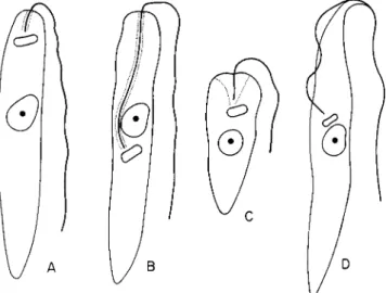
T h e trypanosomatid genera are distinguished on the basis of the morphology of adult stages and morphological forms that occur in their life cycle. Wallace (1961) proposed the following morphological criteria for differentiating strictly entomic genera of the Trypanosomatidae.
T h e genus Leptomonas Kent comprises flagellates with a lanceolate body and with the blepharoplast placed near the anterior end of the body; the flagellum emerges through a reservoir having a short, narrow opening.
T h e genus Herpetomonas Kent resembles Leptomonas morphologi
cally except that in some forms the blepharoplast is posterior to the nucleus; the flagellum always passes through a long reservoir extending from the blepharoplast to the anterior end of the body.
T h e genus Crithidia Leger includes species with short and truncate bodies; a short flagellum passes through a deep, funnel-shaped reservoir.
N o u n d u l a t i n g m e m b r a n e is present.
1 According to a new taxonomic concept of the genus Crithidia the so-called
"crithidial form" having an u n d u l a t i n g membrane no longer applies to this genus.
For this reason, it is suggested to avoid this term and replace it with a new and proper term "blastocrithidial form" derived from the generic n a m e Blastocrithidia Laird.
T h e genus Blastocrithidia Laird includes those flagellates which par
asitize insects exclusively and have the u n d u l a t i n g membrane.
T h e features of these genera are summarized in Fig. 1.
These criteria, morphological in nature, are not a safe basis for identifying entomophilic trypanosomatids found in insects. In cases of
FIG. 1. Morphological features of four strictly entomophilic genera of Trypa
nosomatidae: ( A ) Leptomonas; (B) Herpetomonas; ( C ) Crithidia; (D) Blastocrithidia.
(Original.)
mixed infection by flagellates of two different genera it is impossible to make a proper identification of the parasites involved. Therefore, in any doubtful case, the pure-culture method should be applied and sero
logical tests must be carried out.
T h e n u m b e r of strictly entomophilic species of the family Trypanoso
matidae reported in the literature is close to 120 species. Unfortunately, we do not know how many of them are really valid species. It was a common practice to describe new species of Leptomonas, Herpetomonas, or Crithidia just on the basis of a host without special consideration that it may be only the leptomonad or blastocrithidial stage of digenetic species of Leishmania, Phytomonas, or Trypanosoma found in their insect vector. Several corrections have been already made; for example, the formerly recognized species Crithidia melophagia Flu from Melo- phagus ovinus (Linnaeus) was identified as the crithidial stage of Tryp
anosoma melophagium (Flu) in its insect vector. For the reason men
tioned above, a systematic revision of entomophilic Trypanosomatidae
340 JERZY J . LIPA
seems to be rather urgent, and complex criteria should be worked out including morphological, biochemical, and physiological features of the trypanosomatids.
A n animal or plant disease caused by parasitic flagellates is some
times called "flagellosis" or "flagellatosis." Although the first term is more convenient it is, however, incorrect etymologically; for this reason we shall use the term "nagellatosis" for any disease caused by entomo
philic flagellates. T h i s could be extended and special distinctive names could be applied to distinguish infections caused by different genera, e.g., "leptomonadosis" for infections caused by Leptomonas species. At present, however, it seems unnecessary to employ more than one term
to designate the different flagellatoses.
a. Flagellatoses caused by Leptomonas species. A n u m b e r of flagel
lates were described u n d e r the generic n a m e Leptomonas Kent. At pres
ent about 40 species are known, mostly found in Hemiptera, Diptera, and Siphonaptera. However, information about the pathogenic influence of leptomonads on their insect hosts is very scanty since most papers contain only taxonomic data about the species involved.
T o a group of better-known species belongs Leptomonas pyrrhocoris (Zotta) from the hemipteran Pyrrhocoris apterus (Linnaeus) (Zotta, 1912). T h i s flagellate is a common inhabitant of the gut of larvae and adults of P. apterus, and only in a small percentage of specimens have the flagellates been found also in the salivary glands and in the hemocoel (Lipa, 1958).
T h e flagellates observed in the gut are mostly haptomonads attached to the epithelium by short flagella, though free leptomonad and leish
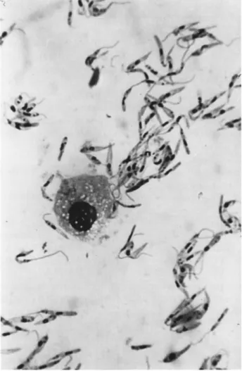
manial forms, the latter frequently encysted, are also numerous. I n the hemolymph the most prevalent are leptomonad forms 15 μ long by 2 to 3 μ wide and having a flagellum about 8 to 12 μ in length (Fig. 2).
T h e pathological influence of L. pyrrhocoris on its natural host is rather weak when it inhabits the gut, and only diarrhea may indicate the presence of infection. I n the case of general infection, however, dis
eased insects are less active and the red color of the body becomes lighter.
T h e hemolymph containing flagellates is thicker and whitish in color instead of the normal light green.
Zotta (1921) found that many insects of different orders might be easily infected with L. pyrrhocoris by inoculation into the body cavity:
Galleria mellonella (Linnaeus) [Lepidoptera], Carausius morosus Brun
ner [Orthoptera], Calliphora sp. [Diptera], Tenebrio molitor Linnaeus [Coleoptera], and Naucoris cimicoides (Linnaeus) and Notonecta glauca Linnaeus [Hemiptera]. In the case of inoculation of L. pyrrhocoris into the hemocoel of larval G. mellonella the flagellates multiplied rapidly
FIG. 2. Leptomonas pyrrhocoris (Zotta) in the h e m o l y m p h of the red soldier bug Pyrrhocoris apterus (Linnaeus). (Original.)
and eventually the larva died. T h e flagellates migrated from the body cavity into the lumen of the intestine, which is the reverse of the migra
tion observed in the natural host P. apterus. A n infected larva of G.
mellonella might be liberated from flagellates by placing it in an in
cubator at 39°C. After 2 days all flagellates in the hemolymph died and the larvae showed no signs of infection. Flagellates developed well in T. molitor and other insects, too, and only in the hemolymph of C.
morosus did they degenerate and die after several hours or a few days.
Zotta and Teodoresco (1933) observed significant changes in the blood formula of G. mellonella infected by L. pyrrhocoris that may
342 JERZY J. LIPA
partly explain the n a t u r e of the pathogenic influence of this parasite on insects. T h e n u m b e r of proleucocytes in the hemolymph of diseased larvae was three times greater than in hemolymph of normal insects. O n the other hand, the n u m b e r of macronucleocytes and micronucleocytes was fifteen to twenty times less as compared with normal hemolymph.
These changes, however, were not only specific to flagellatosis; they were also observed when some inert materials, e.g., China ink, were injected into the body cavity.
Galli-Valerio (1920) observed that L. pyrrhocoris also developed in the plant nectar produced by flowers of Colchicus autumnalis Linnaeus.
However, the statement of Galli-Valerio that flagellates may only over
winter in the plant nectar does not appear to be correct, as the author of this chapter has frequently observed infected bugs during the winter and spring.
Several leptomonads have been described from various species of Hemiptera, but, as they are considered commensals, and because of limited space, they will not be discussed here. O n e such species, however, should be mentioned because of its outstanding features. Gibbs (1957) described Leptomonas serpens Gibbs from Nezara viridula (Linnaeus).
T h i s flagellate inhabits the gut a n d salivary glands of the plant b u g N. viridula, which feeds on the tomato plant, Lycopersicum esculentum Miller. Flagellates may also develop in the plant sap where they are introduced with saliva of infected bugs which feed on plants. Healthy insects become infected while sucking sap of such plants.
Observations m a d e by Galli-Valerio (1920) and Gibbs (1957) are quite unusual for species of Leptomonas which are assumed to be strictly entomophilic. T h i s indicates that the distinction between Leptomonas and Phytomonas may eventually become superfluous.
I n the Lepidoptera, only one leptomonad is known to occur. Paillot (1927) found that caterpillars of Pyrausta nubilalis (Hübner) suffer from flagellatosis caused by Leptomonas pyraustae Paillot. T h e incidence of infection was very low as only 4 larvae of 620 examined were parasitized by this flagellate.
T h e flagellates were observed in the gut and Malpighian tubes, usu
ally as haptomonads attached to the epithelium. T h e pathogenicity of L. pyraustae is not well k n o w n and n o specific external symptoms were described by Paillot. H e observed, however, that infected Malpighian tubes changed color, losing their transparency and becoming slightly hypertrophied.
Several leptomonads have been described from various fleas (Sipho- naptera). A well-known species is Leptomonas ctenocephali (Fantham) from the dog flea, Ctenocephalides canis (Curtis) (Fantham, 1912). It
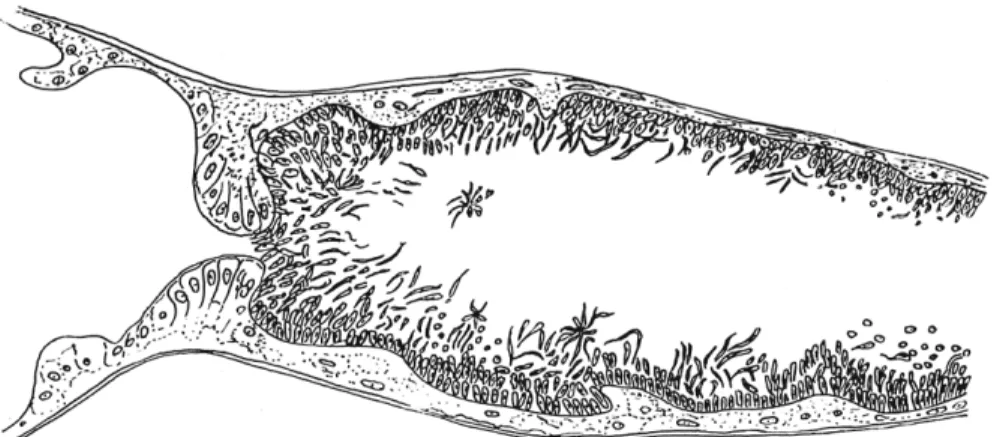
lives in the hindgut, Malpighian tubes, and, rarely, in the midgut. T h e majority of flagellates observed in the h i n d g u t are h a p t o m o n a d in form and attach to the epithelium (Fig. 3). I n the rectum, leishmania and encysted forms are prevalent, being discharged from the body with feces.
Healthy adults or larvae become infected while ingesting the feces con-
FIG. 3. Leptomonas ctenocephali (Fantham) in the hindgut of the dog flea Cteno- cephalides cams (Curtis). (From Moshkovski and Rashina, 1951.)
taining encysted flagellates. T h e pathological influence of L. ctenocephali is not well known, b u t it seems quite logical to assume that such great numbers of flagellates attached to the epithelium of the gut diminishes the capacity for ingestion of food, or, when a b u n d a n t , slows down the function of the Malpighian tubes.
A n u m b e r of leptomonads have been found in various fleas: Ctenoph- thalmus spp., Ceratophyllus spp., Pnlex spp. It seems, however, quite probable that many of them, if not all, might be identical with L. cteno
cephali as their life cycles are very similar.
b. Flagellatoses caused by Herpetomonas species. More than 40 species of flagellates from insects have been assigned to the genus Herpe
tomonas Kent. Some protozoologists do not recognize this genus and con
sider it synonymous with Leptomonas Kent (Grasse, 1952).
H e r p e t o m o n a d species are especially common among Diptera. T h e commonest species is Herpetomonas muscarum Leidy ( = H. muscae- domesticae Stein) found in m o r e t h a n 20 species of flies belonging to dif
ferent genera: Musca Linnaeus, Fannia R o b i n e a u and Desvoidy, Lucilla R o b i n e a u and Desvoidy, Scatophaga Meigen, Neuroctena R o n d a n i , Ho- molomyia Bouche, Cochliomyia Townsend, Phormia Meigen, and others (Becker, 1923b; Wenyon, 1913). A n u m b e r of flagellates related to H.
344 JERZY J . LIPA
muscarum have been described from Drosophila spp. as well (Chatton and Leger, 1911).
T h i s flagellate inhabits the gut of flies, where it occurs in herpetomo- nad and leptomonad forms which are u p to 30 μ long and 2 to 3 μ wide.
Reproduction is by longitudinal binary fission, and the two newly formed cells are connected for a relatively long period of time. T h i s fea
ture was even used by Prowazek (1904), for some time, as a specific criterium for this species. It was assumed that transmission of the flagel
lates is accomplished by cysts that are discharged with excrement from the body. Wallace and Clark (1959), however, on the basis of their study on flagellates of Phaenicia sericata (Meigen) have suggested that so-called cysts of H. muscarum seem to be morphological variants of Crithidia species. According to these authors, identification of these structures as cysts of H. muscarum is explained by the frequency of mixed infections of flies by species of Herpetomonas and Crithidia.
Pathogenicity of H. muscarum to its insect host is not well known.
Recently Kramer (1961) observed a huge n u m b e r of flagellates in the hemolymph of some dead and m o r i b u n d larvae of Musca domestica Linnaeus. Although no particular symptoms were detected, this observa
tion indicates that, u n d e r some circumstances, H. muscarum may invade the hemocoel and cause the death of its host.
T h e larvae of Tipula paludosa Linnaeus, Tipula maxima Poda, and others are sometimes infected by Herpetomonas ludvigi (Kramar). T h i s herpetomonad, originally described by Kramaf (1950) u n d e r the generic name Leptomonas, was the object of a detailed morphological work by Vickerman (1960). However, the pathogenicity of H. ludvigi is not known and requires additional study.
Herpetomonas ludvigi is restricted to the cecum. Leptomonad forms are very elongate, 8 to 26 μ in length, 1 to 2 μ in width, with the flagel- l u m u p to 17 μ long. T h e kinetoplast is bean-shaped in outline, b u t no blepharoplast was observed in any preparation. T h e largest organisms observed were haptomonads attached to the epithelium of the cecum which were u p to 34 μ long. T h e y h a d only a m i n u t e flagellum or lacked even that and were nonmotile. Crithidial forms were 9 to 14 μ long and had a very short flagellum. Trypanosome forms of the same size as the crithidia, had the kinetoplast behind the nucleus, occasionally nearer the posterior end of the body. A few small vacuoles may be present in the cytoplasm. T h e cysts, which h a d a leishmanial shape, were 3 to 4 μ in diameter and had a small nucleus and a closely approximate kineto
plast.
c. Flagellatoses caused by Crithidia and Blastocrithidia. Wallace (1943) suggested that the genus Crithidia also included several species for which
another genus should be erected. Following this suggestion, Laird (1959) separated all the forms that possess an u n d u l a t i n g m e m b r a n e from the former genus Crithidia and placed them into the new genus Blasto
crithidia. T h e species that have a truncate body and deep funnel-shaped reservoirs and no u n d u l a t i n g m e m b r a n e remained in the genus Cri
thidia.
It is not the purpose of this treatise to revise the species formerly rec
ognized as Crithidia, but, whenever feasible, we shall apply the new generic n a m e Blastocrithidia according to the present taxonomic con
cept.
T h e type species of the genus Crithidia is Crithidia fasciculata Leger from Anopheles maculipennis Meigen (Leger, 1902). As demonstrated by Wallace (1943) the life cycle of Crithidia fasciculata has no forms with an u n d u l a t i n g m e m b r a n e as was claimed by Woodcock (1914).
Crithidia fasciculata appears in two morphological forms. T h e active stage is truncate, 6 to 8 μ long and 2 to 3 μ wide whereas the attached forms are small, 3 to 4 μ long, and 2 to 4 μ wide; the flagellum is absent or very short.
T h e occurrence of C. fasciculata appears to be restricted to the adult stage of mosquitoes. N a t u r a l infections of the larval stages were reported by Patton (1907). O n the other hand, Wallace (1943) was not successful in establishing a p e r m a n e n t artificial infection of mosquito larvae but adults were easily infected. I n natural conditions, flagellates are presum
ably transmitted through ingestion of feces or contaminated food (Garnham, 1959).
Crithidia fasciculata can be maintained for years in culture on various media without any loss of infectivity. Wallace and Johnson (1961) dem
onstrated that several strains of C. fasciculata that were maintained in culture for 2 to 44 years still produced heavy infection in artificially infected mosquitoes.
According to Wallace (1943) several species of Crithidia described from mosquitoes are invalid as they are synonyms of C. fasciculata. T h e y include: H. culicis Novy et al. (as cited by Patton, 1907), L. fasciculata Woodcock, L. michiganensis Speer, Η. culicidarum Noguchi and Tilden,
C. anophelis Missiroli, and L. (Strigomonas) fasciculata Lwoff and Lwoff.
A good account on trypanosomatids of mosquitoes was given by Novy et al. (1907).
Laird (1959), in establishing the new genus Blastocrithidia, desig
nated B. gerridis (Patton) as the type species. T h i s flagellate was origi
nally described by Patton (1908) in the genus Crithidia and came from the gut of the water strider, Gerris remigis De Geer.
Blastocrithidia gerridis is a parasite of Gerris spp. occurring in the
346 JERZY J. LIP A
alimentary tract of the host from the stomodeum to the rectum. T h e body of the flagellate is slender, 18 to 60 μ long and 0.9 to 3.2 μ wide.
T h e u n d u l a t i n g m e m b r a n e extends along one side of the body. Repro
duction is by binary fission and transmission of the parasite is by cysts or congenitally. T h e pathogenicity of B. gerridis is unknown.
Among the Lepidoptera only one case of flagellatosis caused by a Blastocrithidia is known. Levadite (1905) found a blastocrithidial
flagellate in the adult female of Bombyx mori (Linnaeus), which he named Blastocrithidia bombycis (Levadite) comb. nov. (== Herpetomonas bombycis Levadite). Another female artificially infected by this blasto- crithidium died on the sixth day after infection and flagellates were observed in its hemocoel. T h e pathogenicity of B. bombycis remained unknown, however, as adults were infected simultaneously by microspo
ridia. T w o types of flagellates were observed in the hemolymph of dis
eased insects: one truncate, 6 to 10 μ in length and 4 μ in width with a flagellum of 20 μ; the other longer, 10 to 12 μ in length and 2 to 3 μ in width with a flagellum 20 μ long. I n the body of some flagellates a digestive vacuole was observed.
Several species having an u n d u l a t i n g m e m b r a n e and previously rec
ognized as Herpetomonas and Crithidia should now be assigned to the genus Blastocrithidia. Some of these species are listed below: B. culicis Novy et al. (— Herpetomonas culicis Novy, MacNeal and Torrey: Wal
lace and Johnson, 1961) from Culex pipiens Linnaeus, B. leptocoridis (McCulloch) comb. nov. ( = Crithidia leptocoridis McCulloch) from Lep- tocoris trivittatus (Say), B. sandoni (Gibbs) comb. nov. ( = Crithidia san- doni Gibbs) from Holopterna alata Linnaeus, B. familiaris (Gibbs) comb, nov. ( = Crithidia familiaris Gibbs) from Cenaeus carnifex (Fabricius).
2. Trypanosomatidae Having Insects and Vertebrates or Plants as Additional Hosts
A n u m b e r of trypanosomatid flagellates are found in the gut of insects, where they live temporarily. These flagellates usually spend only a part of their life cycle in the insect body and the remainder in a ver
tebrate or plant. T h e insect usually serves as the vector and transmits these flagellates from diseased animals and plants to healthy organisms.
T h e r e are three digenetic genera which spend part of their life cycle in insects: Phytomonas Donovan, Leishmania Ross, and Trypano
soma Gruby. T h e stages that occur in the life cycle of these species closely resemble corresponding stages in the life cycle of strictly ento
mophilic genera. Therefore, it is easy to misidentify digenetic genera with monogenetic ones. For this reason the study of the life cycle and final identification of a doubtful flagellate should be m a d e only with p u r e culture material.
T h e genus Phytomonas Donovan, with leptomonad and leishmanial stages in its life cycle, is morphologically indistinguishable from Lepto
monas except that it infect plants also, whereas Leptomonas is restricted entirely to insects. Gibbs (1957), however, demonstrated that Leptomonas serpens Gibbs may develop in the insect body Nezara viridula (Linnaeus) as well as in tomato plants. Several species of Phytomonas described from plants were found to be transmitted by insects (Niechschulz, 1922; Stein
haus, 1947). O n e well-known species of this g r o u p is Phytomonas elmas- siani (Migone) from Asclepias syriaca Linnaeus which, in its distribu
tion, coincides with the range of its vector, Oncopeltus fasciatus (Dallas) (Holmes, 1925).
T h e genus Leishmania Ross also includes species with only lepto
m o n a d a n d leishmanial stages in their life cycle; they are also digenetic and have two hosts—an insect and a vertebrate. In the insect gut the Leishmania develop into the leptomonad form, whereas in the vertebrate, they develop into the intracellular leishmanial form without a flagellum.
Sand flies, Phlebotomus spp. [Diptera], mainly serve as vectors of Leish
mania species.
T h e genus Trypanosoma Gruby includes those species that develop into blastocrithidial, leptomonad, and leishmanial forms in the insect vector, whereas in the vertebrate host, they develop into the trypanoso- mal form.
Most species of Trypanosoma live extracellularly in the gut of their insect vector, b u t some of them, e.g. Trypanosoma lewisi (Kent), develop intracellularly in the gut epithelium of its vector, the n o r t h e r n rat flea, Nosopsyllus fasciatus (Bose). It seems quite probable that in such cases the flagellates h a r m the insect vector (see Volume I, Chapter 8).
3. Entomophilic Protomonadina Other Than Trypanosomatidae Several species of flagellates belonging to the family Bodonidae of the order P r o t o m o n a d i n a inhabit the insect intestine. Retortamonas or- thopterorum (Parisi) has been found in the intestine of Gryllotalpa gryl- lotalpa Linnaeus, Tipula abdominalis Linnaeus, Ectobius lapponicus (Linnaeus) and Blatta orientalis Linnaeus (Semans, 1943). Other Retorta
monas have been found in Phyllophaga spp., Popillia japonica Newman, and in some species of T r i c h o p t e r a and Hemiptera.
B. Entomophilic Flagellates Other Than Protomonadina
A n u m b e r of flagellates from orders other than Protomonadina are associated with insects (Steinhaus, 1947). Although most of them are commensal or symbiotic in habit, some may be pathogenic for insects.
A m o n g the Polymastigida there are a n u m b e r of species found in insects (Steinhaus, 1947). Polymastix melolonthae and related species
348 JERZY J. LIPA
inhabit the intestine of Melolontha melolontha Linnaeus, Popillia ja
ponica Newman, Anomala orientalis Waterhouse, Oryctes nasicornis Lin
naeus, Tipula sp., and other insects. T h e genus Eutrichomastix includes about 6 species from various insects, the best known being E. trichop- terae Mackinnon from Tipula abdominalis Linnaeus, Tipula sp., Lim- nophilus flavicornis Fabricius, and other insects (Morgan, 1944).
Many polymastigids inhabit the gut of termites. Most are symbiotic in habit, b u t some species of Streblomastix are considered to be parasites of termites (Dogel, 1956).
Among the Hypermastigida, several species have been found to be symbiotes or commensals of termites and blattids. Well-known species are Lophomonas striata Bütschli and L. blattarum Stein from the rectum of Gryllotalpa spp., Blatta spp., and Periplaneta spp. (Semans, 1943).
III. SARCODINA
Sarcodina found in insects belong to the order Amoebina and re
semble Amoeba proteus Ehrenberg and other naked amoebae. I n the active stage, pseudopodia serve as locomotory organs and to ingest food.
N o digestive vacuole was observed in the protoplasm of parasitic amoe
bae. T h e life cycle is simple and frequently includes the formation of cysts. Reproduction is by binary fission although multiple division may also occur.
Entomophilic Amoebae
Almost all entomophilic amoebae belong to the families Amoebidae and Endamoebidae. T h e family Amoebidae include three strictly en
tomophilic genera, Malamoeba Taylor and King, Malpighamoeba Prell, and Malpighiella Minchin. However, a few entomophilic species of this family belong to the genus Amoeba Ehrenberg and Hartmanella Alex- eieff. I n the family Endamoebidae several entomophilic species have been reported which belong to four genera: Entamoeba Casagrandi and Barbagallo, Endamoeba Leidy, Endolimax Kuenen and Schwellengrebel, and Dobellina Bishop and T a t e .
Amoebic diseases are sometimes referred to as amoeboses or amoe- biasis. T h e most important and well-known amoebic diseases of insects are observed in honey bees and in grasshoppers.
1. Amoebic Disease of the Honey Bee
T h e amoebic disease of the honey bee, Apis mellifera Linnaeus, was first observed by Maassen in 1916 and the pathogen involved in this dis
ease was described by Prell (1927) u n d e r the name Malpighamoeba (Vahlkampfia) mellificae Prell. T h e genus Malpighamoeba Prell, with
only one species, M. mellificae, closely resembles the hay amoebae of the genus Vahlkampfia, b u t there are certain differences in the structure of the nucleus and the shape of the pseudopodia.
T h e vegetative stages of M. mellificae are variable in shape and size, and their protoplasm is divided into ectoplasm and endoplasm. W i t h i n the endoplasm there is a well-developed nucleus and several granules.
Pseudopodia are thin, sharp, a n d frequently angulate-curved. T h e thick walled cysts are oval, 4 to 8 μ in diameter, and contain one nucleus.
T h e life cycle of the pathogen is not well known. Hasseneim (1952) observed that the time involved between ingestion of a viable cyst and cyst formation was 21 days. T h e parasite develops exclusively in the Malpighian tubes, where it occurs in great numbers in b o t h the vegeta
tive and cyst stage. I n the intestine and in the feces, cysts may be observed.
Reproduction is asexual, exclusively by binary fission. Cyst formation involves some changes of the nucleus a n d of the cytoplasm, which forms a thick wall a r o u n d the body.
Malpighamoeba mellificae is an extracellular parasite and ingests food by pseudopodia which are introduced between or within the cells of the Malpighian tubes. Nuclei of cells that are in contact with the parasite degenerate, and the whole cell is gradually destroyed.
T h e pathological influence of the parasite on the host is perhaps mostly mechanical in nature. Prell (1927) suggested that a great n u m b e r of amoebae and cysts in the l u m e n of Malpighian tubes could hinder their function. Overwintering and immobile bees apparently do not suffer from the infection. However, any disorder, in the function of Malpighian tubes is fatal for actively flying bees. Dead bees are not observed inside a hive, and this indicates that diseased bees die while away from the hive.
T h e disease is quite evident in the spring, and for this reason it is sometimes called a "spring disease." Even bee swarms that appear to overwinter normally may eventually die in spring within 10 to 20 days.
Giordani (1959) observed that seasonal incidence of the disease in Italy was greatest in the spring from March to May, b u t a slight secondary increase in early a u t u m n , in September and October, also occurred.
T h e amoebic disease may occur alone, b u t frequently it is observed in mixed infections with nosematosis, acarine, May sickness, and paral
ysis. Morgenthaler (1939) suggests that amoebic disease alone is not very important, b u t together with nosematosis it is very serious. It can be assumed that bees infected with an amoebic disease are less resistant to other pathogens, an assumption that seems very likely as mixed infec
tions of various types are frequently observed. T h e high mortality of
350 JERZY J. LIPA
bees observed in such cases may also represent a synergistic interaction between the amoeba and other pathogens (Toumanoff, 1951).
Conditions that favor the appearance of the disease are not well known. It is perhaps influenced by unfavorable overwintering, feeding with sugar or small amounts of wax. T h e spread of the disease in a
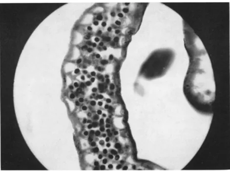
FIG. 4. Cysts of Malpighamoeba (Vahlkampfia) mellificae Prell in the l u m e n of Malpighian tubes of Apis mellifera Linnaeus. (From Giordani, 1959.)
hive is favored by diarrhea that occurs quite frequently, especially in mixed infections with nosematoses. T h i s disease spreads quickly as feces are removed from the hive by ingesting and then cysts are swallowed and the bees become infected.
T h e gradual decrease in the numbers of bees in a hive is a very char
acteristic sign of this disease. Therefore, when a hive has a relatively small n u m b e r of bees as compared to the n u m b e r of brood and a m o u n t of honey, we may suspect that it is infected with an amoebic disease.
Poltev (1953) observed that production of honey in hives with swarms infected by nosematoses and amoeboses was three times lower than in healthy hives. However, when an amoebic disease occurs alone, great reduction in honey production is not observed.
T h e r e are n o characteristic external symptoms of the disease, and diagnosis must be based on a microscopic examination of Malpighian tubes or feces. Infected Malpighian tubes are transparent and distended
because of the great n u m b e r of cysts, which are easily noticeable (Fig. 4).
Cysts are readily distinguished from Nosema, fungus, and yeast spores by their thick wall. Cysts are also found in the intestine and in the feces.
Control methods are complex and include several sanitation measures which, applied together, may give good results. Morison (1931) m a d e several recommendations which may prevent the appearance of the dis
ease: to keep hives in good conditions, to m a i n t a i n a supply of fresh water, to clean the hive inside, and to remove dead bees. Bailey (1955) and Giordani (1959) recommended removing bees from a contaminated hive to a clean one and disinfecting wooden parts or wax with the vapor of hydrochloric acid.
2. Amoebic Diseases of Grasshoppers
A m o n g the several species of amoeba that parasitize various Orthop- tera, the most i m p o r t a n t is Malamoeba locustae (King and Taylor) which causes amoebic disease of Melanoplus spp. and infects about 40 other species of grasshoppers (King and Taylor, 1936; T a y l o r and King, 1937).
T h i s amoeba parasitizes the l u m e n of Malpighian tubes and the cells of the midgut epithelium. I n the cells, the amoebae are surrounded by a large vacuole and food is taken osmotically. Infected tubules are swollen, more or less glossy, and heavily packed with cysts. T h e diameter of diseased tubules increases and the cells become greatly stretched. T h e walls may eventually r u p t u r e and liberate cysts, which are distributed by the hemolymph and thus may be found in different parts of the body. I n such cases, tumorous masses are frequently observed in muscles and fat body surrounded by hemocytes. These masses are about 1 m m in diam
eter and contain a great n u m b e r of cysts.
T h e vegetative stages of M. locustae, which are variable in size and shape, are 5 to 10 μ in diameter. Inside, the endoplasm, one nucleus, a n d u p to 30 granules are present. Pseudopodia are either spherical or fili
form. Cysts, like vegetative stages, are uninuclear a n d oval or slightly elongated. T h e y are thick walled and are 8.5 to 19 μ in length a n d 4.6 to 6.2 μ in width.
Cysts are the infective stage of the disease. T h e n u m b e r of daily cysts discharged with feces by some specimens may range from two to four million. T h e interval between ingestion of a cyst and cyst formation in the Malpighian tubes is about 14 to 18 days. T h e cannibalistic tendency of many grasshoppers greatly favors spread of the disease.
External symptoms of the disease greatly depend on the degree of infection. I n a light infection there is a lack of any particular symptoms, b u t as the disease progresses grasshoppers become less active a n d lose their appetite. Obviously, a great n u m b e r of trophozoites
352 JERZY J. LIPA
and cysts in the Malpighian tubes cause a progressive disorder in the function of tubules and the normal metabolism of the insect is dimin
ished. Heavily infected nymphs and adults become very sluggish, finally entering a lethargic state, and are not able to m a i n t a i n a normal posi
tion. Death is preceded by tetanic convulsions clearly seen in muscles of the legs and mouthparts.
T h e host range of M. locustae is very wide. T h e parasite was orig
inally described from species of Melanoplus: M. mexicanus Saussure, Μ. femurrubrum De Geer and M. differentialis (Thomas). Experiments with artificial infections showed, however, that 37 additional species from other subfamilies were susceptible: Cyrtacanthacrinae with 18 species, Oedipodinae with 14 species, and Acridinae with 5 species.
Taylor and King (1937) demonstrated that the incidence of amoebic disease could be increased in the field in grasshopper populations. T h e y collected feces of diseased insectary-reared grasshoppers which contained cysts and mixed them with b r a n and molasses. Using this as a microbial insecticide they sprayed plants and soil in plots of about 9.5 square meters where the population of grasshoppers was high and slightly in
fected with amoebae (2 infected out of 633 collected insects). Insects col
lected on the eighth week after application were infected in a much higher degree in the sprayed area (20 infected out of 422 collected in
sects). These data indicate that microbial control of grasshoppers with Malamoeba seems feasible b u t requires additional study.
3. Amoebic Diseases of Other Insects
Little is known about the pathogenicity of other entomophilic amoeba from such genera as Entamoeba, Endolimax, Hartmanella, Dobellina, Malpighiella, and others, b u t some of them appear to have pathological potentiality.
Minchin (1910) described Malpighiella refringens Minchin from the northern rat flea Ceratophyllus (— Nosopsyllus) fasciatus (Bosc) which may also infect Ctenocephalides canis (Curtis). Malpighian tubes of fleas are filled with large numbers of trophozoites a n d cysts of this parasite.
T h i s amoeba resembles, in many features, Malpighamoeba mellificae, b u t it differs in the n u m b e r of nuclei inside the cysts which are tetranu- clear.
Keilin (1917), and Bishop and T a t e (1939) studied Dobellina mesnili (Keilin), which frequently occurs as a huge mass in the space between the peritrophic m e m b r a n e and gut epithelium of Trichocerca hiemalis Meigen and T. annulata Meigen. T h e incidence of infection showed great seasonal variations; the greatest n u m b e r of infected insects was ob-
served in J a n u a r y (92 percent) whereas in May the disease disappeared almost completely.
IV. CILIOPHORA
T h e subphylum Ciliophora is conventionally divided into two classes, Ciliata and Suctoria. Corliss (1961a) proposed a new classification in which this subphylum would have only one class Ciliata, the Suctorida being one of nine orders in the subclass Holotricha. Suctorida differ from other holotrichans by the absence, in the adult stage, of cilia; the cilia having been replaced by tentacles.
Entomophilic Ciliata
Several ciliates are found associated with insects, b u t only a few of them are pathogenic for their hosts (Wenyon, 1926; Steinhaus, 1947).
T h e majority of ciliates are endocommensals in the gut of insects, mostly blattids and termites. However, a n u m b e r of them are ectocommensal or ectoparasitic in habit and live attached to the body of aquatic insects, e.g., Epistylidae or Suctoridae. T h e most pathogenic species belong to the order Holotricha and the family Tetrahymenidae, including many facultative parasites, which u n d e r special conditions cause fatal cilia- toses of various aquatic insects.
1. Ciliatoses of Mosquitoes and Chironomids
Corliss (1960) indicated that several ciliates reported from Chiro- nomidae and Culicidae, and described u n d e r different generic names such as Glaucoma Ehrenberg, Lambornella Keilin, Balantidium Clap- erede and Lachman, Protobalantidium, Leptoglena, and Turchiniella Grasse and Boissezon are synonymous with two or three species belong
ing to the genus Tetrahymena. Corliss presented a complete list of all known records of entomophilic ciliates which should be included in the genus Tetrahymena. H e also gave their probable identification with the presently recognized species: Tetrahymena pyriformis (Ehrenberg), Τ.
stegomyiae (Keilin), and T. chironomi Corliss; some species, because of incomplete original descriptions, were merely designated as species of Tetrahymena.
T h e first apparently authentic case of ciliatosis d u e to a tetrahy- menid was observed by L a m b o r n (1921) in Aedes scutellaris (Walker).
T h e pathogen involved in this disease was Tetrahymena stegomyiae (Keilin) originally described by Keilin (1921) u n d e r the generic n a m e Lambornella. Infected by T. stegomyiae, mosquito larvae differed from healthy ones by a different color of the cuticle which m a d e it less trans
parent. T h e fat body was greatly affected by the parasite, a n d some lobes of this tissue were completely destroyed. T h e ciliates were motile in the
354 JERZY J. LIPA
hemolymph and in all parts of the hemocoel. T h e n u m b e r of ciliates was relatively high in the head capsule and in the tracheal gill siphons, where u p to 200 ciliates were observed.
T h e typical shape of T. stegomyiae is pyriform and the length is 60 to 100 μ. Cilia are arranged in 25 to 30 rows uniformly distributed on the surface of the body. Cysts, which are readily noticed on the external surface of the larval body, are oval and 30 to 40 μ in diameter.
Infection is acquired per os by ingesting a cyst. T h e active stage that emerges from the cyst penetrates through the intestinal wall to the body cavity where it begins to multiply rapidly. Once infected by T. stego
myiae, mosquito larvae always die; therefore this ciliate may be con
sidered as a true pathogen. However, Corliss (1960) considers all species of Tetrahymena found in insects as facultative parasites since they all may live freely outside the insect body.
I n addition to its original host, Aedes scutellaris (or as Corliss claims A. alhopictus), Muspratt (1945) found that T. stegomyiae may also infect several other mosquito species: Aedes metallicus Edwards, A. aegypti (Linnaeus), A. calceatus Edwards, A. fulgens Edwards, A. marshalli T h e obald, A. haworthi Edwards, Culex decens T h e o b a l d , and C. nehulosus T h e o b a l d .
T w o other species, T. pyriformis, and T. chironomi, were also found to be pathogenic for several species of Culicidae and Chironomidae.
These species are smaller in size than T. stegomyiae and differ in the n u m b e r of rows of cilia. T h e body of T. pyriformis is pyriform and measures, on an average, 50 by 30 μ (minimal size 8 to 9 μ and maximal size about 100 μ); the cilia are in 17 to 21 rows. T h e body of T. chiro
nomi is also pyriform and measures 40 by 30 μ (never more than 50 μ); the cilia are usually in 24 to 26 rows.
T. pyriformis causes fatal infections among larvae of several chiro- nomids and mosquitoes: Culiseta annulata Schrank, Culicoides pere- grinus Kieffer, Culex pipiens Linnaeus, C. fuscocephalus T h e o b a l d , C.
gelidus Theobald, C. taeniorhynchus trisimialis, C. t. summorosus Dyar, Chironomus plumosus Linnaeus, Wyeomyia smithii Coquillett, and Aedes alhopictus Skuse.
Pathogenicity of T. pyriformis to insects other than Diptera was studied by several authors (Lwoff, 1924; J a n d a and Jirovec, 1938; Erhar- dova, 1952; McLaughlin, 1959). J a n d a and Jirovec inoculated p u r e cul
tures of T. pyriformis into the body cavity of insects of different orders:
Coleoptera, Dermaptera, Diptera, Hemiptera, Orthoptera, and Lepidop
tera. I n the insects studied, with few exceptions, ciliates grew and multi
plied, causing death of the insect hosts. These observations show that T. pyriformis has n o distinct host specificity and u n d e r favorable con
ditions may infect several insects.
McLaughlin (1959) injected larvae and p u p a e of G. mellonella with a determined quantity of T. pyrijormis which ranged from 60 to 200,000 ciliates per individual. Of 70 larvae injected with 200 ciliates, 75 percent died in 5 days and 100 percent in 12 days. Injection of 10 fifth-instar larvae with 100,000 ciliates caused 80 percent mortality in 36 hours and
100 percent in 2 days. P u p a e injected with 60 to 80 ciliates died in 3 to 5 days. Similar dosage injected into the body cavity of 50 additional p u p a e caused the death of 44 in 2 to 7 days. T h e density of parasites in some dead p u p a e ranged from 65,000 to 1,500,000 per individual. Six pupae, from the inoculated g r o u p of 50, emerged as adults within 2 days after inoculation. U p o n examination of these six moths, very few ciliates were found in the body cavity of three, which h a d appeared to be quite normal. T h e other three, which h a d already died, contained 3000 to 12,000 protozoans.
Corliss (1960) showed that T. chironorni causes a fatal infection of Chironomus plumosus Linnaeus. T h e list of hosts also includes Culiseta annulata, Culicoides peregrinus, and Culex pipiens; Corliss, however, in
dicates that these insects are only potential hosts because descriptions in the original reports are too scanty to make p r o p e r identification of the involved ciliates.
O u t of 2149 examined specimens of C. plumosus, 186 contained cili
ates, or about 9 percent infection. I n about half the cases T. chironorni was the sole parasite, and in the other half, double infections of larvae by T. chironorni and T. pyrijormis were observed; T. pyrijormis was never found as the sole parasite. T h e multiplication of the parasite in the hemocoel of a larva is very rapid. A light infection in a chironomid body could give rise to an extremely dense population of ciliates in 48 hours.
I n some heavily infected larvae the density of parasites was estimated at 100,000 to 200,000 ciliates. T h e infected larvae never p u p a t e and finally die within 8 to 41 days after infection.
T h e transmission and mode of infection remain unknown. Infection per os failed, and the only possible way seems to be through some weak
ened parts of the body d u r i n g molting.
Ciliatosis of Aedes sierrensis (Ludlow) caused apparently by Tetrahy- mena pyrijormis was reported by Kellen et al. (1961). T h e hemocoel and anal papillae of mosquito larvae were filled with a great n u m b e r of the ciliates; diseased larvae could be easily distinguished from healthy specimens by their abnormal whitish and o p a q u e color. T h e incidence of infection was very low as only a single individual with ciliatosis was disclosed out of several h u n d r e d fourth-instar larvae of A. sierrensis examined. T h e parasitized larva was active and did not exhibit any gross symptoms of the infection.
Corliss (1961b) reported an apparently new Tetrahymena sp. from
356 JERZY J. LIPA
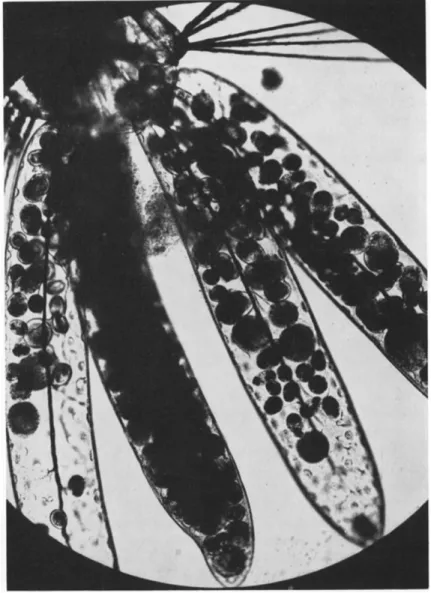
Armigeres (Leicesleria) dentatus Barraud. T h e parasite was observed in great numbers in the body cavity and papillae, b u t no data as to its path
ogenicity are available (Fig. 5). Larvae of a related species A. digitatus Edwards were also observed to suffer from ciliatosis.
Although tetrahymenids are strictly facultative parasites, they cause fatal infections in their hosts; Corliss (1961b) suggests the possibility of
FIG. 5. Anal gills of larval Armigeres (Leicesteria) dentatus infected with the ciliate Tetrahymena sp. (From Corliss, 1961b.)
employing laboratory cultures of these ciliated protozoa in experimental control of natural populations of certain mosquitoes in the tropics.
Several other ciliates have been reported to be associated with mos
quitoes, b u t they do not appear to be true pathogens (Johnson, 1903;
Christophers, 1952; Lipa and Steinhaus, 1962).
2. Ciliatoses of Other Insects
A great n u m b e r of ciliates of different orders were found to be inter
nally or externally associated with various insects (Steinhaus, 1947). Most of them are commensal in habit, e.g., Nyctotherus spp. and Balantidium spp. from the intestine of Blatta orientalis Linnaeus, Blattella germanica (Linnaeus), Gryllotalpa gryllotalpa Linnaeus, and from other Orthoptera and Isoptera (Semans, 1939; Hoyte, 1961).
However, there are numbers of ciliates that cause some h a r m to their insect hosts. Lichtenstein (1921) described a large pyriform holotrichan, Ophryoglena collini Lichtenstein from a larva of Baetis sp. [Ephem- erida]. T h e parasite, about 200 to 300 μ long, destroys various tissues including the gonads. T h e life cycle of Ophryoglena within the body of its host was studied by Codreanu (1930).
Mercier and Poisson (1923) observed an accidental infection of nymphs of Nepa cinerea Linnaeus [Hemiptera] by the holotrichan Col- poda sp. T h e parasite invaded the hemocoel, and characteristic tumors were observed inside the body and on its surface. These tumors con
tained a great n u m b e r of ciliates.
A n u m b e r of Peritricha are reported to be associated with insects mainly as epibionts, and only a few of them cause some h a r m to their hosts (Nenninger, 1948; Stammer, 1948). Lust (1950) reported 38 species of the Epistylidae which were associated with various aquatic Coleop
tera and Hemiptera. T h e four genera of this family, Pyxidium Kent, Opercularia Stein, Orb opercular ia Lust, and Operculariella Stammer serve as an excellent example of gradual adaptation from ectocommen- salism through endocommensalism to endoparasitism. Pyxidium and Opercularia live as ectocommensals on several insects, Orb opercularia may be found in the oral cavity of insects, and Operculariella parasitica Stammer parasitizes the esophagus of aquatic Coleoptera. Elson (1933) observed that Epistylis spp., attached to the body surface of the aquatic beetle Tropisternus californicus LeConte rendered the movement of the elytra difficult and hindered respiration of the beetles.
A m o n g the Suctorida, only a small n u m b e r of species are reported from insects. Dieter (1956) tabulated 20 species of the genus Discophrya which attach to the body of several aquatic Coleoptera. Other suctorians such as Periacineta bucket (Kent) are frequently observed on the body
358 JERZY J. LIPA
of Ranatra linearis (Linnaeus), and Rynchophrya palpans Collin on Hydrophilus piceus (Linnaeus). All suctorians are ectocommensal or ectoparasitic in habit and rarely h a r m their hosts. Nevertheless, when attached to the respiratory or locomotory organs they may slow down some life processes.
REFERENCES
Bailey, L., 1955. Control of amoeba disease by fumigation of combs and by fumi- gallin. Bee World, 36, 162-163.
Becker, E. R. 1923a. T h e intestinal flagellates of insects. In "Problems and Methods of Research in Protozoology" (R. Hegner and J. Andrews, eds.), p p . 248-256.
Macmillan, N e w York.
Becker, E. R. 1923b. Transmission experiments on the specificity of Herpetomonas muscae-domesticae in muscoid flies. / . Parasitol., 10, 23-34.
Bishop, Α., and Tate, P. 1939. T h e morphology and systematic position of Dobel- lina mesnili nov. gen. (Entamoeba mesnili Keilin, 1917). Parasitology, 31, 501-510.
Chatton, E., and Leger, L. 1911. Sur quelques leptomonas de Muscides et leur leptotrypansomes. Compt. rend. soc. biol., 64, 120-121.
Christophers, S. R. 1952. T h e recorded parasites of mosquitoes. Riv. parassitol., 13, 21-28.
Clark, Τ . B. 1959. Comparative morphology of four genera of the Trypanosomatidae.
J. Protozool, 6, 227-232.
Codreanu, R. 1930. Sur la phase interne du cycle evolutif de formes d'Ophryoglena, infusoires endoparasites des larves d'Ephemeres. Compt. rend. acad. sei., 190, 1154-1157.
Corliss, J. O. 1959. Comments o n the systematics and phylogeny of the protozoa.
Syst. Zool, 8 , 169-190.
Corliss, J. O. 1960. Tetrahymena chironomi sp. nov., a ciliate from midge larvae, and the current status of facultative parasitism in the genus Tetrahymena.
Parasitology, 50, 111-153.
Corliss, J. O. 1961a. " T h e Ciliated Protozoa," 310 p p . Pergamon, N e w York.
Corliss, J. O. 1961b. Natural infection of tropical mosquitoes by ciliated protozoa of the genus Tetrahymena. Trans. Roy. Soc. Trop. Med. Hyg., 55, 149-152.
Dieter, M. 1956. Säugeninfusorien auf Wasserkafern. Orion, 11 (9-10), 369-373.
Dogel, V. A. 1956. Symbiotic protozoans of termites and their biological significance for the life of their hosts. In "Infective and Protozoan Diseases of Beneficial and N o x i o u s Insects" (V. I. Poltev and M. S. Paveleva, eds.), p p . 47-62. Selhozgiz, Moskva. [In Russian.]
Elson, J. A. 1933. Protozoans and beetles. Am. Naturalist, 13, 366-368.
Erhardova, B. 1952. U m e l e vyvolany prasitismus volne zijimi provoky. Ceskoslov.
biol, 1 (3), 171-174.
Fantham, Η. B. 1912. Some insect flagellates and the problem of the transmission of Leishmania. Brit. Med. J., I I , 1196-1197.
Galli-Valerio, B. 1920. Le cycle Evolutif probable de YHerpetomonas pyrrhocoris Zotta et Galli-Valerio. Schweiz, med. Wochschr., 50, 401-402.
Garnham, P. C. C. 1959. Some natural protozoal parasites of mosquitoes with spe
cial reference to Crithidia. Trans. 1st Intern. Conf. Insect Pathol, and Biol.
Control, Praha 1958, p p . 287-294.
Gibbs, A. J. 1957. Leptomonas serpens n. sp. parasitic in the digestive tract and salivary glands of Nezara viridula (Pentatomide) and in the sap of Solanum lycopersicum (tomato) and other plants. Parasitology, 4 7 , 297-303.
Giordani, G. 1959. A m o e b a disease of the honey bee, Apis mellifera Linnaeus, and an attempt at its chemical control. / . Insect Pathol., 1, 245-269.
Grasse, P. 1952. Order des Trypanosomides. In "Traite de Zoologie" (P. Grasse, ed.), Vol. 1, Pt. 1, p p . 601-668. Masson, Paris.
Hasseneim, Μ. H. 1952. Some studies on amoeba disease. Bee World, 3 3 , 109-112.
Holmes, F. O. 1925. T h e relation of Herpetomonas elmassiani (Migone) to its plant and insect hosts. Biol. Bull, 4 9 , 323-337.
Hoyte, H. M. D . 1961. T h e Protozoa occurring in the hind-gut of cockroaches. I.
Responses to changes in environment. Parasitology, 5 1 , 415-436.
Janda, V., and Jirovec, O. 1938. Über künstlich hervorgerufenen Parasitismus eines frielebenden Ciliaten Glaucoma piriformis u n d Infektionsversuche mit Euglena gracilis u n d Spirochaeta biflexa. Mem. Soc. Zool. Tchecoslov. Prague, 5 , 34-58.
Johnson, Η. P. 1903. Α study of certain mosquitoes in N e w Jersey and statement of the "mosquito-malaria-theory." Rept. Entomol. Dept. New Jersey Agr. Coll.
Expt. Sta. for 1902 Appendix A, p p . 559-593.
Keilin, D . 1917. U n nouvelle entamibe Entamoeba mesnili n. sp., parasite intestinale d'une larve d'une diptere. Compt. rend. soc. biol., 8 0 , 133-136.
Keilin, D . 1921. O n a new ciliate, Lambornella stegomyiae n. g., n. sp., parasitic in the body cavity of the larvae of Stegomyia scutellaris Walker (Diptera, Nema- tocera, Culicidae). Parasitology, 1 3 , 216-224.
Kellen, W . R., Wills, W., and Lindgren, J. E. 1961. Ciliatosis in Aedes sierrensis (Ludlow). / . Insect Pathol., 3 , 335-338.
King, R. L., and Taylor, A. B . 1936. Malpighamoeba locustae n. sp. (Amoebidae), a protozoan parasite in the malpighian tubes of grasshoppers. Trans. Am. Micro- scop. Soc, 5 5 , 6-10.
Kramaf, J. 1950. Parasites in the larvae of the cranefly Tipula maxima Poda.
Acta Soc. Zool. Bohemoslov., Prague, 1 4 , 55-76.
Kramer, J. P. 1961. Herpetomonas muscarum (Leidy) in the haemocoele of larval Musca domestica. Entomol. News, 7 2 , 165-166.
Laird, M. 1959. Blastocrithidia n. g. (Mastigophora: Protomonadina) for Crithidia (in part), with a subarctic record for B. gerridis (Patton). Can. J. Zool., 3 7 , 749-772.
Lamborn, W . A. 1921. A protozoon pathogenic to mosquito larvae. Parasitology, 1 3 , 213.
Leger, L. 1902. Sur u n flagelle parasite de VAnopheles maculipennis. Compt. rend, soc biol., 5 4 , 354-356.
Levadite, M. 1905. Sur u n nouveaux flagelle parasite du Bombyx mori, Herpetomonas bombycis. Compt. rend. acad. sei., 1 4 1 , 631-635.
Lichtenstein, J. L. 1921. Ophryoglena collini n. sp., parasite coelomique de larves d'ephemeres. Compt. rend. soc. biol., 8 5 , 794-796.
Lipa, J. J. 1958. Pierwotniaki zyjace w roslinach. Wszechswiat, No. 1, 13-15.
Lipa, J. J., and Steinhaus, Ε. Α. 1962. Further report on identifications of Protozoa pathogenic for insects. Acta Parasitol. Polon., 9 , 165-175.
Lust, S. 1950. Symphorionte Peritrichen auf Käfern u n d Wanzen. Zool. Jahrb. Abt.
Systematik, 7 9 , 353-436.
Lwoff, A. 1924. Infection experimentale ä Glaucoma pyriformis (infusoire) chez Galleria mellonella (lepidoptere). Compt. rend. acad. sei., 1 7 8 , 1106-1108.
360 JERZY J. LIPA
McLaughlin, R. E. 1959. Experimental infection of larval and pupal stages of Galleria mellonella (L.) (Pyralidae, Lepidoptera) by the ciliate Tetrahymena pyri
formis (Ehrbg.). / . Protozool., 6 (Supplement), 27.
Mercier, L., and Poisson, R. 1923. U n cas de parasitisme accidentel d'une nepe par un infusoire. Compt. rend. acad. sei., 1 7 6 , 1838-1841.
Minchin, H. A. 1910. On some parasites observed in the rat-flea (Ceratophyllus fasciatus). Festschr. 60, Geburstag R. Hertwigs, 1 , 289-302.
Morgan, Β. B. 1944. Parasite-host list of the genus Eutrichomastix (Protozoa: Flagel- lata). Proc. Helminthol. Soc. Wash. D.C., 1 1 , 58-60.
Morgenthaler, O. 1939. "Die ansteckende Frühjahrsschwindsucht (Nosema-Amöben- Infektion) der Bienen," 32 p p . Sauerländer, Aarau.
Morison, G. D . 1931. Amoebic disease of bees in Great Britain. Bee World, 1 2 , 56.
Moshkovski, S. D., and Rashina, M. G. (eds.) 1951. "Epidemiology and Medical Para
sitology for Entomologists," 455 p p . Medgiz, Moskva. [In Russian.]
Muspratt, J. 1945. Observation on the larvae of treehole breeding Culicini (Diptera:
Culicidae) and two of their parasites. / . Entomol. Soc. S. Africa, 8 , 13-20.
Nenninger, U. 1948. D i e Peritrichen der U m g e b e n von Erlangen mit besonderer Berücksichtigung ihrer Wirtsspezifität. Zool. Jahrb. Abt. Systematik, 7 7 169-266.
Niechschulz, O. 1922. Unsere bisherigen Kenntnisse von der Flagellatenkrankheit der Pflanzen. Z. Pflanzenkrankh. u. Pflanzenschutz, 33, 102-108.
Novy, F. G., MacNeal, W . J., and Torrey, Η. N. 1907. T h e trypanosomes of mos
quitoes and other insects. / . Infectious Diseases, 4, 223-276.
Paillot, A. 1927. Sur deux protozoaires nouveaux parasites des chenilles de Pyrausta nubilalis H b . Compt. rend. acad. sei., 8 5 , 416-420.
Patton, W. S. 1907. Preliminary notes on the life cycle of a species of Herpetomonas found in Culex pipiens. Brit. Med. J., II, 78-80.
Patton, W . S. 1908. T h e life cycle of a species of Crithidia parasitic in the intes
tinal tract of Gerris fossarum Fabr. Arch. Protistenk., 1 2 , 131-146.
Poltev, V. I. 1953. Effect of nosema and amoeba infections on the productivity of honeybee colonies in the Primorsky region. Pchelovodstvo No. 2 , 46-48. [In Rus
sian.]
Prell, Η. 1927. Beitrage zur Kenntniss einer Amöbenseuche der Honigbiene. Z.
angew. Entomol., 1 2 , 163-168.
Prowazek, S. 1904. Die Entwicklung von Herpetomonas einem mit den Trypano
somen verwandten Flagellaten. Arb. kaiserl. Gesundh., 2 0 , 440-452.
Raabe, Z. 1948. Proba rewizji systemu pierwotniakow. Ann. Univ. Mariae Curie- Sktodowska Lubin-Polon. Sect. C, 3, 259-276.
Semans, F. M. 1939. Protozoan parasites of the Orthoptera, with special reference to those of Ohio. II. Description of the protozoan parasites recognized in this study. Ohio J. Sei., 39, 157-181.
Semans, F. M. 1943. Protozoan parasites of the Orthoptera, with special reference to those of Ohio. IV. Classified list of the protozoan parasites of the Orthoptera of the World. Ohio J. Sei., 43, 221-234.
Stammer, H. J. 1948. Eine neue eigenertige entoparasitische Peritriche Oper- culariella parasitica n. g., s. sp., Zool. Jahrb. Abt. Systematik, 7 7 , 163-168.
Steinhaus, Ε. Α. 1947. "Insect Microbiology," 763 p p . Comstock, Ithaca, N e w York.
Steinhaus, Ε. Α. 1949. "Principles of Insect Pathology," 757 p p . McGraw-Hill, N e w York.
Taylor, A. B., and King, R. L. 1937. Further studies on the parasitic amoebae found in grasshoppers. Trans. Am. Microscop. Soc, 5 6 , 172-176.
Toumanoff, C. 1951. "Les maladies des Abeilles." Special issue of Revue Francaise d'Apiculture, N o . 68, 325 pp.
Vickerman, K. 1960. Herpetomonas ludvigi (Kramaf, 1950) n. comb., the tryp- anosomatid parasite of cranefly larvae (Diptera, Tipulidae). Parasitology, 50, 351-363.
Wallace, F. G. 1943. Flagellate parasites of mosquitoes with special reference to Crithidia fasciculata Leger, 1902. / . Parasitol., 29, 196-205.
Wallace, F. G. 1961. Criteria for the differentiation of genera among the trypa- nosmatid parasites of insects. Abstr. 1st Intern. Conf. Protozoologists, Praha, p p . 230-231.
Wallace, F. G., and Clark, Τ . B. 1959. Flagellate parasites of the fly Phaenicia seri- cata (Meigen). / . Protozool., 6 , 40-42.
Wallace, F. G., and Johnson, A. 1961. T h e infectivity of old cultured strains of mosquito flagellates. / . Insect. Pathol., 3, 75-80.
W e n y o n , C. M. 1913. Observations o n Herpetomonas muscae domesticae and some allied flagellates. Arch. Protistenk., 31, 1-34.
W e n y o n , C. M. 1926. "Protozoology," 1563 p p . W i l l i a m W o o d , N e w York.
Woodcock, Η. M. 1914. Further remarks on the flagellate parasites of Culex. Is there a generic type, Crithidia? Zool. Anz. 44, 26-33.
Zotta, G. 1912. Sur un flagelle du type Herpetomonas chez Pyrrhocoris apterus.
Ann. sei. univ. Jassy, 7, 211-223.
Zotta, G. 1921. Sur la transmission experimentale du Leptomonas pyrrhocoris Z.
chez des insectes divers. Compt. rend. soc. biol., 85, 135-137.
Zotta, G., and Teodoresco, A. M. 1933. Formule leucocytaire de la chenille de Galleria mellonella infectes par le Leptomonas pyrrhocoris. Compt. rend. soc.
biol, 114, 314-316.