BrainResearchBulletinxxx (2012) xxx–xxx
ContentslistsavailableatSciVerseScienceDirect
Brain Research Bulletin
j ourna l h o me p a g e:w w w . e l s e v i e r . c o m / l o c a t e / b r a i n r e s b u l l
Research report
Microinjection of RFRP-1 in the central nucleus of amygdala decreases food intake in the rat
Anita Kovács a , Kristóf László a , Rita Gálosi a , Krisztián Tóth a , Tamás Ollmann a , László Péczely a,b , László Lénárd a,b,∗
aInstituteofPhysiology,PécsUniversityMedicalSchool,Pécs,Hungary
bNeurophysiologyResearchGroupoftheHAS,PécsUniversityMedicalSchool,Pécs,Hungary
a r t i c l e i n f o
Articlehistory:
Received22December2011 Receivedinrevisedform31May2012 Accepted1June2012
Available online xxx Keywords:
Amygdala RFRP-1 Antagonist Feeding
a b s t r a c t
SeveralmembersoftheRFamidepeptidefamilyareknowntohaveroleintheregulationoffeeding.For example,neuropeptideFFandprolactin-releasingpeptidecauseanorexigenic,while26RFaandQRFP resultinorexigeniceffectsinrodents.I.c.v.microinjectionofneuropeptideRFRP-1significantlyreduced foodandwaterintakeinchicks.However,feedingrelatedeffectsofRFRP-1havenotbeenstudiedin mammalsyet.Thecentralpartofamygdala(CeA)isessentiallyinvolvedintheregulationoffeedingand bodyweight.RFRP-1positivenervecellsweredetectedintherathypothalamusandRFRP-1immunore- activefiberswereidentifiedintheCeA.RFRPanalogsbindwithrelativelyhighaffinitytotheNPFF1 andNPFF2receptors(NPFF-R).RFRP-1haspotentactivityforNPFF1.SignificantexpressionofNPFF1 wasdetectedintheCeA.ToevaluatetheroleofRFRP-1infeedingregulationratsweremicroinjected withdifferentdosesofRFRP-1andtheirfoodintakewerequantifiedovera60minperiod.Liquidfood intakeofmaleWistarratswasmeasuredafterbilateralintraamygdaloidadministrationofRFRP-1(25, 50or100ng/side,RFRP-1dissolvedin0.15MsterileNaCl/0.4l,respectively).The50ngdoseofRFRP-1 microinjectionsresultedinsignificantdecreaseoffoodintake.The25and100nghadnoeffect.Action of50ng(37.8pmol)RFRP-1waseliminatedby20ng(41.4pmol)RF9NPFF-Rantagonistpretreatment.
Inopen-fieldtest50ngRFRP-1didnotmodifyspontaneouslocomotoractivityandgeneralbehavior ofanimalsdidnotchange.OurresultsarethefirstreportingthatRFRP-1injectedtotheCeAresultin adecreaseofliquidfoodconsumption.Thisisareceptor-linkedeffectbecauseitwaseliminatedbya NPFF-Rselectiveantagonist.
© 2012 Elsevier Inc. All rights reserved.
1. Introduction
RFRP-1 is a member of the RFamide peptide family. To date, five groups of the RFamide peptides have been documented: NPFF (PQRFa) group, PrRP group, LPXRFamides (RFRPs, GnIH), Kisspeptin group and QFRP (26RFa) group (Ukena and Tsutsui, 2005; Fukusumi et al., 2006; Osugi et al., 2006). Several members of the RFamide family, containing a terminal arginine (R) and amidated pheny- lalanine (F), affect appetite-associated processes in a wide range of species (Dockray, 2004; Bechtold and Luckman, 2007). Some of these peptides cause anorexigenic or orexigenic effects in rodents (Murase et al., 1996; Lawrence et al., 2002; Chartrel et al., 2003;
Bechtold and Luckman, 2006; Takayasu et al., 2006). The mam- malian members of the LPXRFamide peptide family are the RFRP-1
∗Correspondingauthorat:InstituteofPhysiologyandNeurophysiologyResearch GroupoftheHAS,PécsUniversityMedicalSchool,Szigetistr.12,P.O.Box99,H-7602 Pécs,Hungary.Tel.:+3672536432;fax:+3672536423.
E-mailaddress:Laszlo.Lenard@aok.pte.hu(L.Lénárd).
and RFRP-3 as well as the RFRP-2. These peptides are derived from a same single precursor protein (Prepro-RFamide-related Peptides).
RFRP-2 sequence was absent in rat and mouse preprotein indi- cating that RFRP-2 has no significant function in these animals (Hinuma et al., 2000). RFRP-3 has emerged as important regula- tor of reproductive function (Yoshida et al., 2003; Murakami et al., 2008; Pineda et al., 2010a,b; Smith and Clarke, 2010). I.c.v. admin- istration of RFRP-1 in rats has also been shown to raise circulating levels of prolactin in a concentration-dependent manner and did not affect the secretion of other pituitary peptides (Hinuma et al., 2000). RFRP-1 acts in the hypothalamus to inhibit dopaminergic neuronal activity (Willis et al., 2003). The RFRP gene was expressed in the caudal hypothalamus including the dorsomedial hypotha- lamus (DMH) and the periventricular nucleus (PerVN) (Fukusumi et al., 2001; Yano et al., 2003). The DMH plays an important role in the control of energy metabolism.
The administration of RFRPs induces c-Fos protein expression in the arcuate nucleus (ARC), which has a key role in the regulation of feeding behavior. I.c.v. administration of RFRP-3 stimulates food intake in male rats and ovariectomized female rats (Yoshida et al.,
0361-9230/$–seefrontmatter© 2012 Elsevier Inc. All rights reserved.http://dx.doi.org/10.1016/j.brainresbull.2012.06.001
to project directly to cells containing CRH or oxytocin (Qi et al., 2009) in the hypothalamus. Furthermore, i.c.v. administration of RFRP-1 or RFRP-3 induces anxiety-related behavior (Kaewwongse et al., 2011). RFRP-1 plays a role in the processing of pain in mice (Yudin et al., 2006), increases the magnitude and duration of spinal morphine anti-nociception in rats (Jhamandas et al., 2006) and negatively affects adipogenesis in human and mouse cell cultures (Herrera-Herrera and Salazar-Olivo, 2008).
Immunohistological assays identified RFRP-1 immunoreactive cell bodies in the supramammillary nucleus of hypothalamus (suMM), PVN, ventral posteromedial thalamic nucleus (ThVPM), medial preoptic area (MPO) as well as RFRP-1 immunoreactive fibers were detected in the bed nucleus of the stria terminalis (BST), nucleus of the vertical limb of the diagonal band (VDB) and central amygdaloid nucleus (CeA). Several results showed that RFRP-1 and RFRP-3 are produced in the same neurons around the dorsome- dial hypothalamus of rats (Fukusumi et al., 2001; Yano et al., 2003, 2004; Yoshida et al., 2003).
Biological responses mediated by RFRPs peptides result from high affinity binding to two NPFF receptors, i.e. NPFF1 receptor and NPFF2 receptor, respectively. The NPFF2 receptors were identified in the amygdala (AMY) (Bonini et al., 2000; Hinuma et al., 2000;
Liu et al., 2001; Engstrom et al., 2003) but they were not found in the CeA (Liu et al., 2001). On the other hand, significant expression of NPFF1 receptors were detected in the CeA and the medial, and basolateral amygdaloid nuclei (Liu et al., 2001).
The amygdala plays an important role in feeding and body weight regulation. Molecular biological (Bonini et al., 2000) and immunohistochemical (Liu et al., 2001) investigations have revealed significant expression of NPFF1 receptors in the CeA. Since i.c.v. applied RFRP-1 decreased food intake in chicks (Newmyer and Cline, 2009) and because the rat CeA contains both RFRP-1 fibers and NPFF1 receptors, we hypothesized that RFRP-1 infusion into the CeA of rats may result in decrease of food consumption. In mammals feeding related effects of RFRP-1, however, have not been examined so far. Therefore, our present experiments were designed to examine liquid food intake of male Wistar rats after bilateral RFRP-1 microinjections into the CeA. It was also studied whether application of NPFF receptor antagonist RF-9 can prevent the effect of RFRP-1 in the CeA. In open-field test it was examined whether RFRP-1 microinjections modify spontaneous locomotor activity or induce anxiety-like behavior.
2. Materialsandmethods
2.1. Subjects
Subjectswere96maleWistarrats(LATI,Gödöll ˝o,Hungary)weighing280–320g atthebeginningofexperiments.Animalswerehousedindividuallyandcaredfor inaccordancewithinstitutional(PécsUniversityMedicalSchool)andinternational standards(EuropeanCommunityCouncilDirective86/609/EEC).Ratswerekeptin alight-andtemperature-controlledroom(12:12hlight–darkcyclewithlightson at06:00a.m.,22±2◦C).Tapwaterandstandardlaboratoryfoodpellets(CRLT/N standardrodentfoodpellet,CharlesRiverLaboratories,Budapest,Hungary)were availableadlibitumbeforeexperiments.Dailyfoodandwaterconsumptionand bodyweightweremeasuredtothenearestgramsandmilliliters,respectively.
Inthefirsttwoexperimentsliquidfoodconsumptionwasstudiedand78ani- malswereused.Fromthe14thpreoperativedayonratsweretrainedforaweek toconsumetheliquiddiet(milk,136.45kJ/100ml,MilkQuick,Debrecen,Hungary).
laboratoryfoodpelletsandtapwaterwereavailableadlibitum.
2.2. Surgery
Rats wereanaesthetizedi.p.with ketamine supplementedwith diazepam (Calypsol,80mg/kgbwandSeduxen,2mg/kgbw,respectively,Richter,Hungary).
Stainlesssteelbilateralguidetubes(22-gauge)werestereotaxicallyimplantedinto theCeA(coordinatesreferringtothebregma:AP:−2.3mm,ML:4.1mmandDV:
6.5mmventralfromthesurfaceofthedura)accordingtostereotaxicatlasofPaxinos andWatson(1986).Thetipsofcannulaewerepositioned0.5mmabovetheintended injectionsite.Cannulaewerefixedtotheskullwithacryliccementandstainless steelscrews.Whennotbeingusedforinjection,theguidetubeswereoccluded withstainlesssteelobturatorsmadeof27-gaugestainlesssteelwire.Animalswere allowedtohaveaminimumof5daysforpostoperativerecoverybeforeexperiments commenced,duringthattimetheywerefrequentlyhandled.
2.3. Experimentalprocedures
2.3.1. Druginjectionsandliquidfoodintakemeasurements
RFRP-1(048-48PhoenixPharmaceuticals)wasdissolvedin0.15Msterilesaline attheappropriatedosesforbilateralintraamygdaloidmicroinjectionsinavol- umeof0.4l.Drugsorvehicle(0.15Msterilesaline)weremicroinjectedthrough a27-gaugestainless-steelinjectiontubeextending0.5mmbelowthetipsofthe implantedguidecannulae.Theinjectioncannulawasattachedviapolyethylenetub- ing(PE-10)toaHamiltonmicrosyringe(25l,Bonaduz,Switzerland).Drugswere injectedfor1minbyautomatedsyringepumps(ColeParmer,USA),andtheinjec- tioncannulawasleftinplaceforanadditional1mintoreducetheamountofdrug drawnupthecannulatrack.Awakenanimalswereinjectedintheirhomecageand thebilateralinjectionproceduretook5min.Followingmicroinjectionsliquiddiet intakewasmeasuredatmillilitersaccuracyevery5minfor30minandthe40th, 50th,60thminute.Datawereanalyzedandpresentedfor60minofmeasurements.
Inthefirst2experimentswithinsubjectdesignwasapplied,i.e.eachanimal servedasitsowncontrol.Inthefirstexperimentwestudiedtheeffectsofdifferent dosesofRFRP-1infoodintake.Thewithinsubjectdesignmeantthatconsumptions ofthesameratweremeasuredandcomparedaftereithervehicleorafteronedose ofRFRP-1.Animalsweremicroinjectedwith25ng(18.93pmol),50ng(37.8pmol) or100ng(75.7pmol)RFRP-1intotheleftandrightCeA.(Inthisreportallthedoses mentionedaremeanttobethedosepersidevalue.)Ascontroltreatmentvehicle solutionwasappliedinthesamevolume.
InthesecondexperimenttheeffectofRF9antagonistwasstudied.Thetotal volumeofinjectionwas0.8l(0.4l+0.4l)persideineachofthecasesof thisexperimentbecauseoftheantagonistpretreatments.Atfirstweexamined theeffectofRFRP-1microinjectionwith extravolume (0.8l). Animalswere treated with 0.4l vehicle15min priorto a second 0.4l vehicle injection (vehicle+vehicle)orto50ngRFRP-1injections(vehicle+50ngRFRP-1).Animals thatweretestedfortheeffectofANTreceived20ng(41.4pmol)NPFFreceptor antagonist(ANT,RF-9,R4282,SigmaChemicalCo.)microinjected15minbefore thevehicle(ANT+vehicle)orvehiclewasmicroinjectedbeforethesecondvehicle injection(vehicle+vehicle).AtexaminationofantagonisttreatmentonRFRP-1, animalsweremicroinjectedwith20ngANTbeforethevehicle(ANT+vehicle)or beforethebilateral50ngRFRP-1application(ANT+50ngRFRP-1),respectively.
Vehiclesolution,ordrugapplicationsweremadeoncounterbalancedmanner (i.e.,applicationswererandomlystartedwithvehicleordrugswithinsubjects).A minimumof3-dayperiodseparatedthedrugorvehicleadministrations.
Inthethirdexperimentbetweensubjectsdesignwasapplied,i.e.open-field activityofvehicletreatedandRFRP-1injectedgroupswasmeasuredandanalyzed.
Animalsweremicroinjectedwith50ngRFRP-1orvehicleintotheleftandrightCeA.
2.3.2. Open-fieldtest
Open-fieldtestwasusedtomeasurethespontaneousmotoractivity.Animals wereputintoa60cm×60cm×60cmgraypaintedcageonedaybefore(Basalactiv- ity)and10minafter(Test)bilateral50ngRFRP-1orvehiclemicroinjections.The groundoftheboxwasdividedinto16identicalsquaresbypaintedlines.Behavior ofeachratwasrecordedbymeansofCCDcamcorder.Resultswereanalyzedby NoldusEthoVisonSystem(NoldusInformationTechnology,TheNetherlands).Dur- ingobservationperiod(5min)thenumberofcrossingsandthedistancemovedwere investigated.InbothgroupsBasalactivityanddatameasuredduringTestsession werecomparedandanalyzed.
A.Kovácsetal./BrainResearchBulletinxxx (2012) xxx–xxx 3
Fig.1.Illustrationofreconstructedinjectionsites.CorrectbilateralinjectionplacementsareindicatedasclosedcirclesintheCeAonpanelA(n=83).Incorrectinjection placementsareindicatedonpanelB(n=13).BrainstructurediagramsofcoronalsectionsareadaptedfromthestereotaxicatlasofPaxinosandWatson(1986).Thenumbers refertoanterior–posteriordistancefrombregmainmm.IdenticalsymbolsonpanelBindicatecoherentinjectionsitesofbilateralinjections.Numbersabovesymbolson panelsAandBindicatenumbersofanimals.C:RepresentativehistologicalpictureofbilateralcorrectinjectionintotheCeA.Cresylvioletstaining.Barbelowpicture:1mm.
2.4. Statisticalanalysis
Allresultswereexpressedasamean±S.E.M.Dataoffeedingrelatedexperi- mentswereevaluatedbyrepeated-measuresanalysisofvariance(ANOVA,SPSSfor Windows11.0).Whentheanalysisofmaineffectand/ortheinteractionshowed significance,ANOVAwasfollowedbypaired-samplesttestanalysis.Thiswasan appropriatemethodbecauseintheseexperimentseachanimalservedasitsown control.Dataoftheopen-fieldexperimentswereanalyzedbytwo-wayANOVAfol- lowedbyTukeyandBonferroniposthoctests.Thestatisticalrejectioncriterionwas setatp<0.05level.
2.5. Histology
Inordertoverifycannulaeplacements,animalswereanaesthetizedwiththe sameprocedureasusedforsurgeryandperfusedtranscardiallywith0.15Msaline followedby10%formalinsolution.Brainswereslicedwithafreezingmicrotome in40msectionsandstainedwithCresylviolet.Injectionsiteswerereconstructed accordingtoastereotaxicatlas(Fig1A–C)(PaxinosandWatson,1986).Onlydata fromratswithcorrectlyplacedcannulaewereanalyzed.Thetrackofcannulaeand thetipsweredeterminedonthebasisofexistenceofdebrisandmoderateglial proliferation.Thirteenofthe96operatedratswereexcludedfromdataanalysis
(Fig.1AandB).Inthefirstandsecondexperiments78animalswereused.Among theserats,in7cases,thecannulatipsweresymmetricallyenteredintotheliquor space,sotheywereoutofthebrainatitsventralsurface.In2cases,cannulatips locatedlaterallyormediallyand1mmabovetheamygdala,soinjectionsweremade inthecaudate-putamenononesideandintheinternalcapsuleontheotherside.In 1case,cannulatipswereplacedlaterallyormediallytothetargetarea,soinjections weremadeinthelateralandbasolateralamygdalaorinthemedialamygdaloid nucleus.In1case,cannulatipsweresymmetricallylocated1mmbelowthetarget area,sobilateralinjectionsweremadeinthebasomedialamygdala.Theseinjections wereineffectivetomodifyfoodintake,butthesefewdataarenotenoughtodraw far-reachinginference.Inthethirdexperiment18ratswereused.Amongtheserats, in2cases,thecannulatipsweresymmetricallyenteredintotheliquorspacesothey wereoutofthebrain.
3. Results
Within 3 days after surgery, the body weight of all animals
reached the preoperative level. Neither hypophagia nor hypodipsia
were observed after the 3rd postoperative day and animals showed
continuous increase in body weight. Food intake tests started from
0 0,5 1 1,5 2 2,5 3 3,5
60`
50`
40`
30' 25' 20' 15' 10' 5' 0'
Time (min)
Food intake (ml/100g bw)
Vehicle+Vehicle ANT+Vehicle
n=10
(E)
0 0,5 1 1,5 2 2,5 3 3,5
60 50`
40`
30' 25' 20' 15' 10' 5'
0'
`
Time (min)
ANT+Vehicle ANT+50ng RFRP-1
n=12 (F)
0 0,5
60`
50`
40`
30' 25' 20' 15' 10' 5' 0'
Time (min)
Food intake (ml/100g bw)
Vehicle+50ng RFRP-10
60`
50`
40`
30' 25' 20' 15' 10' 5' 0'
Time (min)
Food intake (ml/100g bw)
25ng RFRP-1
(B)
0 0,5 1 1,5 2 2,5
60`
50`
40`
30' 25' 20' 15' 10' 5' 0'
Time (min)
Food intake (ml/100g bw)
v ehicle50ng RFRP-1n=11
* * * * * * * *
*
0 0,5 1 1,5 2 2,5
60`
50`
40`
30' 25' 20' 15' 10' 5' 0'
Time (min)
Food intake (ml/100g bw) Food intake (ml/100g bw)
v ehicle100ng RFRP-1
n=9
(C)
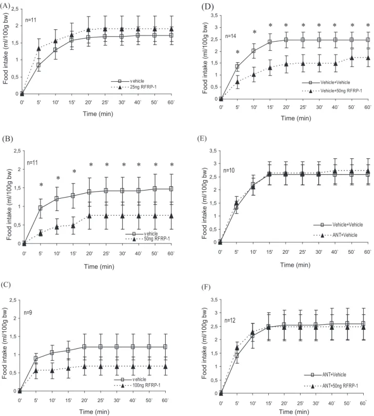
Fig.2.FeedingrelatedeffectsofRFRP-1andRF-9(ANT)intheCeA(A–F).CumulativeliquidfoodintakeafterbilateralapplicationofRFRP-1(A)25ng/side,(B)50ng/side,(C) 100ng/sideorvehiclemicroinjection,(D)cumulativeliquidfoodintakeafterbilateralapplicationofvehicletreatmentfollowedbythemicroinjectionofvehicleor50ng/side RFRP-1,(E)20ngofANTorvehiclewereappliedbilaterallybeforevehiclemicroinjection,and(F)20ngofANTwasappliedbilaterally15minbeforevehicleor50ngRFRP-1 microinjection.Linewithsymbolsrepresentsmeanfoodintakeml/100gbodyweight(±S.E.M.).Symbolabovelinesindicatessignificantdifference(*p<0.05).
the fifth postoperative day. Fig. 2 shows 1 h cumulative evalua- tion of data from measurements after distinct treatments. Values in the figures represent mean liquid food consumption in ml/100 g body weight (
±S.E.M.), and “n” is the number of animals used in the experiments.
In the first experiment effects of different doses of RFRP-1 intraamygdaloid microinjections were examined. As indicated in Fig. 2A, bilateral microinjections of 25 ng RFRP-1 did not mod- ify food intake. After the microinjection of 25 ng RFRP-1 (n = 11), repeated-measures ANOVA showed significant effect of time (F
[8, 80] = 13,079,
p< 0.001), and no significant effect of treatment (F [1, 10] = 0.376,
p> 0.05) nor time
×treatment interaction (F [8, 80] = 0.970,
p> 0.05). Bilateral microinjection of 50 ng RFRP-1 into the CeA resulted in significant liquid food intake reduction (Fig. 2B,
n= 11). ANOVA analysis yielded significant effect of time (F [8, 80] = 3128,
p< 0.004), treatment (F [1, 10] = 29,345,
p< 0.001) and no significant effect of time
×treatment (F [8, 80] = 0.293,
p> 0.05).
Paired-samples
ttest analysis showed significant reduction in liq-
uid food intake at any time points (p < 0.01). In case of 100 ng RFRP-1
treatment food intake somewhat decreased, however ANOVA
A.Kovácsetal./BrainResearchBulletinxxx (2012) xxx–xxx 5
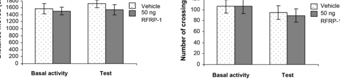
Fig.3. EffectsofRFRP-1intheopen-fieldtest.(A)Columnsrepresentmean(±S.E.M.)distancemovedintheopen-fieldapparatusonedaybefore(Basalactivity)and10min after(Test)bilateral50ngRFRP-1orvehiclemicroinjectionsintotheCeA.(B)Columnsrepresentmean(±S.E.M.)numberofcrossingsduringBasalactivityandTestsessions, respectively.Vehicle:vehicletreatedrats(n=8);50ngRFRP-1:animalsmicroinjectedwith50ngRFRP-1(n=8).
indicated no significant effect of treatment (F [1, 8] = 1.222,
p> 0.05) nor time
×treatment interaction (F [8, 64] = 0.261,
p> 0.05), while the effect of time was significant (F [8, 64] = 2.139,
p< 0.05) (Fig. 2C,
n= 9).
In the second experiment, effects of bilateral intraamygdaloid microinjections of RF9 antagonist were studied. The total volume of injection was 0.8
l (0.4
l + 0.4
l) per side in each of the cases in this experiment, because of the antagonist treatments. Vehi- cle + 50 ng RFRP-1 treatment into the CeA resulted in significant liquid food intake reduction similar to that observed in the first experiment (Fig. 2D,
n= 14). ANOVA analysis yielded significant effect of time (F [8, 104] = 16.999
p< 0.001), treatment (F [1, 13] = 14.093,
p< 0.01) and no significant effect of time
×treatment (F [8, 104] = 1.069,
p> 0.05). Paired-samples
ttest analysis showed significant reduction in liquid food intake at any time points (Fig. 2D,
n= 14,
p= 0.001–0.041). Bilateral microinjections of 20 ng ANT + vehicle treatment into the CeA did not cause changes in food intake compared to the results seen after vehicle + vehicle treat- ment. Repeated-measures ANOVA yielded significant effect of time (F [8, 72] = 15.838,
p< 0.001) and no significant effect of treatment (F [1, 9] = 0.574,
p> 0.05) nor time
×treatment (F [8, 72] = 2.653,
p> 0.05, Fig. 2E,
n= 10). In order to study whether ANT can prevent the effect of RFRP-1, ANT was administered into the CeA bilater- ally 15 min prior the 50 ng RFRP-1 microinjections, respectively.
The equimolar amount of NPFF-receptor antagonist pretreatment prevented the food intake decreasing consequences of the previ- ously effective 50 ng RFRP-1 (Fig. 2F,
n= 12). There was no difference between liquid food consumptions after combined ANT + RFRP- 1 treatment or vehicle microinjection. ANOVA yielded significant effect of time (F [8, 88] = 7.705,
p< 0.001), and not significant effect of treatment (F [1, 11] = 0.001,
p= 0.975) nor time
×treatment inter- action (F [8, 88] = 2.463,
p> 0.05).
In the third experiment, effects of the bilateral intraamygdaloid microinjections of 50 ng RFRP-1 were studied in open-field test.
The distance moved and the numbers of crossings were evaluated.
There were no any alterations in these parameters in the RFRP-1 treated animals compared to vehicle treated controls. ANOVA anal- ysis did not show significant difference between groups (RFRP-1 or vehicle treated animals,
F[1, 28] = 0. 926,
p> 0.05, Fig. 3A) and between sessions (Basal activity or test,
F[1, 28] = 0.450,
p> 0.05, Fig. 3B).
4. Discussion
It is well known that the AMY is essential in the control of hunger motivated behavior (Fonberg, 1966; Lenard and Hahn, 1982; Lenard et al., 1982; Hajnal et al., 1992; Crovetti et al., 1995). It has been described that either hypophagia (Fonberg, 1966; Hajnal et al., 1992) or hyperphagia (Fonberg, 1971) develops after elec- trolytic lesions of differential parts of the AMY. As far as the CeA is concerned, it has been shown that specific catecholaminergic
microlesions reducing the norepinephrine content in this structure produced hyperphagia and weight increase, while dopamine deple- tion caused hypophagia and weight decrease (Lenard and Hahn, 1982; Lenard et al., 1982). Cell-specific microiontophoretic lesions of the CeA with kainic acid, which destroy neurons in the target area but leave the passing fibers intact, also induce hypophagia and weight decrease (Hajnal et al., 1992). The amygdala is recip- rocally connected to the hypothalamus (Oomura et al., 1970) and brainstem having projections to autonomic-related centers such as the dorsal motor nuclei of the vagal nerve, nucleus of the solitary tract and the parabrachial nucleus (Hopkins and Holstege, 1978).
These regions are known to modulate feeding-related autonomic functions and constitute major relay of taste pathways in rodents.
In the amygdala different orexigenic and anorexigenic pep- tides and their receptors have been described. While direct microinjections of bombesin or bombesin-like peptides, such as gastrin-releasing peptide and neuromedin C induced satiety in the CeA (Fekete et al., 2002, 2007), similar application of orexin A increased food intake (Hangodi et al., 2006).
Less is known about feeding related effects of RFamide peptides in the amygdala, however. Members of the PrRP group and RFRP peptides belong to the RFamide peptide family that share exten- sive homology and a common RF (arginine–phenylalanine) amide C terminus. PrRP inhibits food intake in rats after CeA microinjection (Fan et al., 2005). PrRP reduced food consumption and enhanced the normal behavioral satiety sequence, suggesting that it may func- tion as a homeostatic regulator of food intake (Lawrence et al., 2002). Feeding related effects of RFRP peptides have not been stud- ied in mammals with one exception. Namely, orexigenic effects were detected in rats after i.c.v. injection of RFRP-3 (Yoshida et al., 2003; Murakami et al., 2008). As far as the RFRP-1 is concerned, it was applied i.c.v. to chicks and exhibited anorexigenic proper- ties, i.e. RFRP-1 significantly reduced both food and water intake (Newmyer and Cline, 2009). Possible anorexigenic effects of RFRP- 1 in rats, especially in the rat amygdaloid body have not been examined so far. In the rat CeA RFRP-1 immunoreactive fibers and terminals were identified (Fukusumi et al., 2001; Yano et al., 2003, 2004) and NPFF1 receptors were also found there (Yoshida et al., 2003).
Our present experiments, therefore, were designed to exam-
ine the effects of microinjections of RFRP-1 on food intake in the
rat CeA. While bilateral microinjections of 25 ng RFRP-1 did not
modify feeding and 100 ng dose of RFRP-1 had only a tendency to
decrease food intake, 50 ng RFRP-1 significantly reduced liquid food
consumption. It seems, therefore, that 50 ng RFRP-1 is the effective
dose and that similarly to those of other neuropeptides (Huston and
Oitzl, 1989; Fekete et al., 2002) an inverted U-shaped dose–effect
relationship is characteristic in case of RFRP-1. The effect of 50 ng
RFRP-1 was specific because it could be eliminated by NPFF receptor
antagonist. The food intake decreasing effect of RFRP-1 was rapid
and short, lasted for 20 min. Since we used cumulative evaluation,
could be due to anxiogenic effect which was detected after RFRP- 1 and RFRP-3 treatments in rats (Kaewwongse et al., 2011). In these experiments, however, very high dose of RFRP-1 was applied [10
g; 7.5 nmol, which is approximately 200
×higher than the effective dose (50 ng; 37.8 pmol) applied in the present study]. In our experiments, in the open-field test 50 ng RFRP-1 (which signifi- cantly reduced food intake) did not modify spontaneous locomotor activity and the general behavior of animals did not change. These results are against the supposition that food intake decrease seen in our experiments was a consequence of any anxiogenic effect of RFRP-1.
RFRP analogs can bind both NPFF1 and NPFF2 receptors, first identified as cognate receptors for neuropeptide FF (NPFF). Sub- sequent studies have shown that NPFF1 exhibits higher affinity for RFRPs than for neuropeptide FF, and NPFF displays a higher potency for NPFF2. Therefore, NPFF2 is now generally considered to be the endogenous receptor for NPFF, while NPFF1 would be the RFRP receptor (Hinuma et al., 2000; Liu et al., 2001; Engstrom et al., 2003;
Yoshida et al., 2003). The precise role of both NPFF receptor sub- types has not been clarified yet. Distribution of NPFF1 and NPFF2 in several mammalian species indicates that NPFF2 is localized to pain-processing regions, whereas NPFF1 would most likely partic- ipate in neuroendocrine function (Bonini et al., 2000; Zeng et al., 2003; Gouarderes et al., 2004). According to the results of other authors RFRP-1 binds to NPFF2 and is involved in pain modulation responses in the NTS (Pertovaara et al., 2005).
In our experiments prior application of NPFF receptor antag- onist RF9 prevented the food intake reducing effect of RFRP-1 in the CeA. The RF9 is a potent and selective receptor antagonist, which displays both the same affinity and antagonist activity at NPFF1 and NPFF2 subtypes (Simonin et al., 2006). In rats, this com- pound blocks the increase of arterial blood pressure and heart rate evoked by NPFF. Moreover, it prevents the development of delayed and long-lasting paradoxical hyperalgesia induced by daily heroin administration and associated tolerance (Simonin et al., 2006). Cen- tral administration of RF9 evoked a dose-dependent increase of LH and FSH levels in adult male and female rats (Pineda et al., 2010a,b).
The affinity of RF9 was investigated for human NPY receptor and for a subset of related G-protein-coupled receptors, including the three other RFamide receptors (GPR10, GPR54, GPR103), opioid (
,
␦,
) and ORL-1 receptors (Simonin et al., 2006). No competitive activ- ity was observed for RF9 on these receptors at doses up to 10
M, except on
and
, for which it was observed a slight competition at this concentration. These results indicate a good selectivity of RF9 for NPFF receptors, but the RF9 is not selective for NPFF receptor subtypes.
It is important to know that NPFF2 receptors were identified in the amygdala (Bonini et al., 2000; Hinuma et al., 2000; Liu et al., 2001; Engstrom et al., 2003) but were not found in the CeA, while NPFF1 receptors in relatively high density can be found in the amyg- dala including the CeA. This indicates that in the CeA effects of RFRP-1 might be mediated by NPFF1 receptors. The NPFF antag- onist RF9 was applied in equimolar amount (20 ng, 41.4 pmol) to the effective dose of RFRP-1 (50 ng, 37.8 pmol) and prevented the food intake decreasing effect of the peptide. We could not observe behavioral alterations after the antagonist microinjection,
All authors declare that there is not any actual or potential conflict of interest including any financial, personal or other rela- tionships with other people.
Acknowledgments
The authors would like to express their thanks to Anna Schul- teisz, Erzsébet Korona and András Belvárácz for their technical contribution to this work. Thanks are also expressed to Zoltán Petykó for his helps in histology documentation. This study was supported by NKTH-OTKA K 68431, SROP-4.2.2/B-10/1-2010- 0029, SROP-4.2.1/B-10/2/KONV-2010-0002 and by the Hungarian Academy of Sciences.
References
Bechtold,D.A.,Luckman,S.M.,2007.TheroleofRFamidepeptidesinfeeding.Journal ofEndocrinology192,3–15.
Bechtold,D.A.,Luckman,S.M.,2006.Prolactin-releasingpeptidemediatesCCK- inducedsatietyinmice.Endocrinology147,4723–4729.
Bonini,J.A.,Jones,K.A.,Adham,N.,Forray,C.,Artymyshyn,R.,Durkin,M.M.,Smith, K.E.,Tamm,J.A.,Boteju,L.W.,Lakhlani,P.P.,Raddatz,R.,Yao,W.J.,Ogozalek, K.L.,Boyle,N.,Kouranova,E.V.,Quan,Y.,Vaysse,P.J.,Wetzel,J.M.,Branchek, T.A.,Gerald,C.,Borowsky,B.,2000.IdentificationandcharacterizationoftwoG protein-coupledreceptorsforneuropeptideFF.JournalofBiologicalChemistry 275,39324–39331.
Chartrel,N.,Dujardin,C.,Anouar,Y.,Leprince,J.,Decker,A.,Clerens,S.,Do-Rego,J.C., Vandesande,F.,Llorens-Cortes,C.,Costentin,J.,Beauvillain,J.C.,Vaudry,H.,2003.
Identificationof26RFa,ahypothalamicneuropeptideoftheRFamidepeptide familywithorexigenicactivity.ProceedingsoftheNationalAcademyofSciences oftheUnitedStatesofAmerica100,15247–15252.
Crovetti,L.F.,Mancia,M.,Mariotti,M.,Porrini,M.,Spinnler,P.,Testolini,G.,1995.
Foodintakeafteramygdaloidlesionsintherats.NutritionResearch15,565–570.
Dockray,G.J.,2004.Theexpandingfamilyof-Rfamidepeptidesandtheireffectson feedingbehavior.ExperimentalPhysiology89,229–235.
Engstrom,M.,Brandt,A.,Wurster,S.,Savola,J.M.,Panula,P.,2003.Prolactinreleasing peptidehashighaffinityandefficacyatneuropeptideFF2receptors.Journalof PharmacologyandExperimentalTherapeutics305,825–832.
Fan,H.,Wang,Z.,Qlao,P.,Wang,S.,Yan,J.,2005.PrRPinhibitsfoodintakeinrats aftercentralnucleusofamygdalamicroinjection.HenanMedicalResearch1, 28–31.
Fekete,E.,Vigh,J.,Bagi,E.E.,Lenard,L.,2002.Gastrin-releasingpeptidemicroinjected intotheamygdalainhibitsfeeding.BrainResearch955,55–63.
Fekete,E.M.,Bagi,E.E.,Toth,K.,Lenard,L.,2007.NeuromedinCmicroinjectedinto theamygdalainhibitsfeeding.BrainResearchBulletin71,386–392.
Fonberg,E.,1966.Aphagia,producedbydestructionofthedorsomedialamyg- dalaindogs.Bulletindel’AcademiePolonaisedesSciences.SeriedesSciences Biologiques14,719–722.
Fonberg,E.,1971.Hyperphagiaproducedbylateralamygdalarlesionsindogs.Acta NeurobiologiaeExperimentalis(Wars)31,19–32.
Fukusumi,S.,Fujii,R.,Hinuma,S.,2006.RecentadvancesinmammalianRFamide peptides:thediscoveryandfunctionalanalysesofPrRP,RFRPsandQRFP.Pep- tides27,1073–1086.
Fukusumi,S.,Habata,Y.,Yoshida,H.,Iijima,N.,Kawamata,Y.,Hosoya,M.,Fujii,R., Hinuma,S.,Kitada,C.,Shintani,Y.,Suenaga,M.,Onda,H.,Nishimura,O.,Tanaka, M.,Ibata,Y.,Fujino,M.,2001.Characteristicsanddistributionofendogenous RFamide-relatedpeptide-1.BiochimicaetBiophysicaActa1540,221–232.
Gouarderes,C.,Puget,A.,Zajac,J.M.,2004.DetaileddistributionofneuropeptideFF receptors(NPFF1andNPFF2)intherat,mouse,octodon,rabbit,guineapig,and marmosetmonkeybrains:acomparativeautoradiographicstudy.Synapse51, 249–269.
Hajnal,A.,Sandor,P.,Jando,G.,Vida,I.,Czurko,A.,Karadi,Z.,Lenard,L.,1992.Feeding disturbancesandEEGactivitychangesafteramygdaloidkainatelesionsinthe rat.BrainResearchBulletin29,909–916.
Hangodi,O.,Urbán,B.,Bagi,É.E.,Fekete,É.,Tóth,K.,Lénárd,L.,2006.Orexin-A microinjectionmediatedfoodandwaterintakeareantagonizedbyselective orexin-1receptorantagonistinthebednucleusofstriaterminalis.International CongressSeries1291,141–144.
A.Kovácsetal./BrainResearchBulletinxxx (2012) xxx–xxx 7
Herrera-Herrera,M.L.,Salazar-Olivo,L.A.,2008.RFamideneuropeptides inhibit murine and human adipose differentiation. Biochemical and Biophysical ResearchCommunications377,29–34.
Hinuma,S.,Shintani,Y.,Fukusumi,S.,Iijima,N.,Matsumoto,Y.,Hosoya,M.,Fujii, R.,Watanabe,T.,Kikuchi,K.,Terao,Y.,Yano,T.,Yamamoto,T.,Kawamata,Y., Habata,Y.,Asada,M.,Kitada,C.,Kurokawa,T.,Onda,H.,Nishimura,O.,Tanaka, M.,Ibata,Y.,Fujino,M.,2000.Newneuropeptidescontainingcarboxy-terminal RFamideandtheirreceptorinmammals.NatureCellBiology2,703–708.
Hopkins,D.A.,Holstege,G.,1978.Amygdaloidprojectionstothemesencephalon, ponsand medulla oblongata in the cat.Experimental Brain Research32, 529–547.
Huston,J.P.,Oitzl,M.S.,1989.Therelationshipbetweenreinforcementandmemory– parallelsintherewardingandmnemoniceffectsoftheneuropeptidesubstance- P.NeuroscienceandBiobehavioralReviews13,171–180.
Jhamandas,K.,Milne,B.,Sutak,M.,Gouarderes,C.,Zajac,J.M.,Yang,H.Y.,2006.Facil- itationofspinalmorphineanalgesiainnormalandmorphinetolerantanimals byneuropeptideSFandrelatedpeptides.Peptides27,953–963.
Kaewwongse,M.,Takayanagi,Y.,Onaka,T.,2011.EffectsofRFamide-relatedpeptide (RFRP)-1andRFRP-3onoxytocinreleaseandanxiety-relatedbehaviourinrats.
JournalofNeuroendocrinology23,20–27.
Kirby,E.D.,Geraghty,A.C.,Ubuka,T.,Bentley,G.E.,Kaufer,D.,2009.Stressincreases putativegonadotropininhibitoryhormoneanddecreasesluteinizinghormone inmalerats.ProceedingsoftheNationalAcademyofSciencesoftheUnited StatesofAmerica106,11324–11329.
Lawrence,C.B.,Ellacott,K.L.J.,Luckman,S.M.,2002.PRL-releasingpeptidereduces foodintakeandmaymediatesatietysignaling.Endocrinology143,360–367.
Lenard,L.,Hahn,Z.,1982.Amygdalarnoradrenergicanddopaminergicmechanisms intheregulationofhungerandthirst-motivatedbehavior.BrainResearch233, 115–132.
Lenard,L.,Hahn,Z.,Karadi,Z.,1982.Bodyweightchangesafterneurochemical manipulationsoflateralamygdala:noradrenergicanddopaminergicmecha- nisms.BrainResearch249,95–101.
Liu,Q.Y., Guan, X.M.,Martin,W.J., McDonald, T.P.,Clements,M.K.,Jiang,Q.P., Zeng,Z.Z., Jacobson, M.,Williams, D.L.,Yu, H.,Bomford, D., Figueroa,D., Mallee,J.,Wang,R.P.,Evans,J.,Gould,R.,Austin,C.P.,2001.Identificationand characterizationofnovelmammalianneuropeptideFF-likepeptidesthatatten- uatemorphine-inducedantinociception.JournalofBiologicalChemistry276, 36961–36969.
Murakami,M.,Matsuzaki,T.,Iwasa,T.,Yasui,T.,Irahara,M.,Osugi,T.,Tsutsui,K., 2008.HypophysiotropicroleofRFamide-relatedpeptide-3intheinhibitionof LHsecretioninfemalerats.JournalofEndocrinology199,105–112.
Murase,T.,Arima,H.,Kondo,K.,Oiso,Y.,1996.NeuropeptideFFreducesfoodintake inrats.Peptides17,353–354.
Newmyer,B.A.,Cline,M.A.,2009.NeuropeptideSFisassociatedwithreducedfood intakeinchicks.BehaviouralBrainResearch205,311–314.
Oomura,Y.,Ono,T.,Ooyama,H.,1970.Inhibitoryactionoftheamygdalaonthe lateralhypothalamicareainrats.Nature228,1108–1110.
Osugi,T.,Ukena,K.,Sower,S.A.,Kawauchi,H.,Tsutsui,K.,2006.Evolutionaryori- ginanddivergenceofPQRFamidepeptidesandLPXRFamidepeptidesinthe RFamidepeptidefamily.InsightsfromnovellampreyRFamidepeptides.FEBS Journal273,1731–1743.
Paxinos,G.,Watson,C.R.,1986.TheRatBraininStereotaxicCoordinates,seconded.
AcademicPress,NewYork.
Pertovaara,A.,Ostergard, M.,Anko,M.L.,Lehti-Koivunen, S.,Brandt,A.,Hong, W.,Korpi,E.R.,Panula,P.,2005.RFamide-relatedpeptidessignalthroughthe
neuropeptideFFreceptorandregulatepain-relatedresponsesintherat.Neu- roscience134,1023–1032.
Pineda,R.,Garcia-Galiano,D.,Sanchez-Garrido,M.A.,Romero,M.,Ruiz-Pino,F., Aguilar,E.,Dijcks,F.A.,Blomenrohr,M.,Pinilla,L.,vanNoort,P.I.,Tena-Sempere, M.,2010a.CharacterizationoftheinhibitoryrolesofRFRP3,themammalian orthologofGnIH,inthecontrolofgonadotropinsecretionintherat:invivoand invitrostudies.AmericanJournalofPhysiology,EndocrinologyandMetabolism 299,E39–E46.
Pineda,R.,Garcia-Galiano,D.,Sanchez-Garrido,M.A.,Romero,M.,Ruiz-Pino,F., Aguilar,E.,Dijcks,F.A.,Blomenröhr,M.,Pinilla,L.,vanNoort,P.I.,Tena-Sempere, M.,2010b.Characterizationofthepotentgonadotropin-releasingactivityof RF9,aselectiveantagonistofRFamide-relatedpeptidesandneuropeptideFF receptors:physiologicalandpharmacologicalimplications.Endocrinology151, 1902–1903.
Qi,Y.,Oldfield,B.J.,Clarke,I.J.,2009.ProjectionsofRFamide-relatedpeptide-3neu- ronesintheovinehypothalamus,withspecialreferencetoregionsregulating energybalanceandreproduction.JournalofNeuroendocrinology21,690–697.
Simonin,F.,Schmitt,M.,Laulin,J.P.,Laboureyras,E.,Jhamandas,J.H.,MacTavish,D., Matifas,A.,Mollereau,C.,Lauren,P.,Parmentier,M.,Kieffer,B.L.,Bourguignon, J-J.,Simonnet,G.,2006.RF9,apotentandselectiveneuropeptideFFreceptor antagonist,preventsopioid-inducedtoleranceandparadoxicalhyperalgesia.
ProceedingsoftheNationalAcademyofSciencesoftheUnitedStatesofAmerica 103,466–471.
Smith,J.T.,Clarke,I.J.,2010.Gonadotropininhibitoryhormonefunctioninmammals.
TrendsinEndocrinologyandMetabolism21,255–260.
Takayasu,S.,Sakurai,T.,Iwasaki,S.,Teranishi,H.,Yamanaka,A.,Williams,S.C.,Iguchi, H.,Kawasawa,Y.I.,Ikeda,Y.,Sakakibara,I.,Ohno,K.,Ioka,R.X.,Murakami,S., Dohmae,N.,Xie,J.,Suda,T.,Motoike,T.,Ohuchi,T.,Yanagisawa,M.,Sakai,J., 2006.AneuropeptideligandoftheGprotein-coupledreceptorGPR103reg- ulatesfeeding,behavioralarousal,andbloodpressureinmice.Proceedings oftheNationalAcademyofSciencesoftheUnitedStatesofAmerica103, 7438–7443.
Ukena,K.,Tsutsui,K.,2005.AnewmemberofthehypothalamicRF-amidepep- tidefamily,LPXRF-amidepeptides:structure,localization,andfunction.Mass SpectrometryReviews24,469–486.
Willis,K.S.,Cynthia,K.,Charles,K.S.,Henry,W.S.,Brian,L.,Jennifer,R.B.,Meghan, M.T.,2003.Prolactin-releasingpeptideanditshomologRFRP-1actinhypotha- lamusbutnotinanteriorpituitaryglandtostimulatestresshormonesecretion.
Endocrine20,59–66.
Yano,T.,Iijima,N.,Hinuma,S.,Tanaka,M.,Ibata,Y.,2004.Developmentalexpression ofRFamide-relatedpeptidesintheratcentralnervoussystem.Developmental BrainResearch152,109–120.
Yano,T.,Iijima,N.,Kakihara,K.,Hinuma,S.,Tanaka,M.,Ibata,Y.,2003.Localization andneuronalresponseofRFamiderelatedpeptidesintheratcentralnervous system.BrainResearch982,156–167.
Yoshida,H.,Habata,Y.,Hosoya,M.,Kawamata,Y.,Kitada,C.,Hinuma,S.,2003.Molec- ularpropertiesofendogenousRFamide-relatedpeptide-3anditsinteraction withreceptors.BiochimicaetBiophysicaActa1593,151–157.
Yudin,Y.K.,Tamarova,Z.A.,Krishtal,O.A.,2006.Peripherallyappliedneuropeptide SFisequallyalgogenicinwildtypeandASIC3–/–mice.NeuroscienceResearch 55,421–425.
Zeng,Z.Z.,McDonald,T.P.,Wang,R.P.,Liu,Q.Y.,Austin,C.P.,2003.Neuropeptide FFreceptor2(NPFF2)islocalizedtopain-processingregionsintheprimate spinalofthemedullacordandthelowerleveloblongata.JournalofChemical Neuroanatomy25,269–278.