Research report
Microinjection of RFRP-1 in the central nucleus of amygdala decreases food intake in the rat
Anita Kovácsa,
Kristóf Lászlóa,
Rita Gálosia,
Krisztián Tótha,
Tamás Ollmanna,
László Péczelya, b,
László Lénárda, b,
a Institute of Physiology, Pécs University Medical School, Pécs, Hungary
b Neurophysiology Research Group of the HAS, Pécs University Medical School, Pécs, Hungary
Abstract
Several members of the RFamide peptide family are known to have role in the regulation of feeding. For example, neuropeptide FF and prolactin-releasing peptide cause anorexigenic, while 26RFa and QRFP result in orexigenic effects in rodents. I.c.v. microinjection of neuropeptide RFRP-1 significantly reduced food and water intake in chicks. However, feeding related effects of RFRP-1 have not been studied in mammals yet. The central part of amygdala (CeA) is essentially involved in the regulation of feeding and body weight. RFRP-1 positive nerve cells were detected in the rat hypothalamus and RFRP-1 immunoreactive fibers were identified in the CeA. RFRP analogs bind with relatively high affinity to the NPFF1 and NPFF2 receptors (NPFF-R). RFRP-1 has potent activity for NPFF1. Significant expression of NPFF1 was detected in the CeA. To evaluate the role of RFRP-1 in feeding regulation rats were microinjected with different doses of RFRP-1 and their food intake were quantified over a 60 min period. Liquid food intake of male Wistar rats was measured after bilateral intraamygdaloid administration of RFRP-1 (25, 50 or 100 ng/side, RFRP-1 dissolved in 0.15 M sterile NaCl/0.4 μl, respectively). The 50 ng dose of RFRP-1 microinjections resulted in significant decrease of food intake. The 25 and 100 ng had no effect. Action of 50 ng (37.8 pmol) RFRP-1 was eliminated by 20 ng (41.4 pmol) RF9 NPFF-R antagonist pretreatment. In open-field test 50 ng RFRP-1 did not modify spontaneous locomotor activity and general behavior of animals did not change. Our results are the first reporting that RFRP- 1 injected to the CeA result in a decrease of liquid food consumption. This is a receptor-linked effect because it was eliminated by a NPFF-R selective antagonist.
Highlights
► RFRP-1 belonging to the RFamide peptide family was microinjected to the amygdala. ► RFRP-1 in 50 ng dose decreases liquid food intake in rats. ► NPFF receptor antagonist RF9 pretreatment prevents RFRP-1 effect in the amygdala. ► These results are the first to show feeding related effects of RFRP-1 in mammals.
Keywords
Amygdala;
RFRP-1;
Antagonist;
Feeding
1. Introduction
RFRP-1 is a member of the RFamide peptide family. To date, five groups of the RFamide peptides have been documented: NPFF (PQRFa) group, PrRP group, LPXRFamides (RFRPs, GnIH), Kisspeptin group and QFRP (26RFa) group (Ukena and Tsutsui, 2005, Fukusumi et al., 2006 and Osugi et al., 2006). Several members of the RFamide family, containing a terminal arginine (R) and amidated phenylalanine (F), affect appetite-associated processes in a wide range of species (Dockray, 2004 and Bechtold and Luckman, 2007). Some of these peptides cause anorexigenic or orexigenic effects in rodents (Murase et al., 1996, Lawrence et al., 2002, Chartrel et al., 2003, Bechtold and Luckman, 2006 and Takayasu et al., 2006). The mammalian members of the LPXRFamide peptide family are the RFRP-1 and RFRP-3 as well as the RFRP-2. These peptides are derived from a same single precursor protein (Prepro- RFamide-related Peptides). RFRP-2 sequence was absent in rat and mouse preprotein indicating that RFRP-2 has no significant function in these animals (Hinuma et al., 2000).
RFRP-3 has emerged as important regulator of reproductive function (Yoshida et al., 2003, Murakami et al., 2008, Pineda et al., 2010a, Pineda et al., 2010b and Smith and Clarke, 2010).
I.c.v. administration of RFRP-1 in rats has also been shown to raise circulating levels of prolactin in a concentration-dependent manner and did not affect the secretion of other pituitary peptides (Hinuma et al., 2000). RFRP-1 acts in the hypothalamus to inhibit dopaminergic neuronal activity (Willis et al., 2003). The RFRP gene was expressed in the caudal hypothalamus including the dorsomedial hypothalamus (DMH) and the periventricular nucleus (PerVN) (Fukusumi et al., 2001 and Yano et al., 2003). The DMH plays an important role in the control of energy metabolism.
The administration of RFRPs induces c-Fos protein expression in the arcuate nucleus (ARC), which has a key role in the regulation of feeding behavior. I.c.v. administration of RFRP-3 stimulates food intake in male rats and ovariectomized female rats (Yoshida et al., 2003 and Murakami et al., 2008). It has been shown, however, that RFRP-1 applied i.c.v. to chicks (Gallus gallus) significantly reduced both food intake and water intake (Newmyer and Cline, 2009).
Administration of RFRPs activates neurons in the locus coeruleus (LC) and nucleus tractus solitarii (NTS), which play important roles in neuroendocrine and behavioral stress responses.
Subsequent to stressful stimuli the percentage of RFRP neurons expressing c-Fos protein and the expression of RFRP mRNA in the hypothalamus are increased (Kirby et al., 2009). RFRP
fibers are observed in the hypothalamic paraventricular nucleus (PVN) and appear to project directly to cells containing CRH or oxytocin (Qi et al., 2009) in the hypothalamus.
Furthermore, i.c.v. administration of RFRP-1 or RFRP-3 induces anxiety-related behavior (Kaewwongse et al., 2011). RFRP-1 plays a role in the processing of pain in mice (Yudin et al., 2006), increases the magnitude and duration of spinal morphine anti-nociception in rats (Jhamandas et al., 2006) and negatively affects adipogenesis in human and mouse cell cultures (Herrera-Herrera and Salazar-Olivo, 2008).
Immunohistological assays identified RFRP-1 immunoreactive cell bodies in the supramammillary nucleus of hypothalamus (suMM), PVN, ventral posteromedial thalamic nucleus (ThVPM), medial preoptic area (MPO) as well as RFRP-1 immunoreactive fibers were detected in the bed nucleus of the stria terminalis (BST), nucleus of the vertical limb of the diagonal band (VDB) and central amygdaloid nucleus (CeA). Several results showed that RFRP-1 and RFRP-3 are produced in the same neurons around the dorsomedial hypothalamus of rats (Fukusumi et al., 2001, Yano et al., 2003, Yano et al., 2004 and Yoshida et al., 2003).
Biological responses mediated by RFRPs peptides result from high affinity binding to two NPFF receptors, i.e. NPFF1 receptor and NPFF2 receptor, respectively. The NPFF2 receptors were identified in the amygdala (AMY) (Bonini et al., 2000, Hinuma et al., 2000, Liu et al., 2001 and Engstrom et al., 2003) but they were not found in the CeA (Liu et al., 2001). On the other hand, significant expression of NPFF1 receptors were detected in the CeA and the medial, and basolateral amygdaloid nuclei (Liu et al., 2001).
The amygdala plays an important role in feeding and body weight regulation. Molecular biological (Bonini et al., 2000) and immunohistochemical (Liu et al., 2001) investigations have revealed significant expression of NPFF1 receptors in the CeA. Since i.c.v. applied RFRP-1 decreased food intake in chicks (Newmyer and Cline, 2009) and because the rat CeA contains both RFRP-1 fibers and NPFF1 receptors, we hypothesized that RFRP-1 infusion into the CeA of rats may result in decrease of food consumption. In mammals feeding related effects of RFRP-1, however, have not been examined so far. Therefore, our present experiments were designed to examine liquid food intake of male Wistar rats after bilateral RFRP-1 microinjections into the CeA. It was also studied whether application of NPFF receptor antagonist RF-9 can prevent the effect of RFRP-1 in the CeA. In open-field test it was examined whether RFRP-1 microinjections modify spontaneous locomotor activity or induce anxiety-like behavior.
2. Materials and methods
2.1. Subjects
Subjects were 96 male Wistar rats (LATI, Gödöllő, Hungary) weighing 280–320 g at the beginning of experiments. Animals were housed individually and cared for in accordance with institutional (Pécs University Medical School) and international standards (European Community Council Directive 86/609/EEC). Rats were kept in a light- and temperature- controlled room (12:12 h light–dark cycle with lights on at 06:00 a.m., 22 ± 2 °C). Tap water and standard laboratory food pellets (CRLT/N standard rodent food pellet, Charles River Laboratories, Budapest, Hungary) were available ad libitum before experiments. Daily food and water consumption and body weight were measured to the nearest grams and milliliters, respectively.
In the first two experiments liquid food consumption was studied and 78 animals were used.
From the 14th preoperative day on rats were trained for a week to consume the liquid diet (milk, 136.45 kJ/100 ml, Milk Quick, Debrecen, Hungary). Graduated drinking cylinders with 1.0 ml divisions fitted with a glass sipper spout attached to a permanent point at the front of each home cage were used for measuring milk ingestion. Milk was available between 08:00 a.m. and 12:00 a.m., and it was used to overcome neophobia and to accustom the rats to the palatable complex food. From the 7th preoperative day rats were maintained on liquid diet, which was available for only 3 h from 08:00 a.m. In the remaining time standard laboratory food pellets were available ad libitum. This feeding schedule was maintained until the end of the experiments. Our method (Fekete et al., 2007) made exact consumption measurement possible in 5 min intervals with ml accuracy without disturbing animals in their cages. Rats were excluded from any experiments if their liquid food intake did not show stable baseline.
In the third experiment open-field activity was studied in 18 rats. Standard laboratory food pellets and tap water were available ad libitum.
2.2. Surgery
Rats were anaesthetized i.p. with ketamine supplemented with diazepam (Calypsol, 80 mg/kg bw and Seduxen, 2 mg/kg bw, respectively, Richter, Hungary). Stainless steel bilateral guide tubes (22-gauge) were stereotaxically implanted into the CeA (coordinates referring to the bregma: AP: −2.3 mm, ML: 4.1 mm and DV: 6.5 mm ventral from the surface of the dura) according to stereotaxic atlas of Paxinos and Watson (1986). The tips of cannulae were positioned 0.5 mm above the intended injection site. Cannulae were fixed to the skull with acrylic cement and stainless steel screws. When not being used for injection, the guide tubes were occluded with stainless steel obturators made of 27-gauge stainless steel wire. Animals were allowed to have a minimum of 5 days for postoperative recovery before experiments commenced, during that time they were frequently handled.
2.3. Experimental procedures
2.3.1. Drug injections and liquid food intake measurements
RFRP-1 (048-48 Phoenix Pharmaceuticals) was dissolved in 0.15 M sterile saline at the appropriate doses for bilateral intraamygdaloid microinjections in a volume of 0.4 μl. Drugs or vehicle (0.15 M sterile saline) were microinjected through a 27-gauge stainless-steel injection tube extending 0.5 mm below the tips of the implanted guide cannulae. The injection cannula was attached via polyethylene tubing (PE-10) to a Hamilton microsyringe (25 μl, Bonaduz, Switzerland). Drugs were injected for 1 min by automated syringe pumps (Cole Parmer, USA), and the injection cannula was left in place for an additional 1 min to reduce the amount of drug drawn up the cannula track. Awaken animals were injected in their home cage and the bilateral injection procedure took 5 min. Following microinjections liquid diet intake was measured at milliliters accuracy every 5 min for 30 min and the 40th, 50th, 60th minute. Data were analyzed and presented for 60 min of measurements.
In the first 2 experiments within subject design was applied, i.e. each animal served as its own control. In the first experiment we studied the effects of different doses of RFRP-1 in food intake. The within subject design meant that consumptions of the same rat were measured and compared after either vehicle or after one dose of RFRP-1. Animals were microinjected with
25 ng (18.93 pmol), 50 ng (37.8 pmol) or 100 ng (75.7 pmol) RFRP-1 into the left and right CeA. (In this report all the doses mentioned are meant to be the dose per side value.) As control treatment vehicle solution was applied in the same volume.
In the second experiment the effect of RF9 antagonist was studied. The total volume of injection was 0.8 μl (0.4 μl + 0.4 μl) per side in each of the cases of this experiment because of the antagonist pretreatments. At first we examined the effect of RFRP-1 microinjection with extra volume (0.8 μl). Animals were treated with 0.4 μl vehicle 15 min prior to a second 0.4 μl vehicle injection (vehicle + vehicle) or to 50 ng RFRP-1 injections (vehicle + 50 ng RFRP-1). Animals that were tested for the effect of ANT received 20 ng (41.4 pmol) NPFF receptor antagonist (ANT, RF-9, R4282, Sigma Chemical Co.) microinjected 15 min before the vehicle (ANT + vehicle) or vehicle was microinjected before the second vehicle injection (vehicle + vehicle). At examination of antagonist treatment on RFRP-1, animals were microinjected with 20 ng ANT before the vehicle (ANT + vehicle) or before the bilateral 50 ng RFRP-1 application (ANT + 50 ng RFRP-1), respectively. Vehicle solution, or drug applications were made on counterbalanced manner (i.e., applications were randomly started with vehicle or drugs within subjects). A minimum of 3-day period separated the drug or vehicle administrations.
In the third experiment between subjects design was applied, i.e. open-field activity of vehicle treated and RFRP-1 injected groups was measured and analyzed. Animals were microinjected with 50 ng RFRP-1 or vehicle into the left and right CeA.
2.3.2. Open-field test
Open-field test was used to measure the spontaneous motor activity. Animals were put into a 60 cm × 60 cm × 60 cm gray painted cage one day before (Basal activity) and 10 min after (Test) bilateral 50 ng RFRP-1 or vehicle microinjections. The ground of the box was divided into 16 identical squares by painted lines. Behavior of each rat was recorded by means of CCD camcorder. Results were analyzed by Noldus EthoVison System (Noldus Information Technology, The Netherlands). During observation period (5 min) the number of crossings and the distance moved were investigated. In both groups Basal activity and data measured during Test session were compared and analyzed.
2.4. Statistical analysis
All results were expressed as a mean ± S.E.M. Data of feeding related experiments were evaluated by repeated-measures analysis of variance (ANOVA, SPSS for Windows 11.0).
When the analysis of main effect and/or the interaction showed significance, ANOVA was followed by paired-samples t test analysis. This was an appropriate method because in these experiments each animal served as its own control. Data of the open-field experiments were analyzed by two-way ANOVA followed by Tukey and Bonferroni post hoc tests. The statistical rejection criterion was set at p < 0.05 level.
2.5. Histology
In order to verify cannulae placements, animals were anaesthetized with the same procedure as used for surgery and perfused transcardially with 0.15 M saline followed by 10% formalin solution. Brains were sliced with a freezing microtome in 40 μm sections and stained with Cresyl violet. Injection sites were reconstructed according to a stereotaxic atlas (Fig 1A–C)
(Paxinos and Watson, 1986). Only data from rats with correctly placed cannulae were analyzed. The track of cannulae and the tips were determined on the basis of existence of debris and moderate glial proliferation. Thirteen of the 96 operated rats were excluded from data analysis (Fig. 1A and B). In the first and second experiments 78 animals were used.
Among these rats, in 7 cases, the cannula tips were symmetrically entered into the liquor space, so they were out of the brain at its ventral surface. In 2 cases, cannula tips located laterally or medially and 1 mm above the amygdala, so injections were made in the caudate- putamen on one side and in the internal capsule on the other side. In 1 case, cannula tips were placed laterally or medially to the target area, so injections were made in the lateral and basolateral amygdala or in the medial amygdaloid nucleus. In 1 case, cannula tips were symmetrically located 1 mm below the target area, so bilateral injections were made in the basomedial amygdala. These injections were ineffective to modify food intake, but these few data are not enough to draw far-reaching inference. In the third experiment 18 rats were used.
Among these rats, in 2 cases, the cannula tips were symmetrically entered into the liquor space so they were out of the brain.
Fig. 1. Illustration of reconstructed injection sites. Correct bilateral injection placements are indicated as closed circles in the CeA on panel A (n = 83). Incorrect injection placements are indicated on panel B (n = 13). Brain structure diagrams of coronal sections are adapted from the stereotaxic atlas of Paxinos and Watson (1986).
The numbers refer to anterior–posterior distance from bregma in mm. Identical symbols on panel B indicate coherent injection sites of bilateral injections. Numbers above symbols on panels A and B indicate numbers of animals. C: Representative histological picture of bilateral correct injection into the CeA. Cresyl violet staining.
Bar below picture: 1 mm.
Figure options
3. Results
Within 3 days after surgery, the body weight of all animals reached the preoperative level.
Neither hypophagia nor hypodipsia were observed after the 3rd postoperative day and animals showed continuous increase in body weight. Food intake tests started from the fifth postoperative day. Fig. 2 shows 1 h cumulative evaluation of data from measurements after distinct treatments. Values in the figures represent mean liquid food consumption in ml/100 g body weight (±S.E.M.), and “n” is the number of animals used in the experiments.
Fig. 2. Feeding related effects of RFRP-1 and RF-9 (ANT) in the CeA (A–F).
Cumulative liquid food intake after bilateral application of RFRP-1 (A) 25 ng/side, (B) 50 ng/side, (C) 100 ng/side or vehicle microinjection, (D) cumulative liquid food intake after bilateral application of vehicle treatment followed by the microinjection of vehicle or 50 ng/side RFRP-1, (E) 20 ng of ANT or vehicle were applied bilaterally before vehicle microinjection, and (F) 20 ng of ANT was applied bilaterally 15 min before vehicle or 50 ng RFRP-1 microinjection. Line with symbols represents mean food intake ml/100 g body weight (±S.E.M.). Symbol above lines indicates significant difference (*p < 0.05).
Figure options
In the first experiment effects of different doses of RFRP-1 intraamygdaloid microinjections were examined. As indicated in Fig. 2A, bilateral microinjections of 25 ng RFRP-1 did not modify food intake. After the microinjection of 25 ng RFRP-1 (n = 11), repeated-measures
ANOVA showed significant effect of time (F [8, 80] = 13,079, p < 0.001), and no significant effect of treatment (F [1, 10] = 0.376, p > 0.05) nor time × treatment interaction (F [8, 80] = 0.970, p > 0.05). Bilateral microinjection of 50 ng RFRP-1 into the CeA resulted in significant liquid food intake reduction (Fig. 2B, n = 11). ANOVA analysis yielded significant effect of time (F [8, 80] = 3128, p < 0.004), treatment (F [1, 10] = 29,345, p < 0.001) and no significant effect of time × treatment (F [8, 80] = 0.293, p > 0.05). Paired- samples t test analysis showed significant reduction in liquid food intake at any time points (p < 0.01). In case of 100 ng RFRP-1 treatment food intake somewhat decreased, however ANOVA indicated no significant effect of treatment (F [1, 8] = 1.222, p > 0.05) nor time × treatment interaction (F [8, 64] = 0.261, p > 0.05), while the effect of time was significant (F [8, 64] = 2.139, p < 0.05) (Fig. 2C, n = 9).
In the second experiment, effects of bilateral intraamygdaloid microinjections of RF9 antagonist were studied. The total volume of injection was 0.8 μl (0.4 μl + 0.4 μl) per side in each of the cases in this experiment, because of the antagonist treatments. Vehicle + 50 ng RFRP-1 treatment into the CeA resulted in significant liquid food intake reduction similar to that observed in the first experiment (Fig. 2D, n = 14). ANOVA analysis yielded significant effect of time (F [8, 104] = 16.999 p < 0.001), treatment (F [1, 13] = 14.093, p < 0.01) and no significant effect of time × treatment (F [8, 104] = 1.069, p > 0.05). Paired-samples t test analysis showed significant reduction in liquid food intake at any time points (Fig. 2D, n = 14, p = 0.001–0.041). Bilateral microinjections of 20 ng ANT + vehicle treatment into the CeA did not cause changes in food intake compared to the results seen after vehicle + vehicle treatment. Repeated-measures ANOVA yielded significant effect of time (F [8, 72] = 15.838, p < 0.001) and no significant effect of treatment (F [1, 9] = 0.574, p > 0.05) nor time × treatment (F [8, 72] = 2.653, p > 0.05, Fig. 2E, n = 10). In order to study whether ANT can prevent the effect of RFRP-1, ANT was administered into the CeA bilaterally 15 min prior the 50 ng RFRP-1 microinjections, respectively. The equimolar amount of NPFF- receptor antagonist pretreatment prevented the food intake decreasing consequences of the previously effective 50 ng RFRP-1 (Fig. 2F, n = 12). There was no difference between liquid food consumptions after combined ANT + RFRP-1 treatment or vehicle microinjection.
ANOVA yielded significant effect of time (F [8, 88] = 7.705, p < 0.001), and not significant effect of treatment (F [1, 11] = 0.001, p = 0.975) nor time × treatment interaction (F [8, 88] = 2.463, p > 0.05).
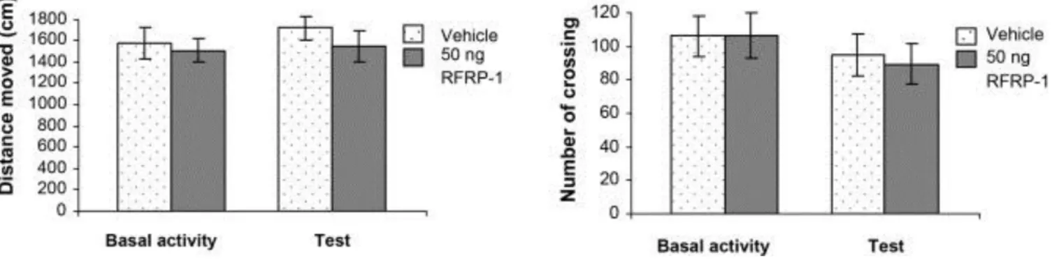
In the third experiment, effects of the bilateral intraamygdaloid microinjections of 50 ng RFRP-1 were studied in open-field test. The distance moved and the numbers of crossings were evaluated. There were no any alterations in these parameters in the RFRP-1 treated animals compared to vehicle treated controls. ANOVA analysis did not show significant difference between groups (RFRP-1 or vehicle treated animals, F [1, 28] = 0. 926, p > 0.05, Fig. 3A) and between sessions (Basal activity or test, F [1, 28] = 0.450, p > 0.05, Fig. 3B).
Fig. 3. Effects of RFRP-1 in the open-field test. (A) Columns represent mean (±S.E.M.) distance moved in the open-field apparatus one day before (Basal activity) and 10 min after (Test) bilateral 50 ng RFRP-1 or vehicle microinjections into the CeA. (B) Columns represent mean (±S.E.M.) number of crossings during Basal activity and Test sessions, respectively. Vehicle: vehicle treated rats (n = 8); 50 ng RFRP-1: animals microinjected with 50 ng RFRP-1 (n = 8).
Figure options
4. Discussion
It is well known that the AMY is essential in the control of hunger motivated behavior (Fonberg, 1966, Lenard and Hahn, 1982, Lenard et al., 1982, Hajnal et al., 1992 and Crovetti et al., 1995). It has been described that either hypophagia (Fonberg, 1966 and Hajnal et al., 1992) or hyperphagia (Fonberg, 1971) develops after electrolytic lesions of differential parts of the AMY. As far as the CeA is concerned, it has been shown that specific catecholaminergic microlesions reducing the norepinephrine content in this structure produced hyperphagia and weight increase, while dopamine depletion caused hypophagia and weight decrease (Lenard and Hahn, 1982 and Lenard et al., 1982). Cell-specific microiontophoretic lesions of the CeA with kainic acid, which destroy neurons in the target area but leave the passing fibers intact, also induce hypophagia and weight decrease (Hajnal et al., 1992). The amygdala is reciprocally connected to the hypothalamus (Oomura et al., 1970) and brainstem having projections to autonomic-related centers such as the dorsal motor nuclei of the vagal nerve, nucleus of the solitary tract and the parabrachial nucleus (Hopkins and Holstege, 1978). These regions are known to modulate feeding-related autonomic functions and constitute major relay of taste pathways in rodents.
In the amygdala different orexigenic and anorexigenic peptides and their receptors have been described. While direct microinjections of bombesin or bombesin-like peptides, such as gastrin-releasing peptide and neuromedin C induced satiety in the CeA (Fekete et al., 2002 and Fekete et al., 2007), similar application of orexin A increased food intake (Hangodi et al., 2006).
Less is known about feeding related effects of RFamide peptides in the amygdala, however.
Members of the PrRP group and RFRP peptides belong to the RFamide peptide family that share extensive homology and a common RF (arginine–phenylalanine) amide C terminus.
PrRP inhibits food intake in rats after CeA microinjection (Fan et al., 2005). PrRP reduced food consumption and enhanced the normal behavioral satiety sequence, suggesting that it may function as a homeostatic regulator of food intake (Lawrence et al., 2002). Feeding
related effects of RFRP peptides have not been studied in mammals with one exception.
Namely, orexigenic effects were detected in rats after i.c.v. injection of RFRP-3 (Yoshida et al., 2003 and Murakami et al., 2008). As far as the RFRP-1 is concerned, it was applied i.c.v.
to chicks and exhibited anorexigenic properties, i.e. RFRP-1 significantly reduced both food and water intake (Newmyer and Cline, 2009). Possible anorexigenic effects of RFRP-1 in rats, especially in the rat amygdaloid body have not been examined so far. In the rat CeA RFRP-1 immunoreactive fibers and terminals were identified (Fukusumi et al., 2001, Yano et al., 2003 and Yano et al., 2004) and NPFF1 receptors were also found there (Yoshida et al., 2003).
Our present experiments, therefore, were designed to examine the effects of microinjections of RFRP-1 on food intake in the rat CeA. While bilateral microinjections of 25 ng RFRP-1 did not modify feeding and 100 ng dose of RFRP-1 had only a tendency to decrease food intake, 50 ng RFRP-1 significantly reduced liquid food consumption. It seems, therefore, that 50 ng RFRP-1 is the effective dose and that similarly to those of other neuropeptides (Huston and Oitzl, 1989 and Fekete et al., 2002) an inverted U-shaped dose–effect relationship is characteristic in case of RFRP-1. The effect of 50 ng RFRP-1 was specific because it could be eliminated by NPFF receptor antagonist. The food intake decreasing effect of RFRP-1 was rapid and short, lasted for 20 min. Since we used cumulative evaluation, differences were significant for 60 min. However, food intake of the animals was quantified over 180 min period after microinjection of RFRP-1. It is interesting to note that animals failed to compensate for the early decrease in intake, even after 180 min (data are not shown), an effect which might be originated from the food intake decreasing effect of the drug seen during the first part of the experiment. The relatively short effect of RFRP-1 on feeding might be result of a short half-life of the peptide. However, this value is not yet known.
One may suppose that food intake reducing property of RFRP-1 could be due to anxiogenic effect which was detected after RFRP-1 and RFRP-3 treatments in rats (Kaewwongse et al., 2011). In these experiments, however, very high dose of RFRP-1 was applied [10 μg;
7.5 nmol, which is approximately 200× higher than the effective dose (50 ng; 37.8 pmol) applied in the present study]. In our experiments, in the open-field test 50 ng RFRP-1 (which significantly reduced food intake) did not modify spontaneous locomotor activity and the general behavior of animals did not change. These results are against the supposition that food intake decrease seen in our experiments was a consequence of any anxiogenic effect of RFRP-1.
RFRP analogs can bind both NPFF1 and NPFF2 receptors, first identified as cognate receptors for neuropeptide FF (NPFF). Subsequent studies have shown that NPFF1 exhibits higher affinity for RFRPs than for neuropeptide FF, and NPFF displays a higher potency for NPFF2. Therefore, NPFF2 is now generally considered to be the endogenous receptor for NPFF, while NPFF1 would be the RFRP receptor (Hinuma et al., 2000, Liu et al., 2001, Engstrom et al., 2003 and Yoshida et al., 2003). The precise role of both NPFF receptor subtypes has not been clarified yet. Distribution of NPFF1 and NPFF2 in several mammalian species indicates that NPFF2 is localized to pain-processing regions, whereas NPFF1 would most likely participate in neuroendocrine function (Bonini et al., 2000, Zeng et al., 2003 and Gouarderes et al., 2004). According to the results of other authors RFRP-1 binds to NPFF2 and is involved in pain modulation responses in the NTS (Pertovaara et al., 2005).
In our experiments prior application of NPFF receptor antagonist RF9 prevented the food intake reducing effect of RFRP-1 in the CeA. The RF9 is a potent and selective receptor
antagonist, which displays both the same affinity and antagonist activity at NPFF1 and NPFF2 subtypes (Simonin et al., 2006). In rats, this compound blocks the increase of arterial blood pressure and heart rate evoked by NPFF. Moreover, it prevents the development of delayed and long-lasting paradoxical hyperalgesia induced by daily heroin administration and associated tolerance (Simonin et al., 2006). Central administration of RF9 evoked a dose- dependent increase of LH and FSH levels in adult male and female rats (Pineda et al., 2010a and Pineda et al., 2010b). The affinity of RF9 was investigated for human NPY receptor and for a subset of related G-protein-coupled receptors, including the three other RFamide receptors (GPR10, GPR54, GPR103), opioid (μ, δ, κ) and ORL-1 receptors (Simonin et al., 2006). No competitive activity was observed for RF9 on these receptors at doses up to 10 μM, except on μ and κ, for which it was observed a slight competition at this concentration. These results indicate a good selectivity of RF9 for NPFF receptors, but the RF9 is not selective for NPFF receptor subtypes.
It is important to know that NPFF2 receptors were identified in the amygdala (Bonini et al., 2000, Hinuma et al., 2000, Liu et al., 2001 and Engstrom et al., 2003) but were not found in the CeA, while NPFF1 receptors in relatively high density can be found in the amygdala including the CeA. This indicates that in the CeA effects of RFRP-1 might be mediated by NPFF1 receptors. The NPFF antagonist RF9 was applied in equimolar amount (20 ng, 41.4 pmol) to the effective dose of RFRP-1 (50 ng, 37.8 pmol) and prevented the food intake decreasing effect of the peptide. We could not observe behavioral alterations after the antagonist microinjection, however, further experiments with application of different doses of RF9 are needed to clarify this question.
In conclusion, our results showed that RFRP-1 injected into the CeA of rats results in food intake decrease. This is a NPFF-1 linked effect, because it could be prevented by NPFF- receptor antagonist RF9. Our results are the first to demonstrate in rats that intraamygdaloid microinjection of RFRP-1 exhibits anorexic properties.
Conflict of interest
All authors declare that there is not any actual or potential conflict of interest including any financial, personal or other relationships with other people.
Acknowledgments
The authors would like to express their thanks to Anna Schulteisz, Erzsébet Korona and András Belvárácz for their technical contribution to this work. Thanks are also expressed to Zoltán Petykó for his helps in histology documentation. This study was supported by NKTH- OTKAK 68431, SROP-4.2.2/B-10/1-2010-0029, SROP-4.2.1/B-10/2/KONV-2010-0002 and by the Hungarian Academy of Sciences.
References
1.
o Bechtold and Luckman, 2007
o D.A. Bechtold, S.M. Luckman
o The role of RFamide peptides in feeding
o Journal of Endocrinology, 192 (2007), pp. 3–15
o Bechtold and Luckman, 2006
o D.A. Bechtold, S.M. Luckman
o Prolactin-releasing peptide mediates CCK-induced satiety in mice
o Endocrinology, 147 (2006), pp. 4723–4729
o View Record in Scopus
|
Full Text via CrossRef
| Cited By in Scopus (28) 2.
o Bonini et al., 2000
o J.A. Bonini, K.A. Jones, N. Adham, C. Forray, R. Artymyshyn, M.M. Durkin, K.E. Smith, J.A. Tamm, L.W. Boteju, P.P. Lakhlani, R. Raddatz, W.J. Yao, K.L. Ogozalek, N. Boyle, E.V. Kouranova, Y. Quan, P.J. Vaysse, J.M. Wetzel, T.A. Branchek, C. Gerald, B. Borowsky
o Identification and characterization of two G protein-coupled receptors for neuropeptide FF
o Journal of Biological Chemistry, 275 (2000), pp. 39324–39331
o View Record in Scopus
|
Full Text via CrossRef
| Cited By in Scopus (193) 3.
o Chartrel et al., 2003
o N. Chartrel, C. Dujardin, Y. Anouar, J. Leprince, A. Decker, S. Clerens, J.C.
Do-Rego, F. Vandesande, C. Llorens-Cortes, J. Costentin, J.C. Beauvillain, H.
Vaudry
o Identification of 26RFa, a hypothalamic neuropeptide of the RFamide peptide family with orexigenic activity
o Proceedings of the National Academy of Sciences of the United States of America, 100 (2003), pp. 15247–15252
o
4.
o Crovetti et al., 1995
o L.F. Crovetti, M. Mancia, M. Mariotti, M. Porrini, P. Spinnler, G. Testolini
o Food intake after amygdaloid lesions in the rats
o Nutrition Research, 15 (1995), pp. 565–570
o
5.
o Dockray, 2004
o G.J. Dockray
o The expanding family of -Rfamide peptides and their effects on feeding behavior
o Experimental Physiology, 89 (2004), pp. 229–235
o
6.
o Engstrom et al., 2003
o M. Engstrom, A. Brandt, S. Wurster, J.M. Savola, P. Panula
o Prolactin releasing peptide has high affinity and efficacy at neuropeptide FF2 receptors
o Journal of Pharmacology and Experimental Therapeutics, 305 (2003), pp. 825–
832
o View Record in Scopus
|
Full Text via CrossRef
| Cited By in Scopus (46) 7.
o Fan et al., 2005
o H. Fan, Z. Wang, P. Qlao, S. Wang, J. Yan
o PrRP inhibits food intake in rats after central nucleus of amygdala microinjection
o Henan Medical Research, 1 (2005), pp. 28–31
o View Record in Scopus
| Cited By in Scopus (1) 8.
o Fekete et al., 2002
o E. Fekete, J. Vigh, E.E. Bagi, L. Lenard
o Gastrin-releasing peptide microinjected into the amygdala inhibits feeding
o Brain Research, 955 (2002), pp. 55–63
o Article
|
PDF (256 K)
|
View Record in Scopus
| Cited By in Scopus (12) 9.
o Fekete et al., 2007
o E.M. Fekete, E.E. Bagi, K. Toth, L. Lenard
o Neuromedin C microinjected into the amygdala inhibits feeding
o Brain Research Bulletin, 71 (2007), pp. 386–392
o Article
|
PDF (548 K)
|
View Record in Scopus
| Cited By in Scopus (12) 10.
o Fonberg, 1966
o E. Fonberg
o Aphagia, produced by destruction of the dorsomedial amygdala in dogs
o Bulletin de l’Academie Polonaise des Sciences. Serie des Sciences Biologiques, 14 (1966), pp. 719–722
o View Record in Scopus
| Cited By in Scopus (6) 11.
o Fonberg, 1971
o E. Fonberg
o Hyperphagia produced by lateral amygdalar lesions in dogs
o Acta Neurobiologiae Experimentalis (Wars), 31 (1971), pp. 19–32
o View Record in Scopus
| Cited By in Scopus (29) 12.
o Fukusumi et al., 2006
o S. Fukusumi, R. Fujii, S. Hinuma
o Recent advances in mammalian RFamide peptides: the discovery and functional analyses of PrRP, RFRPs and QRFP
o Peptides, 27 (2006), pp. 1073–1086
o Article
|
PDF (253 K)
|
View Record in Scopus
| Cited By in Scopus (60)
13.
o Fukusumi et al., 2001
o S. Fukusumi, Y. Habata, H. Yoshida, N. Iijima, Y. Kawamata, M. Hosoya, R.
Fujii, S. Hinuma, C. Kitada, Y. Shintani, M. Suenaga, H. Onda, O. Nishimura, M. Tanaka, Y. Ibata, M. Fujino
o Characteristics and distribution of endogenous RFamide-related peptide-1
o Biochimica et Biophysica Acta, 1540 (2001), pp. 221–232
o Article
|
PDF (587 K)
|
View Record in Scopus
| Cited By in Scopus (82) 14.
o Gouarderes et al., 2004
o C. Gouarderes, A. Puget, J.M. Zajac
o Detailed distribution of neuropeptide FF receptors (NPFF1 and NPFF2) in the rat, mouse, octodon, rabbit, guinea pig, and marmoset monkey brains: a comparative autoradiographic study
o Synapse, 51 (2004), pp. 249–269
o View Record in Scopus
|
Full Text via CrossRef
| Cited By in Scopus (36) 15.
o Hajnal et al., 1992
o A. Hajnal, P. Sandor, G. Jando, I. Vida, A. Czurko, Z. Karadi, L. Lenard
o Feeding disturbances and EEG activity changes after amygdaloid kainate lesions in the rat
o Brain Research Bulletin, 29 (1992), pp. 909–916
o Article
|
PDF (1046 K)
|
View Record in Scopus
| Cited By in Scopus (18) 16.
o Hangodi et al., 2006
o O. Hangodi, B. Urbán, É.E. Bagi, É. Fekete, K. Tóth, L. Lénárd
o Orexin-A microinjection mediated food and water intake are antagonized by selective orexin-1 receptor antagonist in the bed nucleus of stria terminalis
o International Congress Series, 1291 (2006), pp. 141–144
o Article
|
PDF (81 K)
|
View Record in Scopus
| Cited By in Scopus (3) 17.
o Herrera-Herrera and Salazar-Olivo, 2008
o M.L. Herrera-Herrera, L.A. Salazar-Olivo
o RFamide neuropeptides inhibit murine and human adipose differentiation
o Biochemical and Biophysical Research Communications, 377 (2008), pp. 29–
34
o Article
|
PDF (710 K)
|
View Record in Scopus
| Cited By in Scopus (5) 18.
o Hinuma et al., 2000
o S. Hinuma, Y. Shintani, S. Fukusumi, N. Iijima, Y. Matsumoto, M. Hosoya, R.
Fujii, T. Watanabe, K. Kikuchi, Y. Terao, T. Yano, T. Yamamoto, Y.
Kawamata, Y. Habata, M. Asada, C. Kitada, T. Kurokawa, H. Onda, O.
Nishimura, M. Tanaka, Y. Ibata, M. Fujino
o New neuropeptides containing carboxy-terminal RFamide and their receptor in mammals
o Nature Cell Biology, 2 (2000), pp. 703–708
o View Record in Scopus
|
Full Text via CrossRef
| Cited By in Scopus (268) 19.
o Hopkins and Holstege, 1978
o D.A. Hopkins, G. Holstege
o Amygdaloid projections to the mesencephalon, pons and medulla oblongata in the cat
o Experimental Brain Research, 32 (1978), pp. 529–547
o View Record in Scopus
| Cited By in Scopus (256) 20.
o Huston and Oitzl, 1989
o J.P. Huston, M.S. Oitzl
o The relationship between reinforcement and memory – parallels in the rewarding and mnemonic effects of the neuropeptide substance-P
o Neuroscience and Biobehavioral Reviews, 13 (1989), pp. 171–180
o Article
|
PDF (954 K)
|
View Record in Scopus
| Cited By in Scopus (49) 21.
o Jhamandas et al., 2006
o K. Jhamandas, B. Milne, M. Sutak, C. Gouarderes, J.M. Zajac, H.Y. Yang
o Facilitation of spinal morphine analgesia in normal and morphine tolerant animals by neuropeptide SF and related peptides
o Peptides, 27 (2006), pp. 953–963
o
22.
o Kaewwongse et al., 2011
o M. Kaewwongse, Y. Takayanagi, T. Onaka
o Effects of RFamide-related peptide (RFRP)-1 and RFRP-3 on oxytocin release and anxiety-related behaviour in rats
o Journal of Neuroendocrinology, 23 (2011), pp. 20–27
o
23.
o Kirby et al., 2009
o E.D. Kirby, A.C. Geraghty, T. Ubuka, G.E. Bentley, D. Kaufer
o Stress increases putative gonadotropin inhibitory hormone and decreases luteinizing hormone in male rats
o Proceedings of the National Academy of Sciences of the United States of America, 106 (2009), pp. 11324–11329
o
24.
o Lawrence et al., 2002
o C.B. Lawrence, K.L.J. Ellacott, S.M. Luckman
o PRL-releasing peptide reduces food intake and may mediate satiety signaling
o Endocrinology, 143 (2002), pp. 360–367
o
25.
o Lenard and Hahn, 1982
o L. Lenard, Z. Hahn
o Amygdalar noradrenergic and dopaminergic mechanisms in the regulation of hunger and thirst-motivated behavior
o Brain Research, 233 (1982), pp. 115–132
o
26.
o Lenard et al., 1982
o L. Lenard, Z. Hahn, Z. Karadi
o Body weight changes after neurochemical manipulations of lateral amygdala:
noradrenergic and dopaminergic mechanisms
o Brain Research, 249 (1982), pp. 95–101
o
27.
o Liu et al., 2001
o Q.Y. Liu, X.M. Guan, W.J. Martin, T.P. McDonald, M.K. Clements, Q.P.
Jiang, Z.Z. Zeng, M. Jacobson, D.L. Williams, H. Yu, D. Bomford, D.
Figueroa, J. Mallee, R.P. Wang, J. Evans, R. Gould, C.P. Austin
o Identification and characterization of novel mammalian neuropeptide FF-like peptides that attenuate morphine-induced antinociception
o Journal of Biological Chemistry, 276 (2001), pp. 36961–36969
o
28.
o Murakami et al., 2008
o M. Murakami, T. Matsuzaki, T. Iwasa, T. Yasui, M. Irahara, T. Osugi, K.
Tsutsui
o Hypophysiotropic role of RFamide-related peptide-3 in the inhibition of LH secretion in female rats
o Journal of Endocrinology, 199 (2008), pp. 105–112
o
29.
o Murase et al., 1996
o T. Murase, H. Arima, K. Kondo, Y. Oiso
o Neuropeptide FF reduces food intake in rats
o Peptides, 17 (1996), pp. 353–354
o
30.
o Newmyer and Cline, 2009
o B.A. Newmyer, M.A. Cline
o Neuropeptide SF is associated with reduced food intake in chicks
o Behavioural Brain Research, 205 (2009), pp. 311–314
o
31.
o Oomura et al., 1970
o Y. Oomura, T. Ono, H. Ooyama
o Inhibitory action of the amygdala on the lateral hypothalamic area in rats
o Nature, 228 (1970), pp. 1108–1110
o
32.
o Osugi et al., 2006
o T. Osugi, K. Ukena, S.A. Sower, H. Kawauchi, K. Tsutsui
o Evolutionary origin and divergence of PQRFamide peptides and LPXRFamide peptides in the RFamide peptide family. Insights from novel lamprey RFamide peptides
o FEBS Journal, 273 (2006), pp. 1731–1743
o
33.
o Paxinos and Watson, 1986
o G. Paxinos, C.R. Watson
o The Rat Brain in Stereotaxic Coordinates
o (second ed.)Academic Press, New York (1986)
o
34.
o Pertovaara et al., 2005
o A. Pertovaara, M. Ostergard, M.L. Anko, S. Lehti-Koivunen, A. Brandt, W.
Hong, E.R. Korpi, P. Panula
o RFamide-related peptides signal through the neuropeptide FF receptor and regulate pain-related responses in the rat
o Neuroscience, 134 (2005), pp. 1023–1032
o
35.
o Pineda et al., 2010a
o R. Pineda, D. Garcia-Galiano, M.A. Sanchez-Garrido, M. Romero, F. Ruiz- Pino, E. Aguilar, F.A. Dijcks, M. Blomenrohr, L. Pinilla, P.I. van Noort, M.
Tena-Sempere
o Characterization of the inhibitory roles of RFRP3, the mammalian ortholog of GnIH, in the control of gonadotropin secretion in the rat: in vivo and in vitro studies
o American Journal of Physiology, Endocrinology and Metabolism, 299 (2010), pp. E39–E46
o
36.
o Pineda et al., 2010b
o R. Pineda, D. Garcia-Galiano, M.A. Sanchez-Garrido, M. Romero, F. Ruiz- Pino, E. Aguilar, F.A. Dijcks, M. Blomenröhr, L. Pinilla, P.I. van Noort, M.
Tena-Sempere
o Characterization of the potent gonadotropin-releasing activity of RF9, a selective antagonist of RFamide-related peptides and neuropeptide FF receptors: physiological and pharmacological implications
o Endocrinology, 151 (2010), pp. 1902–1903
o
37.
o Qi et al., 2009
o Y. Qi, B.J. Oldfield, I.J. Clarke
o Projections of RFamide-related peptide-3 neurones in the ovine hypothalamus, with special reference to regions regulating energy balance and reproduction
o Journal of Neuroendocrinology, 21 (2009), pp. 690–697
o
38.
o Simonin et al., 2006
o F. Simonin, M. Schmitt, J.P. Laulin, E. Laboureyras, J.H. Jhamandas, D.
MacTavish, A. Matifas, C. Mollereau, P. Lauren, M. Parmentier, B.L. Kieffer, J-J. Bourguignon, G. Simonnet
o RF9, a potent and selective neuropeptide FF receptor antagonist, prevents opioid-induced tolerance and paradoxical hyperalgesia
o Proceedings of the National Academy of Sciences of the United States of America, 103 (2006), pp. 466–471
o
39.
o Smith and Clarke, 2010
o J.T. Smith, I.J. Clarke
o Gonadotropin inhibitory hormone function in mammals
o Trends in Endocrinology and Metabolism, 21 (2010), pp. 255–260
o
40.
o Takayasu et al., 2006
o S. Takayasu, T. Sakurai, S. Iwasaki, H. Teranishi, A. Yamanaka, S.C.
Williams, H. Iguchi, Y.I. Kawasawa, Y. Ikeda, I. Sakakibara, K. Ohno, R.X.
Ioka, S. Murakami, N. Dohmae, J. Xie, T. Suda, T. Motoike, T. Ohuchi, M.
Yanagisawa, J. Sakai
o A neuropeptide ligand of the G protein-coupled receptor GPR103 regulates feeding, behavioral arousal, and blood pressure in mice
o Proceedings of the National Academy of Sciences of the United States of America, 103 (2006), pp. 7438–7443
o
41.
o Ukena and Tsutsui, 2005
o K. Ukena, K. Tsutsui
o A new member of the hypothalamic RF-amide peptide family, LPXRF-amide peptides: structure, localization, and function
o Mass Spectrometry Reviews, 24 (2005), pp. 469–486
o
42.
o Willis et al., 2003
o K.S. Willis, K. Cynthia, K.S. Charles, W.S. Henry, L. Brian, R.B. Jennifer, M.T. Meghan
o Prolactin-releasing peptide and its homolog RFRP-1 act in hypothalamus but not in anterior pituitary gland to stimulate stress hormone secretion
o Endocrine, 20 (2003), pp. 59–66
o
43.
o Yano et al., 2004
o T. Yano, N. Iijima, S. Hinuma, M. Tanaka, Y. Ibata
o Developmental expression of RFamide-related peptides in the rat central nervous system
o Developmental Brain Research, 152 (2004), pp. 109–120
o Article
|
PDF (1477 K)
|
View Record in Scopus
| Cited By in Scopus (7) 44.
o Yano et al., 2003
o T. Yano, N. Iijima, K. Kakihara, S. Hinuma, M. Tanaka, Y. Ibata
o Localization and neuronal response of RFamide related peptides in the rat central nervous system
o Brain Research, 982 (2003), pp. 156–167
o Article
|
PDF (1645 K)
|
View Record in Scopus
| Cited By in Scopus (47) 45.
o Yoshida et al., 2003
o H. Yoshida, Y. Habata, M. Hosoya, Y. Kawamata, C. Kitada, S. Hinuma
o Molecular properties of endogenous RFamide-related peptide-3 and its interaction with receptors
o Biochimica et Biophysica Acta, 1593 (2003), pp. 151–157
o Article
|
PDF (146 K)
|
View Record in Scopus
| Cited By in Scopus (74) 46.
o Yudin et al., 2006
o Y.K. Yudin, Z.A. Tamarova, O.A. Krishtal
o Peripherally applied neuropeptide SF is equally algogenic in wild type and ASIC3–/– mice
o Neuroscience Research, 55 (2006), pp. 421–425
o Article
|
PDF (232 K)
|
View Record in Scopus
| Cited By in Scopus (3) 47.
o Zeng et al., 2003
o Z.Z. Zeng, T.P. McDonald, R.P. Wang, Q.Y. Liu, C.P. Austin
o Neuropeptide FF receptor 2 (NPFF2) is localized to pain-processing regions in the primate spinal of the medulla cord and the lower level oblongata
o Journal of Chemical Neuroanatomy, 25 (2003), pp. 269–278
o Article
|
PDF (781 K)
|
View Record in Scopus
| Cited By in Scopus (15)
Corresponding author at: Institute of Physiology and Neurophysiology Research Group of the HAS, Pécs University Medical School, Szigeti str. 12, P.O. Box 99, H- 7602 Pécs, Hungary. Tel.: +36 72 536 432; fax: +36 72 536 423.
Copyright © 2012 Elsevier Inc. All rights reserved.