1. Spectrophotometric Method
Principle
The degradation of hyaluronic acid by bacterial hyaluronidase ** results in the almost quantitative yield o f a disaccharide, which has been characterized as a 4,5-unsaturated uronide (D-A
4
-glucosyl- pyranosyl-uronic acid-p[l->3]-2-acetamino-2-deoxy-D-glucose)
4
*. Like the h o m o l o g o u s oligo
saccharides it has an absorption maximum around 230 ma
2
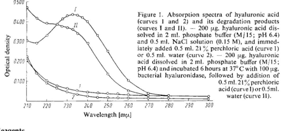
K Hyaluronic acid has a low absorption at this wavelength (Fig. 1).
0.500
Figure 1. Absorption spectra of hyaluronic acid (curves 1 and 2) and its degradation products (curves I and II). — 200 pig. hyaluronic acid dis
solved in 2 ml. phosphate buffer ( M / 1 5 ; p H 6.4) and 0.5 ml. N a C l solution (0.15 M), and immed
iately added 0.5 ml. 21 % perchloric acid (curve 1) or 0.5 ml. water (curve 2). — 200 ag. hyaluronic acid dissolved in 2 ml. phosphate buffer ( M / 1 5 ; p H 6.4) and incubated 6 hours at 37°C with 100 £j»g.
bacterial hyaluronidase, followed by addition o f 0.5 ml. 21% perchloric acid (curve I) orO. 5ml.
i
2 water (curve 11).2W 220 230 2L0 250 260 270 280 290 300 Wavelength [ma]
Reagents
1. Potassium dihydrogen phosphate,
K H 2 P O 4 ,A. R.
2. Disodium hydrogen phosphate, N a 2 H P 0 4 « 2 H 2 0 , A. R.
3. Sodium chloride, A. R.
4. Perchloric acid, A. R., sp. gr. 1.67 (ca. 70% w/w) 5. Bacterial hyaluronidase
from Staphylococcus aureus or streptococci
2
*. Isolation, see p. 92.
6. Potassium hyaluronate
obtained from umbilical cord or vitreous humor and purified
2
*. Commercial preparation, see p. 1021.
** Hyaluronatelyase.
{)
H. Gibian: Mucopolysaccharide und Mucopolysaccharidasen. F. Deuticke, Vienna 1959, p. 90.
2
> H. Greiling, Hoppe-Seylers Z. physiol. Chem. 309, 239 [1957].
3
* H. Greiling, Th. Giinther and T. Eberhard, Hoppe-Seylers Z. physiol. Chem. 319, 161 [I960].
4
* A. Linker, K. Meyer and Ph. Hoffmann, J. biol. Chemistry 219, 13 [1956].
Hyaluronic Acid
Helmut Greiling
Formerly hyaluronic acid was determined by means of turbidimetric methods. However, the results depended on the degree of polymerization of the hyaluronic acid. Other methods are based o n the hydrolysis of hyaluronic acid and the quantitative determination of its constituents
1
*. All these methods are unspecific for hyaluronic acid.
In contrast, the enzymatic methods described h e r e
2
.
3
* are specific and virtually independent of the degree of polymerization.
88 Section B : Estimation of Substrates Purity of the e n z y m e preparation
The hyaluronidase preparation should have a specific activity of at least 20 units**. Contamin
ation with the glycolytic enzymes does not interfere with the determination. Peptidase should not be detectable.
Preparation of Solutions (for ca. 20 determinations) I. Phosphate buffer (M/15; pH 6.4):
In a 250 ml. volumetric flask, dilute 66 ml. of a solution of disodium hydrogen phos
phate (11.876 g./litre) with a solution of potassium dihydrogen phosphate (9.078 g./
litre).
II. Sodium chloride (0.15 M):
Dissolve 0.9 g. NaCl, A. R., in distilled water and make up to 100 ml.
III. Perchloric acid (ca. 20% w/v):
Dilute ca. 13 ml. HCIO4, A. R., (sp. gr. 1.67) to 75 ml. with distilled water.
IV. Bacterial hyaluronidase (ca. 1 mg. protein/ml.):
Dissolve 7 mg. dry preparation in 7 ml. 0.15 M NaCl.
V. Potassium hyaluronate (200 pig./ml.):
Dissolve 20 mg. potassium hyaluronate in 100 ml. phosphate buffer (solution I).
Stability of the s o l u t i o n s
The solutions are stored, stoppered, in a refrigerator atO—4°C. Under these conditions the hyaluro
nidase solution shows no substantial loss of activity within 3 months. The other solutions are stable indefinitely so long as no bacterial growth occurs.
Procedure
Experimental material in solution may be used directly without further preparation, dissolve or suspend solid matter in NaCl solution (II).
Enzymatic reaction
The assay mixture, set up in conical centrifuge tubes, consists of:
1. 2.0 ml. buffer (solution I) 0.5 ml. sample
0.5 ml. NaCl solution (II)
Experimental and standard tubes
I) 2. 1.5 ml. buffer (solution I)
0.5 ml. potassium hyaluronate solution (V) 0.5 ml. sample
0.5 ml. NaCl solution (II) 3. 1.0 ml. buffer (solution I)
1.0 ml. potassium hyaluronate solution (V) 0.5 ml. sample
0.5 ml. NaCl solution (II)
Control tube
4. 2.0 ml. buffer (solution I) 0.5 ml. sample
0.6 ml. NaCl solution (II)
*) According t o
2
* a unit is the amount of enzyme contained in 1 ml. which increases the optical density of a solution containing 1 mg. hyaluronic acid/ml. by 0.100 in 100 sec. at 230 mpt. and 30° C, and with a 1 cm. light path.
Heat all the solutions for 10 min. at 70° C. This preliminary treatment can be omitted if the sample contains no protein.
After cooling solutions 1—3 add 0.1 ml. bacterial hyaluronidase (solution IV). Then incubate all the tubes for 6 hours at 37°C (water bath).
Deproteinization a n d spectrophotometric m e a s u r e m e n t s
After the 6 hour incubation add 0.5 ml. perchloric acid (solution III) to tubes 1—4, mix well, centrifuge for 20 min. at 5000g. Decant the supernatant fluids into silica cuvettes and measure the optical density at 230 mu against control tube 4. Ei, E2 and E3 are the optical densities of experimental tubes 1—3, E2 and E3 contain known amounts of hyaluronic acid.
Calculations
Beer's law is obeyed between 20 and 700 pig. potassium hyaluronate
3
*. For the calculations Ei is related to the optical density found for 100 pig. potassium hyaluronate. That is E2—Ei or E3—E2 or (E3—Ei)/2. The average is:
( E
2
- E i ) + ( E3
- E 2 )Ei00 {Ag. = ~ The amount o f hyaluronic acid in 1 ml. of sample is given by:
Ei x 100 x 2
= (jLg. potassium hyaluronate/ml. sample E100 {xg.
Example
Sample: 0.5 ml. ascites serum. After incubation and deproteinization, measured at 230 mpt against control tube 4. Ei = 0.250; E
2
- 0.470; E3
= 0.700; ( E2
- E i ) = 0.220; ( E3
- E2
) - 0.230.Eioofig. = (0.220 + 0.230)/2 - 0.225 0.250 x 100
^ = 111 pig. potassium hyaluronate/0.5 ml. ascites serum.
Sources of Error
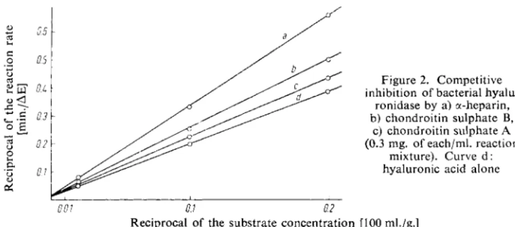
Substances which absorb at 230 mpi and are not precipitated by perchloric acid, for instance high concentrations of nucleotides, interfere with the determination. a-Heparin and the chondroitin sulphates A and B competitively inhibit bacterial hyaluronidase (Fig. 2).
Figure 2. Competitive inhibition of bacterial hyalu
ronidase by a) a-heparin, b) chondroitin sulphate B,
c) chondroitin sulphate A (0.3 mg. of each/ml. reaction
mixture). Curve d:
hyaluronic acid alone
Reciprocal of the substrate concentration [100 ml./g.]
In the presence of large amounts of these substances the enzymatic determination must be preceded by an isolation of the hyaluronic acid.
90 Section B : Estimation of Substrates
Specificity and Dependence of the Method on the Degree of Polymerization
a-Heparin, chondroitin sulphate A and B are not decomposed by bacterial hyaluronidase. Chon
droitin is only hydrolysed to a slight extent. Moreover, the p H optimum of the chondroitin hydrolysis is in the acid range (pH 5.7 with the same ionic strength of 0.12). Contamination of the hyaluronic acid with up to 3 0 % chondroitin does not interfere with the determination
5
*. So far chrondroitin has only been found in the cornea
6
*
7
*.
Table 1 shows that the method is independent of the molecular weight up to a mean of about 20 for the degree o f polymerization of the hyaluronic acid. With an average molecular weight o f 1 600 the optical density change after 6 hours incubation with bacterial hyaluronidase is about 1 2 % t o o low, which can be explained by the decrease in the number o f hydrolysable bonds with lower degrees of polymerization.
Table 1. Michaelis constants, maximum velocities and optical density changes o n incubation of 150 pig. hyaluronic acid of different degrees of polymerization, with 100 pig. bacterial hyaluronidase for 6 hours. — The mean degree o f polymerization was calculated according t o :
2 x hexosamine(pimolesN-acetylglucosamine/mg.) reducing value (pimoles /mg.)
Degree o f
t K l i / r v M n I
Ew 2 3
o/150pig.polymerization 7 °
L w t
;
r r
^m x a
, after 6 hrs.
(mean) <
m e a n
> [g./100 ml.] [jxg./mm.]
i n c u b a t i o n
184 32973 l . l x l 0 ~ i 67 0.282
108 19 354 1.0x10⁻¹ 66 0.274
88 15 770 1.1 x 1 0- 1 67 0.291 72 12902 1.0x10⁻¹ 67 0.280
36 6451 1.0x10⁻¹ 68 0.283
25 4480 0.95x10⁻¹ 67 0.275 20 3 584 1.0x10⁻¹ 67 0.286 8 1434 1.0x10⁻¹ 66 0.247
2. Colorimetric Method
Principle
Bacterial hyaluronidase degrades hyaluronic acid quantitatively to an unsaturated disaccharide, thus liberating 7V-acetylglucosamine end groups. These can be determined colorimetrically with the Morgan-Elson reaction: Thus o n heating N-acetylglucosamine in alkali, furan derivatives are formed, which react with /7-dimethylaminobenzaldehyde to form a red dye
8
*. The condensation product of p-dimethylaminobenzaldehyde and the disaccharide from hyaluronic acid has two absorption maxima at 544 mpi and 585 mpi (Fig. 3) if the reaction is carried out according to Leloir^. In the method de
scribed here the optical density of the reaction mixture is measured at 585 mpi.
9 a
*
Reagents
1.—6. as for the spectrophotometric method (p. 87).
Additional:
7. Boric acid, A. R.
8. Potassium hydroxide, A. R.
5
* H. Greiling, Th. Gunther and T. Eberhard, unpublished.
6) E. A. Davidson and K. Meyer, J. biol. Chemistry 211, 605 [1954].
7)
K. Meyer, E. A. Davidson, A. Linker and P. Hoffmann, Biochim. biophysica Acta 21, 506 [1956].
8* R. Kuhn, Angew. Chem. 69, 23 [1957].
9) J. L. Reissig, J. L. Strohminger and L. F. Leloir, J. biol. Chemistry 217, 959 [1955].
9a) H. Greiling, Z. Rheumaforsch. 20, 298 [1961].
9. Acetic acid, A. R.
10. Hydrochloric acid, A. R., ION 11. /7-Dimethylaminobenzaldehyde, A. R.
a
O
# 0 540 560 580 600 Wavelength [mu]
Fig. 3. Absorption spectrum of the Morgan-Elson reaction products of the disaccharide formed from hyaluronic acid by bacterial h y a l u r o n i d a s e
9
l Pure hyaluronic acid has no absorption between 520 and 600 mu.
Preparation of Solutions
Solutions I—V as for the spectrophotometric method (p. 88).
Additional:
VI. Potassium tetraborate (0.8 M):
Dissolve 24.7 g. boric acid and 43.87 g. potassium hydroxide in distilled water and make up to 500 ml.
VII. /7-Dimethylaminobenzaldehyde reagent:
Dissolve 10 g. /?-dimethylaminobenzaldehyde in a mixture of 100 ml. acetic acid and 12.5 ml. 10N H Q . Immediately before use dilute 1:10 with acetic acid.
Stability of the s o l u t i o n s
The /7-dimethylaminobenzaldehyde solution must be stored in a dark bottle and should be prepared freshly each week.
Procedure
Enzymatic reaction
As for the spectrophotometric method (p. 88).
D e p r o t e i n i z a t i o n and colour reaction
After 6 hours incubation with the enzyme add to each tube 0.5 ml. perchloric acid (solution III),
mix thoroughly and centrifuge for 20 min. at 5 000 g.
Pipette into test tubes:
2 ml. supernatant
0.6 ml. potassium tetraborate solution (VI).
Heat for 3 min. in a boiling water bath and then cool 15 min. in an ice bath. Potassium per- chlorate precipitates out. Pipette into test tubes:
1 ml. KClC>4-free supernatant
3 ml. /?-dimethylaminobenzaldehyde solution (VII).
92
Section B : Estimation of SubstratesIncubate for 20 min. at 37° C. Read the optical densities of solutions 1—3 against control solution 4 in 1 cm. cuvettes at 585 ma. The optical densities E 2 and E 3 of the solutions 2 and 3 are standards.
Calculations
Calculate the hyaluronic acid content of the sample by means of the standards (see p. 89).
Example
The reaction mixture contained 0.1 ml. o f synovial fluid which was diluted with 0.4 ml. sodium chlor
ide solution. After the colour reaction the following values were measured at 585 m[i against the control cuvette 4 :
Ei = 0.257; E
2
= 0.388; E3
= 0.523; ( E2
- Ei) = 0.131; ( E3
- E2
) = 0.135_ 0 . 1 3 1 + 0 . 1 3 5 . . . . -fcioofxg. = 2
= u
-
1 3 3
0.257 X 100 ^ . ,0 ^ 3 3
t l / x
, , . ,a
.„=
^
J
P °
t a s s i u m
hyaluronate/0.1 ml. synovial fluid
Discussion
The colorimetric method has the advantage over the spectrophotometric method that the measure
ments can be made in the presence o f nucleotides, amino acids and small amounts of protein. In this method also the mixture must be heated to 70° C before incubating with bacterial hyaluronidase in order to remove inhibitors.
The colorimetric method is more suitable for the determination of hyaluronic acid in body fluids than the spectrophotometric method, which is more suitable for hyaluronic acid solutions low in protein.
Appendix
Isolation of bacterial h y a l u r o n i d a s e (hyaluronate lyase)
Hyaluronidase is produced in the growth phase by strains o f streptococci, staphylococci, pneumo- cocci and Clostridium perfringens. The isolation of a bacterial hyaluronidase from a strain o f group A streptococci is described below.
G r o w
1 0
* the organisms in casein hydrolysate with the addition of hyaluronic acid from umbilical cords (0.2%). Centrifuge the 24 hour culture at high speed at 0 ° C . A d d a m m o n i u m sulphate to the supernatant and collect the protein fraction precipitating between i /
3
a n
d
2
h saturation by centri- fuging at high speed. Dissolve the precipitate in distilled water and dialyse against 0.005 M phosphate buffer (pH 7). Adsorb the contents of the dialysis sac onto a column o f DEAE-cellulose and elute with: (a) 0.02 M phosphate buffer (pH 6); (b) 0.05 M N a H
2
P 04
solution and (c) solutions o f 0.05 M N a H2
P 04
in 0.02 M to 0.1 M N a C l .The preparation contains 4 isoenzymes o f hyaluronidase. The main fraction with the highest specific activity is eluted with 0.05 M NaH
2
PC>4 in 0.05 M N a C l . Dialyse this eluate and then freeze- dryiD. Enzyme preparations obtained in this way have a specific activity o f about 100 u n i t s1 2
V m g . 10) H. J. Rogers, Biochem. J. 40, 583 [1946].
I D H. Greiling, Osterr. Chemiker-Ztg. 63, 285 [1962].
12) H. Greiling, Hoppe-Seylers Z. physiol. Chem. 309, 239 [1957].