Inhibitions in the Citric Acid Cycle
J. H. Quastel
I. Introduction 473 II. Inhibitors of Succinic Dehydrogenase 475
A. Malonate 475 B. Effects of Thiol Reactants . . 476
C. Effects of Fluoride 478 D. Effects of Cyanide 479 E . Effects of Action of Iron Chelators 480
F. Effects of High Oxygen Tension 480
G. Effects of Arsenicals 481 H. Effects of Iodoacetate 481 I. Effects of Quinones 482 J. Effects of Pyrophosphate 482 K. Effects of Oxalacetate 482 L. Mitochondrial Respiration in Presence of Succinate and the
Effects of Metabolic Inhibitors 483 M. Inhibitive Effects of Various Succinic Acid Analogues 486
N. Antimycin A Inhibition 487 O. Oligomycin Inhibition 488 III. Fumarate Conversion to L-Malate; Inhibitions of Fumarase . . . . 488
IV. Inhibition of Malate Metabolism 489
V. Oxalacetate Metabolism 491 VI. Citrate Breakdown; Inhibition of Aconitase 491
A. Fluoroacetate Inhibition 491 B. Oxalomalate Inhibition 494 VII. Metabolism of α-Ketoglutarate 495
VIII. Isocitrate Oxidation 496
References 496
I. INTRODUCTION
Studies of the biological oxidation of succinate and of its products, fumarate and L-malate, gave rise to observations fundamental for the
473
development of modern concepts of the mechanisms involved in respira
tory processes, particularly those embraced by the citric acid cycle. The demonstration (1) that pyruvic acid is a normal product of the biological oxidation of fumaric acid and therefore of its precursor, succinic acid
[2-5), led to a study of the relations existing between succinic and fumaric acid. This resulted in the rinding, with bacterial suspensions (5), that a reversible equilibrium exists between succinate and fumarate in presence of succinic dehydrogenase, and later in the observation that substances exist, other than the substrates succinate and fumarate, that have an affinity for this enzyme. It was found (6) that molecules related chemically in structure to the substrate may have an affinity for the enzyme [or, as then designated, its active center ( 7 ) ] and may compete with the natural substrate and thus inhibit its metabolism. This observa
tion was the foundation of the principle of competitive inhibition, which was early used in an investigation of the effects of succinate and its analogues on the growth of Escherichia coli (8).
Various substances of the structure R C H2C O O H inhibit the activity of succinic dehydrogenase, but of these malonic acid (6) is the best known. I t inhibits the enzyme competitively (6), and its high specificity of action has made it an important tool in the investigation of respiratory processes. Its action as a competitive inhibitor of succinic dehydrogenase, shown originally with suspensions of E. coli, was found to apply to mam
malian tissues (9) and to soluble succinic dehydrogenase (10-12). There are, however, wide differences between the degrees of inhibition of the rates of oxygen consumption brought about by malonate in the presence of succinate with different organisms and tissues, these being largely due to the differences between rates of oxidation of succinate and of fumarate (or L-malate) with these cells. When the latter rate is large, relatively little effect of malonate on the rate of total oxygen uptake is seen, owing to the fact that, in spite of the inhibition of succinic dehydrogenase, suffi
cient fumarate (or L-malate) is formed to enable subsequent oxidative reactions to proceed optimally (9). Thus, malonate exercises but little inhibition of oxygen consumption of suspensions of Bacillus subtilis or Micrococcus lysodeikticus in the presence of succinate under conditions where it almost completely suppresses the rate of oxygen consumption of rabbit muscle or brain tissue. The observations (18) that malonate inhibi
tion of respiration may be overcome by the addition of fumarate, that fumarate has a catalytic effect on cell respiration (18, H), that malonate inhibition leads aerobically to the formation of succinate, that in the pres
ence of malonate a variety of substrates, e.g., malate, oxalacetate, citrate, and α-ketoglutarate, is converted to succinate, and that oxalacetate and
pyruvate together can give rise to citrate in the presence of tissues, led to the formulation of the citric acid cycle (15, 16) as a major respiratory process.
The fact that succinic dehydrogenase is the only enzyme that is in
hibited to a marked extent by malonate at low concentrations (up to 1 mM) at physiological succinate concentrations is supported by the observation that injection of malonate leads to the accumulation of suc
cinate in animal tissues and body fluids (17, 18).
II. INHIBITORS OF SUCCINIC DEHYDROGENASE
A. M a l o n a t e
Study of the properties of succinic dehydrogenase may be simplified by replacement of methylene blue (19) as hydrogen acceptor by ferricyanide (20), whose reduced product is not autoxidizable and whose reduction, measured either manometrically or spectroscopically, makes it possible to assay affinities of succinate or its analogues to the enzyme. Using ferri
cyanide as a hydrogen acceptor, it was found (21) that the Km of suc
cinic dehydrogenase in six different heart muscle preparations has values ranging from 2.5 Χ 1 0 ~4 Μ to 5.3 Χ 1 0 ~4 M, while the inhibitory constant Ki of malonate has values ranging from 5.4 χ 1 0 ~6 Μ to 9.8 X 1 0 ~6 M. The ratio of affinities of malonate and succinate varies from 4.7 to 60, depending on the nature and concentration of the hydrogen acceptor. The variations are explained as being due to differences in the values of the rate constant for decomposition of the enzyme-substrate complex to give the products of reaction. It was concluded (21) that the true ratio of affinities of succinic dehydrogenase for malonate and suc
cinate, in the absence of further reactions of succinate, is about 3. Ferri
cyanide may react, however, at several points in the succinic oxidase chain, and it is considered that phenazine methosulfate provides a more reliable means for assay of succinic dehydrogenase activities in a variety of preparations (22, 23). The kinetic constants, using the phenazine methosulfate method, of succinic dehydrogenase at pH 7.6 and 38° are given (10) as follows: Km values for succinate, for heart, yeast, and Proteus vulgaris are 1.3 X 1 0 ~3 ikf, 1.0 X 1 0 ~3M , and 1.3-2 χ 1 0 ~3Μ , respectively, while the Km values at 38° for fumarate for these prepara
tions are 1.9 χ 1 0 ~3Μ , 1.03 X 1 0 ~3M , and 1.8 X 10"3ikf, respectively.
The corresponding value for malonate Κι is given as 4.1 χ 1 0 ~5 Μ at 38°
and 2.5 Χ 1 0 ~5 Μ at 20° for beef heart succinic dehydrogenase, and as 1 χ 1 0 ~5Μ at 38° for a purified yeast enzyme (10).
With an isolated soluble succinic dehydrogenase preparation (10, 12) (and ferricyanide at pH 7.8 with bovine serum albumin present) at 20-23°, it was found (11) that Km (succinate) is 1.2 X 10~3M, and K{
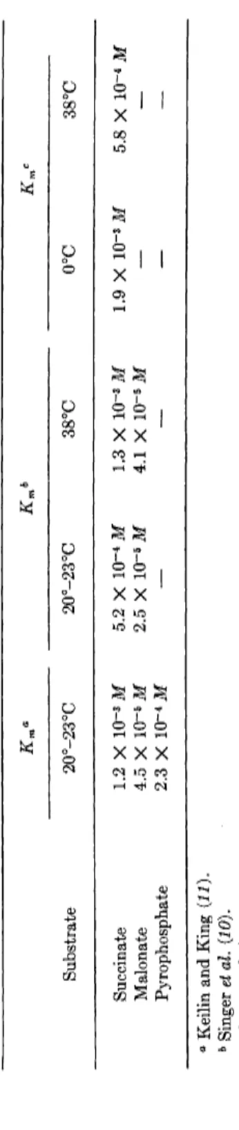
(malonate) is 4.5 Χ Ι Ο- 5 M. The ferricyanide method was chosen to avoid the complication due to the formation of hydrogen peroxide during the reoxidation of reduced phenazine methosulfate. Some values, collected by Keilin and King (11), for the Michaelis constants of succinic dehydro
genase are given in Table I. The affinities of the soluble enzyme for malonate and pyrophosphate differ from those reported with earlier preparations of the enzyme (24), the affinity for malonate being about five times that for pyrophosphate (11).
The affinity of malonate to succinic dehydrogenase is also shown by its ability (in common with succinate and fumarate) to protect the sulfhydryl groups of the dehydrogenase from oxidation by oxidized gluta
thione (25, 26). Cystine is also inhibitory to the succinoxidase system (27), the extent of inhibition in tissue homogenates being dependent on the concentration of the dicarboxylic acids present, particularly fumarate and malonate (28). It had earlier been shown that fumarate and its analogues (e.g., succinate, malate) can protect the enzyme fumarase from the toxic action of dyestuffs and certain trypanocidal agents (29, SO). Such protection indicates an affinity for the enzyme active center, if combination between the protector molecule and the toxic agent does not occur. The protection of succinic dehydrogenase by malonate takes place with very low concentrations of the latter, much lower than those necessary for the inhibition of the succinic dehydrogenase (26).
Protection also occurs, as would be expected, with succinate and fumarate.
Pyrophosphate also protects, but with less efficiency.
B. Effects of Thiol Reactcmts
Copper ions, maleic acid, and alloxan are inhibitory to the enzyme, presumably through their combination with the thiol groups of succinic dehydrogenase (26); malic, lactic, and α-glycerophosphate dehydro
genases are much less sensitive to these inhibitors.
Succinic dehydrogenase is also inhibited by sodium diethyldithiocar- bamate ( D D C ) , this being due, not to chelation with a metal, but to the oxidized form of D D C , namely, tetraethyldithiocarbamyl disulfide
[ ( C2H5)2N C S - S S - S C N ( C2H5)2] derived from D D C by an oxidation catalyzed by the cytochrome system (31). The oxidized product is
TABLE I MICHAELIS CONSTANTS OF SUCCINIC DEHYDROGENASE Km« Kmb Km< Substrate 20°-23°C 20°-23°C 38°C 0°C 38°C Succinate 1.2 Χ 10~3 Μ 5.2 Χ ΙΟ"4 Μ 1.3 Χ 10~3 Μ 1.9 Χ ΙΟ"3 Μ 5.8 Χ 10~4 Μ Malonate 4.5 Χ 10~5 Μ 2.5 Χ ΙΟ"6 Μ 4.1 Χ 10~5 Μ — — Pyrophosphate 2.3 Χ 10~4 Μ — — — — ° Keilin and King (11). b Singer et al. (10). c Wangei al. (12).
inhibitory, presumably by its ability to oxidize the thiol groups of the enzyme (11). Tetrathionate is also inhibitory presumably for the same reason.
The well-known thiol reactant n-chloromercuribenzoate inactivates soluble succinic dehydrogenase. The enzyme may be reactivated by a short incubation with cyanide (10). Thus, conclusions as to the thiol nature of succinic dehydrogenase obtained from studies with particulate preparations of the enzyme are confirmed by studies with the soluble enzyme preparation.
Alloxan inhibition of the citric acid cycle (32), which occurs at low concentrations, e.g., 0.03 mM, is not wholly due to a suppression of suc
cinic oxidase activity. Succinate oxidation is less sensitive to alloxan than that of pyruvate, α-ketoglutarate, or L-malate by rat kidney homogenates.
Protection against inhibition may be brought about by the presence of 0.15 m l nicotinicadenine dinucleotide ( N A D ) or 0.6 m l glutathione.
Alloxan inhibition of yeast fermentation (38) is also suppressed by the addition of N A D , and the diabetogenic action of alloxan in rats may be diminished by preliminary injection of nicotinamide, an effect probably due to an elevation of the N A D level (34, 85). These facts point to an interference of alloxan with NAD-linked metabolic systems, and may account for the uncoupling action of alloxan on oxidative phosphorylation (32, 36). Conceivably, alloxan may compete with N A D as a hydrogen acceptor for some dehydrogenase system. It should be noted that mod
erately high concentrations of N A D H and N A D have been held to be competitive inhibitors with respect to ferricyanide (87), which can act at more than one point in the N A D H oxidase chain, either directly with N A D H (20, 88) or at a site probably identical with that taken by cytochrome c (87).
C. Effects of Fluoride
Fluoride and phosphate each have weak inhibitory effects on succinic dehydrogenase and compete with succinate for the enzyme; a mixture of the two may be strongly inhibitory (40). A study of the kinetics of the inhibitions indicates that one phosphate ion and one fluoride ion are in
volved in the inhibition. It is pointed out (40) that inhibitions by fluoride often involve two fluoride ions, e.g., that of enolase (41) and the oxidation of quinol by cytochrome c and cytochrome oxidase (42). It has been sug
gested (43) that succinic dehydrogenase is a manganese compound on the basis of the fluoride inhibition, but this seems unlikely as managanese ions have but little effect on the enzyme activity (40). The suggestion
that the enzyme is a calcium complex (44), both because of the fluoride inhibition and of the activating action of calcium ions, is also unlikely, as it is known that the calcium activation is connected with the manner in which the enzyme preparation is made (81,4&) ( f or instance, no activating effect of calcium is seen with the enzyme in presence of phosphate-free buffers). Aluminum and chromium ions and those of the rare earths (46, 47) increase the rate of oxidation of succinate in the presence of a variety of tissue preparations. The suggestion was made (48) that the activating effect of calcium on succinic dehydrogenase is due to its activa- tion of an enzyme hydrolyzing N A D [whose presence produces oxal- acetate, a potent inhibitor of succinic dehydrogenase (45, 4&)]j but opposed to this suggestion was the demonstration that calcium ions are able to exert their effects in the absence of N A D from the enzyme preparation (81).
Fluoride, in comparison with other inhibitors, is not a particularly powerful inhibitor of succinic dehydrogenase; but if the substrate, suc- cinate, is used at the low concentrations normally found in cells, fluoride becomes a strong inhibitor. It is conceivable, in fact, that the entire inhibitory effect of fluoride on cell respiration (42) may be due to its effect on succinic dehydrogenase (40). It has been concluded (40) that succinic dehydrogenase is the only component of the succinic oxidase system which is susceptible to fluoride, although the isolated enzyme is apparently less sensitive than the complete respiratory system, possibly because the enzyme within the cell is not working at optimal activity.
D. Effects of C y a n i d e
Cyanide, in low concentrations, inhibits the oxidation of succinate by the cell oxidase system by combination with cytochrome oxidase (cyto- chrome a3) . It therefore prevents the reoxidation of other components of the cytochrome system (cytochrome b, cx, c, and a ) . It has no effect on the anaerobic oxidation of succinate by methylene blue and other hydro- gen (or electron) acceptors. However, cyanide, at a relatively high con- centration (0.1 M) brings about a considerable inhibition (50%) of succinic dehydrogenase activity (50). It appears (51) that the enzyme is slowly and irreversibly inactivated by cyanide, the rate depending on temperature and concentration of cyanide. It may be protected from such inactivation by succinate (in its role as hydrogen donor) or by sodium dithionite, but not by malonate or by fumarate. The inactivation is due (11) to a reaction of cyanide with a disulfide group of the succinic dehydrogenase, which is independent of the thiol groups already known
to be implicated in the activity of the enzyme (25, 26). Thus, the enzyme needs for its activity not only flavin adenine nucleotide, two or four iron atoms, and two thiol groups per molecule but an additional disulfide group {10-12). The succinic dehydrogenase of a particulate preparation of brain tissue or of yeast cells, unlike that of a heart muscle preparation, is not affected by incubation with cyanide (52). This is held (11) to be due to a more efficient protection of the enzyme's disulfide group, which may play a role as a carrier of hydrogen via the flavin prosthetic group to the cytochrome system.
Cyanide may therefore react with the components of the succinic oxidase system in the following ways: (a) rapid reversible reaction with the trivalent iron of cytochrome oxidase, (b) slow and reversible reaction with the trivalent iron of cytochrome c, and (c) slow and irreversible reaction with a disulfide group of succinic dehydrogenase.
E. Effects of Action of Iron Chelators
While the role of the iron in succinic dehydrogenase activity is still problematical (58), it is known that certain iron-chelating agents exert inhibitory effects. Thus, 8-oxyquinoline, O-phenanthroline, and α,α'- dipyridyl as well as crystalline /^-globulin are inhibitory (54). It is suggested (10) that O-phenanthroline, for example, forms a ternary com
plex with the enzyme, the complex having about 70% of the original activity in assays requiring the participation of the enzymic iron (e.g., with ferricyanide, phenazine methosulfate) and full activity in those assays where the carrier reacts with the flavin ( F M N , diethylsafranine).
Possibly, the iron may be the immediate electron donor to ferricyanide and phenazine methosulfate (10) [see, however (58) ].
I t should be noted that hematin inhibits succinic dehydrogenase ac
tivity, concentrations of 3 X 1 0 ~4 Μ and 6 X 1 0 -4 Μ inhibiting the activity of a soluble preparation by 75% and 100%, respectively (11).
Its mode of action is unknown.
F. Effects of High O x y g e n Tension
Brain cell respiration is inhibited at high tensions of oxygen, the rate of respiration falling off to a greater extent in the presence of oxygen than in air, particularly when the respiration is allowed to occur in the presence of glucose, lactate, or pyruvate (55). Apparently, the most vulnerable enzyme is the thiol enzyme, pyruvic oxidase (55). Respiration
in presence of succinate is also sensitive to the inhibitory effect of oxygen.
Protection may be afforded by the presence of malonate (1 mM) or manganese ions (0.25 mM) (56). It is reported that, at pressures of 4-7 atmospheres oxygen, the thiol groups of succinate are slowly oxidized
(56-58).
G. Effects of Arsenicals
Investigations reported to the Medical Research Council (U.K.) in 1940, showed that trivalent organic arsenoso compounds are highly in
hibitory to thiol enzymes, particularly pyruvic oxidase, choline dehydro
genase, and succinic dehydrogenase (59, 60). 2-Amino-4-arsenosophenol is a highly effective inhibitor, its effects being neutralized by the addition to the arsenoso compound of thiol compounds such as glutathione and cysteine. The pentavalent arsenic compounds, such as tryparsamide or atoxyl, are inert until they are reduced to the trivalent form. The organic arsenoso compounds establish an equilibrium with the thiol enzymes as their toxicities do not increase with time, and the activities of some of the enzymes are restored by addition of thiol compounds (59, 60). The inhibitory effects of the organic arsenoso compounds are greater than those due to arsenite at equivalent concentrations. Succinate protects its dehydrogenase from inactivation by the arsenoso compound (59, 60).
Many thiol enzymes are attacked by trivalent arsenicals (59-61), but of these the brain pyruvic oxidase system seems to be most sensitive, 50% inhibition occurring with 17 μΜ lewisite (62). The trivalent organic arsenicals were first used as thiol reagents in 1935 (63); their addition compounds with the thiol compounds readily dissociate on increase of pH.
With dithiol compounds, such as reduced lipoic acid, the trivalent arseni
cals form stable ring structures, the combination not being reversed by subsequent addition of a monothiol compound. The inhibition of pyruvate oxidase by a trivalent arsenical such as lewisite, which is presumably due to an attack on the lipoate cofactor, cannot be reversed by subsequent addition of cysteine (64). It can, however, be reversed by certain dithiol compounds, notably 2,3-dimercaptopropanol (BAL) (65).
H. Effects of I o d o a c e t a t e
While iodoacetate, a well-known thiol reagent, has little or no effect on the activity of succinic dehydrogenase, its ester, ethyl iodoacetate, is a highly effective inhibitor (inhibition occurring at 0.7 mM) (66). For
information on the effects of lachrymators and chemical warfare sub
stances on thiol enzymes, the reader is referred to a variety of reviews (67-73) on this subject, details of which are outside the scope of this chapter.
I. Effects of Quinones
Succinic oxidase is inhibited by quinones (74), benzoquinone (5 X 1 0 ~5 M ) bringing about 80% inhibition (61). The inhibition is suppressed by the addition of glutathione (2 m M ) . p-Benzoquinone inhibition takes place noncompetitively in a heart muscle preparation in which the succinate-methylene blue system is mediated by ubiquinone and other active quinones (75).
Doubtless, the quinones are largely effective by their reactions with enzyme thiol groups (76, 77). A large number of naphthoquinone deriva
tives (antimalarials) have been tested on a succinic oxidase preparation, but no correlation of the inhibitors with their therapeutic effects has been found (78).
J. Effects of Pyrophosphate
Pyrophosphate, known as an inhibitor of cell respiration (79), inhibits succinic dehydrogenase competitively. It appears to act in a manner similar to that of malonate, by competition with succinate by virtue of its adjacent acid groups (24, 51). Kinetic studies with a soluble succinic dehydrogenase preparation confirm the fact that pyrophosphate is a com
petitive inhibitor of succinate (11), and a value for Km (pyrophosphate) is given in Table I.
K. Effects of Ο χ α I a c e t a t e
Since the observation of the competitive inhibitory action of malonate on succinic dehydrogenase, many studies have been made of the effects of other succinic acid analogues. The most important of these is oxalacetic acid.
Oxalacetate is probably the most potent competitive inhibitor of suc
cinic dehydrogenase both in its soluble and its particulate form (45, 80-82) [Κι 1.5 X 1 0 -6 Μ (82)]. Under conditions in which oxalacetate accumulates, succinate oxidation is inhibited (83, 84).
The importance of the inhibitory action of oxalacetate lies in the fact that, because of this, the substance may exert a rate-controlling effect on cell respiration, or that aspect of it controlled by the citric acid cycle.
It is known (45}49) that the addition of N A D to a suspension of washed and ground pig heart greatly reduces its succinic dehydrogenase activity.
This is attributed {45) to the formation of oxalacetate from succinate by the action of succinic dehydrogenase and of malate dehydrogenase in
conjunction with N A D . The oxalacetate formed, even in very small quantities, inhibits succinic dehydrogenase. The addition of N A D a s e {85) to the heart preparation removes the inhibitory effect due to N A D (86), but the inhibitory effect is retained if nicotinamide is added at the same time to diminish the activity of the N A D a s e (86). The addition of N A D to a brain preparation has no effect on the latter's succinic dehydrogenase activity, and this was attributed to a rapid breakdown of oxalacetate (45). It is more likely, however, that the lack of effect is due to the high NADase activity of the brain preparation. In the presence of nicotina
mide, the inhibitory effect of N A D on brain succinic dehydrogenase is evident (86).
The rate-limiting effect of oxalacetate on succinate metabolism in the intact cell is seen in a study of the conversion of succinate-2,3-C1 4 into radioactive amino acids in brain cortex slices in the absence and presence of glucose (87). In the presence of an ample supply of acetyl CoA, such as is afforded by the presence of glucose, the yields of radioactive C 02 and amino acids, particularly glutamate and glutamine, are much in
creased above those found in the absence of glucose. The fraction of radioactive succinate converted to radioactive C 02 is increased threefold by the addition of glucose, and at the same time the yield of labeled aspartate (formed by transamination of oxalacetate) is almost halved.
These results are satisfactorily explained by the conclusion that succinate metabolism is retarded by the formation of oxalacetate, whose removal by condensation into the citric acid cycle allows succinate metabolism to be enhanced, aspartate yield to be reduced, and glutamate and glutamine formation to be increased.
Inhibition of succinic oxidase by oxalacetate (0.2 μΜ) may be pre
vented (48) by addition of L-glutamate, which converts it to aspartate by transamination.
L. Mitochondrial Respiration in Presence of Succinate a n d the Effects of Metabolic Inhibitors
Although, as already pointed out, the effects of glucose on the oxida-
tion of succinate, and on the conversion of the latter to aspartate, to glutamate, and to glutamine in brain cortex slices, may be explained by the conclusion that the formation of oxalacetate is a controlling factor in succinate metabolism (87), the use of cell fractions, such as mitochondria, makes it possible to study in more detail the somewhat complicated factors involved in succinate oxidation and the effects of inhibitors.
Succinate brings about the reduction of N A D in mitochondria and it has been suggested that this involves a reversal of oxidative phosphoryla
tion (87a-k). A different mechanism, competition between succinate and N A D H for a common rate-limiting component of the respiratory chain, has also been suggested (871, m ) , and it has been disputed as to whether a reversal of oxidative phosphorylation plays a major role in succinate oxidations in mitochondria or homogenates (87n). Competition between substrates for oxidizing catalysts presumably plays an important role in cell oxidations although this may be more observable in cell fractions than in the intact cell. Many years ago (87o) it was observed that when a mixture of succinate and lactate was added to minced guinea pig brain, the oxidation of succinate was retarded by an amount proportional to the oxidation of the lactate. The explanation given at that time was that there was competition between the substrates for an intracellular hydrogen carrier.
Recent observations have given rise to the conclusion that the reduction of N A D by succinate requires energy made available by oxidative phos
phorylation, but not necessarily in the form of ATP, as such reduction can occur (89) in the presence of concentrations of oligomycin which can inhibit synthesis of A T P but not that of energy-rich intermediates which precede A T P formation (111, 112, 114). In the absence of oligomycin, a competition for an energy-rich intermediate possibly occurs between a reaction leading, on one hand, to the formation of A T P and a reaction
(involving succinate and N A D ) leading, on the other hand, to the for
mation of N A D H2. Thus, according to this hypothesis, the continued utilization of N A D H2 (as, for example, in the reductive amination of α-ketoglutarate to L-glutamate) leads to a diminution in the rate of formation of A T P (39,114).
When beef heart mitochondria are incubated anaerobically with a mixture of succinate, N A D , and ATP, the rate of formation of N A D H2 is dependent on the concentration of A T P (87q). This may be suppressed by suitable concentrations of Amytal, oligomycin, and uncouplers of oxidative phosphorylation. Both the ATP-dependent reduction of N A D by succinate and the fumarate oxidation of N A D H2 are inhibited by
malonate, Amytal, and high concentrations of antimycin (87r)} the results supporting the concept of reversible oxidative phosphorylation in the N A D H region of the respiratory chain. The N A D H dehydrogenase system would appear to be implicated as Amytal is known to inhibit the reduction of coenzyme Q)1 by N A D H (87s).
The proposition {87t) that aerobic oxidation of succinate by rat liver mitochondria requires A T P is supported by the rinding that preincuba- tion of the mitochondria in an arsenate medium (free of phosphate) leads to depression of succinic oxidase activity when measured in the presence of 2,4-dinitrophenol or dicoumarol, and that addition of A T P , after the preincubation, restores the oxidation. However, the restoration by A T P occurs only after a time lag; the inactivation is not specific for succinate oxidation nor is the reactivation specific for A T P (87u). Moreover, the reactivation by A T P is not sensitive to oligomycin. It has been sug- gested (87u) that the phenomena in question are due to changes in mito- chondrial structure.
Doubt, however, is thrown on the possibility that succinate oxidation in mitochondria is necessarily linked with oxidative phosphorylation by the demonstration that a mixture of 0.5 mM dinitrophenol and 1.25 mM Amytal, which strongly inhibits oxidative phosphorylation, may not inhibit the reduction of acetoacetate by succinate (87n). 2,4-Dinitro- phenol stimulates respiration of a rat liver homogenate in presence of succinate at 0.05 mM and inhibits it at 0.5 mM.
Amytal, which does not normally inhibit succinate oxidation, abolishes both the stimulatory and inhibitory effects of dinitrophenol on the res- piration of liver homogenate and on succinate oxidation (87n). It does not reverse the inhibition of oxidative phosphorylation by dinitrophenol.
In this connection, it may also be observed that barbiturates suppress the stimulated respiration of rat brain cortex slices (in a glucose-saline medium) brought about by the addition of 2,4-dinitrophenol (87v). It is possible to explain this by the known suppression by barbiturates and other anesthetics of ADP-dependent N A D H2 oxidation (see Chapter 33).
The stimulation of liver homogenate respiration brought about by Amytal in presence of succinate and an inhibitory concentration of 2,4-dinitrophenol is abolished by the addition of ATP. The evidence (87n) supports the conclusion that, in presence of Amytal, electron transport from N A D H is no longer controlled by A D P or phosphate or both (see also Chapter 33). The activity of purified succinic dehy- drogenase is not affected by dinitrophenol.
The steady-state concentration of oxalacetate controls the rate of oxygen uptake in isolated mitochondria in presence of succinate or
L-malate (87p), and the removal of oxalacetate by transamination with L-glutamate stimulates the rate of oxygen consumption (87p). The addi
tion of ATP (magnesium salt) prevents the inhibition by added oxal
acetate of the oxidation of succinate in rat kidney homogenates (83; see also 82). It is held that this may be due to diminished penetration of oxalacetate into the mitochondria (39).
The suggestion that oxalacetate may be the immediate cause of the inhibition of succinate oxidation by the presence of a relatively high concentration of dinitrophenol (87w) is apparently not borne out by experiment which shows that less than 0.05 m M oxalacetate accumulates under these conditions (87n). But, it should be pointed out that, with guinea pig liver mitochondria, the addition of glutamate greatly stimu
lates the rate of oxidation of L-malate, and it is evident that undetectable concentrations (less than 0.03 mM) of oxalacetate are able to inhibit malate dehydrogenase (87x).
There is a large increase in the rate of oxygen uptake in presence of succinate when oxidative phosphorylation is uncoupled, both with guinea pig (87x) and rat liver mitochondria (87z), and the evidence obtained from studies of guinea pig liver mitochondria indicates that oxidation of succinate is controlled by the rate of formation of A T P (87x). More
over, in presence of 2,4-dinitrophenol, added oxalacetate (which nor
mally gives only a limited inhibition of succinate oxidation by mito
chondria) gives a complete suppression of succinate oxidation. There, thus, appears to be an energy requirement for local removal of oxal
acetate at the succinate oxidase site (83,87xy87y). In mammary gland mitochondria the oxidation of succinate is controlled by the rate of removal of oxalacetate (87x).
M. Inhibitive Effects of Various Succinic Acid A n a l o g u e s
It was pointed out (6) in the study of bacterial succinic dehydrogenase that other structures related to succinate, besides malonate, had inhibitory effects on this enzyme, though malonate was the most effective of those tested. Among such substances were glutaric acid, β-phenylpropionic acid, phenylacetic acid, and tricarballylic acid.
Alkylsuccinic acids are inhibitory, the inhibition varying with the length of the alkyl chain (88). 1,2-Ethanedisulfonate and β-sulfopropio- nate in concentrations between 3.3 mM and 20 m M inhibit succinic dehydrogenase almost as well as malonate (89).
Arsenoacetic acid ( C O O H C H2A s 03H2) acts as a competitive inhibi
tor of protozan succinic dehydrogenase (90), but it is apparently without effect on a succinic dehydrogenase preparation from mouse or rat (91).
The additions of competitive inhibitors (e.g., maleate, itaconate, or malonate) to a soluble succinic dehydrogenase preparation give changes in the absorption spectrum of the enzyme preparation that are similar to those brought about by fumarate (92, 93). Malate and oxalacetate have relatively large effects, but pyrophosphate has no action. These results suggest the possibility that the spectrum is that of a flavin semiquinone (92, 93).
Malondialdehyde has a slight inhibitory effect on succinate and oxal
acetate utilization by rat liver homogenate (93a).
N. Antimycin A Inhibition
Antimycin A inhibits the flow of electrons from succinate (or D P N H ) to cytochrome c but not to other acceptors in mitochondrial preparations (94, see also 95), and there appears to be an equivalence between the flavin content of the enzyme preparation and the antimycin titer (96). This evi
dence, taken together with the inhibitory effects of ethyl urethan at high concentrations [for 0.36 Μ ethyl urethan inhibits oxidation through the cytochrome system by about 90% and through phenazine methosulfate by about 40% (11) ] and those of 2,3-dimercaptopropanol (BAL) in the pres
ence of oxygen (97-99), suggests the existence of one or more carriers additional to cytochrome b and c. These inhibitions have been believed to be associated with the same factor (commonly known as the "Slater"
factor) in the respiratory chain, but there seems to be evidence that the antimycin- and BAL-sensitive sites are not identical (100). Antimycin A is also held to inhibit electron flow between succinate and D P N (101).
The Slater factor has been identified at various times with either cyto
chrome b or c; but this is unlikely to be the case, as antimycin A inter
feres with the reaction between cytochrome b and c without affecting the spectrum of either (102). The inhibition of succinic acid oxidation in the respiratory chain by antimycin A in fact appears to be localized between cytochromes b and Ci (11, 103). With 0.3 mg of a soluble prep
aration of succinic dehydrogenase, 0.1 mg antimycin A had no effect on the oxidation of succinate (11) by phenazine methosulfate as an au- toxidizable hydrogen acceptor (see also 104-107). However, it affects the properties of cytochrome b in disrupted beef heart mitochondria
(103).
Ο . Oligomycin Inhibition
Succinate-linked pyridine nucleotide reduction may occur in mitochon
dria, and this is inhibited by small concentrations of Amytal (see also Chapter 33). Oligomycin can also affect the N A D reduction [except in the presence of glutamate (108)], but partial or complete oligomycin insensitivity depends on the concentrations of A T P or internal high energy intermediates (109-111); the latter are supplied during succinate oxidation. Oligomycin interferes with A T P formation (112, 113) and blocks the interaction of A T P with components of the respiratory chain
(112). It can, in fact, prevent the stimulating action of A T P on metabolic processes dependent on malate oxidation (114). Respiratory inhibition by oligomycin in rat liver slices is largely released by 2,4-dinitrophenol suggesting that, as in isolated mitochondria, the oligomycin-sensitive respiration is coupled to the formation of intermediates of oxidative phosphorylation (114a).
III. FUMARATE CONVERSION TO L-MALATE;
INHIBITIONS OF FUMARASE
Fumarase plays an essential role in the citric acid cycle by bringing about the conversion of fumarate to L-malate. It is very widely distrib
uted in bacteria, molds, yeasts, and animal tissues and even in red blood cells (115); in fact, human blood is one of the richest sources of fumarase.
Most cells seem to be so amply supplied with the enzyme that it is un
likely to play any rate-limiting role in the citric acid cycle. Specific inhibitors of the enzyme would have to be very effective indeed to bring about marked diminutions in the rate of operation of the citric acid cycle.
Phosphate ions bring about a marked activation of fumarase (115-117), but the activation varies according to the preparation of the enzyme and the pH (115). They have a protective effect against the toxic action of certain dyestuffs [e.g. brilliant green (115)].
A variety of anions, di- or trivalent (e.g., arsenate, citrate) activates fumarase, the activation being a function of pH (118). This is held to be due to a combination of the anion with a basic group adjacent to either of the two ionizable groups in the active center of fumarase (118).
Fumarase is particularly sensitive to the action of acid dyes such as Congo red, trypan blue, methyl violet. Congo red will exert inhibitory effects at a concentration of 1.2 χ 1 0 ~5 Μ (115). The presence of pro
teins diminishes the dye toxicity, owing to their rapid combination with
the dyes. Substances related in structure to trypan blue or trypan red, such as Bayer 205 (suramin) and the S-carbamides of 2-naphthylamine- disulfonic acid, are highly inhibitory to fumarase {119), and there seems to be some relation between fumarase inhibition and trypanocidal ac
tivity. However, it must not be supposed that the toxicity of the higher S-carbamides to fumarase is any explanation for their trypanocidal activi
ties, as some of them, which are toxic to fumarase, have no trypanocidal action (119). As reported in 1932 (SO), fumarase is protected from the toxic action of dyes by its substrate fumarate and by its analogues, suc
cinate and malate, that combine reversibly with the enzyme. Competitive inhibitors of fumarase are D-malate, citrate, D-tartrate, L,a-hydroxy-/?- sulfopropionate, maleate, mesaconate, transaconitate, succinate, malonate, adipate, glutarate, glycine (120). Substances that are inactive as inhibi
tors include crotonate, acetoacetate, acetate, butyrate, and acetylenedi- carboxylate (120). It would appear that the presence of two acidic groups is necessary for attachment to the active center of fumarase, but it is important to note that removal of the α-hydrogen atoms from fumarate
(as in acetylenedicarboxylate) renders the substance inactive.
It seems possible that the high toxicity of acid (sulfonated) dyes, referred to previously, to fumarase may be due to combination with the receptor groups at the active center, with which the acid groups of fumarate and its analogues also combine.
Many investigations have been carried out on the activity of fumarase and its mode of action (115, 120-12S), and it is evident that this enzyme presents many advantages for studies of the manner in which enzymic hydration of the double bond is brought about.
IV. INHIBITION OF MALATE METABOLISM
Just as the metabolism of lactate is inhibited by lactic acid analogues, e.g., pyruvic acid, tartronic acid, glyceric acid, glyoxylic acid, mandelic acid, raeso-tartaric acid, oxalic acid (6), so is the metabolism of malate affected by malic acid analogues, which resemble both the lactic acid analogues and the succinic acid analogues.
Tartronate and oxalacetate are inhibitors of malate oxidation in a pig heart preparation (124)and in a partially purified pigeon liver prepara
tion, using the anaerobic ferricyanide system, tartronate is approximately 1000 times more inhibitory of malate oxidation than in a pig heart preparation (125). It is of interest to note that, with the liver preparation, citric acid, maleic acid, isocitric acid, and malonic acid have inhibitory
effects (125) j indicating some affinity of these molecules to the malic dehydrogenase. Thus, isocitrate (10 mM) and malonate (20 mM) inhibit malate oxidation by 55% and 30%, respectively, in the presence of 20 mM DL-malate.
β-Fluorooxalacetate is a powerful inhibitor of malic dehydrogenase (and of glutamic-aspartic transaminase) (126). Relatively high concen
trations of fluorooxalacetate also inhibit the reduction of triphosphopyri- dine dinucleotide ( N A D P ) by L-malate at pH 7.4 by a preparation of
"malic enzyme" of chicken liver. Moreover, the NADP-dependent decar
boxylation of oxalacetate at pH 4.5 is inhibited by fluorooxalacetate (127). Apparently, this inhibitor reacts nonenzymically with N A D H and N A D P H , and with some analogues of these coenzymes, to form fluorescent addition compounds. Oxalacetate is inert in this respect (127). The addi
tion compounds are without influence at pH 7.5 on the action of malic and glucose-6-phosphate dehydrogenases and of the malic enzyme.
The malic enzyme (128), which is very widely distributed and which catalyzes the reaction [Eq. ( 1 ) ]
Pyruvate + C 02 + NADPH + H+ <=± Malate + N A D P (l) tends to favor the synthesis of malate, rather than its breakdown, owing
to the high value of the N A D P H / N A D P ratio in the cell. This enzyme is inhibited by the malic acid analogues oxalacetate, malonate, tartronate, oxalate, mesoxalate, meso-tartrate, and fluoromalate (129-182). In this connection, reference may be made to the inhibitory action of meso- tartrate on pyruvate metabolism, in rat kidney slices, and rat kidney mitochondria (183). This inhibition is reversed by the addition of small quantities of fumarate, malate, or citrate. The evidence indicates that meso-tartrate acts by inhibiting dicarboxylic acid formation from pyruvate.
Malate synthetase, which catalyzes formation of malate from glyoxylic acid and acetyl CoA (184), requires magnesium ions for maximal ac
tivity and is competitively inhibited by analogues of glyoxylic acid, viz., oxalate (Kt = 1.9 Χ 1 0 ~5 Μ ) , fluoroacetate (K* = 2.5 X 1 0 ~4 M ) , and glycolate (Κ{ = 3.1 χ 1 0 ~4 M ) (135). The Km for glyoxylate was found to be 9.3 χ Ι Ο- 5 Μ (in a preparation from yeast).
Mention may be made here of the enzyme isocitratase (186), which controls the reversible reaction between isocitrate and its products, suc
cinate and glyoxylate. This enzyme is noncompetitively inhibited by succinate (137); the sensitivity to succinate varies with the source of the enzyme (134). Isocitratase is also powerfully inhibited by oxalacetate
(138), but pyruvate, malate, fumarate, and aspartate are without effect.
V. OXALACETATE METABOLISM
I t is reported that oxalacetate decarboxylase is inhibited by malonate (139-141), even at a concentration of 10 m M (141) and by D - and L - malate. At a concentration of 30 mM, malonate is reported as inhibiting the conversion of oxalacetate to citrate with a liver preparation (82).
Oxalacetate metabolism plays a basic role in the citric acid cycle, for oxalacetate acts as a catalyst in the oxidation of acetate to C 02, in the conversion of L-glutamate to L-aspartate, in the synthesis of fatty acids from sugars, and in controlling the oxidation of succinate and L-malate.
With guinea pig liver mitochondria (87x) its utilization is relatively con
stant under a variety of conditions, e.g., presence or absence of succinate or 2,4-dinitrophenol. Its removal by acetyl CoA to give citrate or malonyl CoA, or by glutamate to yield aspartate and α-ketoglutarate is important for determining the steady-state level.
VI. CITRATE BREAKDOWN; INHIBITION OF ACONITASE
The reversible transformations of citric acid into cis-aconitic acid and d-isocitric acid, catalyzed by aconitase (142, 14$), are an essential part of the series of reactions involved in the citric acid cycle. As is well known, the key reaction of the cycle is the condensation of oxalacetate with acetyl CoA to form a tricarboxylic acid (144-146)-
While the succinate cytochrome oxidase system is always found in mitochondria, aconitase is found mainly in the supernatant in the liver but exclusively in the mitochondria in brain (147).
The enzyme is inhibited by copper and mercury ions, cyanide, and sulfide at relatively low concentrations (148). Aconitase, in contrast to fumarase, is unstable in solution on storage even in the refrigerator (148).
The enzyme substrates, citrate or cis-aconitate, protect it from inactiva
tion. It is fairly stable in frozen tissues. It is reported to be dependent on ferrous ions for optimal activity.
The analogue, irans-aconitate, which is not activated by aconitase, inhibits the enzyme presumably competitively (14&).
A. Fluoroacetate Inhibition
A good deal of literature has now accumulated on the subject of fluoro
acetate inhibition of cell respiration, and much of it is summarized in a
recent review (150). Only the outstanding facts that bear on the mecha
nism of fluoroacetate inhibition will be considered here.
Fluoroacetate, known as a rat poison because of a selective toxicity to rodents (151), inhibits the oxidation of acetate in animal tissues and causes an accumulation of acetate in the presence of pyruvate (152). It was thought to be a specific, perhaps competitive, inhibitor of acetate oxidation (152). However, an accumulation of citrate takes place in kidney homogenates in the presence of fluoroacetate (153, 154), and a very large accumulation of citrate takes place in vivo in a variety of tissues when fluoroacetate is injected into rats, pigeons, guinea pigs, and frogs (155, 156). It was suggested (157-159) that fluoroacetate is not directly responsible for the accumulation of citric acid but that there is enzymic synthesis to a component affecting the citric acid cycle, possibly fluorocitrate (158). Fluorocitrate was eventually identified as a product formed on incubation of tissue homogenates with oxalacetate or fumarate and fluoroacetate (160), and it was shown (161) that fluorocitrate specifii- cally inhibits aconitase, thus accounting for citric acid accumulation.
Fluoroacetate itself has no effect on aconitase (162). Apparently, fluoro
citrate is more effective in vivo than in vitro using soluble preparations of aconitase (62). Fluorocitrate, which inhibits the enzyme competitively (161), has a lesser effect on a soluble preparation of the enzyme than in a mitochondrial preparation, and it is thought possible that this may be due to accumulation of fluorocitrate in the mitochondria (163). There is a lesser effect of synthetic fluorocitrate than enzyme-synthesized fluoro
citrate, at equal concentrations, on citrate disappearance by a kidney preparation, and this is attributed to the presence in the former product of inactive isomers of fluorocitrate. On the other hand, synthetic fluorocitrate is a more powerful inhibitor of purified aconitase than the enzymic preparation (150).
Fluoroacetyl CoA reacts with oxalacetate in the presence of the purified condensing enzyme to form fluorocitrate (164). The initial reaction is slower than with acetyl CoA, and there is competitive inhibition. How
ever, a pigeon liver acetate-activating system does not form fluorocitrate from fluoroacetate and oxalacetate (150). Acetate inhibits the synthesis of fluorocitrate by kidney particles in the presence of malate and fluoro
acetate, but the evidence appears to indicate that fluoroacetate and acetate are not necessarily activated at the same enzyme (150). Pigeon brain tissue, when used as a homogenate, does not synthesize fluorocitrate from fluoroacetate; nor does the presence of fluorocitrate give rise to citrate accumulation. When the brain tissue is more finely ground and
reinforced with A T P and magnesium ions, the presence of fluorocitrate causes citrate accumulation in the presence of pyruvate {165).
Acetate acts as a protector to some animals (e.g. mice) against the toxic effects in vivo of fluoroacetate, and ethanol is effective with mice, guinea pigs, and rabbits (166) but not in dogs. The most effective protectors are glycerol monoacetate and acetamide (167,168). Presumably, these effects are due to competition between acetate (or acetyl CoA) and fluoroacetate
(or fluoracetyl CoA).
Fluoroacetate also accelerates acetoacetate formation in liver in vitro (169), and this is held to be due to an inhibition in the citric acid cycle (150). An effect of fluoroacetate in vivo is to increase blood sugar levels (170), and in fact an injection of 8 m g / k g of sodium fluoroacetate into fasted rats gives a rise in ketone bodies followed by a rise in blood sugar (171). Possibly, these observations may be explained by an inter- ference with the citric acid cycle preventing the normal utilization of acetyl CoA, so that more of the latter is converted to acetoacetate. It is already established (172) that malonate, added to liver slices in presence of acetate, causes accumulation of acetoacetate. This phenomenon was held, at first, to be due to inhibition of acetoacetate breakdown. Subse- quent work showed, however, that inhibition of acetoacetate breakdown by rat kidney cortex slices by malonate was reversed by the addition of fumarate (173), indicating that the breakdown of acetoacetate is depend- ent on the operation of the citric acid cycle. The fact that the inhibition was reversed also by lactate and alanine is consistent with this view, as both substances give rise to pyruvate and thence to oxalacetate. It follows that inhibition of the citric acid cycle with a diminished utilization of acetyl CoA will affect acetoacetate formation.
The convulsive effects due to fluoroacetate administration appear not to be due to citrate accumulation (150, 17^). Convulsions occur when there is little increase in citrate in the brain, and introduction of fluoro- acetate or fluorocitrate directly into the cortex causes changes in the electrical patterns in various areas without increased citrate levels in these areas. Fluorocitrate convulsions in mice cannot be stopped by intravenous calcium administration (175). Nor is there evidence that convulsions are due to defective acetylcholine metabolism (150, 176), although bovine brain tissue is reported capable of forming fluoroacetylcholine from fluoroacetate (177). Fluoroacetate has little or no effect on the respiration of mammalian brain tissue (178, 179), and there seems to be no evidence that fluoroacetate gives rise to fluorocitrate in brain (150). In view of this it is difficult to understand cerebral effects of fluoroacetate as being due to significant inhibitions of the cerebral citric acid cycle.
It should be noted in this connection that the enzymes involved in the citric acid cycle and subsequent hydrogen transfer, as well as pyruvic oxidase, are unaffected by fluoroacetate (even at concentrations as high as 16.6 mM) (152, 157). The respiration of pigeon brain homogenate, in the presence of pyruvate, is unaffected by fluoroacetate (16.6 mM) (157).
A relatively large increase in the ammonia level of dog brain has been reported (180, 181) as preceding the onset of convulsions in fluoroacetate poisoning. Recent work (182) indicates that fluoroacetate, at concentra
tions (e.g. 0.1 mM) that have no effect on rat brain cortex respiration (stimulated or unstimulated) in vitro, greatly reduces glutamine forma
tion from glucose and the cerebral utilization of ammonium ions. There is no inhibition of glutamate formation, indicating that there is little interference with the citric acid cycle, for glutamate is derived by trans
amination from α-ketoglutarate. The mechanism by which fluoroacetate affects cerebral utilization of ammonium ions is at present unknown.
Possibly, this phenomenon is related to the convulsive effects of fluoro
acetate administration.
A study (183) of the effects of fluoroacetate on the metabolism of Rhodaspirillum rubrum has shown that this substance (0.8 mM) strongly inhibits the photometabolism of butyrate, pyruvate, and oxalacetate, but it also affects the photoreduction of carbon dioxide by hydrogen, a process apparently independent of the citric acid cycle.
Fluoroacetate (1 mM) inhibits nitrogen fixation and ammonia meta
bolism in Azotobacter (184), a process counteracted by acetate, but not by α-ketoglutarate. Whether this inhibition is brought about by an in
hibitory effect on the citric acid cycle is unknown.
In studies of the metabolism of Aspergillus terreus, it is shown (185) that fluoroacetate (0.2 mM) increases the molar conversion of both glu
cose and citrate to itaconate (arsenite, 10 mM, on the other hand, inhibits the conversion of glucose to itaconate, with accumulation of pyruvate and acetaldehyde).
B. O x a l o m a l a t e Inhibition
Oxalomalate, which may be formed nonenzymically from glyoxylate and oxalacetate, is a competitive inhibitor of aconitase (186, 187), and it is possible that inhibitory effects of glyoxylate on the oxidation of com
ponents in the citric acid cycle may be partly due to the formation of oxalomalate (187).
VII. METABOLISM OF
α-ΚETOGLUTARATΕ
a-Ketoglutarate plays an important role in the operation of the citric acid cycle. It is not only broken down to succinate by mechanisms in
cluding the participation of guanosine diphosphate and adenosinediphos- phate (so that for every molecule of α-ketoglutarate oxidized, one of A T P is formed), but it is transaminated to L-glutamate, the precursor of γ-aminobutyric acid and glutamine. Inhibitors of a-ketoglutarate metabolism may be expected, therefore, to affect the velocity of the citric acid cycle.
Parapyruvate ( C O O H C ( O H ) (CH3) C H2C O COOH), a structural analogue of α-ketoglutarate, inhibits specifically the oxidation of a-keto
glutarate in various tissue preparations (188-190). It diminishes respira
tion of muscle homogenates in presence of pyruvate, the inhibition being restored by the addition of succinate and fumarate, with accumulation of α-ketoglutarate (190). Apparently, those stages involved in the formation of α-ketoglutarate from fumarate in the citric acid cycle proceed more rapidly in the presence of parapyruvate and compensate for the loss of activity in the steps between α-ketoglutarate and fumarate. a-Keto
glutarate dehydrogenase activity is also inhibited by ions of heavy metals (191, 192), the inhibition being readily reversed by BAL. However, in
hibitions of the enzyme by cobalt and copper ions are not reversed by BAL, and this may be explained by chelation with dihydrolipoic acid
(198).
Arsenite is an effective inhibitor of α-ketoglutarate oxidation (108).
A method of selectively inhibiting one step in the citric acid cycle is to omit phosphate and add 2,4-dinitrophenol. Under these conditions, the step of α-ketoglutarate conversion to succinate is inhibited because the substrate-linked phosphorylation step, in contrast to respiratory- chain phosphorylation, is not uncoupled by 2,4-dinitrophenol (193a, 193b). It has been shown (87%) that the aerobic oxidation of α-keto
glutarate in rat liver mitochondria in presence of 2,4-dinitrophenol requires phosphate but not A D P for maximal activity. Arsenate, which abolishes α-ketoglutarate-linked phosphorylation, is able to replace phos
phate. The lack of requirement of A D P is explained as being due to con
tinuous regeneration of mitochondrially bound A D P from A T P by dinitrophenol-activated ATPase. Phosphate (and not A D P ) is also needed for the optimal rate of α-ketoglutarate dismutation in presence of ammonium ions and rat liver mitochondria (193c).
VIII. ISOCITRATE OXIDATION
Estradiol increases the conversion of both pyruvate-2-C1 4 and acetate- 1 - C1 4 to C1 402 by slices of placenta, and homogenates of placenta also respond to the in vitro addition of estradiol, the production of α-ketoglu
tarate from citrate being increased about 50%. The estradiol apparently stimulates specifically isocitric dehydrogenase (194, 195) and activates the reversible reaction [Eq. (2) ]
Isocitrate + N A D ^ NADH + H+ + C 02 + α-ketoglutarate (2) Stilbestrol is an inhibitor of this system, apparently competing with estradiol for the receptor group on the enzyme (194, 195).
The activating agent is 17/?-estradiol, and 17a-estradiol is a potent competitive inhibitor. It is suggested that estradiol combines with isocitric dehydrogenase, converting it from an inactive to an active form and thus affecting the operation of the citric acid cycle (194, 195).
In addition, it may be pointed out that crude extracts of Aspergillus niger contain both N A D - and NADP-linked isocitric dehydrogenases and that 2'-adenylic acid inhibits the N A D P isocitric dehydrogenase but not the NAD-linked enzyme (196). The accumulation of citric acid is also reported to inhibit the isocitric dehydrogenase of A. niger (197).
A heart muscle preparation (from horse or pig) contains isocitrate dehydrogenase, requiring N A D (or N A D P ) and M g + + needed for decarboxylation of oxalosuccinate. Magnesium ions may be replaced by catalytic amounts of acetyl CoA, which reacts with oxalosuccinate to form α-ketoglutarate and malonyl CoA which is a precursor of acetyl CoA (198).
REFERENCES
1. J. H. Quastel, Biochem. J. 18, 365 (1924).
2. F. Batelli and L. Stern, Biochem. Z. 30,172 (1911).
3. T. Thunberg, Zentr. Physiol. 31, 98 (1916).
4. H. Einbeck, Biochem. Z. 95, 296 (1919).
5. J. H. Quastel and M. D. Whetham, Biochem. J. 18, 519 (1924).
6. J. H. Quastel and W. R . Wooldridge, Biochem. J. 22, 689 (1928).
7. J. H. Quastel and W. R . Wooldridge, Biochem. J. 21, 1224 (1927).
8. J. H. Quastel and W. R . Wooldridge, Biochem. J. 23, 115 (1929).
9. J. H. Quastel and Α. Η. M. Wheatley, Biochem. J. 25, 117 (1931).
10. T. P. Singer, Ε. B. Kearney, and V. Massey, Advances in Enzymol. 18, 65 (1957).
11. D. Keilin and Τ. E. King, Proc. Roy. Soc. B152, 163 (1960).
12. Τ. Y. Wang, C. L. Tsou, and Y. L. Wang, Sci. Sinica (Peking) 5, 73 (1956).
13. A. Szent-Gyorgyi, Z. physiol Chem. Hoppe-Seyler's 236, 1 ( 1 9 3 5 ) ; 244, 105 (1936).
14. F. J. Stare and C. A. Baumann, Proc. Roy. Soc. B121, 338 (1936).
15. H. A. Krebs and W. A. Johnson, Enzymologia 4,148 (1937).
16. H. A. Krebs and L. V. Egglestone, Biochem. J. 34, 442 (1940).
17. H. A. Krebs, E. Salvin, and W. A. Johnson, Biochem. J. 32,113 (1938).
18. H. Busch and V. R. Potter, J. Biol. Chem 198, 71 (1952).
19. T. Thunberg, Skand. Arch. Physiol 40, 1 (1920).
20. J. H. Quastel and Α. Η. M. Wheatley, Biochem. J. 32, 936 (1938).
21. Μ. B. Thorn, Biochem. J. 54, 540 (1953).
22. T. P. Singer and Ε. B. Kearney, Biochim. et Biophys. Acta 15, 151 (1954).
23. T. P. Singer and Ε. B. Kearney, J. Biol. Chem. 219, 963 (1956).
24. L. F. Leloir and M. Dixon, Enzymologia 11, 81 (1937).
25. F. G. Hopkins and E. J. Morgan, Biochem. J. 32, 611 (1938).
26. F. G. Hopkins, E. J. Morgan, and C. Lutwak-Mann, Biochem. J. 32, 1829 (1938).
27. S. R. Ames and C. A. Elvehjem, Proc. Soc. Exptl Biol. Med. 57, 108 (1944).
28. S. R. Ames and C. A. Elvehjem, Arch. Biochem. 5,191 (1944).
29. J. H. Quastel, Biochem. J. 25, 898 (1931).
30. J. H. Quastel, Proc. Roy. Soc. B i l l , 294 (1932).
31. D. Keilin and E. F. Hartree, Biochem. J. 44, 205 (1949).
32. E. S. Younathan, J. Biol. Chem. 237, 608 (1962).
33. C. J. Kensler, S. O. Dexter, and C. P. Rhoads, Cancer Research 2, 1 (1942).
34. A. Lazarow, J. Liambies, and A. J. Tausch, J. Lab. Clin. Med. 36, 249 (1950).
35. N. O. Kaplan, A. Goldin, S. R. Humphreys, Μ. M. Ciotti, and F. E. Stol- zenbach, J. Biol. Chem. 219, 287 (1956).
36. G. Bhattacharya, Proc. Natl. Inst. Sci. India 21B, 210 (1955).
37. S. Minakami, R. L. Ringler, and T. P. Singer, J. Biol Chem. 237, 569 (1962).
38. E. Haas, Biochem. Z. 291, 79 (1937).
39. E. C. Slater, Chem. Weekblad 58, No. 52 (1962).
40. E . C. Slater and W. D. Bonner, Biochem. J. 52, 185 (1952).
41. O. Warburg and W. Christian, Biochem. Z. 310, 384 (1942).
42. H. Borei, Arkiv. Kemi, Mineral. Geol. 20A, No. 8 (1945).
43. L. Massart, Z. physiol. Chem. Hoppe-Seyler's 258, 190 (1939).
44. V. R. Potter and W. C. Schneider, J. Biol. Chem. 142, 543 (1942).
45. D. Keilin and E. F. Hartree, Proc. Roy. Soc. B129, 277 (1940).
46. B. L. Horecker, E. Stolz, and T. R. Hogness, J. Biol. Chem. 128, 251 (1939).
47. W. C. Schneider and V. R. Potter, J. Biol. Chem. 149, 217 (1943).
48. K. F. Swingle, A. E. Axelrod, and C. A. Elvehjem, J. Biol. Chem. 145, 581 (1942).
49. V. R. Potter, Arkiv. Kemi, Mineral. Geol. 13B, No. 7 (1939).
50. I. Banga and E. Porges, Z. physiol. Chem. Hoppe-Seyler's 254, 200 (1938).
51. C. L. Tsou, Biochem. J. 49, 512 (1951).
52. A. Giuditta and T. P. Singer, J. Biol. Chem. 234, 666 (1959).