CULTURE OF HUMAN MONOCYTES IN MICROPLATES AND ENZYMATIC ASSAYS FOR FOLLOWING THEIR MATURATION
Lois B. Epstein Karen Yu Lawrence P. Chong Constance C. Reese
INTRODUCTION
Over the past several years, we cultured human macrophages on a large scale in Leighton tubes. We obtain these cells by maturation in vitro from human monocytes obtained from
peripheral blood. Such macrophage cultures are very versatile and have readily lent themselves for study in three distinct areas of research in our laboratory: (a) as a model system for the investigation of enzyme replacement and other modes of therapy for patients with lysosomal storage diseases (1);
(b) in studies to determine the cellular origin and the nature of cellular interactions involved when interferon is produced by leukocytes in response to mitogens or specific antigens (for review, see 2 - 5) ; and (c) for exploration on the many non- antiviral actions of interferon (6 - 9). However, the Leighton tube macrophage culture system we employed was very costly, not only in terms of medium and serum but also in requiring large donations of blood. Thus, this chapter will detail our new
METHODS FOR STUDYING Copyright © 1981 by Academic Press, Inc.
MONONUCLEAR PHAGOCYTES 4 9 All rights of reproduction in any form reserved.
ISBN 0-12-044220-5
micromethod for the preparation of human monocyte-derived macrophage cultures (10) and a recently developed fluorometric micromethod for monitoring the maturation of monocytes to mac- rophages by following the specific activity of two lysosomal enzymes, 3-galactosidase and ß-N-acetylglucosaminidase.
II. REAGENTS
A. Reagents for Preparation of Macrophage Cultures 1. McCoy's 5a medium (Grand Island Biological Co.
[Gibco], Santa Clara, California) is prepared with 100 U peni- cillin and 100 ug streptomycin/ml, respectively, and is used throughout the procedure. For diluting plasma, McCoy's medium containing 5 U/ml phenol-free sodium heparin (Lipo-Hepin, Riker Laboratories, Northridge, California) is prepared. For washing the macrophages, McCoy's medium is supplemented with
10% AB positive serum, and, for the culture of macrophages, the medium is supplemented with 30% AB positive serum.
2. Dextran (MW 500,000, Sigma Chemical Co., St. Louis, Missouri) is prepared as a 4.5% solution in pH 7.4 phosphate- buffered saline and is used for sedimenting erythrocytes to obtain leukocyte-rich plasma.
3. NH4CI - Tris solution is prepared by combining 9 ml of 0.83% NH4CI with 1 ml of Tris buffer (pH 7.65). This solution is used to free leukocyte-rich plasma of any residual erythro- cytes remaining after dextran sedimentation.
B. Reagents for Lysosomal Enzyme Assays Distilled water is used throughout.
1. Buffers for Enzyme Assays
(a) Sodium acetate buffer (0.1 M, pH 4.5) is used in the assay of 3-galactosidase. Prepare a 1 M acetic acid solution by combining 5.75 ml glacial acetic acid with 94.25 ml water.
Prepare a 1 M solution of sodium acetate by combining 13.61 gm (0.1 mole) sodium acetate with 100 ml water. To prepare the buffer, combine in a beaker 6.25 ml of 1 M acetic acid and
3.7 ml of 1 M sodium acetate. Add 90.05 ml water. Using a pH meter, add glacial acetic acid dropwise until pH 4.5 is reached.
To sterilize the buffer, filter it through a Millipore filter (pore size 0.22 ym) into a sterile 100-ml bottle. Store at room temperature.
(b) Citrate phosphate buffer (1 if, pH 4.6) is used in the assay of ß-iV-acetylglucosaminidase. Prepare a 0.2 if solution of Na2P04 by combining 14.198 gm Na2PC>4 and 500 m l water.
Prepare a 0.2 M citric acid solution by combining 21.015 gm citric acid and 500 ml water. Prepare a 2 if KCl solution by combining 37.28 gm KCl and 250 m l water. To prepare the buffer combine in a beaker 49.3 ml of the 0.2 if N a2P 0 4 with 25.5 m l of the 0.2 if citric acid and 13.3 m l 2 if K C l . Add water to a total volume of 100 m l . To sterilize the buffer, filter it through a Millipore filter (pore size 0.22 ym) into a sterile 100-ml bottle. Check pH of aliquot. Store at room temperature.
(c) Glycine - NaOH buffer (0.4 if, pH 10.3) is used to ter- minate the enzymatic reactions and to enhance the fluorescence of the liberated product, 4-methylumbelliferone (see below for details of enzymatic reactions). Dissolve 30.028 gm glycine
(MW 75.07) in 900 m l sterile deionized double-distilled water.
Add 13 gm NaOH. Check pH on pH meter and add extra NaOH pellets if necessary. Store at room temperature.
2. Substrate Buffer Solutions
Substrates obtained from Sigma Chemical Co., St. Louis, Missouri.
(a) 4-Methylumbelliferyl-3-D-galactopyranoside is the sub- strate for the assay of fB-galactosidase. Weigh out 16.9 m g and dissolve in 100 m l sodium acetate buffer, pH 4.5, in a volumetric flask. Transfer this volume to a 250-ml Erlenmeyer flask and heat the solution until the substrate dissolves.
Aliquot the solution to 4-ml sterile disposable Pyrex tubes and store the tubes at -70°C. Thaw tubes on the day of assay in the amount desired, and heat gently to dissolve substrate if necessary.
(b) 4-Methylumbelliferyl-2-acetamido-2-deoxy-3-iV-acetyl- glucopyranoside is the substrate for the assay of ß-iV-acetyl- glucosaminidase. Weigh out 18.95 m g and dissolve in 100 ml citrate - phosphate buffer, pH 4.6. This solution is light sensitive and so must b e kept in the dark. Aliquot the solu- tion into aluminum foil-wrapped, 4 m l , sterile - capped, plastic test tubes. Store the tubes at -70°C. Thaw tubes on the day of assay in the amount desired.
(c) 4-Methylumbelliferone Standard Solution (4-MUSS).
This solution is used to generate data for a standard curve against which is compared the enzyme-induced hydrolysis of substrates. The molecular weight of 4-methylumbelliferone is 176.2. Weigh out 2.643 mg of the crystalline substance and dissolve by adding absolute ethanol drop by drop. Add to the solution 0.01 N H2SO4 until the final volume equals 100 m l , and the solution is 0.15 mif. Store the solution in a dark bottle, as it is light sensitive.
To obtain the data for the standard curve, prepare the dilutions on the day of the assay in semidarkness as follows.
Make a 1/50 dilution to be used as a stock solution by com- bining 2 ml of the 0.15 mM solution with 98 ml sterile twice- distilled water. Make all subsequent dilutions in a 0.4 M glycine - NaOH, pH 10.3 in Pyrex glass dilution tubes as shown in the tabulation below.
Dilutions 1/50 1/100 1/200 1/500 1/1000 1/5000 1/7500 1/10,000 1/20,000
Preparation
4-MUSS (ml) + Buffer (ml) 2 of 0.15 mM +
1 of 1/50 + 1 of 1/100 + 0.05 of 1/50 + 0.1 of 1/50 + 0.1 of 1/1000 + 0.1 of 1/1000 + 0.1 of 1/1000 + 0.1 of 1/10000 +
98 1 1 0.45 1.9 0.4 0.65 0.9 0.1
Final
concentration (nm/ml) 3
1.5 0.75 0.30 0.15 0.03 0.02 0.15 0.0075
Cover the tubes with a Parafilm and put them in the dark until they are read in the spectrophotometer.
Reagents for Protein Determinations on Cell Lysates 1. Stock solutions
2% NaC03 in 0.1 N NaOH 0.5% CUSO4
1% Na tartarate
Folin - Ciocalteau phenol reagent (Harleco, Gibbs- town, New Jersey), dilute with equal volume of dis- tilled water on day of assay
Bovine serum albumin (BSA) (Sigma Chemical Co., St. Louis, Missouri)
0.1% Triton in 0.85% saline
2. Preparation of BSA standard dilutions. The concentra- tion of our original stock of BSA is 50 mg/ml. The concentra- tion of our working stock is 1 mg/ml, made in distilled water
(see tabulation below).
Solution Preparation Final designation BSA (ml) + distilled H2O (ml) Concentration (mg/ml)
a b c d e f h 9 i k j 1 m
1 of original stock + 49 0.75
0.75 0.5 0.85 0.8 0.5 0.5 0.5 0.5 0.5 0.5 0.5
of a of a of b of c of e of d of e of f of h of i of j of 1
+ + +
■f
+ +
-f
■f
-f
+ + +
0.25 0.75 0.5 0.85 0.2 0.5 0.5 0.5 0.5 0.5 0.5 0.5
1 0.75 0.5 0.375 0.25 0.20 0.188 0.125 0.100 0.0625 0.0469 0.0313 0.0156
III. PROCEDURE
A. Preparation of Macrophage Microcultures
Withdraw venous blood into plastic syringes previously heparinized with Lipo-Hepin (100 U/10 ml blood) and transfer 10 ml aliquots to 16 x 150 mm round bottomed, plastic, screw- topped tubes that contain 4 ml 4.5% dextran in PBS. Incubate the dextran - blood mixture for 21 min at 37°C and transfer the leukocyte-rich plasma to 50 ml plastic centrifuge tubes to which an equal volume of plain McCoy's medium has been added.
Centrifuge the diluted, leukocyte-rich plasma at 1000 rpm for 10 min and discard the supernatant fluid. Resuspend the pel- let in 10 ml wash medium, recentrifuge as above, and discard the supernatant wash medium. Lyse the erythrocytes that did not settle during the dextran treatment by an 8-min exposure to NH4CI in Tris buffer at 37°C. Remove the erythrocyte ghosts by centrifugation at 1000 rpm for 5 min. Discard the super- natant and resuspend the pellet in 10 mg of culture medium and mix well. Perform a leukocyte count and make a Wright's
stained cytocentrifuge preparation (Shandon-Elliott cytocentri- fuge, London, England). Adjust the cell concentration to 10 x 10^ cells/ml with the culture medium.
Perform cell suspension transfers with a Minitek pipetter (BBL Microbiology Systems, Becton, Dickinson and Co., Cockeys- ville, Maryland). Prepare the appropriate number of Nunclon Micro Test Plates (Nunc Products, Irvine Scientific Sales Co., Irvine, California) according to the design of the experimental
protocol. If the cells are to be used to prepare cell lysates for enzyme activity measurements, allow one plate for each day of harvest (i.e., 3 plates, one to be harvested at day 3, one at day 5, one at day 7) and place 0.2-ml aliquots of the cell suspension containing 2 x 10^ leukocytes in the desired number of wells in alternate rows on each plate. Allow replicate wells for each enzyme assay, i.e., four to five or more.
Incubate the microplates for 2 - 2.5 hr at 37°C in a humi- dified 5% C02 atmosphere. To remove nonadherent cells, aspirate the supernatant fluids with a finely tapered Pasteur pipette with the tip bent at a 90° angle. Wash each well with 0.05 ml of wash medium, using gentle application of the Minitek pi- petter. Aspirate the wash medium and refeed the cells with 0.15 ml culture medium. Incubate the microplates for 3 days at 37°C in a humidified 5% CO2 atmosphere. Wash all the plates to be harvested at day 5 or day 7 on day 3, and refeed with fresh culture medium. The preparation of cell lysates at the time of harvest is detailed below.
B. Preparation of Cell Lysates
The following procedure is used to prepare lysates from cells harvested at 3, 5, and 7 days after initiation of cul- tures. Aspirate the culture medium from each well on the plates to be harvested with bent-tip Pasteur pipettes. Wash the cells to be harvested that day twice with wash medium, using a Pasteur pipette. Wash another two times with PBS, using Pasteur pipettes. Aspirate the PBS. Examine the wells with an inverted microscope and grade each well from 1 to 6, to have a record of the relative density of cells and degree of maturation. This information is useful later to help ex- plain infrequent single samples with spurious enzyme results.
Add 0.05 ml of 0.1% Triton in 0.85% saline to each well with the Minitek gun. Freeze-thaw the cells by putting culture plates on the flat surface of a piece of Dry Ice. Once com- pletely frozen, remove from Dry Ice. Let the frozen pellet thaw and immediately return plate to Dry Ice. Repeat the
freeze-thaw procedure six times. Transfer with a Finn pipette thawed cell lysates into 0.5 ml microtubes (Brinkman 2236430-8), which are labeled with the day of harvest, donor, and well num- ber. Store cell lysates at -70°C until the day of assay.
C. Enzyme Assay
Prepare 0.5 ml Brinkman microtubes for the assay by label- ing them with regard to enzyme to be assessed, well number, and a, b, or c for triplicate determinations on a given well. For
example, tubes for assay of ß-galactosidase would be labeled ß-Gal-1-a, ß-Gal-1-b, ß-Gal-1-c, and assay of the same en- zyme from well No. 2 would be labeled ß-Gal-2-a, ß-Gal-2-b, ß-Gal-2-c. Also label additional tubes as reagent controls for each enzyme to be assayed. Nunclon plates serve as excel- lent tube holders.
Meanwhile, thaw substrate buffer reagents, i.e., the 4-methylumbelliferyl-3-D-galactopyranoside in sodium acetate buffer, pH 4.5, and the 4-methylumbelliferyl-2-acetamido-2- deoxy-3-D-glucopyranoside in citrate - phosphate buffer, pH 4.6. Warm the former slightly to dissolve the substrate.
Equilibrate the substrate buffers in a stable 37°C water bath.
In addition, as described in Section II.B.2.C, prepare the dilutions of the 4-methylumbelliferone for use in obtaining the standard and keep tubes in the dark until read with the spectrofluorometer.
Thaw out cell lysates from five wells from the 3-, 5-, or 7-day harvest and place the tubes in an ice bath. If neces- sary, centrifuge at 4°C at 1700 rpm for 10 min to pellet any debris. Prepare a solution of 1 mg/ml BSA in 0.85% saline with which to dilute the cell lysates. The presence of BSA stabilizes the enzymatic reaction. Dilute a given lysate either 1/5, 1/10, or 1/20 with the BSA - saline solution
(where enzyme activities are expected to be high, i.e., at 7 days, we usually use the 1/10 or 1/20 dilutions).
Using a P200 Pipetman (Rainin Instruments Co., Inc.,
Emeryville, California), pipette 40 yl of each substrate buffer into the appropriate reaction microtubes. Set an automatic timer for 60 min. Immediately, using the P20 Pipetman, add 10 yl of the first tube of diluted cell lysate into the first set of reaction tubes (e.g., all of those from well No. 1).
Do not add cell lysates to the control reagent tubes until later (see below). Vortex the first set of reaction tubes and put in rack in 37°C bath.
Record the time and add the second diluted cell lysate to the second set of reaction tubes, vortex, put in 37°C bath, and record the time. Continue until all sets of the cell ly- sates are added. Incubate the reaction tubes for 60 min.
During this incubation period, it is convenient to read the 4-methylumbelliferone standard solution (MUSS) dilutions from which the standard curve will be generated.
When the 60 min is up, add 200 yl 0.4 M glycine - NaOH buffer to the reaction tubes to terminate the reaction. Add this in the same order as the cell lysate dilutions and with the same amount of time between each cell lysate addition and vortex.
At this time, add 10 yl of the appropriately diluted cell lysates to the controls, which contain 40 yl of enzyme sub- strate and 200 yl of 0.4 Ä glycine - NaOH buffer.
Fluorescence is measured with a model SPF 125 spectrofluoro- meter (American Instrument Co., Silver Springs, Maryland) with excitation and emission wavelengths set at 368 and 448 nm, re- spectively. Microcuvettes are employed and the machine is zeroed with 0.4 M glycine - NaOH buffer. Between samples the cuvettes are rinsed with twice-distilled deionized water. En- zyme activity is expressed as nanomoles of 4-methylumbelli- ferone released per hour per milligram of protein.
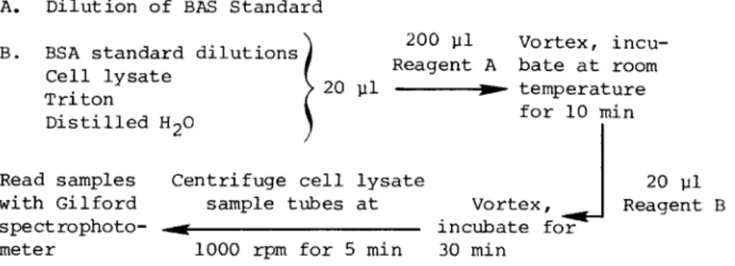
D. Assay of Protein in Cell Lysates
This technique is a micro modification of the method orig- inally described by Lowry et al. (11). Prepare fresh on the day of the assay a solution that contains 0.1 ml of 0.5%
CuS04/ 0.1 ml of 1% Na tartarate, and 10 ml of 2% NaC03 in 0.1 N NaOH (reagent A). Also prepare fresh a solution that contains 5 ml of Folin-Ciocalteau phenol reagent and 5 ml distilled H2O (reagent B ) .
Then set up 0.5-ml Brinkman microtubes for reaction, al- lowing one blank tube for distilled water, as a blank for the BSA dilutions employed for the protein standard curve, and another blank tube for 0.1% Triton in 0.85% saline, which serves as a blank for the cell lysates.
With a Pipetman, add 20 μΐ of original cell lysates to microtubes. Add 20 yl distilled water to the tube designated as BSA blank and 20 yl Triton in saline to the tube designated as blank for cell lysates. In separate reagent tubes, add 20 yl BSA standard dilutions (see Section U.C. 2) . With the Pipetman, add 200 yl of reagent A to each microtube; vortex and let stand at room temperature for 10 min. Add 20 yl of reagent B to each microtube; Vortex and let stand at room tem- perature for 30 min. Centrifuge out the green precipitate in the cell lysate sample tubes and their blanks by centrifuging the tubes at 1000 rpm for 5 min.
Read the samples within the next 2 hr on a Gilford spectro- photometer with visible light source at 750 nm. Clean the cu- vettes with distilled water, alcohol, and acetone between samples.
IV. CALCULATION OF DATA
Construct a BSA standard linear curve by plotting concen- tration of BSA in milligrams per milliliter on the abscissa and optical density readings at 750 nm on the ordinate. Use this curve to determine the concentration of protein in the various
cell lysates in milligrams per milliliters. Prepare another graph by plotting the values for n moles per milliliter 4-methylumbelliferone standard solution on the abscissa and values for optical density obtained from the Aminco fluoro- meter on the ordinate. From this graph, the values for n moles per milliliter of 4-methylumbelliferone (reaction product) liberated can be obtained. Then, correct for the fact that in the reaction mixture (glycine - NaOH) there is only 0.25 ml instead of 1 ml and for the fact that only 10 μΐ of cell lysate was used at a dilution of either 1/5, 1/10, or 1/20 of the original cell lysate. The resultant value can be expressed as nanomoles product per hour per milligram of protein.
V. CRITICAL COMMENTS
For clarity, the procedures described above are shown below.
(a) Preparation of Cell Lysate dextran sedimentation
Blood Macrophage microcultures
cell lysate
stored in , Ί Ί Ί
o -^-day cell lysate
3rd, 5th, and 7th freeze-thaw 3rd, 5th and
^ 7th day harvest-
(b) Enzyme Assay thawed
Cell lysate Centrifuge at 4 C 1700 rpm
for 10 min —
200 μΐ, pH 10.3, glycine, NaOH buffer
Incubate 37 C vortex 40 μΐ enzyme - for 1 hr - ^ substrates -^
Read i n Aminco
► SPF-125
10 μΐ cell lysate
(c) Micro Lowry P r o t e i n Assay
A. Dilution of BAS Standard
τ,οτν .- Λ Λ Α-Λ ,_· ^ 2 0° Vl Vortex, incu- B. BSA standard dilutions à , ' _ ,Ί , I Reagent A bate at room Cell lysate I .. _
T r i t o n > 2 0 P1 ^ temperature
Distilled H20 1
Read samples Centrifuge cell lysate
with Gilford sample tubes at Vortex, spectrophoto- - ^ incubate for meter 1000 rpm for 5 min 30 min
20 μΐ Reagent B
Table I summarizes the results of our microassay for ß-galactosidase on the monocyte cultures prepared from the blood of 13 normal donors. Each value in the table represents the mean ± S.E. of the results of 4 or 5 wells studied per donor at a given time point.
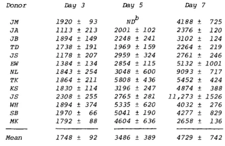
Table II summarizes the results of our microassay for
$-N-acetylglucosaminidase obtained in a similar manner. Both sets of data indicate an increase in lysosomal enzyme activity from 3 to 7 days, and illustrate that this assay can be used as a biochemical parameter of the maturation of human mono- cyte s to macrophages in vitro.
Table III compares the results of the data obtained with our enzyme microassays with that previously published for our macro culture system and enzyme macro assays (6). They are in excellent agreement.
The success of these techniques depends on several factors.
Careful aspiration and addition of culture medium is essential for obtaining optimal macrophage cultures that are >99% pure as indicated by their phagocytic capacity (10). It is impor- tant to wash away nonadherent cells without disturbing macro- phage adherence. Wells with >50% of their surface area covered with cells are suitable for use, although it is important to note that the results expressed are independent of cell number or cell size, but are related instead to milligrams of protein.
In preparing standard curves for BSA dilutions or MUSS di- lutions, it is important to include dilutions that span the entire range of lowest and highest readings obtained for either protein concentration data or enzyme activity data from the cell lysates.
These micromethods are far more versatile than the macro- methods previously employed in our laboratory. In addition they are more economical, and as only 5 - 10 ml blood are
TABLE J . Change in β-Galactosidase Activity3 During Monocyte Maturation
Donor Day 3 Day 5 Day 7
JM JA JB TD JS EW NL TK KS JS WH SB MK
Mean SE 259
93 225 191 148 164 194 154 143 233 220 215 198 187
± ±
± ±
± ±
± ±
± ±
± ±
±
±
10 13 13 20 24 20 19 19 9 44 68 10 5 13
805 243 338 262 378 391 401 527 316 362 503 433 420 414
± ±
± ±
± ±
± ±
± ±
± ±
±
±
57 20 22 19 50 10 55 35 18 34 58 9 45 40
413 247 330 254 365 502 1484 791 565 1889 352 497 360 619
± 81
± 17
± 13
± 25
± 37
± 100
± 63
± 122
± 33
± 238
± 40
± 8
± 6
± 139
3A c t i v i t y in nmol/hr/mg protein.
TABLE II. Change in $-N-Acetylglucosaminidase Activity0 During Monocyte Maturation
Donor Day 3 Day 5 Day 7
JM JA JB TD JS EW NL TK KS JS WH SB MK Mean
1920 1113 1894 1738 1178 1384 1843 1864 1830 2308 1894 1970 1792 1748
±
± ±
± ±
± ±
± ±
± ±
± ±
±
93 213 149 191 207 134 254 211 114 255 374 66 88 92
ND 2001 2248 1969 2959 2854 3048 5808 3196 2765 5335 5041 4604 3486
± ±
± ±
± ±
± ±
± ±
± ±
±
102 241 159 324 115 600 436 24 7 281 620 190 636 389
4188 2376 3102 2264 2761 5132 9093 5452 4874 11,273
4032 4277 2658 4729
±
± ±
± ±
± ±
± ±
± ±
± ±
±
725 120 124 219 246 1001 717 424 388 1526 276 829 136 742
Activity in nmol/hr/mg protein.
W , not done.
Lysosomal Enzyme Activities
Day 3 Day 5 Day 7
Enzyme Macro Micro Macro Micro Macro Micro
$-Galactosidase 142 ± 16 187 ± 13 312 ± 24 414 ± 40 402 ± 65 619 ± 139
&-N-Acetylglucosaminidase 1503 ± 304 1748 ± 92 3604 ± 420 3486 ± 389 5093 ± 747 4729 ± 742
aData previously published in reference (6).
bActivity in nmol/hr/mg protein.
required per patient, these techniques can now be applied to the study of monocyte function in a wide variety of disease states.
Acknowledgments
This work was supported by NIH Grant CA 27903 and Grant 6-126 from the March of Dimes Birth Defects Foundation. The authors wish to thank Dr. Charles Epstein and Ms. Georgianne Tucker for their advice and helpful suggestions concerning microenzyme assays, Nancy McManus for editorial assistance, and Mary Evelyn Rose for typing the manuscript.
REFERENCES
1. S. Yatsiv, L. B. Epstein, and C. J. Epstein. Monocyte- derived macrophages: An in vitro system for studying hereditary lysosomal storage diseases. Pediatr. Res. 12:
939-944, 1978.
2. L. B. Epstein. Mitogen and antigen induction of Inter- feron in vitro and in vivo. Tex. Rep. Biol. Med. 35:
43-56, 1978.
3. L. B. Epstein. The ability of macrophages to augment in vitro mitogen- and antigen-stimulated production of interferon and other mediators of cellular immunity by lymphocytes. In "Immunobiology of the Macrophage"
(D. S. Nelson, ed.), pp. 201-234. Academic Press, New York, 1976.
4. L. B. Epstein and A. J. Ammann. Evaluation of T lympho- cyte effector function in immunodeficiency diseases:
Abnormality in mitogen stimulated interferon in patients with selective IgA deficiency. J. Immunol. 112: 617- 626, 1974.
5. L. B. Epstein and M. J. Cline. Chronic lymphocyte leuke- mia: Studies on mitogen-stimulated interferon as a new technique for assessing T lymphocyte effector function.
Clin. Exp. Immunol. 16: 553-563, 1974.
6. S. H. S. Lee and L. B. Epstein. Reversible inhibition by interferon of the maturation of human peripheral blood monocytes to macrophages. Cell. Immunol. 50: 177-190, 1980.
7. L. B. Epstein, S. H. S. Lee, and C. J. Epstein. Enhanced sensitivity of trisomy 21 monocytes to the maturation- inhibiting effect of interferon. Cell. Immunol. 50:
191-194, 1980.
8. J. Weil, L. B. Epstein, and C. J. Epstein. Synthesis of
interferon-induced polypeptides in normal and chromosome 21-aneuploid human fibroblasts: Relationship to relative sensitivities in antiviral assay. J. Interferon Res. 1:
111-124, 1980.
L. B. Epstein and C. J. Epstein. T lymphocyte function and sensitivity to interferon in trisomy 21. Cell. Im- munol. 51: 303-318, 1980.
D. Goldblatt, N. H. McManus, and L. B. Epstein. A micro- method for preparation of human macrophage cultures for the study of lymphocyte-macrophage interaction in immune interferon production and blastogenesis. Immunopharma- cology 1: 13-20, 1978.
0. H. Lowry, W. J. Rosebrough, A. L. Farr, and R. J. Ran- dall. Protein measurement with the Folin phenol reagent.
J. Biol. Chem. 193: 265-275, 1971.