Acetaldehyde
Determination with Alcohol Dehydrogenase from Yeast
Hans-Ulrich Bergmeyer Principle
A l c o h o l dehydrogenase ( A D H ) catalyses the reaction:
(1) CH3CHO + D P N H + H+
T
1 C2H5OH + D P N +A t p H 6.5 the equilibrium o f the reaction lies so far to the right that acetaldehyde reacts virtually quantitatively. The method described here is based o n the work o f
1 - 3
) .
Reagents *)
1. Dipotassium hydrogen phosphate, K2HPO4 2. Reduced diphosphopyridine nucleotide, DPNH
disodium salt, D P N H - N a 2 - Commercial preparation, see p. 1011.
3. Sodium hydrogen carbonate, A. R., 1 % (w/v) 4. Perchloric acid, A. R., sp. gr. 1.67; ca. 70% (w/w) 5. Alcohol dehydrogenase, ADH
crystalline, from yeast, suspension in 2.4 M a m m o n i u m sulphate solution (containing 3 % (w/v) N a 4 ? 2 0 7 and 1 % (w/v) glycine, p H ca. 8). Commercial preparation, see p. 969.
Purity of the e n z y m e preparations
The A D H should have a specific activity of at least 180 units **>/mg. It should not be contami
nated with more than 0.02 % o f any o f the glycolytic enzymes (relative to the specific activity of the A D H ) .
Preparation of Solutions (for ca. 15 determinations) I. Phosphate solution (1.1 M K 2 H P 0 4 ) :
Dissolve 2.874 g. K2HPO4 in doubly distilled water and make up to 15 ml.
II. Reduced diphosphopyridine nucleotide (ca. 3 x 10~
3
M (3-DPNH):
Dissolve 2.5 mg. DPNH-Na2 in 1 % NaHCC>3 solution and make up to 1 ml.
III. Alcohol dehydrogenase, ADH (10 mg. protein/ml.):
Dilute the stock suspension with 2.4 M ammonium sulphate solution (containing 3 % (w/v) N a 4 P 2 0 7 and 1 % (w/v) glycine, pH ca. 8).
IV. Perchloric acid (ca. 6 % w/w):
Dilute 5.2 ml. 70% HCIO4 (sp. gr. 1.67) to 100 ml. with doubly distilled water.
Stability of the solutions
Solution IV is stable indefinitely. S o is solution I, providing that n o bacterial contamination occurs.
The D P N H solution is stable for about 14 days at 0 to 4 ° C and about 4 weeks in the frozen state.
The A D H suspension should not be frozen; it is stable for several months at 0 to 4 ° C .
*) Complete reagent kits are available commercially, see p. 1035.
**) A unit is the amount of enzyme which converts 1 u.mole of substrate in 1 min. at 25° C.
D / . Sell, Diploma Thesis, Universitat Munich, 1953.
2
> E. Racker in S. P. Colowick and N. O. Kaplan: Methods in Enzymology. Academic Press, N e w York 1957, Vol. Ill, p. 295.
3) H. Holzer, E. Holzer and G. Schulz, Biochem. Z. 326, 385 [1955].
Procedure
Collection of b l o o d and deproteinization
Acetaldehyde boils at 20.2° C; therefore to avoid loss of acetaldehyde from biological mater
ial at body temperature, it must be mixed with ice-cold perchloric acid as quickly as possible and preferably with exclusion of air. Only after cooling to a low temperature should the sample be homogenized and deproteinized.
Collect blood according to
Klein4
)
as follows:
Pipette into a centrifuge tube with a 10 ml. graduation near to the top (the air space above the fluid should be as small as possible when a rubber stopper is inserted)
5 ml. perchloric acid solution (IV)
and cool to at least 2°C. Obtain blood from a lightly constricted elbow vein with a winged cannula. The cannula is connected to a polyethylene, polyvinyl or rubber tube, which is im
mersed in the perchloric acid so that it touches the bottom of the centrifuge tube. Allow the blood to run into the centrifuge tube up to the 10 ml. graduation mark and dilute exactly
to 10 ml. with blood
by slowly withdrawing the tube. Lightly clamp the tube to reduce the blood flow. Imme
diately stopper the tube with a clean rubber stopper and allow to stand for 10 min. in an ice bath. Only then mix the perchloric acid and the blood; if necessary, stir with a glass rod.
Centrifuge, stoppered, for 10 min. at ca. 3000 g.
Pipette into a 10 ml. centrifuge tube:
2.0 ml. deproteinized sample 0.7 ml. cold phosphate solution (I).
Allow to stand for 10 min. in an ice bath, rapidly filter through soft filter paper. The filtrate is buffered at pH ca. 6.5; use 2 ml. for the assay.
Spectrophotometric m e a s u r e m e n t s
Wavelength: 340 or 366mo.; cuvettes with lids, light path: 1 cm.; final volume: 2.1 ml.;
room temperature. Measure against air or water.
Pipette quickly into the cuvette:
2.00 ml. deproteinized and buffered sample 0.05 ml. DPNH solution (II).
Mix rapidly with a plastic rod flattened at one end, put on the cuvette lid and read the optical density Ei. Mix in
0.05 ml. ADH suspension (III),
replace the cuvette lid and after 3 — 5 min. read the optical density E2. Ei— E2 = AE is used for the calculations.
Calculations
According to the following formula (refer to p. 37).
A E x V A E—- = = umoles acetaldehyde/assay mixture X 2.1 ,
f J
,J
,£ X d £
where V = assay volume (2.1 ml.) d = light path (1 cm.)
£ = extinction coefficient of D P N H (£340 = 6.22 cm.2/jjimole; £366 = 3.3 cm.
2
/[xmole).
4
> H. Klein and / . Korzis, Die Medizinische 345 [1958].
T o obtain the acetaldehyde content per ml. of blood it is necessary to allow for the dilution occurring on deproteinization and neutralization o f the perchloric acid extract. Blood contains ca. 8 0 % of its weight as water; 1 ml. blood weighs 1.06 g. Taking 5 ml. blood = 5.3 g., this gives 5.3 X 80/100 + 5 = 9.24 ml. o f extract after deproteinization, o f which 2 ml. is brought to p H 6.5 with 0.7 ml. phosphate solution (total volume 2.7 ml.). 2 ml. of this solution is taken for the assay. The total dilution is ( 9 . 2 4 / 5 )X( 2 . 7 / 2 )X( 1 / 2 ) = 1.247:1. T o convert to u.moles acetaldehyde/ml. blood multiply by 1.247.
Therefore for 366 m\x:
A E x 2.1 x 1.247 3.3 for 340 mu,:
A E x 2.1 x 1.247
A E X 0.794 = [xmoles acetaldehyde/ml. blood
A E X 0.421 = (xmoles acetaldehyde/ml. blood 6.22
T o convert to u.g. it is necessary to multiply by the molecular weight of acetaldehyde (44).
Sensitivity
If it is assumed that an optical density difference at 340 m\L of A E = 0.010 can be read with sufficient accuracy, then as little as ca. 4 x 10~
3
[xmoles or 0.18 ug. acetaldehyde/ml. blood can be determined quantitatively.
Specificity and Sources of Error
Apart from acetaldehyde, yeast A D H reacts with the following aldehydes, though at considerably slower rates: glycolaldehyde, formaldehyde, propionaldehyde, butyraldehyde, valeraldehyde, iso- butyraldehyde and glyceraldehyde. The presence o f these aldehydes in the sample leads to non- constant end-points with the acetaldehyde reaction. In such cases, the optical density E2 is extra
polated to zero time (see p. 39).
If the A D H preparation is contaminated with glycolytic enzymes (e.g. lactic dehydrogenase), then extra D P N H may be oxidized due to the reduction o f metabolites in the sample (e.g. pyruvate).
The most frequent source of error is evaporation of acetaldehyde during the preparation o f the sample, therefore always work with stoppered containers and keep all solutions cold (up to the time of the measurements).
High concentrations of ethanol in the sample interfere, because the reaction does not proceed to completion. Ethanol in human blood up to lethal concentrations does not interfere.
Determination with Aldehyde Dehydrogenase from Yeast
Frank Lundquist
The determination of acetaldehyde in b l o o d and other biological fluids is of interest in connection with ethanol metabolism. Enzymatic methods employing alcohol dehydrogenase
1
), liver aldehyde dehydrogenase
2
) or yeast aldehyde dehydrogenase
3
) have been described. The assay with alcohol dehydrogenase (see p. 290) has disadvantages, especially in the presence of high concentrations of ethanol. The assay with aldehyde dehydrogenase from beef liver is liable to error when applied to biological material, because the preparation is contaminated with lactic dehydrogenase, which has so far proved difficult to remove.
1) H. Holzer, E. Holzer and G. Schulz, Biochem. Z. 326, 385 [1955].
2
) E. Racker in S. P. Colowick and N. O. Kaplan: Methods in Enzymology. Academic Press, N e w York 1957, Vol. Ill, p. 295.
3) F. Lundquist, Biochem. J. 68, 172 [1958].
Principle
Aldehyde dehydrogenase from y e a s t
4
.
5
) catalyses the reaction:
(1) Acetaldehyde + D P N + acetate + D P N H + H+
The enzyme requires a relatively high concentration of potassium ions for its action. The kinetic properties of the enzyme are very different from those of the liver aldehyde dehydrogenase. In contrast to the liver enzyme, the affinity of the yeast enzyme for acetaldehyde is l o w
6
) . Although the equilibrium of the reaction lies far in favour of acetate and D P N H , a quantitative oxidation of acetaldehyde is not possible with the yeast enzyme. Therefore the rate of the reaction, which is proportional to the concentration over a limited range, is used to measure the amount o f acetaldehyde present. Also refer to p. 6.
Reagents
1. Metaphosphoric acid, HPO3, A. R. (as sticks) 2. Potassium hydroxide, 1.25 or 1.5 M
3. Acetaldehyde, A. R.
4. Diphosphopyridine nucleotide, DPN
free acid; commercial preparation, see p. 1010.
5. Tris-hydroxymethyl-aminomethane, tris 6. Ethylene-diamine-tetra-acetic acid, EDTA
disodium salt, E D T A - N a
2
H2
- 2 H2
0 .7. Mercaptoethanol
8. Potassium chloride, A. R.
9. Aldehyde dehydrogenase from yeast
preparation, see Appendix, p. 296.
Purity of the e n z y m e preparations
Enzyme preparations obtained according to the procedure o f Black
4
*
5
) contain n o interfering enzymes. During the preparation a check must be made to discover whether alcohol dehydro
genase has been completely removed.
Preparation of Solutions
Prepare all solutions with fresh, glass distilled or deionized water.
I. Metaphosphoric acid (0.75 M):
Powder 12 g. metaphosphoric acid and dissolve in 100 ml. distilled water. The solution contains large amounts of sodium pyrophosphate, but this does not interfere with the assay. Store the solution, stoppered, at 4°C. Due to the rapid hydrolysis of the acid the solution must be prepared freshly every few days. Immediately before use titrate the solution with standard alkali.
II. Potassium hydroxide (1.25 M):
Dissolve 70 g. KOH in 100 ml. distilled water. 0.5 ml. of this solution are required for the neutralization of 2 ml. deproteinized plasma. When the metaphosphoric acid is partially hydrolysed, it is advisable to dilute a 1.5 M KOH stock solution so that 0.5 ml. of the alkali neutralizes 2 ml. plasma filtrate.
4) S. Black, Arch. Biochem. Biophysics 34, 86 [1951].
5)
S. Black in S. P. Colowick and N. O. Kaplan: Methods in Enzymology. Academic Press, N e w York 1955, Vol. I, p. 508.
6) E. Racker, J. biol. Chemistry 177, 883 [1949].
III. Potassium chloride (1.0 M):
Dissolve 74.5 g. KC1 in distilled water and make up to 1000 ml.
IV. Tris buffer (1 M ; p H 8.0):
Dissolve 30 g. tris-hydroxymethyl-aminomethane in about 150 ml. distilled water, adjust to pH 8.0 (glass electrode) with ca. 25 ml. 5 N H Q and dilute to 250 ml. with distilled water.
V. Ethylene-diamine-tetra-acetic acid, EDTA (0.1 M):
Dissolve 3.7 g. EDTA-Na2H 2 -2 H2O in distilled water and make up to 100 ml.
VI. Mercaptoethanol (0.1 M):
Dissolve 0.78 g. mercaptoethanol in distilled water and make up to 100 ml.
VII. Diphosphopyridine nucleotide (0.015 M (3-DPN):
Dissolve 100 mg. (3-DPN in distilled water and make up to 10 ml.
VIII. DPN-buffer mixture:
Immediately before use mix 3 volumes solution III, 6 volumes solution IV, 0.5 volumes solution V, 0.5 volumes solution VI and 0.5 volumes solution VII.
IX. Acetaldehyde stock solution (2 M):
Prepare an approximately 10% solution of freshly distilled acetaldehyde in distilled water. Determine the exact concentration by titration (reaction with sodium bi
sulphite and titration of the excess bisulphite with iodine solution). The solution is stable for long periods at 4°C. It should be titrated every two months and prepared afresh every six months.
X. Aldehyde dehydrogenase (10000 units *Vml.):
Thaw the frozen enzyme solution (see Appendix, p. 296) and dilute with distilled water.
Stability of the s o l u t i o n s
Store all the solutions in a refrigerator (4°C). Keep all the bottles tightly stoppered to avoid contami
nation with aldehydes. The D P N solution should be prepared freshly every fortnight. The other solutions are stable for long periods.
Procedure
The method can be used for the analysis of plasma or serum as well as for organs. Collect blood in plastic centrifuge tubes, immediately place in an ice bath and stopper with plastic stoppers. Centrifuge the blood for 10 min. at 0°C (2000g). Under these conditions the blood does not coagulate.
Deproteinization
Prepare stoppered centrifuge tubes (glass, about 8 ml. capacity) with 2 ml. cold meta
phosphoric acid solution (I) and add 2 ml. freshly collected plasma. After vigorous shaking, centrifuge (10 min. at 2000 g) and pipette 2 ml. of the clear supernatant into a cold, glass- stoppered tube. Neutralize with 0.5 ml. KOH solution (II). If necessary, the sample can be left at this stage overnight in a refrigerator without any appreciable loss of acetaldehyde.
*) See Appendix, p. 296.
Spectrophotometric m e a s u r e m e n t s
Wavelength: 340 mu.; silica cuvettes, light path: 1cm.; final volume: 3.025 ml. Measure against a cuvette containing water. Each series of measurements should be carried out at a constant temperature (15°C) and with the same amount of enzyme.
Before the measurements keep all the solutions in an ice bath. Cover the cuvettes with polyethylene film to prevent the evaporation of acetaldehyde.
For each series of measurements prepare a
reagent blank.For this, dilute metaphosphoric acid solution (I) 1:1 with distilled water and neutralize with KOH (solution II). Use 2 ml.
of this neutralized solution for the assay instead of the deproteinized sample. The optical density increases by less than 0.010 in 10 min. if the reagents contain no acetaldehyde.
Prepare
standardswith solutions of known acetaldehyde content (1—10 pig.) as follows:
dilute the acetaldehyde stock solution (IX) 1:4000 to 1:400 with distilled water. Add 0.1 ml.
of each of these dilutions (corresponding to 1 —10 pig. acetaldehyde) to the assay mixtures (see "Method").
Method:
Pipette successively into the cuvettes:
2 ml. deproteinized sample (neutralized, diluted metaphosphoric acid in the blank*
0.025 ml. acetaldehyde solution in the standards) 1 ml. DPN-buffer mixture (solution VIII).
Mix and read the optical density Ei. Rapidly mix in
0.025 ml. aldehyde dehydrogenase solution (X) (at least 250 units),
start a stopwatch and again read the optical density after exactly 10 min. E2 — Ei = AE is used for the calculations.
Calculations
The A E value for the reagent blank must be subtracted from those of the samples and standards.
The corrected values are proportional to the acetaldehyde concentration, providing they are smaller than 0.25/10 min. It follows that:
AEA
K
~ — X S = A [pig. acetaldehyde in the cuvette]A E s where
A E ^
=
optical density difference o f sample containing acetaldehyde concentration A A E s = optical density difference of the standard containing acetaldehyde concentration S
S = concentration of acetaldehyde in standard cuvette The acetaldehyde concentration in the plasma sample is:
AEA
- r — — X S X 3.75 = A [u,g. acetaldehyde/ml. plasma]
A E s
3.75 = 2.5 X 1.5 —- (dilution o n deproteinization) X (dilution in assay) Sensitivity
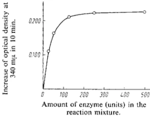
With constant substrate concentration the rate o f the reaction is not linearly proportional to the enzyme concentration (Fig. 1). With high enzyme concentrations the rate of reaction depends only on the concentration of substrate. It is preferable to use these large amounts of enzyme because this will eliminate the effect of small amounts of inhibitory compounds. A s little as 0.1—0.2 u,g. acetalde-
hyde/ml. plasma can be detected quantitatively, while about 1 u.g. can still be determined accurately.
The measured optical density difference corresponds to about a 50 % oxidation o f acetaldehyde to acetate.
Specificity and Sources of Error
Other aldehydes, for example, propionaldehyde, butyraldehyde and benzaldehyde, also react with yeast aldehyde dehydrogenase; formaldehyde, glyceraldehyde, salicylaldehyde and ketones d o not react. Since the enzyme preparation is relatively impure, it might contain other D P N - d e p e n d e n t dehydrogenases and so react with other metabolites of plasma. However, this does not appear to be the case with the enzyme prepared according to the description given below.
Fig. 1. Relation between the rate of the reaction and the concentration of yeast aldehyde dehydrogenase.
The reaction mixture consisted of 7.4 \xg. acet
aldehyde, 2 ml. neutralized metaphosphoric acid and 1 ml. DPN-buffer mixture (solution VIII). The points are averages of two estimations. They have been corrected for the reagent blank and the different amounts of enzyme.
0 WO 200 300 LOO 500 Amount of enzyme (units) in the
reaction mixture.
Appendix
A l d e h y d e d e h y d r o g e n a s e f r o m y e a s t The method o f Black
4
*
5
) has been slightly modified
3
). Extract dried baker's yeast or an acetone-dried powder o f fresh yeast at r o o m temperature with phosphate buffer and maintain the p H at 8.6 (with N H 4 O H ) . A l l o w to stand overnight at 4 ° C , centrifuge, warm the clear supernatant (pH 6.6, citric acid) to 55°C for 15 min. and then cool rapidly. Centrifuge off the precipitate, adjust the supernatant to p H 4.7 and allow to stand for 2 hours at 4 ° C . The precipitate which forms contains the enzyme.
Dissolve in phosphate buffer containing cysteine, re-precipitate at p H 4.7, dissolve, adjust the solution to p H 6.3 and remove the slight precipitate.
The preparation is fairly free from alcohol dehydrogenase. A b o u t 2 0 0 0 0 0 units are obtained from 100 g. yeast, ca. 800000 units from 100 g. acetone-dried powder. Small portions of enzyme solution containing about 10000 units are stored frozen at — 2 0 ° C ; they are stable for several months.
*) A unit is the amount of enzyme which, under the assay conditions described here (3 ml. reaction mixture), increases the optical density at 340 mu. by 0.001 in 1 min.