Research Article
4-Hydroxy-2-nonenal Alkylated and Peroxynitrite Nitrated Proteins Localize to the Fused Mitochondria in Malpighian Epithelial Cells of Type IV Collagen Drosophila Mutants
András A. Kiss,
1Nikoletta Popovics,
1Zsolt Boldogk y i,
1Katalin Csiszár,
2and Mátyás Mink
11Institute of Medical Biology, University of Szeged, Somogyi B. U. 4, 6720 Szeged, Hungary
2John A. Burns School of Medicine, University of Hawaii, 1960 East West Road, Honolulu, HI 96822, USA
Correspondence should be addressed to M´aty´as Mink; mink@bio.u-szeged.hu
Received 4 September 2017; Revised 3 December 2017; Accepted 2 January 2018; Published 30 January 2018
Academic Editor: Daniela Grifoni
Copyright © 2018 Andr´as A. Kiss et al. This is an open access article distributed under the Creative Commons Attribution License, which permits unrestricted use, distribution, and reproduction in any medium, provided the original work is properly cited.
Background. Human type IV collagenopathy is associated with mutations within theCOL4A1and to a less extent theCOL4A2genes.
The proteins encoded by these genes form heterotrimers and are the highest molar ratio components of the ubiquitous basement membrane. The clinical manifestations of theCOL4A1/A2mutations are systemic affecting many tissues and organs among these kidneys. In order to uncover the cellular and biochemical alterations associated with aberrant type IV collagen, we have explored the phenotype of the Malpighian tubules, the secretory organ and insect kidney model, incol4a1collagen gene mutants of the fruit fly Drosophila melanogaster. In Malpighian epithelial cells ofcol4a1mutants, robust mitochondrial fusion indicated mutation-induced stress. Immunohistochemistry detected proteins nitrated by peroxynitrite that localized to the enlarged mitochondria and increased level of membrane peroxidation, assessed by the amount of proteins alkylated by 4-hydroxy-2-nonenal that similarly localized to the fused mitochondria. Nuclei within the Malpighian epithelium showed TUNEL-positivity suggesting cell degradation. The results demonstrated thatcol4a1mutations affect the epithelia and, consequently, secretory function of the Malpighian tubules and provide mechanistic insight intocol4a1mutation-associated functional impairments not yet reported in human patients and in mouse models with mutantCOL4A1.
1. Introduction
Basement membranes (BMs) are nanoscale sheets of extra- cellular matrices that play essential roles in multiple organs including muscle homeostasis, structures, and integrity of the dermal and ocular system, neuromuscular junctions, and blood filtration in the kidneys. The most abundant structural components of BMs include laminins, collagen IV, nidogens, perlecan, and agrin [1]. The ubiquitous mam- malian BMs consist of heterotrimeric type IV collagens with (COL4A1)2COL4A1 composition. The clinical presentation of patients withCOL4A1mutation is systemic with numerous affected organs and tissues including the eyes, brain, the
vascular system, skeletal muscles, and kidneys [2, 3]. A dis- tinct form of type IV collagenopathy, Hereditary Angiopathy, Nephropathy, Aneurysms, and Muscle Cramps (HANAC) syndrome, is caused by N-terminal mutations within the COL4A1gene. The renal manifestation of the same mutations in mouse models includes albuminuria, hematuria, glomeru- lar cysts, and delays in glomerulogenesis and podocyte differentiation [4].
We have identified an allelic series of dominant, temperature-sensitive, antimorph mutations in the cognate col4a1 gene of the fruit fly, Drosophila melanogaster. The col4a1+/− heterozygotes are viable and fertile at permissive temperature of 20∘C but die at 29∘C. In these mutants, we
Volume 2018, Article ID 3502401, 8 pages https://doi.org/10.1155/2018/3502401
impacts protein functions and can be detected as a species- independent antigene [8]. Peroxynitrite can also remove a hydrogen atom from polyunsaturated fatty acids resulting in the formation of aldehydes, conjugated dienes, and hydrop- eroxyradicals that trigger a free radical chain reaction and membrane lipid damage by lipoperoxidation [9].
The main product of membranous polyunsaturated fatty acid peroxidation is the reactive 4-hydroxy-2-nonenal, HNE [10]. The reactivity of HNE with proteins relies on Michael addiction and, by modifying histidine residues, generates alkyl-conjugated polypeptides also detectable as species- independent antigens [11]. As there is no direct laboratory test to estimate lipid peroxidation, measurements of HNE-conju- gated protein levels currently serve as surrogates [12]. The bulk of peroxynitrite reacts rapidly with carbon dioxide, present at ∼1 mM in cells, forming the unstable product nitrosoperoxycarbonate (ONOOCO2−), one-third of which decomposes into carbonate (CO3−∙) and NO2∙radicals and two-thirds into the neutral NO3−and CO2[13].
Insect Malpighian tubules serve as secretory organs.
These renal tubules lack a vascular blood system and float freely in the hemocoel (blood-filled body cavity). The tubules are surrounded by BM and consist of two epithelial cell types, the metabolically active principal and the intercalated stellate cells [14]. The insect renal system is aglomerular, and urine is formed by active transport rather than by selective reabsorption of ultrafiltrate as in vertebrates. While the insect tubule system represents an intermediate towards the glomerular kidney, it fulfills the same basic functions of transport, excretion, and osmoregulation [15].
We have recently shown that the col4a1 Drosophila mutants develop stress fibers in their Malpighian cells and aberrantly express cell-surface integrin receptors [16]. In the present study, we have extended our research to address altered posttranslational protein modifications by peroxyni- trite and 4-hydroxy-2-nonenal in the Malpighian tubules.
The col4a1 mutants demonstrated heavy protein tyrosine nitration and protein-histidine alkylation that localized to the enlarged and fused mitochondria as signs of mitochondrial stress. HNE-protein adducts colocalized with the cytoplasmic membrane that was accompanied by cell degeneration in the tubules performing TUNEL-positivity, collectively suggest- ing that these aberrant processes are integral parts ofcol4a1- associated pathology.
2. Materials and Methods
2.1. Maintenance of Drosophila Strains. Wild-type Oregon flies and col4a1 mutant stock with theDTS-L3 allele were maintained at 20∘C and 29∘C on yeast-cornmeal-sucrose- agar food, consisting of nipagin to prevent fungal infection.
Triton X, dissolved in PBS, and washed three times in PBS.
Blocking was achieved in 5% BSA dissolved in PBS for 1 hour and washed three times in PBS.
2.2. Immunostaining and Antibodies. Nuclei in the dissected Malpighian tubules were counterstained by 1𝜇g/ml 4,6- diamino-2-phenylindole (DAPI) in 20𝜇l PBS, 12 min in dark.
F-actin was stained by 1 unit Texas Red-X Phalloidin (ThermoFisher) in 20𝜇l PBS for 20 min. A-Mannopyranosyl and a-glucopyranosyl residues as membrane markers were stained by Concanavalin A, Alexa Fluor 594 Conjugate (ThermoFisher) in 20𝜇l PBS for 1 hour. We used 1𝜇l mouse monoclonal anti-3-nitrotyrosine [39B6] (Abcam) in 20𝜇l PBS for 1 hour and stained 4-hydroxynonenal conjugate by 1𝜇l mouse monoclonal anti-4-hydroxynonenal antibody (Abcam) in 20𝜇l PBS for 1 hour. Primary mouse antibodies were visualized by 1𝜇l F(ab) 2-Goat Anti-Mouse IgG (H + L) Cross Adsorbed Secondary Antibody conjugated with Alexa Fluor 488 (ThermoFisher) in 20𝜇l PBS for 1 hour and 1𝜇l Goat Anti-Mouse IgG (H + L) Cross Adsorbed Secondary Antibody, Alexa Fluor 350, in 20𝜇l PBS for 1 hour. Mitochon- dria were visualized by the mitochondrially targeted EYFP (mito-GFP) following appropriate crosses [17].
2.3. Confocal Microscopy. Photomicrographs of the Mal- pighian tubules were generated by confocal laser scanning fluorescence microscopy (Olympus Life Science Europa GmbH, Hamburg, Germany). Microscope configuration was the following: objective lens: UPLSAPO 60x (oil, NA: 1.35);
sampling speed: 8𝜇s/pixel; line averaging: 2x; scanning mode: sequential unidirectional; excitation: 405 nm (DAPI), 543 nm (Texas Red), and 488 nm (Alexa Fluor 488); laser transmissivity: 7% were used for DAPI, 42% for Alexa Fluor 488 and 52% for Texas Red.
2.4. TUNEL-Labelling. Terminal deoxyribonucleotide trans- ferase-mediated dUTP-fluorescein conjugate nick end la- belling (TUNEL) was carried out by using the in situ cell death detection kit (Roche) as recommended. Embryos of mutant and control flies were incubated at 20∘C or 29∘C and L3-stage larvae collected. Nuclei in the Malpighian tubules were counterstained by 1𝜇g/ml 4,6-diamidino-2- phenylindole (DAPI). Labellings were visualized by a Hund- Wetzlar fluorescence microscope by using FITC or DAPI filters.
3. Results
3.1. Heavy Protein Nitration in col4a1 Mutants. We have pre- viously demonstrated that thecol4a1mutant flies synthesize peroxynitrite at higher concentration as part of their antimi- crobial immune response under restrictive conditions [7].
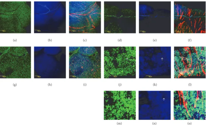
(a) (b) (c) (d) (e) (f)
(g) (h) (i) (j) (k) (l)
(m) (n) (o)
Figure 1: Protein nitration in Malpighian epithelial cells. Color code: mitochondria, green; nitrated proteins, blue; actin, red. (a) Wild-type flies incubated at 20∘C, mitochondria, (b) nitrated proteins, and (c) merge. Note a stellate cell in (c). ((d), (e), (f)) Wild-type flies incubated at 29∘C and displayed in the same order. ((g)–(i)) Mutant flies, incubated at 20∘C, and ((j)–(l)) mutant flies, incubated at 29∘C. Photomicrographs are displayed in the same order as in the upper row. Localization of nitrated proteins to mitochondria is shown in (c), (i), (f), and (l). Uneven distribution and fusion of mitochondria are demonstrated in (j). White arrows in (j), (k), and (l) pointing the region displayed in higher magnification in (m), (n), and (o). White arrows in (m), (n), and (o) showing regions with no/few mitochondria and the lack of staining, demonstrating localization to mitochondria with nitrated proteins indirectly.
Peroxynitrite is produced by the diffusion-driven reaction of nitric oxide (NO) in the presence of oxidants such as the mitochondrial-derived radical superoxide anion, O2∙−. The sources of NO are at extramitochondrial sites and the dis- solved gas diffuses into mitochondria, reacts with O2∙−, and disrupts protein functions by protein tyrosine nitration [18].
We therefore expected accumulation nitrated proteins in the mitochondria ofcol4a1mutant flies following incubation at 29∘C.
We did not observe gross alterations in the Malpighian tubules of the mutants compared to control flies; mito- chondria were distributed evenly in the cytoplasm and the fluorescent light intensities used to record nitrated proteins in the mutants were comparable to the control animals (Figures 1(a)–1(c) and Figures 1(g)–1(i)), following incubation at permissive condition. However, under restrictive condi- tions (29∘C), we noted marked differences in the Malpighian tubules of mutant flies. While mitochondria in the epithe- lial cells of wild-type Malpighian tubules remained evenly distributed with no shape alteration at this temperature (Figure 1(d)), in mutants, mitochondrial fusion and uneven distribution were observed (Figure 1(j)). The level of nitrated proteins was remarkably higher in mutants in comparison
with the control (Figure 1(k) versus Figure 1(e)) and these signals localized to the mitochondria (Figures 1(f) and 1(l)).
3.2. High Levels of Alkylated Proteins in the Mutants. The level of lipid peroxidation was determined indirectly by the accumulation of HNE-protein adducts. Results showed comparable amounts of alkylated proteins in the epithelial cells of mutant Malpighian tubules at permissive condition (Figure 2(b) versus Figure 2(h)), and the appearance of mitochondria remained unaffected in both mutants and controls under these conditions (Figure 2(a) versus Fig- ure 2(g)). In mutants under restrictive conditions (29∘C), uneven distribution and fusion of mitochondria occurred (Figure 2(j) versus Figure 2(d)), the mutants produced more HNE-protein adducts (Figure 2(k) versus Figure 2(e)), and the alkylated proteins localized to mitochondria (Figures 2(f) and 2(l)).
3.3. Protein-HNE Adducts Associate with Cytoplasmic Mem- brane. We next explored the involvement of the cytoplas- mic membrane incol4a1-associated pathology. We recorded numerous alkylation sites in form of punctate staining in colocalization with the cytoplasmic side of the membrane
(a) (b) (c) (d) (e) (f)
(g) (h) (i) (j) (k) (l)
(m) (n) (o)
Figure 2: Protein-HNE adducts in Malpighian epithelium. Color code: mitochondria, green; protein-HNE adducts, blue; actin, red. (a) Mitochondria of wild-type flies incubated at 20∘C, (b) protein-HNE adducts, and (c) merge. ((d), (e), (f)) Wild-type flies incubated at 29∘C, shown in the same order. ((g)–(i)) Mutant flies, incubated at 20∘C, and ((j)–(l)) mutant flies, incubated at 29∘C. The order of photomicrographs is as in upper row. Note mitochondrial fusion in (j) and actin stress fibers in (l). White arrow in (j), (k), and (l) showing the portion displayed in higher magnification in (m), (n), and (o). White arrow in (m), (n), and (o): point regions with no/few mitochondria and the lack of staining, demonstrating localization of alkylated proteins to mitochondria indirectly.
and apparent perinuclear accumulation in the Malpighian epithelial cells in the mutants at permissive conditions (20∘C) (Figures 3(e) and 3(f)). This staining pattern was ampli- fied upon shift to restrictive temperature (29∘C) and the HNE-conjugated proteins appeared within the cytoplasmic membrane indicating direct membrane damage by lipid peroxidation (Figures 3(g) and 3(h)). In the control flies the cytoplasmic membrane remained intact and protein-HNE adducts appeared in the vicinity of the membrane at both permissive and restrictive conditions (Figures 3(a)–3(d)).
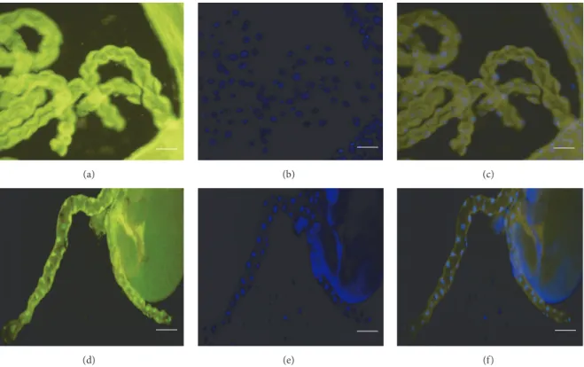
3.4. Cell Degeneration Detected by TUNEL-Positivity. The epithelial cells of the Malpighian tubules proved to be TUNEL-positive in mutants at 29∘C (Figures 4(d)–4(f)), but not at 20∘C (Figures 4(a)–4(c)). These observations further supported our earlier observations of cell death affecting multiple tissues incol4a1mutants.
4. Discussion
Drosophilamodels provide useful tools for determining the pathomechanistic details, functional alterations, and some of the genotype-phenotype correlations of human monogenic disorders [19] including mutations associated with disorders
of the kidneys as some of the human genes known to be asso- ciated with inherited nephrotic syndromes play conserved roles in renal functions from flies to humans [20]. There are nephrotic manifestations of humanCOL4A1mutations of the Hereditary Angiopathy, Nephropathy, Aneurysms, and Muscle Cramps (HANAC) syndrome [21] and recent research revealed glomerular hyperpermeability and adult onset glomerulocystic kidney disease in association withCOL4A1 mutations [4]. Some of the mechanistic elements in context of type IV collagen mutations, such as oxidative stress, have also been demonstrated [22]. However, evidence for chronic inflammation and posttranslational protein modifications are scarce and so far demonstrated only in Drosophila col4a1 mutants [7, 16].
Mitochondrial fusion occurs under situations of cellular stress. Merging of the contents of partially damaged mito- chondria is interpreted as a complementation mechanism rescuing impaired organelles and function [23]. Our prior results demonstrated signs of cellular stress in the form of actin stress fibers in the Malpighian epithelial cells ofcol4a1 mutantDrosophila[16]. Results of the current study show that mutation-associated stress induced mitochondrial hyperfu- sion also occurs under restrictive condition with the enlarged organelles unevenly distributed within cells resulting either
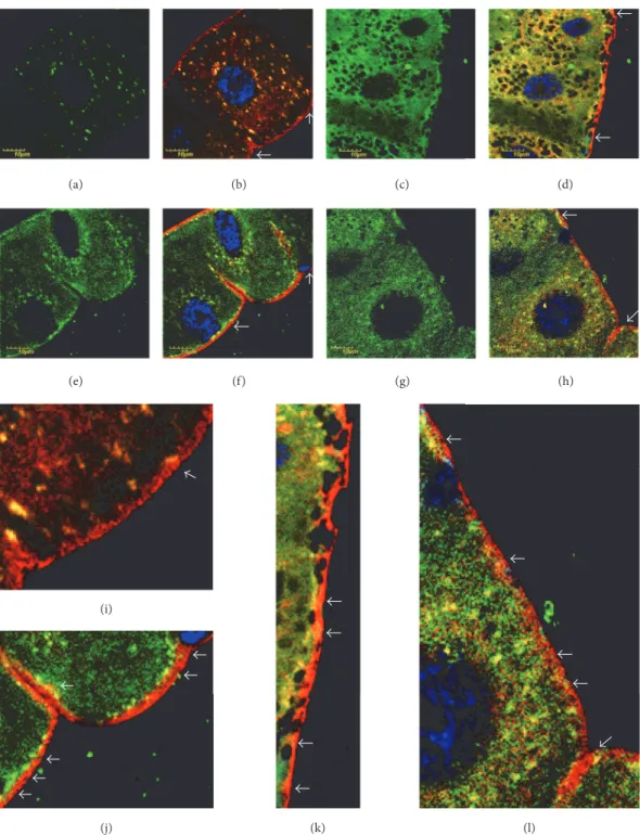
(e) (f) (g)
(j) (i)
(k) (l)
(h)
(a) (b) (c) (d)
Figure 3: Cytoplasmic membrane-associated HNE-modified proteins in mutants. Color code: protein-HNE adducts, green; cytoplasmic membrane red; nuclei, blue. (a) Protein-HNE adducts in wild-type flies incubated at 20∘C. (b) Overlay with membrane staining. (c) Protein- HNE adducts in wild-type flies incubated at 29∘C. (d) Merged with membrane staining. ((e), (f), (g), (h)) Representative mutant, incubated at 20 or 29∘C presented in the same order as in upper row. White arrows in (b), (d), (f), and (h). Regions presented in higher magnification in (i), (j), (k), and (l), respectively. White arrows in (i), (j), (k), and (l) show association of the cytoplasmic membrane with alkylated proteins.
Note the notorious infiltration of HNE-modified proteins into the membrane in (l), which occurs at a less extent in (k). The membrane of wild-type animals is free of alkylated proteins (i), while they associate closely with the membrane in the mutant (j), incubated at permissive temperature.
(a) (b) (c)
(d) (e) (f)
Figure 4: Fluorescence micrographs demonstrating TUNEL-positivity in Malpighian tubules. (a) TUNEL-staining of a Malpighian tubule ofcol4a1+/−L3-larva incubated at 20∘C; (b) DAPI-staining; (c) merge, tubules appearing TUNEL-negative. (d) TUNEL-positive Malpighian tubule of acol4a1+/−L3-larva incubated at 29∘C; (d) DAPI; (e) merge. Scale bars: (a)–(c) 50𝜇m, (d)–(f) 100𝜇m.
in areas apparently lacking mitochondria or in organelle- enriched areas. A further consequence ofcol4a1mutation is the accumulation of nitrated and alkylated proteins in the mutants that localize to both normal and fused mitochondria.
This observation indicates a peroxynitrite-mediated nitrosative stress incol4a1mutants that produce peroxynitrite at higher concentration [7]. We thus suggest that the elevated peroxynitrite level likely causes excess protein tyrosine nitra- tion; however, this reaction does not deplete peroxynitrite in col4a1mutants. Indeed, the still available peroxynitrite can initiate membrane damage by lipid peroxidation producing HNE, which in turn alkylates proteins by the mechanism of Michael addiction [8]. Direct association of alkylated proteins with the epithelial cell membrane of mutant Malpighian tubules supports this scenario. Furthermore, the mutation- induced stress directs the epithelia towards degeneration as demonstrated by the TUNEL-positivity of the nuclei.
The data presented here strongly suggest a central role for peroxynitrite incol4a1-associate defects. In wild-type animals and under physiological conditions, the nitrosoperoxycar- bonate pathway is the preferential reaction of peroxynitrite, as the main decay products of nitrosoperoxycarbonate, nitrate anion, and carbon dioxide do not exert protein or membrane modification effects (Figure 5) [13]. In mutants, however, peroxynitrite is present above physiological concentrations, and it produces excess protein tyrosine nitration and forms
HNE leading to protein alkylation, lipid peroxidation, mem- brane damage, aberrant mitochondria, epithelial cell death, and Malpighian tubule dysfunction.
5. Conclusions
Drosophilawithcol4a1mutation synthesize peroxynitrite as a part of their stress response above physiological concentra- tions. The excess peroxynitrite triggers heavy protein tyrosine nitration and protein alkylation adversely affecting protein function; it also initiates membrane lipid peroxidation and mitochondrial fusion. In control animals, these posttransla- tional protein modifications remain at physiological levels by utilizing the nitrosoperoxycarbonate pathway to neutralize peroxynitrite. We suggest that the posttranslational protein modifications detected in the col4a1 mutant Drosophila model are integral parts ofcol4a1-associated pathology and represent pathomechanistic details that have not yet been addressed in human or mouseCOL4A1mutants.
Conflicts of Interest
The authors declare that there are no conflicts of interest regarding the publication of this paper.
Protein tyrosine nitration Elevated in mutant
Membrane peroxidation, 4-hydroxy-2-nonenal (HNE) production, protein alkylation by HNE Elevated in mutant
Nitric monoxide (NO) + Diffusion-driven production of superoxide anion (/2∙−) peroxynitrite (ONOO−)
ONOO−
(nitrosoperoxy- carbonate), mainly decomposes into
Effective in wt ONOO−+ CO2= ONOO#/2−
CO2and./3−
Figure 5: Schematic representation of peroxynitrite effects in wild-type flies shifting towards the neutralizing nitrosoperoxycarbonate pathway and incol4a1mutants towards protein nitration and alkylation involving membrane peroxidation.
Acknowledgments
This research was supported by the Hungarian Scientific Research Fund OTKA, Contract no. NN 108283 to M´aty´as Mink, and by the New National Excellence Program, Con- tract no. UNKP-17-3-I-SZTE-35 to Andr´as A. Kiss.
References
[1] A. Pozzi, P. D. Yurchenco, and R. V. Iozzo, “The nature and biology of basement membranes,”Matrix Biology, vol. 57-58, pp.
1–11, 2017.
[2] M. Jeanne and D. B. Gould, “Genotype-phenotype correlations in pathology caused by collagen type IV alpha 1 and 2 muta- tions,”Matrix Biology, vol. 57-58, pp. 29–44, 2017.
[3] D. S. Kuo, C. Labelle-Dumais, and D. B. Gould, “COL4A1 and COL4A2 mutations and disease: insights into pathogenic mech- anisms and potential therapeutic targets,”Human Molecular Genetics, vol. 21, no. 1, pp. R97–R110, 2012.
[4] Z. Chen, T. Migeon, M.-C. Verpont et al., “HANAC Syndrome Col4a1 Mutation Causes Neonate Glomerular Hyperpermeabil- ity and Adult Glomerulocystic Kidney Disease,”Journal of the American Society of Nephrology : JASN, vol. 27, no. 4, pp. 1042–
1054, 2016.
[5] I. Kelemen-Valkony, M. Kiss, J. Csiha et al., “Drosophila basement membrane collagen col4a1 mutations cause severe myopathy,”Matrix Biology, vol. 31, no. 1, pp. 29–37, 2012.
[6] I. Kelemen-Valkony, M. Kiss, K. Csisz´ar, and M. Mink,Inherited Myopathies, Nova Publishers, New York, NY, USA, 2012.
[7] M. Kiss, A. A. Kiss, M. Radics et al., “Drosophila type IV collagen mutation associates with immune system activation and intestinal dysfunction,”Matrix Biology, vol. 49, pp. 120–131, 2016.
[8] F. J. Schopfer, P. R. S. Baker, and B. A. Freeman, “NO-dependent protein nitration: A cell signaling event or an oxidative inflam- matory response?”Trends in Biochemical Sciences, vol. 28, no.
12, pp. 646–654, 2003.
[9] A. Denicola and R. Radi, “Peroxynitrite and drug-dependent toxicity,”Toxicology, vol. 208, no. 2, pp. 273–288, 2005.
[10] S. N. A. Hussain, G. Matar, E. Barreiro, M. Florian, M. Divan- gahi, and T. Vassilakopoulos, “Modifications of proteins by 4- hydroxy-2-nonenal in the ventilatory muscles of rats,”American Journal of Physiology-Lung Cellular and Molecular Physiology, vol. 290, no. 5, pp. L996–L1003, 2006.
[11] S. Dalleau, M. Baradat, F. Gu´eraud, and L. Huc, “Cell death and diseases related to oxidative stress: 4-hydroxynonenal (HNE) in the balance,”Cell Death & Differentiation, vol. 20, no. 12, pp.
1615–1630, 2013.
[12] G. Juric-Sekhar, K. Zarkovic, G. Waeg, A. Cipak, and N.
Zarkovic, “Distribution of 4-hydroxynonenal-protein conju- gates as a marker of lipid peroxidation and parameter of malignancy in astrocytic and ependymal tumors of the brain,”
TUMORI, vol. 95, no. 6, pp. 762–768, 2009.
[13] K. M. Nash, A. Rockenbauer, and F. A. Villamena, “Reactive nitrogen species reactivities with nitrones: Theoretical and experimental studies,”Chemical Research in Toxicology, vol. 25, no. 8, pp. 1581–1597, 2012.
[14] J. A. T. Dow and M. F. Romero, “Drosophila provides rapid modeling of renal development, function, and disease,”Amer- ican Journal of Physiology-Renal Physiology, vol. 299, no. 6, pp.
F1237–F1244, 2010.
[15] K. W. Beyenbach and P. L.-F. Liu, “Mechanism of fluid secretion common to aglomerular and glomerular kidneys,” Kidney International, vol. 49, no. 6, pp. 1543–1548, 1996.
[16] A. A. Kiss, N. Popovics, G. Szab´o, K. Csisz´ar, and M. Mink,
“Altered stress fibers and integrin expression in the Malpighian epithelium of Drosophila type IV collagen mutants,”Data in Brief, vol. 7, pp. 868–872, 2016.
[17] D. R. LaJeunesse, S. M. Buckner, J. Lake, C. Na, A. Pirt, and K. Fromson, “Three new Drosophila markers of intracellular membranes,”BioTechniques, vol. 36, no. 5, pp. 784–790, 2004.
[18] R. Radi, “Nitric oxide, oxidants, and protein tyrosine nitration,”
Proceedings of the National Acadamy of Sciences of the United States of America, vol. 101, no. 12, pp. 4003–4008, 2004.
[21] E. Plaisier, O. Gribouval, S. Alamowitch et al., “COL4A1 mutations and hereditary angiopathy, nephropathy, aneurysms, and muscle cramps,”The New England Journal of Medicine, vol.
357, no. 26, pp. 2687–2695, 2007.
[22] Y. Weng, D. J. Dilworth, R. T. Libby, S. W. John, and D. B. Gould,
“Mutant COL4A1 triggers oxidative stress in a genetic model of AMD,”Matrix Biology, vol. 27, p. 39, 2008.
[23] R. J. Youle and A. M. van der Bliek, “Mitochondrial fission, fusion, and stress,”Science, vol. 337, no. 6098, pp. 1062–1065, 2012.
Submit your manuscripts at https://www.hindawi.com
Hindawi Publishing Corporation
http://www.hindawi.com Volume 2014
Anatomy
Research International
Peptides
Hindawi Publishing Corporation
http://www.hindawi.com Volume 2014
Hindawi Publishing Corporation http://www.hindawi.com
International Journal of
Volume 201
=RRORJ\
Hindawi Publishing Corporation
http://www.hindawi.com Volume 2014
Molecular Biology International
Genomics
International Journal of
Hindawi Publishing Corporation
http://www.hindawi.com Volume 2014
The Scientific World Journal
Hindawi Publishing Corporation
http://www.hindawi.com Volume 2014
Hindawi Publishing Corporation
http://www.hindawi.com Volume 2014
Bioinformatics
Advances inMarine Biology
Journal ofHindawi Publishing Corporation
http://www.hindawi.com Volume 2014
Hindawi Publishing Corporation
http://www.hindawi.com Volume 2014
Signal TransductionJournal of
Hindawi Publishing Corporation
http://www.hindawi.com Volume 2014
BioMed
Research International
Evolutionary Biology
International Journal of
Hindawi Publishing Corporation
http://www.hindawi.com Volume 2014
Hindawi Publishing Corporation
http://www.hindawi.com Volume 2014
Biochemistry Research International
Archaea
Hindawi Publishing Corporation
http://www.hindawi.com Volume 2014
Hindawi Publishing Corporation
http://www.hindawi.com Volume 2014
Genetics
Research International
Hindawi Publishing Corporation
http://www.hindawi.com Volume 2014
Advances in
Virology
Hindawi Publishing Corporation http://www.hindawi.com
Nucleic Acids
Journal ofVolume 2014
Stem Cells International
Hindawi Publishing Corporation
http://www.hindawi.com Volume 2014
Hindawi Publishing Corporation
http://www.hindawi.com Volume 2014
Enzyme Research
Hindawi Publishing Corporation
http://www.hindawi.com Volume 2014
International Journal of