doi: 10.3389/fmicb.2019.02743
Edited by:
Agostinho Carvalho, University of Minho, Portugal
Reviewed by:
Rebecca Anne Hall, University of Birmingham, United Kingdom Chantal Fradin, Université de Lille, France
*Correspondence:
Attila Gácser gacsera@bio.u-szeged.hu Héctor M. Mora-Montes hmora@ugto.mx
Specialty section:
This article was submitted to Fungi and Their Interactions, a section of the journal Frontiers in Microbiology
Received:26 August 2019 Accepted:11 November 2019 Published:28 November 2019
Citation:
Hernández-Chávez MJ, Clavijo-Giraldo DM, Novák Á, Lozoya-Pérez NE, Martínez-Álvarez JA, Salinas-Marín R, Hernández NV, Martínez-Duncker I, Gácser A and Mora-Montes HM (2019) Role of Protein Mannosylation in the Candida tropicalis-Host Interaction. Front. Microbiol. 10:2743.
doi: 10.3389/fmicb.2019.02743
Role of Protein Mannosylation in the Candida tropicalis-Host Interaction
Marco J. Hernández-Chávez1, Diana M. Clavijo-Giraldo1, Ádám Novák2, Nancy E. Lozoya-Pérez1, José A. Martínez-Álvarez1, Roberta Salinas-Marín3, Nahúm V. Hernández1, Iván Martínez-Duncker3, Attila Gácser2,4* and
Héctor M. Mora-Montes1*
1Departamento de Biología, División de Ciencias Naturales y Exactas, Campus Guanajuato, Universidad de Guanajuato, Guanajuato, Mexico,2Department of Microbiology, University of Szeged, Szeged, Hungary,3Laboratorio de Glicobiología Humana y Diagnóstico Molecular, Universidad Autónoma del Estado de Morelos, Cuernavaca, Mexico,4MTA-SZTE
“Lendület” Mycobiome Research Group, University of Szeged, Szeged, Hungary
Mannans are components of the fungal wall attached to proteins viaN- orO-linkages. In Candida albicans, Och1 is anα1,6-mannosyltransferase that adds the first mannose unit to theN-linked mannan outer chain; whereas Pmr1 is an ion pump that imports Mn2+
into the Golgi lumen. This cation is the cofactor of Golgi-resident mannosyltransferases, and thus Pmr1 is involved in the synthesis of both N- and O-linked mannans.
Since we currently have limited information about the genetic network behind the Candida tropicalis protein mannosylation machinery, we disrupted OCH1 and PMR1 in this organism. The C. tropicalis pmr11 and och11 mutants showed increased doubling times, aberrant colony and cellular morphology, reduction in the wall mannan content, and increased susceptibility to wall perturbing agents. These changes were accompanied by increased exposure of bothβ1,3-glucan and chitin at the wall surface of both mutant strains. Our results showed thatO-linked mannans are dispensable for cytokine production by human mononuclear cells, but N-linked mannans and β1,3- glucan are key ligands to trigger cytokine production in a co-stimulatory pathway involving dectin-1 and mannose receptor. Moreover, we found that theN-linked mannan core found on the surface ofC. tropicalis och11 null mutant was capable of inducing cytokine production; and that a mannan-independent pathway for IL-10 production is present in the C. tropicalis-mononuclear cell interaction. Both mutant strains showed virulence attenuation in the Galleria mellonella and the mouse model of systemic candidiasis. Therefore, mannans are relevant for cell wall composition and organization, and for theC. tropicalis-host interaction.
Keywords: cell wall, innate immunity, host-fungus interplay, virulence, protein glycosylation
INTRODUCTION
Candidiasis, a superficial or deep-seated infection, is caused by members of theCandidagenus, and thus farCandida albicansis the most frequent species isolated from infected tissues. In most of the cases, superficial candidiasis is a benign disease with low morbidity rates; however, systemic candidiasis is a life-threatening condition that is associated with significant rates of morbidity and mortality (Brown et al., 2012).Candida tropicalisis among the causative agents of candidiasis that
are usually isolated from lesions. This species is commonly found in tropical countries and causes 33–48% of systemic candidiasis (Kothavade et al., 2010;Wang et al., 2016). It is a regular causative agent of candidiasis in neutropenic patients and in recent years has shown increased resistance to antifungal drugs, in particular to fluconazole (Kothavade et al., 2010;Zuza-Alves et al., 2017).
The secreted macromolecules, the capsule, and the cell wall are the fungal components that participate in the early stages of the host-fungus interaction and are key players in the establishment of an immune response against the fungal pathogen. The cell wall ofC. albicanshas been thoroughly characterized and significant amount of information is already available about its role during the interaction with components of the immune system (Díaz- Jiménez et al., 2012;Gow and Hube, 2012;Hall and Gow, 2013;
Hall et al., 2013; West et al., 2013; Estrada-Mata et al., 2015;
Netea et al., 2015; Erwig and Gow, 2016; Navarro-Arias et al., 2016;Perez-Garcia et al., 2016; Hernández-Chávez et al., 2017;
Garcia-Carnero et al., 2018). TheC. albicanscell wall is composed of chitin,β1,3- andβ1,6-glucans that are regarded as structural polysaccharides, localized closer to the plasma membrane, and covered by an outer layer composed of N- and O-linked mannoproteins (Klis et al., 2001). Thus far, we have evidence indicating that most of the cell wall components are engaged with innate immune receptors that signaling involved in cytokine production and phagocytosis (Martinez-Alvarez et al., 2014). The N-linked mannans are recognized by mannose receptor, DC- SIGN, Mincle, dectin-2, and dectin-3 (Netea et al., 2006;Cambi et al., 2008;Wells et al., 2008;Saijo et al., 2010;Zhu et al., 2013);
the O-linked mannans interact with TLR4 (Netea et al., 2006), β1,3-glucan with dectin-1 and TLR2 (Brown and Gordon, 2001;
Netea et al., 2006), and chitin with the mannose receptor, TLR9, and NOD2 (Wagener et al., 2014).
Since genomic analyses have demonstrated that C. albicans andC. tropicalisare closely related species (Butler et al., 2009), it is assumed the cell wall of both organisms should be similar.
So far, it has been reported the presence of chitin, β1,6- and β1,3-glucans, andN-linked mannans that contain lateral chains composed of α1,2- and β1,2-mannose units (Kobayashi et al., 1994; Bizerra et al., 2011; Mesa-Arango et al., 2016; Navarro- Arias et al., 2019). Moreover, the proportion of polysaccharides composing the cell wall is similar for both C. albicans and C. tropicalis(Navarro-Arias et al., 2019). TheN-linked mannan outer chain is also modified with phosphomannan, a β1,2- oligomannoside attached to the glycan by a mannosyl-phosphate residue (Kobayashi et al., 1994), which is more abundant in the C. tropicalis cell wall than in C. albicans (Navarro-Arias et al., 2019). In quantitative terms, C. tropicalis has a similar amount of cell wall protein than C. albicans, but higher wall porosity, suggesting shorter mannan branches (Navarro-Arias et al., 2019). Despite the structure has not been described, the C. tropicaliscell wall containsO-linked mannans, which are as abundant as those found in C. albicans (Navarro-Arias et al., 2019). Even though the cell wall structure ofC. tropicalisis similar to that described for C. albicans, these subtle differences may lead to differential recognition of both pathogens by components of the immune system. C. tropicalis induces higher levels of pro- and anti-inflammatory cytokines than C. albicans when
interacting with human peripheral blood mononuclear cells (PBMCs) (Navarro-Arias et al., 2019), with a strong dependence on dectin-1 engagement with its ligand to induce cytokine production (Duan et al., 2018; Navarro-Arias et al., 2019).
In addition, C. tropicalis is more readily phagocytosed by human monocyte-derived macrophages, thanC. albicanscells, in a phosphomannan-dependent mechanism (Hernandez-Chavez et al., 2018;Navarro-Arias et al., 2019). WhenC. tropicalisand C. albicansinteract with dendritic cells, only the former is capable of inducing the formation of some fungipods (Neumann and Jacobson, 2010). In contrast with our current knowledge in the C. albicans-host interaction, the protection against the systemic disease caused by C. tropicalisdoes not require IL-17 signaling but the CARD9-dependent production of TNF-αthat enhances the antifungal ability of neutrophils (Whibley et al., 2015).
Besides the importance of the immune cell-Candida interaction, mannans are key players in maintaining the cell wall integrity, cellular and colonial morphology, as well as in determining biofilm formation and virulence (Bates et al., 2005, 2006, 2013; Munro et al., 2005; Prill et al., 2005; Mora- Montes et al., 2007, 2010; Hall et al., 2013; West et al., 2013;
Estrada-Mata et al., 2015; Navarro-Arias et al., 2016, 2017;
Perez-Garcia et al., 2016).
The Golgi-resident P-type ATPase (EC: 7.2.2.10), Pmr1, is an ion pump that imports the mannosyltransferase cofactor Mn2+ into the Golgi lumen, allowing proper modification of both N- and O-linked mannans by Golgi- resident mannosyltransferases (Bates et al., 2005). In Botrytis cinerea, C. albicansandC. guilliermondii, disruption ofPMR1 affected the cell wall composition and proper elongation of both N- andO-linked mannans (Bates et al., 2005;Plaza et al., 2015;
Navarro-Arias et al., 2016) and thus, theCandidanull mutants stimulated poor cytokine production by human PBMCs and dendritic cells, reduced uptake by macrophages, and showed virulence attenuation (Netea et al., 2006; Cambi et al., 2008;
McKenzie et al., 2010;Navarro-Arias et al., 2016).
The OCH1 encodes a Golgi-resident α1,6-mannosyl- transferase (EC: 2.4.1.232) that primes the elaboration of the N-linked mannan outer chain (Martinez-Duncker et al., 2014).
Loss of this gene in bothC. albicansandC. parapsilosisincreased the sensitivity to cell wall perturbing agents, affected the cell wall composition, the ability to stimulate cytokine production by human PMBCs and dendritic cells, and the uptake by macrophages (Bates et al., 2006;Netea et al., 2006;Cambi et al., 2008;McKenzie et al., 2010;Perez-Garcia et al., 2016). Similar to thepmr11null mutants, theoch11 mutants showed virulence attenuation, but only defects in the structure of N-linked mannans, with normal O-linked mannans decorating the cell surface (Bates et al., 2006; Perez-Garcia et al., 2016). Besides the genetic approach to assess the relevance of mannans in the fungal biology, the chemical removal of O-linked mannans by β-elimination or the enzymatic trimming ofN-linked mannans with endoglycosidase H (endo H) have positively impacted in our current knowledge about the versatile functions of mannans in the biology of Candida spp. and other fungal species (Hamada et al., 1981;Hazen and Glee, 1994;Mormeneo et al., 1994; Goins and Cutler, 2000; Spreghini et al., 2003;
Navarro-Arias et al., 2016, 2017, 2019; Perez-Garcia et al., 2016;
Martinez-Alvarez et al., 2017;Lozoya-Perez et al., 2019).
Here, to assess the relevance of mannans in the biology of C. tropicalis, we generated single null mutants in the genesOCH1 andPMR1and conducted the phenotypical characterization with an emphasis on the cell wall composition and status of the protein glycosylation pathways. In addition, the ability to stimulate cytokine production by human PBMCs, and the virulence in both mouse andGalleria mellonellamodels were evaluated.
RESULTS
Identification and Disruption of Candida tropicalis PMR1 and OCH1
The C. tropicalis PMR1 and OCH1 sequences were identified following a standard BLAST analysis at the NCBI website1, using the protein sequences of C. albicans Pmr1 (GenBank accession code XP_720380) and Och1 (GenBank accession code AOW28617) as a query. The putative ortholog of C. albicans Pmr1 was the product encoded by the locus EER31186 (GenBank accession code EER31186). The open reading frame (ORF) spans 2760 bp with no putative introns identified and is predicted to encode a polypeptide of 919 amino acids, with 88 and 95%
identity and similarity to C. albicans Pmr1, respectively. The putative protein is predicted to fold in nine transmembrane domains, to contain the canonical motif 352DKTGTLT that includes the aspartic acid involved in the phosphorylation of P-type ATPases (Lutsenko and Kaplan, 1995;Bates et al., 2005), the type A and C cation transport domains, and a putative dehalogenase domain; all these traits already identified in the C. albicans Pmr1 (Bates et al., 2005). For the case of OCH1, the putative ortholog ofC. albicansOch1 was the gene product of the locus EER33436 (GenBank accession code EER33436), whose ORF spans 1131 bp with no intron predicted. The encoded protein spans 376 amino acids, with 72 and 85%
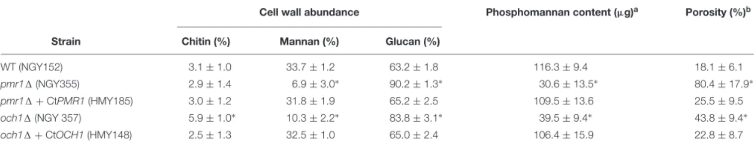
identity and similarity to C. albicans Och1, respectively, is predicted to be a type-II transmembrane protein belonging to the glycosyltransferase family 32, and contains the sequence180DXD, a common signature sequence of proteins that use divalent cations, such as Mn2+ as a cofactor during the transference of monosaccharides from a nucleotide-activated sugar to oligo and polysaccharides (Bates et al., 2006). To test these hypotheses generated by bioinformatic means, we next complemented C. albicans pmr11andoch11 null mutants (Bates et al., 2005, 2006) with the corresponding C. tropicalis ORFs under the control of the strongC. albicans ACT1promoter (Barelle et al., 2004). As previously reported (Bates et al., 2005, 2006), the C. albicans pmr11 andoch11null mutants showed defects in the cell wall composition, with low mannan and phosphomannan content and increased glucan levels (Table 1). Chitin content was only modified in theoch11null mutant but not inpmr11 cells (Table 1). Moreover, in line with the reduction in mannan content, both mutant strains showed increased cell wall porosity (Table 1). Expression of C. tropicalis PMR1 or OCH1 in the
1http://www.ncbi.nlm.nih.gov/
correspondingC. albicansnull mutant restored this phenotype to levels comparable to those found in the wild-type (WT) control strain (Table 1). Collectively, these data suggest thatC. tropicalis PMR1 and OCH1are the functional orthologs of C. albicans PMR1andOCH1, respectively.
Next, to generate aC. tropicalisstrain lacking eitherPMR1 or OCH1 the SATI flipper methodology was used to disrupt both alleles of these two genes. The disruption cassettes were constructed in the pSFS2 plasmid (Reuß et al., 2004) and contained 1500 bp regions upstream and downstream the targeted ORF for homologous recombination. For both genes, disruption of the first allele was confirmed by PCR, cells underwent marker recycling, used in the second round of transformation with the same disruption cassette, and again transformation marker recycling, generating thepmr11and the och11null mutants (Table 2). To generate reintegrant control strains, either PMR1 or OCH1 ORF plus regulatory regions were cloned into pSFS2 and this construction used to transform thepmr11oroch11null mutant, respectively. The integration of constructions into the targeted loci was confirmed by PCR (data not shown).
Both thepmr11andoch11null mutants showed defects in the cell morphology, forming cell aggregates, which were more prominent in the former (Figure 1). The reintegration of one allele of the disrupted gene reverted the phenotype of the null mutant strains, with a cell morphology similar to that displayed by the WT control strain (Figure 1). In addition, both mutant strains showed defects in the colony morphology when grown on a solid culture medium, forming colonies with irregular borders and a wrinkled surface (Figure 1). Again, the reintegrant control strains showed a phenotype similar to the WT strain (Figure 1).
These changes were accompanied by changes in the doubling time at 28◦C, with the null mutant strains tending to grow slower than the WT and reintegrant control strains (doubling time of the WT control strain 1.49 ± 0.19 h, pmr11 strain 2.25 ± 0.24 h, pmr11 + PMR1 strain 1.47± 0.25 h, och11 strain 2.22±0.27 h, andoch11+OCH1strain 1.52±0.12 h, p <0.05 when compared the null mutant strain with the WT strain or the corresponding reintegrant strain), and both the pmr11andoch11null mutants, but not the WT or reintegrant control strains, were unable to grow at 42◦C (data not shown).
The Candida tropicalis pmr1 1 and och11 Null Mutants Have Defects in the Cell Wall and Protein Glycosylation Pathways
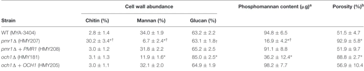
To assess the role of eitherPMR1orOCH1in the C. tropicalis wall composition, cell walls were isolated, cleaned, acid- hydrolyzed, and analyzed by High-Performance Anion-Exchange Chromatography with Pulsed Amperometric Detection (HPAEC-PAD). The cell wall composition of the WT control strain was similar to that recently reported for C. tropicalis, with a large amount of glucan, followed by mannan, and a small proportion of chitin (Hernandez-Chavez et al., 2018;
Table 3). Thepmr11 null mutant showed about 10-fold more chitin content than the WT control strain, a significant 5-fold
TABLE 1 |Complementation ofCandida albicans pmr11andoch11null mutants with the functional orthologs ofCandida tropicalis.
Cell wall abundance Phosphomannan content (µg)a Porosity (%)b
Strain Chitin (%) Mannan (%) Glucan (%)
WT (NGY152) 3.1±1.0 33.7±1.2 63.2±1.8 116.3±9.4 18.1±6.1
pmr11(NGY355) 2.9±1.4 6.9±3.0∗ 90.2±1.3∗ 30.6±13.5∗ 80.4±17.9∗
pmr11+CtPMR1(HMY185) 3.0±1.2 31.8±1.9 65.2±2.5 109.5±13.6 25.5±9.5
och11(NGY 357) 5.9±1.0∗ 10.3±2.2∗ 83.8±3.1∗ 39.5±9.4∗ 43.8±9.4∗
och11+CtOCH1(HMY148) 2.5±1.3 32.5±1.0 65.0±2.4 106.4±15.9 22.8±8.7
aµg of Alcian Blue bound/OD600= 1,bRelative to DEAE-Dextran,∗P<0.05, when compared to the WT or complemented strains.
TABLE 2 |Strains used in this work.
Strain Organism Origin Genotype References
MYA-3404 C. tropicalis ATCC Wild type ATCC
HMY206 C. tropicalis Derived from MYA-3404 As ATCC MYA-3404, butpmr11:sat1/PMR1 This work
HMY207 C. tropicalis Derived from HMY206 As ATCC MYA-3404, butpmr11:sat1/pmr11:sat1 This work
HMY208 C. tropicalis Derived from HMY207 As ATCC MYA-3404, butpmr11:sat1/pmr11:sat1-PMR1 This work
HMY179 C. tropicalis Derived from MYA-3404 As ATCC MYA-3404, butoch11:sat1/OCH1 This work
HMY181 C. tropicalis Derived from HMY179 As ATCC MYA-3404, butoch11:sat1/och11:sat1 This work
HMY205 C. tropicalis Derived from HMY181 As ATCC MYA-3404, butoch11:sat1/och11:sat1-OCH1 This work NGY152 C. albicans Derived from CAI-4 ura31-iro11:imm434/ura31-iro11:imm434;RPSI/rps11:CIp10 Brand et al., 2004 NGY98 C. albicans Derived from NGY97 ura31-iro11:imm434/ura31-iro11:imm434;pmr11:hisG/pmr11:hisG Bates et al., 2005
NGY355 C. albicans Derived from NGY98 As NGY98, butRPSI/rps11:CIp10 Bates et al., 2005
HMY185 C. albicans Derived from NGY98 As NGY98, butRPSI/rps11:pACT1-CtPMR1 This work
NGY205 C. albicans Derived from NGY204 ura31-iro11:imm434/ura31-iro11:imm434;och11:hisG/och11:hisG Bates et al., 2006
NGY357 C. albicans Derived from NGY205 As NGY205, butRPSI/rps11:CIp10 Bates et al., 2006
HMY148 C. albicans Derived from NGY205 As NGY205, butRPSI/rps11:pACT1-CtOCH1 This work
FIGURE 1 |Cell and colony morphology ofCandida tropicalis pmr11andoch11null mutants. The upper panels show the cell morphology after growth at 28◦C for 15 h in YPD medium, demonstrating cell aggregates in both null mutant strains. Scale bar, 10µm. The lower panels show colony morphology after 3 days of growth at 28◦C on YPD plates. Both null mutants formed colonies with irregular borders and a wrinkled surface. Scale bar, 1 mm. Strains used are MYA-3404 (WT), HMY207 (pmr11), HMY208 (pmr11+PMR1), HMY181 (och11), and HMY205 (och11+OCH1).
reduction in the mannan levels, and no changes in the glucan content (Table 3). The och11 null mutant showed a 3-fold reduction in the mannan content, no changes in the chitin levels and a significant 1.3-fold increment in the glucan content (Table 3). Interestingly, the changes in the content of these three cell wall components were different when both null mutants were compared (Table 3). In both cases, the reintegrant control strains restored the content of these wall components to levels
similar to those found in the WT control strain (Table 3). The phosphomannan content and the porosity to DEAE-dextran are wall parameters that have been correlated with defects in the protein glycosylation pathways (Mora-Montes et al., 2007, 2010; Cheng et al., 2011; Navarro-Arias et al., 2016, 2017;Perez-Garcia et al., 2016;Lozoya-Perez et al., 2019). The phosphomannan content was reduced in both null mutants, and this was accompanied by an increment in the cell wall porosity
TABLE 3 |Cell wall composition ofCandida tropicalisWT,pmr11,och11, and reintegrant control strains.
Cell wall abundance Phosphomannan content (µg)a Porosity (%)b
Strain Chitin (%) Mannan (%) Glucan (%)
WT (MYA-3404) 2.8±1.4 34.0±1.9 63.2±2.2 94.8±6.5 51.5±4.7
pmr11(HMY207) 30.2±3.4∗† 6.7±2.4∗† 63.1±1.8† 16.9±4.2∗† 92.9±5.8∗
pmr11+PMR1(HMY208) 3.0±1.2 31.8±2.2 65.2±2.5 91.1±8.8 51.9±9.7
och11(HMY181) 3.1±1.3 11.9±1.6∗ 85.0±2.5∗ 36.2±12.4∗ 88.8±2.7∗
och11+OCH1(HMY205) 3.0±1.1 32.1±2.0 64.9±1.9 98.2±7.7 56.9±10.4
aµg of Alcian Blue bound/OD600= 1,bRelative to DEAE-Dextran,∗P<0.05, when compared to the WT or complemented strains,†P<0.05, when compared to och11null mutant.
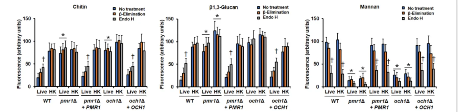
(Table 3). Again, both reintegrant strains showed a phenotype similar to the WT strain (Table 3). To further explore defects in the mannosylation pathways in either the pmr11 or the och11null mutant, cells were subjected to treatment with endo H orβ-elimination to removeN-linked orO-linked mannans, respectively (Navarro-Arias et al., 2016, 2019; Lozoya-Perez et al., 2019). The N-linked and O-linked mannan content in the WT control strain was similar to that recently reported (Navarro-Arias et al., 2019), with most of the cell wall mannan bound to proteins by N-linkages (Figure 2). The pmr11 null mutant showed a low level of both mannans, whereas theoch11 null mutant had a reduction of N-linked mannans and an increment in the O-linked mannan content (Figure 2). The mannan content was different when both null mutant strains were compared (Figure 2). The reintegrant control strains displayed a phenotype similar to the WT strain, confirming that the defects found in the mannosylation pathways are associated with the disruption of the genes under analysis (Figure 2). To further confirm the decreased content of cell wall mannan in both mutants, we labeled these oligosaccharides with fluorescein isothiocyanate-concanavalin A conjugate (Con A-FITC) and inspected the cells under fluorescent microscopy. The lectin strongly bound to the mannan of the WT cells, but a weak interaction of this with the cell wall of either the pmr11 or the och11 null mutant was observed, confirming of findings of decreased mannan content in these mutants (Figure 3 and Supplementary Figure S1). The reintegrant control strains showed a similar ability to bind Con A-FITC than the WT cells (Figure 3 and Supplementary Figure S1). Since heat inactivation of cells artificially exposes β1,3-glucan and chitin at the cell wall surface (Gow et al., 2007; Mora-Montes et al., 2011; Estrada-Mata et al., 2015; Navarro-Arias et al., 2016, 2017, 2019; Perez-Garcia et al., 2016;Hernandez-Chavez et al., 2018); we next repeated the experiment but using heat-killed (HK) cells for fluorescent labeling. This cell treatment did not have an impact on the ability of Con A-FITC to bind the cell wall (Figure 3 and Supplementary Figure S1). When these experiments were performed withβ-eliminated cells we observed a small reduction in the ability of the lectin to bind cell walls, but this was not significant (P > 0.05;Figure 3). When endo H-treated cells were used for interaction with Con A-FITC there was a significant reduction in the binding of the lectin by both the live and HK WT and reintegrant control cells (Figure 3). In the case of both mutants, the treatment with
FIGURE 2 |Cell wall mannan content in theCandida tropicalis pmr11and och11null mutants. Cells were treated with endoglycosidase H or β-elimination to removeN-linked mannans orO-linked mannans, respectively, the trimmed oligosaccharides were concentrated and the carbohydrate content was quantified as described in methods. Data are means±SD of three independent experiments performed in duplicates. Strains used are MYA-3404 (WT), HMY207 (pmr11), HMY208 (pmr11+PMR1), HMY181 (och11), and HMY205 (och11+OCH1).∗P<0.05, when compared with either the WT or reintegrant control cells.†P<0.05, when compared with the och11null mutant.
endo H decreased the binding of Con A-FITC, but this was not significant (Figure 3).
Next, we analyzed the organization of the structural polysaccharides chitin and β1,3-glucan within the cell wall, using fluorescence labeling with fluorescein isothiocyanate-wheat germ agglutinin conjugate (WGA-FITC) and an IgG Fc-Dectin-1 chimera, respectively (Graham et al., 2006;Mora-Montes et al., 2011;Marakalala et al., 2013;Estrada-Mata et al., 2015;Navarro- Arias et al., 2016, 2017, 2019; Hernandez-Chavez et al., 2018).
Both lectins barely bound to chitin and β1,3-glucan localized in the cell wall of the WT and the re-integrant control strains (Figure 3andSupplementary Figures S2, S3). Cells from either the pmr11 or the och11 null mutant were significantly more labeled than the WT or reintegrant control cells with both lectins, suggesting exposure of chitin and β1,3-glucan at the cell wall surface (Figure 3andSupplementary Figures S2, S3). The HK WT and reintegrant control cells showed an increased ability to bind both lectins (Figure 3andSupplementary Figures S2, S3),
FIGURE 3 |Fluorescent labeling of cell wall chitin,β1,3-glucan, and mannan in theCandida tropicalis pmr11andoch11null mutants. Live or heat-killed (HK) yeast cells were labeled with either fluorescein isothiocyanate-wheat germ agglutinin conjugate that binds chitin, IgG Fc-Dectin-1 chimera that bindsβ1,3-glucan, or fluorescein isothiocyanate-concanavalin A conjugate that binds mannan, as described in the methods, inspected under fluorescence microscopy, and the fluorescence associated to 300 individual cells recorded. In addition, cells were previouslyβ-eliminated or treated with endoglycosidase H (endo H) before labeling with lectins.∗P<0.05, when compared with live and HK cells from the WT strain.†P<0.05, when compared with no treated cells. Strains used are MYA-3404 (WT), HMY207 (pmr11), HMY208 (pmr11+PMR1), HMY181 (och11), and HMY205 (och11+OCH1).
FIGURE 4 |Susceptibility to cell wall perturbing agents in theCandida tropicalis pmr11andoch11null mutants. Yeast cells were incubated in YPD broth supplemented with different concentrations of Congo red, Calcofluor white, or Hygromycin B, and cell growth was determined after incubation for 24 h at 30◦C. For normalization, growth results are shown as a percentage of those obtained with the same strain grown in the absence of any perturbing agent. Data are
means±SD of three independent experiments performed in duplicates. Both null mutant strains showed increased susceptibility to the three cell wall perturbing agents analyzed, and were significantly different to either the WT or the reintegrant control strain (P<0.01 when compared by two-way ANOVA). Moreover, the increased susceptibility of the null mutants was different when compared to each other (P<0.05 when compared by two-way ANOVA). Strains used are MYA-3404 (WT), HMY207 (pmr11), HMY208 (pmr11+PMR1), HMY181 (och11), and HMY205 (och11+OCH1).
suggesting that the modest interaction between the lectins and live cells was due to inaccessibility of lectins to polysaccharides, i.e., these are in the inner part of the cell wall. The chitin labeling in both thepmr11andoch11null mutants was similar to that observed in live cells, and β1,3-glucan labeling was increased upon heat inactivation (Figure 3). As in the case of mannan labeling, theβ-elimination treatment did not affect the chitin or β1,3-glucan labeling (Figure 3). Upon incubation with endo H, the lectin binding to both polysaccharides from live and HK null mutant strains was similar to the untreated cells, but an increased binding to both chitin andβ1,3-glucan was observed in the walls of live WT and reintegrant control cells (Figure 3). Collectively these data confirm that both polysaccharides are exposed at the cell surface of thepmr11andoch11null mutants. In addition, they are in line with the increment in glucan content quantified by HPAEC-PAD.
It has been previously reported thatCandidamutants in either thePMR1or theOCH1genes have increased susceptibilities to cell wall perturbing agents (Bates et al., 2005, 2006;Navarro-Arias
et al., 2016; Perez-Garcia et al., 2016). Therefore, we analyzed the effect of this kind of compounds on the growth of the C. tropicalis pmr11 and och11 null mutants. Congo red, a compound that disturbs the glucan within the cell wall (Kopecka and Gabriel, 1992), had a modest effect on the growth of the WT and reintegrant control strains but both thepmr11andoch11 null mutants showed increased susceptibility to this stressor (Figure 4). The WT and reintegrant control strains showed a dose-dependent increased susceptibility to both Calcofluor white and hygromycin B, and this effect was more prominent in the presence of the latter (Figure 4). Calcofluor white binds to chitin; while increased susceptibility to hygromycin B has been reported in yeast cells with defects in the protein glycosylation pathways (Dean, 1995; Mora-Montes et al., 2011). Again, the pmr11andoch11null mutants had increased susceptibility to these perturbing agents (Figure 4). Interestingly, when compared to the ability of both mutants to grow in presence of these three cell wall perturbing agents, the och11 null mutant was more affected than the strain werePMR1was disrupted (P<0.05 in all
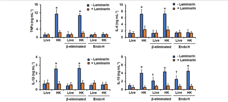
FIGURE 5 |Cytokine production by human PBMCs stimulated withCandida tropicalis. Yeast cells and human PBMCs were co-incubated for 24 h, the supernatant saved and used to quantify the levels of secreted TNFα, IL-6, IL-1β, and IL-10. Results (means±SD) were obtained using samples from seven donors, each assayed in duplicate wells, and the wild type strain MYA-3404.∗P<0.05, when compared with live cells under the same treatment;†P<0.05, when compared with live cells with no treatment. HK, heat-inactivated cells; Endo-H, endoglycosidase H.
cases;Figure 4). Altogether, these data indicate that loss of either PMR1orOCH1affects theC. tropicalisprotein glycosylation and the cell wall composition, organization, and fitness.
Removal of Cell Wall Mannans Affects the Ability of Candida tropicalis to Stimulate Cytokine Production by Human PBMCs
Next, we assessed the importance of cell wall mannan on the C. tropicalis-host interaction, analyzing the ability of this fungal species to stimulate cytokine production by human PBMCs.
We first compared the cytokine profile induced by the WT strain treated with endo H orβ-elimination, to removeN-linked or O-linked mannans, respectively (Navarro-Arias et al., 2016;
Perez-Garcia et al., 2016). Moreover, since in other Candida speciesβ1,3-glucan is one of the major fungal ligands to stimulate cytokine production and is normally buried behind the mannan layer, and therefore inaccessible to engage with dectin-1 (Gow et al., 2007;Estrada-Mata et al., 2015;Navarro-Arias et al., 2016, 2017, 2019; Perez-Garcia et al., 2016), we also compared the ability of live and HK cells to stimulate cytokine production.
Live cells stimulated low levels of TNFα, IL-1β, IL-6, and IL-10, but PMBCs interacting with HK cells produced a significantly higher level of these cytokines (Figure 5). The experimental setting where PBMCs were pre-incubated with laminarin and challenged with HK cells stimulated cytokine levels similar to those with the live cells (Figure 5). Thus, the high cytokine levels induced by HK cells were associated with the engagement of dectin-1 with its ligand, as reported for other fungal cells (Estrada-Mata et al., 2015;Navarro-Arias et al., 2016, 2017, 2019;
Perez-Garcia et al., 2016). Removal of O-linked mannans via β-elimination did not affect the TNFα, IL-1β, or IL-6 profiles, but live cells with noO-linked mannans on the surface tended to simulate higher IL-10 levels than untreated cells (Figure 5).
The trimming ofN-linked mannans with endo H only affected the ability of live cells to stimulate IL-10, which was higher, when compared with the levels stimulated with untreated cells (Figure 5). The endo H-treated HK cells were not able to stimulate a strong production of TNFα, IL-1β, or IL-6; while the levels of IL-10 were not affected when compared with those stimulated with the untreated HK cells (Figure 5). It is possible to hypothesize that treatment with endo-H could expose cell wall proteins to the extracellular compartment, promoting detachment of these macromolecules from the wall, accounting for the changes in the ability to stimulate cytokine production.
However, these is unlikely, as the wall protein content of cells was not affected upon treatment with endo-H (132.4 ± 21.4, 144.1±17.2, 122.9±13.9, and 136.8±15.6µg of protein mg of cell wall−1for live, live+endo H-, HK, HK+endo H-treated cells, respectively).
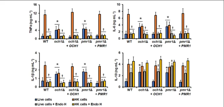
In similar experiments, the ability of thepmr11andoch11 null mutant to stimulate cytokine production by human PBMCs was also assessed. Live cells of either thepmr11oroch11null mutant stimulated higher TNFα, IL-6, IL-1β, and IL-10 when compared with the WT live cells (Figure 6). For the three pro- inflammatory cytokines quantified, the stimulated levels using HK mutant cells were similar to those observed in the system with live cells and not sufficient enough to be comparable to those stimulated with the WT HK cells (Figure 6). For IL-10, the cytokine level stimulated with HK cells was comparable with that observed with live mutant cells, but slightly higher, and
FIGURE 6 |Cytokine production by human PBMCs stimulated withCandida tropicalis och11andpmr11null mutants. Yeast cells and human PBMC were co-incubated for 24 h, the supernatant saved and used to quantify the levels of secreted TNFα, Il-6, IL-1β, and IL-10. Results (means±SD) were obtained using samples from seven donors, each assayed in duplicate wells.∗P<0.05, when compared with WT control cells under the same treatment;†P<0.05, when compared with cells without treatment with endo H. HK, heat-inactivated cells; Endo-H, endoglycosidase H. Strains used are MYA-3404 (WT), HMY207 (pmr11), HMY208 (pmr11+PMR1), HMY181 (och11), and HMY205 (och11+OCH1).
therefore statistically similar to the levels stimulated with the WT HK cells (P= 0.627 when WT andoch11null mutant cells are compared; P = 0.295 when WT andpmr11 null mutant cells are compared). Both live and HK cells of the reintegrant control strains for OCH1 and PMR1 showed an ability to stimulate cytokine production similar to WT control cells (Figure 6). It is worthy of note that it was expected that the cytokine profile stimulated with endo-H-treated WT cells was similar to that observed with cells lacking OCH1but results from Figures 5, 6clearly showed this was not the case. Since Och1 is a protein localized within the Golgi complex, it is expected that the N-linked mannan core is properly assembled and transferred to glycoproteins, as demonstrated in C. albicans (Bates et al., 2006). Therefore, the cell surface of anoch11null mutant strain will display the N-linked mannan core on the wall surface, which contrasts with the surface of endo-H-treated cells where all theN-linked mannan, including the oligosaccharide core, is absent. Thus, we next compared the ability to stimulate cytokine production of the null mutants subjected to endo H treatment.
For both live and HK cells treated with endo H, the levels of the proinflammatory cytokines TNFα, IL-6, and IL-1βwere similar to those produced in the system where live untreated cells were included (Figure 6). For IL-10 stimulation, theoch11null mutant did not show any difference in the ability to stimulate this cytokine when compared to the system where endo H was not included (Figure 6). In the case of thepmr11null mutant, live cells treated with endo H stimulated significantly higher levels of this cytokine, when compared to the system that not included the glycosidase (Figure 6). The reintegrant control strains for both
genes showed a similar ability to stimulate cytokine production to that observed with the WT control cells (Figure 6). To further explore the differences between endo-H-treated WT cells and the och11 null mutant we performed interaction experiments in the presence of the N-linked mannan core Man9GlcNAc2 (M9) (Mora-Montes et al., 2004) and measured the cytokine production. Human PBMCs pre-incubated with M9 and then challenged with live WT cells stimulated similar cytokine levels than cells without the preincubation step; however, in similar experiments using HK cells the amount of TNFα, IL-6 and IL- 1βwas significantly diminished, when compared with the system without M9 (Figure 7and data not shown). For the case of IL- 10, pre-incubation with M9 did not affect the cytokine levels stimulated by either live or HK cells (Figure 7). In the case of both live and HKoch11null mutant cells, the levels of TNFα, IL- 6, and IL-1βwere significantly reduced upon pre-incubation with M9 (Figure 7and data not shown). As the WT cells, the levels of IL-10 stimulated with either live and HKoch11null mutant cells were not affected by pre-incubation with M9 (Figure 7).
The reintegrant control strain stimulated similar cytokine levels to those observed with the WT strain (Figure 7). The pmr11 null mutant showed a similar ability to stimulate cytokine production than the och11 null strain (data not shown). This antagonistic ability of M9 during cytokine production suggested the involvement of mannose receptor (MR) in the PBMC-yeast interaction, as this pattern recognition receptor senses branched oligosaccharides with alpha glycosidic bonds, as M9 (Netea et al., 2006). Therefore, we explored the contribution of this receptor in the C. tropicalis recognition. Human PBMCs preincubated
with anti-MR antibodies were challenged with either WT or mutant cells. The anti-MR antibodies did not affect the ability to produce IL-10 but negatively affected the TNFα, IL-6, and IL- 1β levels stimulated with HK WT cells (Figure 7and data not shown). For the case of the och11 null mutant, the presence of anti-MR antibodies within the system had little impact on the production of IL-10 but reduced the production of TNFα, IL-6, and IL-1β stimulated with live cells (Figure 7 and data not shown). Although the pro-inflammatory cytokine levels were reduced in the presence of anti-MR and HK cells of theoch11 null mutant, this was not statistically significant (Figure 7and data not shown). The control system with an isotype-matched antibody or with the reintegrant control strain shown cytokine levels similar to those stimulated with the WT cells.
Finally, we assessed the contribution of dectin-1 during the interaction withoch11null cells. As in the case of the WT strain, the TNFα, IL-6, IL-1β, and IL-10 levels stimulated by either live or HK mutant cells was significantly reduced (Figure 7and data not shown). Similarly, the cytokine production stimulated by live or HKpmr11cells was negatively affected by pre-incubation with laminarin (data not shown), stressing the importance of dectin- 1 engagement withβ1,3-glucan during theC. tropicalis-human PBMC interaction.
PMR1 and OCH1 Are Required for Candida tropicalis Virulence in Both Galleria mellonella and the Mouse Model of Systemic Candidiasis
To assess the relevance of protein mannosylation in the C. tropicalisvirulence, we first infected larvae of G. mellonella and evaluated the survival rate of these animals. The animals infected with WT and the reintegrant control strains showed similar survival curves, with the total of the animal population dead after 8 days of infection when injected with the WT strain (Figure 8). For the case of the reintegrant strains, the total of the animal population succumbed after 10 days post- infection, and this difference was not statistically significant (Figure 8; P = 0.19 and 0.27, when the curve associated to WT cells is compared with either pmr11 + PMR1 or och11 +OCH1, respectively). The survival curves of animals infected with either the pmr11 or the och11 null mutant were similar and showed virulence attenuation, with 50% of the animal population surviving at the end of the observation period (Figure 8). The fungal burdens in the inoculated animals indicated a defect in the null mutant strains to colonize the host tissues, as this was lower than that associated to the WT or the reintegrant control strains (1.3×10⁷±0.6×10⁷cells mL−1, 0.7 × 10⁷ ± 0.2 × 10⁷, 1.1 × 10⁷ ± 0.4 × 10⁷cells mL−1, 0.6×10⁷±0.4×10⁷, 1.0×10⁷±0.6×10⁷cells mL−1for WT, pmr11,pmr11+PMR1,och11,och11+OCH1, respectively.
P<0.05 when compared to the null mutant strains with the WT control strain).
We next used a murine model of non-lethal disseminated candidiasis as previously reported (Ifrim et al., 2014; Navarro- Arias et al., 2016; Perez-Garcia et al., 2016), to evaluate the virulence in thisin vivosetting. After 3 days of infection, BALB/c
mice infected with the pmr11 null mutant had lower fungal burdens in the spleen, kidneys, brain, and liver, when compared to either the WT control cells or the reintegrant control strain (Figure 9). Similarly, the fungal burden of animals infected with the och11null mutant strain was lower to that observed in animals infected with the WT strain, but interestingly, the och11+OCH1reintegrant control strain behaved like the null mutant strain, and fewer colony-forming units were recovered from the spleen, kidneys, brain, and liver of infected animals (Figure 10). Collectively, these results indicate thatPMR1and OCH1have a significant role in the interaction ofC. tropicalis with eitherG. mellonellaor the mouse.
DISCUSSION
So far, the knowledge we have about the genetic machinery behind protein glycosylation and cell wall assembly in C. tropicalis is limited, likely because of the close genetic relationship with C. albicans, a thoroughly studied model of fungal cell wall synthesis. Previously, we have genetically addressed the relevance of protein phosphomannosylation during theC. tropicalis-host interaction and found that the role of this wall component in the interaction with macrophages is different from that described inC. albicans(Hernandez-Chavez et al., 2018). This report represents the first genetic approach to address the relevance ofC. tropicalis N- andO-linked mannan for cell wall composition, organization, and interaction with the host.
TheO-linked mannosylation pathway includes the Pmt and Mnt transferases that belong to gene families composed each of team of five members with functional redundancy (Mora-Montes et al., 2009;Martinez-Duncker et al., 2014). This has represented a challenge to assess the contribution of each of the family members to bothO-linked mannan elaboration and the study of the host- fungus interaction (Munro et al., 2005;Prill et al., 2005;Mora- Montes et al., 2010). Therefore, we chose to disruptPMR1to get indirect insights on the contribution ofO-linked mannans to the C. tropicalis-host interaction.
The bioinformatics analysis and the heterologous complementation in C. albicans strongly suggest that the ORFs we have analyzed are indeed the functional orthologs of C. albicans PMR1 andOCH1.The C. albicansgenome also contains HOC1, a gene encoding for a mannosyltransferase that is part of the M-Pol II complex, involved in the elongation of the α1,6-backbone of the N-linked mannan outer chain (Martinez-Duncker et al., 2014). BothOCH1andHOC1encode products with significant similarity in the primary sequences but they are unable to complement each other and have signature sequences that differentiate them from each other (Lambou et al., 2010; Lozoya-Perez et al., 2019; see Supplementary Figure S4). Bioinformatics analysis of theC. tropicalisgenome could identify the putative gene encoding Hoc1, whose primary structure is closer to C. albicans Hoc1 than Och1. Moreover, a sequence analysis differentiated Och1 proteins from Hoc1 proteins, being grouped in different clades (Supplementary Figure S4). Therefore, it is unlikely that we have identified the functional ortholog of HOC1 instead of OCH1. In addition,
FIGURE 7 |Cytokine production by human PBMCs preincubated with M9, anti-MR, or laminarin and stimulated with theCandida tropicalis och11null mutant.
human PBMCs were pre-incubated with M9, anti-MR, or laminarin as described in the Materials and Methods section and then stimulated with either live or heat-killed (HK) yeast cells for 24 h. The supernatant of the interactions was saved and used to quantify cytokine production by ELISA. In the experiments using anti-MR antibodies, control assays with PBMCs pre-incubated with an irrelevant IgG were included as a control. Results (means±SD) were obtained using samples from seven donors, each assayed in duplicate wells.∗P<0.05, when compared with the corresponding live or HK untreated cells. M9, Man9GlcNAc2; MR, mannose receptor. Strains used are MYA-3404 (WT), HMY181 (och11), and HMY205 (och11+OCH1).
FIGURE 8 |Mortality curve ofGalleria mellonellalarvae infected with either the Candida tropicalis pmr11oroch11null mutant strains. Each larva was infected with 2×10⁷yeast cells and survival was monitored daily.
Experiments were performed three times, with a total of 30 larvae per group (10 larvae for each experiment). PBS, control group injected only with PBS.
Strains used are MYA-3404 (WT), HMY207 (pmr11), HMY208
(pmr11+PMR1), HMY181 (och11), and HMY205 (och11+OCH1). The null mutant strains showed a significant difference in the ability to kill larvae when compared to the parental or reintegrant control strains (P<0.05 in all cases).
if this C. tropicalis ORF was the true ortholog of HOC1, it would be difficult to explain the phenotype restoration of the C. albicans och11 null mutant expressing C. tropicalis HOC1, as the M-Pol II complex works after Och1 has added the first mannose residue to theN-linked mannan outer chain backbone (Martinez-Duncker et al., 2014).
Both the C. tropicalis pmr11 and och11 null mutants displayed phenotypical traits characteristic of yeast mutants with defects in the protein mannosylation pathways: increased doubling rates, abnormal cell and colony morphology, decreased mannan content, changes in the cell wall composition and organization, and fitness to resist the action of wall perturbing agents (Bates et al., 2005, 2006, 2013; Munro et al., 2005; Mora-Montes et al., 2007, 2010; Hall et al., 2013;
Navarro-Arias et al., 2016;Perez-Garcia et al., 2016). InCandida
guilliermondii, loss of PMR1 affected the cellular growth, changing from a radial growth that maintains the yeast cell shape, to a unilateral one, establishing the growth of pseudohyphae (Navarro-Arias et al., 2016). Our results are in line with those found in C. albicans andSaccharomyces cerevisiae, where cells grow as yeast cells upon PMR1 disruption (Antebi and Fink, 1992; Bates et al., 2005), stressing that C. guilliermondii has different mechanisms to mobilize calcium within the cell to those found inC. albicansorC. tropicalis.
Interestingly, the changes in the composition and organization of structural polysaccharides were not similar in the mutant strains under study, but comparable to those reported in C. albicans (Bates et al., 2005, 2006). One discrepancy between C. albicans and C. tropicalis though is the chitin content in the och11null mutant strains, as its abundance in the former species was doubled but not affect in the C. tropicalis strain (Bates et al., 2006). Since all the components of the core of the cell wall integrity pathway (the master regulator to control the wall composition and for triggering compensatory mechanisms when this structure is affected) are present within theC. tropicalis genome (Butler et al., 2009), it is likely this controls the cell wall remodeling upon aggression by stressors or genetic modifications that affect the cell wall structure (Dichtl et al., 2016). It has been described that the repertoire of receptors that activate this signaling pathway and its ability to engage with ligands is species- specific (Dichtl et al., 2016), which may offer an explanation to the differential effect ofOCH1loss on the chitin content in both C. albicansandC. tropicalis. In both cases, the decreased amount of mannan content may account for the exposure of this wall polymer on the surface.
It was anticipated that the och11 null mutant had defects only in the N-linked mannan synthesis; while the pmr11 mutant in both N- and O-linked mannan synthesis, a fact that was confirmed in our study. Therefore, it is feasible to hypothesize thatO-linked mannans have a significant role in the
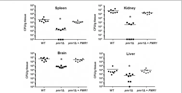
FIGURE 9 |TheCandida tropicalis pmr11null mutant has decreased virulence in a mouse model of non-lethal systemic candidiasis. Wild-type Balb/c mice were infected with 1×10⁶yeast cells and the fungal burdens in the spleen, kidneys, brain, and liver were determined after 3 days of infection. Data are expressed as colony forming units (CFU) g−1tissue (mean). Results are pooled data from two separate experiments with eight mice per group.∗P<0.05 when compared to animals infected with the WT control strain. Strains used are MYA-3404 (WT, closed circle), HMY207 (pmr11, closed square), and HMY208 (pmr11+PMR1, closed triangle).
FIGURE 10 |TheCandida tropicalis och11null mutant has decreased virulence in a mouse model of non-lethal systemic candidiasis. Same legend asFigure 9, but strains used are MYA-3404 (WT), HMY207 (pmr11), HMY208 (pmr11+PMR1). MYA-3404 (WT, closed circle), HMY181 (och11, closed square), and HMY205 (och11+OCH1, closed triangle).∗P<0.05, when compared to animals infected with the WT control strain.
signaling pathways that control the cell wall integrity pathway (Levin, 2005). Despite the contribution of N-linked mannans in the activation of this pathway has been largely documented, the detailed mechanisms behind the contribution of O-linked mannans to this signaling pathway are poorly understood (Levin, 2005).
As reported in C. albicans (Netea et al., 2006; Gow et al., 2007), C. tropicalis O-linked mannans were dispensable for stimulation of cytokine production, beingN-linked mannans and β1,3-glucan the major stimuli. In the case of IL-10 stimulation, we found that mannans were not required for this cytokine production, in fact, removal of these cell wall components had a positive effect on the IL-10 levels. It has been reported that different to other cytokines, engagement of dectin-1 with β1,3-glucan is enough to signaling the production of IL-10, representing a mannan-independent pathway for stimulation of this anti-inflammatory cytokine (Reid et al., 2009). Our results confirm this mannan-independent mechanism of IL-10 production also occurs in theC. tropicalis-PBMC interaction. The activation of this signaling pathway is the most likely explanation to the fact that the och11 and the pmr11 null mutants were capable of inducing more IL-10 production that the control strains in the live form, as most of theβ1,3-glucan was exposed on the cell surface of the mutants. This increment in the amount of superficial β1,3-glucan is also the likely explanation of the increased ability of the live mutant strains to stimulate higher levels of proinflammatory cytokines, stressing once again that the major fungal player in cytokine production is β1,3-glucan.
Alternatively, it is possible to speculate that removal of mannans by disruption of eitherOCH1orPMR1increments the exposure of cell wall phospholipomannan on the cell surface, accounting for the increased ability to stimulate cytokine production. The presence of phospholipomannan has been described in the cell wall of both C. albicans and C. tropicalis (Cantelli et al., 1995). In the former, it has been demonstrated that stimulates pro-inflammatory cytokines via engagement with TLR2 and galectin-3 (Jouault et al., 2003, 2006). Further experiments are required to assess the contribution of phospholipomannan in the ability of theoch11andpmr11mutants to stimulate cytokine production. Nonetheless, our results clearly suggest that like in otherCandidaspeciesC. tropicalismannans are maskingβ1,3- glucan, precluding interaction with dectin-1 (Wheeler and Fink, 2006;Gow et al., 2007;Navarro-Arias et al., 2016;Perez-Garcia et al., 2016). It is worthy of note that despite the null mutant cells have more β1,3-glucan exposed at the cell surface, this did not lead to a higher stimulation of pro-inflammatory cytokines, even after heat inactivation of cells. Similar observations have been reported forC. albicans,C. parapsilosis, andC. guilliermondiinull mutants with defects in mannan elaboration and higher levels of β1,3-glucan at the cell wall (Netea et al., 2006;Mora-Montes et al., 2007, 2010;Navarro-Arias et al., 2016;Perez-Garcia et al., 2016).
Our results showed that MR plays a role as important as that described for dectin-1 in the stimulation of the proinflammatory cytokines analyzed in this work, and our observations point out that this receptor engages withN-linked mannans, as reported inC. albicans(Netea et al., 2006). Therefore, it is tempting to speculate that even though signaling via dectin-1 is important for
cytokine production, the interaction of this lectin with its ligand should be part of a co-stimulation network where other receptors, or at least MR, are involved in cytokine production. In line with this hypothesis, it has been reported the synergistic interaction between dectin-1 with TLR-2, TLR-4, TLR-5, TLR-7 or TLR-9 during cytokine stimulation (Reid et al., 2009).
It is worthy of mention that despite the fact thatβ-elimination can removeO-linked mannans from the wall, this alkali treatment could affect protein structures and can be released to the medium (Mormeneo et al., 1994), potentially affecting the outcome of the immune cell-fungus interaction. However, no pattern- recognition receptor on the surface of immune cells has been described to engage with the polypeptide backbone of a protein cell wall (Netea et al., 2008).
Another interesting observation is the fact that theC. albicans och11null mutant barely stimulates cytokine production (Bates et al., 2006), whereas here, loss of OCH1 had a mild effect on the ability of C. tropicalisto stimulate cytokine production, highlighting a subtle but different relevance ofN-linked mannans in cytokine stimulation by these species. We found that the presence of the N-linked mannan core on the C. tropicalis surface is likely to account for this observation and that this oligosaccharide has the ability to block the stimulation of cytokines most likely via MR. It was reported that inC. albicans theN-linked mannan core is further modified in theoch11null mutant with two to sevenα1,2-mannose units (Bates et al., 2006), which contrast with the glycans isolated fromS. cerevisiae och11 null mutant that added only one mannose unit to theN-linked mannan core (Bates et al., 2006). It is tempting to speculate that inC. tropicalisthisN-linked mannan core is elongated with more mannose residues than inC. albicans, which accounts for its ability to stimulate higher cytokine levels to that observed withC. albicans. If this was true, the number of mannose units modifying theN-linked mannan core and the kind of glycosidic linkages involved remain to be addressed.
Both null mutant strains showed decreased virulence in both the G. mellonella and murine models of systemic candidiasis, similar to otherCandidaspecies where mutants in eitherOCH1 or PMR1 have been generated (Bates et al., 2005; Navarro- Arias et al., 2016; Perez-Garcia et al., 2016). However, in the mouse model, the reintegrant control strain forOCH1failed to colonize as the WT control strain. Since in all the phenotypical analysis this strain behaved like the WT control strain, it is unlikely this could be related to the generation of the strains.
Haploinsufficiency has been described in C. albicans (Chaillot et al., 2017), and a significant reduction of mannosyltransferase activity has been reported for the heterozygous strain inOCH1 (Bates et al., 2006). Therefore, we hypothesize that inC. tropicalis the gene could be haploinsufficient and the enzyme activity provided by one OCH1 copy could be enough to display a normal phenotype under the conditions tested but insufficient to adapt to the mouse milieu. Nevertheless, our results clearly showed virulence attenuation in the mouse model uponOCH1 disruption. Since both mutant strains showed defects in the doubling time and wall fitness, it is likely the whole-cell fitness is compromised by disruption of any of the genes under study, which is likely to account to the defects of the null mutants to