Root Organization Shapes the Rhizosphere Microbiome
Julius Durr
1, Guilhem Reyt
2, Stijn Spaepen
3,4, Sally Hilton
1, Cathal Meehan
2, Wu Qi
5,
Takehiro Kamiya
5, Paulina Flis
2, Hugh G. Dickinson
6, Attila Feher
7, Umashankar Shivshankar
8, Shruti Pavagadhi
8, Sanjay Swarup
8, David Salt
2, Gary D. Bending
1and Jose Gutierrez-Marcos
1,*
1School of Life Sciences, University of Warwick, Coventry CV4 7AL, UK
2Division of Plant and Crop Sciences, Future Food Beacon of Excellence & School of Biosciences, University of Nottingham, Nottingham LE12 5RD, UK
3Department of Plant Microbe Interactions & Cluster of Excellence on Plant Sciences (CEPLAS), Max Planck Institute for Plant Breeding Research, Carl-von- Linne-Weg 10, K€oln 50829, Germany
4Centre for Microbial and Plant Genetics, Leuven Institute for Beer Research, University of Leuven, Gaston Geenslaan 1 B-3001, Belgium
5Graduate School of Agricultural and Life Sciences, University of Tokyo, Tokyo 113-8657, Japan
6Department of Plant Sciences, University of Oxford, Oxford OX1 3RB, UK
7Institute of Plant Biology, Biological Research Centre of the Hungarian Academy of Sciences, Temesvari krt. 62, Szeged H-6726, Hungary
8Department of Biological Sciences, National University of Singapore, 14 Science Drive 4, 117543, Singapore
*Corresponding author: E-mail, j.f.gutierrez-marcos@warwick.ac.uk
The Casparian strip (CS) constitutes a physical diffusion barrier to water and nutrients in plant roots, which is formed by the polar deposition of lignin polymer in the endodermis tissue.
The precise pattern of lignin deposition is determined by the scaffolding activity of membrane-bound Casparian Strip do- main proteins (CASPs), but little is known of the mechanism (s) directing this process. Here, we demonstrate that Endodermis-specific Receptor-like Kinase 1 (ERK1) and, to a lesser extent, ROP Binding Kinase1 (RBK1) are also involved in regulating CS formation, with the former playing an essential role in lignin deposition as well as in the localization of CASP1.
We show that ERK1 is localized to the cytoplasm and nucleus of the endodermis and that together with the circadian clock regulator, Time for Coffee (TIC), forms part of a novel signaling pathway necessary for correct CS organization and suberization of the endodermis, with their single or combined loss of func- tion resulting in altered root microbiome composition. In add- ition, we found that other mutants displaying defects in su- berin deposition at the CS also display altered root exudates and microbiome composition. Thus, our work reveals a com- plex network of signaling factors operating within the root endodermis that establish both the CS diffusion barrier and influence the microbial composition of the rhizosphere.
Keywords: Endodermis • Metabolome • Microbiome • Phosphorylation • Signaling • Suberin.
Introduction
Plant roots are specialized structures essential for plant growth and survival. Roots control the selective uptake of nutrients and water from the soil, and also prevent the passive diffusion and entry of pathogens and toxins (Hawes et al. 1998, Parniske 2008), while attracting beneficial microorganisms through the secretion of certain compounds into the soil (Sasse et al. 2018).
This selectivity is determined by the root architecture and its ability to confer a barrier between the vascular cylinder and the outer cell layers, primarily cortex and epidermis, which are connected to the soil via the apoplast to control the uptake and the exudation of compounds. The apoplastic barrier pro- vided by the Casparian strip (CS) is contained within the endo- dermis; the innermost cortical cell layer surrounding the vasculature (Bonnett 1968,Nagahashi et al. 1974). These speci- alized groups of cells possess highly localized ring-like lignin deposits in the radial and transverse cell walls surrounding the endodermis (Naseer et al. 2012), which seals the cell wall and breaks the apoplastic connection between the vasculature and the outer cell layer.
The endodermal cell wall undergoes further modification by the incorporation of suberin—a hydrophobic polymer com- posed of long chain fatty acids and glycerol embedded with waxes (Baxter et al. 2009), which constitutes a second barrier.
The suberin lamellae ensure that, in fully differentiated endo- dermal cells, nutrients can only enter or leave the vasculature through specialized transmembrane carriers or special unsuber- ized passage cells (Andersen et al. 2018, Doblas et al. 2017a, Graca and Santos 2007). The extent of suberin deposition is dynamic and mediated by abscisic acid and ethylene in re- sponse to nutrient stresses (Barberon et al. 2016) or in the case of a defective CS (Wang et al. 2019). Further, suberization of the CS shows high plasticity in contrast to lignification of the CS.
In Arabidopsis, the formation of the CS apoplastic barrier is regulated by the transcription factor MYB36, which controls the expression of major genes involved in CS formation during the initial stages of endodermis differentiation (Kamiya et al.
2015). Thefirst major MYB36-dependent initiation step that gives rise to CS formation is the polar localization offive redun- dant CASPARIAN STRIP DOMAIN PROTEINs (CASP1-5) at the
Plant Cell Physiol.62(2): 248–261 (2021) doi:10.1093/pcp/pcaa170, Advance Access publication on 22 January 2021, available online at https://academic.oup.com/pcp
#The Author(s) 2021. Published by Oxford University Press on behalf of Japanese Society of Plant Physiologists.
This is an Open Access article distributed under the terms of the Creative Commons Attribution License (http://creativecommons.org/licenses/by/4.0/), which permits unrestricted reuse, distribution, and reproduction in any medium, provided the original work is properly cited.
Rapid Paper
Downloaded from https://academic.oup.com/pcp/article/62/2/248/6104897 by guest on 01 September 2021site of CS initiation (Roppolo et al. 2011). The CASPs form a platform to recruit proteins involved in polar lignin deposition, such as endodermis-specific peroxidase PEROXIDASE64 and NADPH oxidase F (RbohF) (Lee et al. 2013). Several mutants have been identified that affect CASP localization to the endo- dermis. The two receptor-like kinase mutantsschengen 1(sgn1) andschengen 3(sgn3), and the dirigent domain-containing pro- tein mutantenhanced suberin 1(esb1) exhibit only a discon- tinuous CS, whileCASPexpression and lignification of the CS domain remain unaffected (Alassimone et al. 2016, Hosmani et al. 2013, Pfister et al. 2014). By contrast, other CS mutants, such aslord of the rings 1and2(lotr1andlotr2), display relatively strong ectopic localization of CASP proteins outside of the CS domain and irregular lignification (Kalmbach et al. 2017, Li et al. 2017).
Despite thesefindings, the precise mechanisms underlying formation of the endodermal barriers, and in particular CS lig- nification, remain poorly understood. To further understand these events, we performed a reverse genetic screen to identify novel signaling components involved in endodermis develop- ment. Here, we report two cytoplasmic receptor-like kinases that specifically accumulate in the root endodermis and are involved in the formation of the CS apoplastic barrier. We show that these kinases facilitate the polar localization of CASP1 and the polar deposition of lignin at the CS domain.
In addition, we provide evidence that the circadian clock regu- lator protein, TIME FOR COFFEE (TIC), is a downstream target of these kinases and is also involved in CS organization. Finally, we also show that the correct deposition of lignin and suberin in the endodermis is critical for the release of root exudates and for selective recruitment of beneficial microbes to roots.
Results
Identi
fication of two cytoplasmic receptor-like kinases implicated in the formation of the endodermal barriers
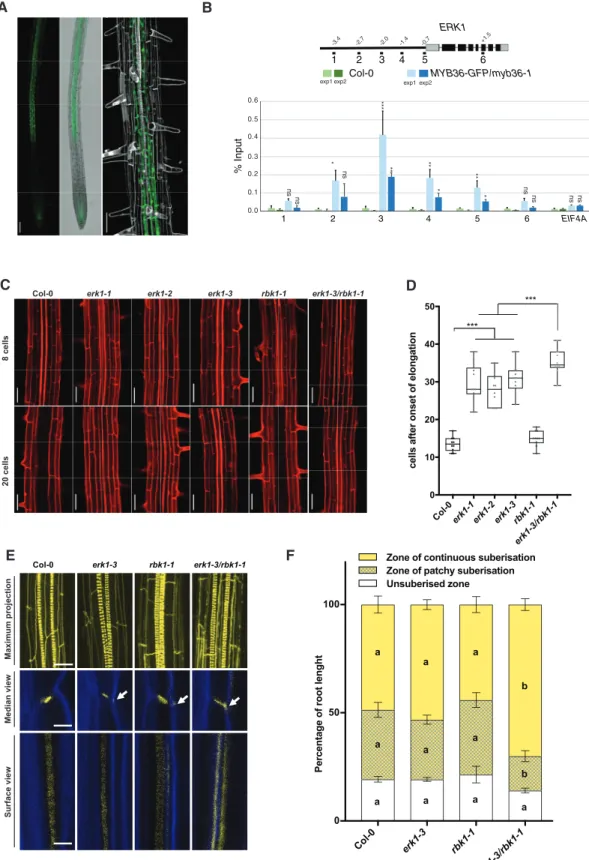
To identify novel components of the signaling pathway impli- cated in the formation of the CS, we carried out an in silico analysis of receptor-like kinases (RLK) that are under the con- trol of MYB36—a transcription factor implicated in endoder- mis gene expression and CS formation (Kamiya et al. 2015). This analysis identified a candidate, RLK (At5g65530/ARLCK VI_A3), hereafter named ENDODERMIS RECEPTOR KINASE1 (ERK1) (Supplementary Fig. S1A). We generated ERK1-GFP lines, which revealed ERK1 accumulation in the cytoplasm and nucleus of endodermal root cells only; its expression was first detected in the late elongation zone and was highest in the region where thefirst xylem bundles are formed (Fig. 1A). Using chromatin immunoprecipitation, we also determined that MYB36 directly targets a discrete region of the ERK1 promoter (Fig. 1B). We investigated the expression of the closest homo- logue of ERK1, ROP binding kinase1 (RBK1), and found that it was largely restricted to the root endodermis (Supplementary Fig. S1B). Both ERK1 and RBK1 lack the extracellular and trans- membrane domains usually present in other receptor-like
kinases (Afzal et al. 2008) but have a conserved serine/threonine kinase domain that is activated by phosphorylation through small monomeric G proteins of the plant-specific Rho family (RAC/ROP) (Huesmann et al. 2012). While genetic analyses of ERK1 and RBK1 have previously revealed their roles in trichome branching and in the control of basal resistance to powdery mildew (Huesmann et al. 2012,Reiner et al. 2015), and in auxin- responsive cell expansion (Enders et al. 2017) and pathogen response (Molendijk et al. 2008), respectively, their roles in endodermal function are unknown. We therefore isolated in- dependent T-DNA insertion lines for both genes (Supplementary Fig. S1C) and assessed mutants for defects in apoplastic barrier diffusion determined by leakage analysis of the apoplastic tracer propidium iodide (PI) into the stele of roots (Reiner et al. 2015). We found that all ERK1 mutant lines (erk1-1/SALK_148741, erk1-2/SALK_010841 and erk1-3/
SALK_060966) showed defects in the formation of the apoplas- tic barrier in the endodermis (Fig. 1C, D). We were able to restore the apoplastic barrier defects observed in erk1-3 by complementation with a wild-type ERK1 genomic fragment (Supplementary Fig. S1D), suggesting that the CS defects observed are due to the loss ofERK1function. On the other hand, we found that RBK1 mutants (rbk1-1/SALK_043441) did not display any obvious defects (Fig. 1C, D). Notably, erk1-3/
rbk1-1mutants showed a significantly stronger defective apo- plastic barrier phenotype than either of the individual single mutants, as measured by PI penetration (Fig. 1C, D), suggesting thatrbk1-1has an additive effect in the absence ofERK1.
To determine of the cause of the apoplastic barrier defects found inerk1mutants, we investigated the cell wall deposition in CS by transmission electron microscopy (TEM). We found that the cell wall deposition at the CS was enlarged inerk1-3 mutants compared to wild-type (Supplementary Fig. S1E). To further investigate the endodermis defects found in erk1 mutants, we then examined the pattern of lignin and cellulose deposition in the CS using dyes and confocal microscopy.
Compared to wild-type roots, the single erk1-3 and rbk1-1 mutants showed a slight accumulation of ectopic lignin in the corners of endodermal cells, though ectopic lignin was more apparent inerk1-3/rbk1-1mutants (Fig. 1E). Given that precocious and ectopic deposition of suberin has also been reported in the endodermis of other mutants harboring similar disruptions to the CS, such asesb1-1,casp1-1/casp3-1,myb36-2 andlcs2-1(Hosmani et al. 2013,Kamiya et al. 2015,Li et al. 2017, Roppolo et al. 2011), we also assessed suberin content in our mutants. We found that suberization in singleerk1-3andrbk1-1 mutants was normal, whileerk1-3/rbk1-1roots showed prema- ture suberization and resulted in an enlarged deposition of continuous suberin in early differentiated endodermal cells (Fig. 1F). Collectively, these data suggest that both ERK1 and, to a lesser extent, RBK1 are necessary for the correct deposition of lignin and suberin in the root endodermis.
Downstream targets of ERK1 are required for CS barrier formation
It has been reported that ERK1 is capable of phosphorylating proteins in vitro (Reiner et al. 2015). Therefore, to identify the
Downloaded from https://academic.oup.com/pcp/article/62/2/248/6104897 by guest on 01 September 2021
Col-0 erk1-1 erk1-2 erk1-3 rbk1-1 erk1-3/rbk1-1
8 cells20 cells
Col-0 erk1-3 rbk1-1 erk1-3/rbk1-1
Maximum projectionMedian viewSurface view
Col-0erk1-1 erk1-2 erk1-3rbk1-1 erk1-3/rbk1-1
cells after onset of elongation
Col-0 erk1-3 rbk1-1
erk1-3/rbk1-1
Percentage of root lenght
Unsuberised zone Zone of patchy suberisation Zone of continuous suberisation
0 10 20 30 40 50
***
***
0 50 100
a a
a
b
a a
a
b
a a a
a
A
C D
E F
B
% Input
1 2 3 4 5 6 EIF4A
ERK1
1 2 3 4 5 6
Col-0 MYB36-GFP/myb36-1
0.6
0.4 0.3 0.2 0.1 0.0 0.5
***
exp1exp2 exp1exp2
** ** *
* *
ns **
ns ns ns ns ns ns
-3.4 -2.7 -2.0 -1.4 -0.7 +1.5
Fig. 1 Loss of CS integrity and disruption of the apoplastic barrier inerk1andrbk1mutants. (A) Confocal microscopy images of roots expressing ERK1–GFP. Cell walls stained with propidium iodide (grey). Bar¼200lM. (B) Chromatin immunoprecipitation shows that MYB36 binds the promoter ofERK1(n¼3). EIF4A was used as control. ChIP-qPCR data are shown as means ± S.D from 3 technical replicates. Student'st-test, *P<0.1,
**P<0.05, ***P<0.001 and n.s. indicate no significance. (C) Lack of endodermal diffusion barrier inerk1andrbk1mutants visualized by presence of propidium iodide (red) in stele. Bar¼50lM. (D) Quantification of PI penetration into the stele quantified as number of endodermal cells from the (continued)
Downloaded from https://academic.oup.com/pcp/article/62/2/248/6104897 by guest on 01 September 2021
downstream targets of ERK1 in the endodermis, we performed a mass spectrometry-based quantitative phosphoproteomics analysis using roots from wild-type (Col-0) anderk1-3plants.
We identified100 proteins displaying over 1.5-fold significant change in phosphorylation (Supplementary Table S1). To re- veal the potential targets and pathways affected, we performed a Gene Ontology (GO) analysis and found sequences associated with the terms ‘response to abscisic acid’, ‘establishment of localization’ and ‘translation’ to be significantly enriched (P<0.001) (Supplementary Fig. S2A). One of these proteins was RBOHD, which has been recently found to be part of ROS production and lignification in the CS (Fujita et al. 2020).
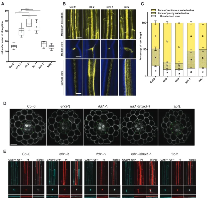
Notably, TIME FOR COFFEE (TIC) and TOM-LIKE6 (TOL6) were found to be differentially phosphorylated in erk1-3 (Supplementary Table S1andFig. S2B). Although these pro- teins have been implicated in different aspects of plant devel- opment, they have not been linked to CS formation. We assessed the permeability of the apoplastic tracer PI into the stele in the respective mutant backgrounds and observed a large delay in the formation of the PI block in two independent ticmutants, which was significantly greater than in theerk1-3 mutant allele (Fig. 2A). By contrast, we did not see any increased permeability in tol6-1or in combinations of other TOLmutants (Fig. 2AandSupplementary Fig. S3A). To evalu- ate if the apoplastic barrier defect observed inticmutants is a generic circadian clock defect, we analyzed three other circa- dian clock mutants (elf3-4,cca1-1andelf4-7)and overexpression lines (ELF3-OX and CCA1-OX); however, none of these lines showed any differences in apoplastic permeability with respect to the wild-type (Supplementary Fig. S3B). We then inspected the deposition of lignin in the endodermis ofticandtolmutants and found that bothtic-2and the quintupletolQlines showed strong ectopic deposition of lignin in the lateral margins of endodermal cells (Fig. 2B). Intriguingly, we found that onlytic mutants showed precocious accumulation of suberin in the endodermis (Fig. 2C). Collectively, these results suggest that ERK1 and TIC act in the same pathway and are responsible for the organization of the CS. We then assessed PI penetration, lignin deposition and suberin accumulation in mutants oferk1- 3in combination withtic-2 and general CS mutantsmyb36-2, sgn3-3, esb1-1andcasp1-3/casp3-1. We did notfind significant additive effects for PI penetration and lignin deposition in double erk1-3/tic2mutants compared with singleerk1-3mutants, nor in any of the other double mutants compared with the single mutants, except for erk1-3/sgn3-3, which seemed to contain slightly less lignin than sgn3-3 (Supplementary Fig. S4A, B).
When assessing suberin accumulation, again, no major differen- ces were observed; however, triple and double mutants oferk1-3
with casp1-3/casp3-1 and myb36-2, respectively, appeared to have slightly less suberin (Supplementary Fig. S4C).
Two recently identified stele-expressed peptides, CASPARIAN STRIP INTEGRITY FACTORS 1 & 2 (CIF1 & 2), which bind to the SGN3 receptor and promote CS formation, have been found to enhance suberin deposition in wild-type plants and induce CS mislocalization as well as overlignification (Doblas et al. 2017b, Nakayama et al. 2017). To determine whether ERK1 and RBK1 kinases are linked with the CIF/
SGN3 signaling pathway, we assessed lignin deposition, CASP1-GFP expression and suberin accumulation in response to exogenous application of the CIF peptide. Our data show that CIF2 induced suberinization, ectopic polymerization and mislocalization of CASP1-GFP similar to wild-type, suggesting that the role of ERK1/RBK1 in suberin accumulation may be part of an additional compensatory pathway independent of the SGN3/CIF pathway (Supplementary Fig. S5).
The ectopic deposition of lignin inerk1-3/rbk1-1andtic-2 mutants raises the possibility that the CS machinery is mislo- calized in these mutants. To evaluate this hypothesis, we assessed the localization of the CASP1-GFP reporter in different mutant backgrounds. We did not observe any major defects in the cellular organization of root optical cross-sections in any of the mutants tested (Fig. 2B). However, in transverse root cross- sections, we noticed that inerk1-3/rbk1-1mutants, localization of CASP1-GFP was not restricted to the CS, but displayed a broader distribution (Fig. 2D, E). In addition, we found in erk1-3/rbk1-1 mutants that the deposition of lignin was not limited to the CS domain, but instead it was broadly distributed in the endodermal cell wall (Fig. 2D, E). Taken together, these data suggest that ERK1 and RBK1 affect the polar localization of CASP proteins and the deposition of lignin in the endodermis.
The endodermal defects in
erk1erk1-3and
tic-2lead to ionomic changes
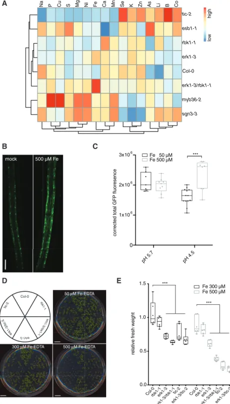
Root suberization and the CS have been shown to play a role in environmental adaptation by acting as a physical barrier to prevent unfavorable inward and outward leakage of ions be- tween the xylem and the soil (Barberon et al. 2016). In previous studies, it was shown that several CS-defective mutants exhibit changes in concentrations of multiple elements. Therefore, we compared the ionomic profiles of erk1-3, rbk1-1 and tic-2 mutants with other mutants known to be defective in CS func- tion. A principal-component analysis of elemental concentra- tions in the leaves showed that the ionomic profile of theerk1-3 andrbk1-1 mutants was similar to wild-type, whereas theerk1- 3/rbk1-1 double mutant was more similar toesb1-1but distinct to either myb36-2 or sgn3-3 mutants (Supplementary Fig.
Fig. 1Continued
first fully expanded cell (n¼10). Differences between groups were determined by pairedt-test, ***P<0.001. (E) Three-dimensional maximum projections of CS autofluorescence. Spiral structures in the center of the root are xylem (top). Longitudinal section of lignin deposition sites (bottom). Cleared roots were stained with basic fuchsin (yellow; lignin) and Calcofluor White (blue; cellulose). Although both of these dyes stain cell walls, basic fuchsin primarily interacts with lignin and Calcofluor White with cellulose. White arrows indicate the dispersed deposition of lignin at the CS. (F) Quantitative analysis of suberin accumulation. Suberin was stained withfluorol yellow 088. The endodermal cell suberin was counted from the onset of elongation to the junction (base) between root and hypocotyl (n¼6). Individual letter shows significant differences using Mann– Whitney test between the same zones (P<0.01).
Downloaded from https://academic.oup.com/pcp/article/62/2/248/6104897 by guest on 01 September 2021
S6A). Unlikesgn3-3, esb1-1andmyb36-2have enhanced lignifi- cation and suberization (Hosmani et al. 2013). Further analysis of the ionomic data revealed thaterk1-3/rbk1-1 mutants accu- mulated higher levels of iron in their roots compared with wild- type plants (Fig. 3A). Because iron is more soluble in acidic soils than in neutral soils, we reasoned that this response may reflect
an adaptation to ensure the growth and survival of plants under unfavorable mineral conditions. We therefore tested the effect of iron on ERK1-GFP accumulation and found that ERK1-GFP expression in the endodermis increased in response to excessive iron in a pH-dependent manner (Fig. 3B, C). Because a previous genetic screen for iron homeostasis mutants identified TIC as a
Col-0 tic-2 tol6-1 tolQ
Maximum projectionMedian viewSurface view
erk1-3/rbk1-1
Col-0 erk1-3 rbk1-1 tic-2
tic-2 rbk1-1
Col-0 erk1-3 erk1-3/rbk1-1
Col-0 tic-2 tol6-1 tolQ
Percentage of root lenght
Unsuberised zone Zone of patchy suberisation Zone of continuous suberisation
Col-0 erk1-3 tic-2 tol6-1 tolQ
cells after onset of elongation
0 10 20 30 40
50 ***
***
0 50 100
a b
a
a a
a
b a
a a
a a
D
A B C
E
CASP1-GFP PIPI merge
b
b a tic-3 tic-3
CASP1-GFP PI merge CASP1-GFP PI merge CASP1-GFP PI merge CASP1-GFP PI merge
Fig. 2 TICandTOLare downstream targets of the ERK1 signaling pathway implicated in Casparian Strip formation. (A) Quantification of PI penetration into the stele quantified as number of endodermal cells from the first fully expanded cell (n¼ 10). Differences between groups were determined by pairedt-test, ***P<0.001. (B) Three-dimensional maximum projections of CS autofluorescence. Spiral structures in the center of the root are xylem (top). Longitudinal section of lignin deposition sites (bottom). Cleared roots were stained with basic fuchsin (yellow; lignin) and Calcofluor White (blue; cellulose). (C) Quantitative analysis of suberin accumulation. Suberin was stained withfluorol yellow 088.
The endodermal cell with suberin was counted from the onset of elongation to the junction (base) between root and hypocotyl (n¼6). Individual letter shows significant differences using Mann–Whitney test between the same zones (P<0.01). (D) Confocal microscopy images of cross-section from roots expressing CASP1–GFP. Cell walls stained with PI (grey). Bar¼20lM. (E) Three-dimensional maximum projections of the mature endodermis expressing CASP1–GFP and stained with basic fuchsin (lignin, red) (top) on cleared roots. Median and surface view of mature endodermal cells expressing CASP1–GFP and stained with basic fuchsin (bottom). White arrows indicate dispersed localization of CASP1-GFP and lignin.
Downloaded from https://academic.oup.com/pcp/article/62/2/248/6104897 by guest on 01 September 2021
regulator of FERRITIN1 (Duc et al. 2009), we tested the growth response of these mutants under high iron growth conditions.
We found that botherk1-3andtic-2displayed high sensitivity to elevated iron conditions (Fig. 3D, E). Moreover, we also found
thatsgn3-3, but not inmyb36-2, was highly sensitivity to high iron growth conditions (Supplementary Fig. S6B). Collectively, these results suggest that these mutants are compromised in their responses tofluctuating iron levels most likely due to the
sgn3-3 myb36-2 erk1-3/rbk1-1 Col-0 erk1-3 rbk1-1 esb1-1 tic-2
Na P Cu S Mg Ni Fe Ca Mn Se K Zn As Li B Co lowhigh
pH 5.7 pH 4.5
0 1x10⁶ 2x10⁶ 3x10⁶
corrected total GFP fluoresence
***
Fe 50 µM Fe 500 µM mock 500 µM Fe
A
B
D E
C
Col-0rbk1-1erk1-3 erk1-3/rbk1-1
tic-2 erk1-3/tic-2
Col-0rbk1-1erk1-3 erk1-3/rbk1-1
tic-2 erk1-3/tic-2 0.0
0.5 1.0 1.5
relative fresh weight
Fe 300 µM Fe 500 µM
***
***
Col-0 tic-2
k1-3/tic-2 er erk1-3
k1er /rb -3 k1-1 rbk1-1
50 µM Fe-EDTA
300 µM Fe-EDTA 500 µM Fe-EDTA
Fig. 3 ERK1andTICmutants display notable ionomic changes and sensitivity to excess iron. (A) Heat-map showing differences in ion accumulation in shoots from wild-type and CS mutants (n¼ 15). (B) Induction of ERK1-GFP expression in root endodermis by elevated iron-EDTA. (C) Quantification of ERK1-GFP expression in endodermis by iron-EDTA at different pH (n¼10). Differences between groups were determined by pairedt-test, ***P<0.001. (D) Sensitivity of plants grown in vitro with different iron concentrations. (E) Relative mean fresh weight of plants grown in media with elevated iron-EDTA. (n¼6). Differences between groups were determined by pairedt-test, ***P<0.001.
Downloaded from https://academic.oup.com/pcp/article/62/2/248/6104897 by guest on 01 September 2021
inward and outward leakage resulting from their defective endodermal barriers.
The root endodermal barrier in
fluences the structure of the root microbial community
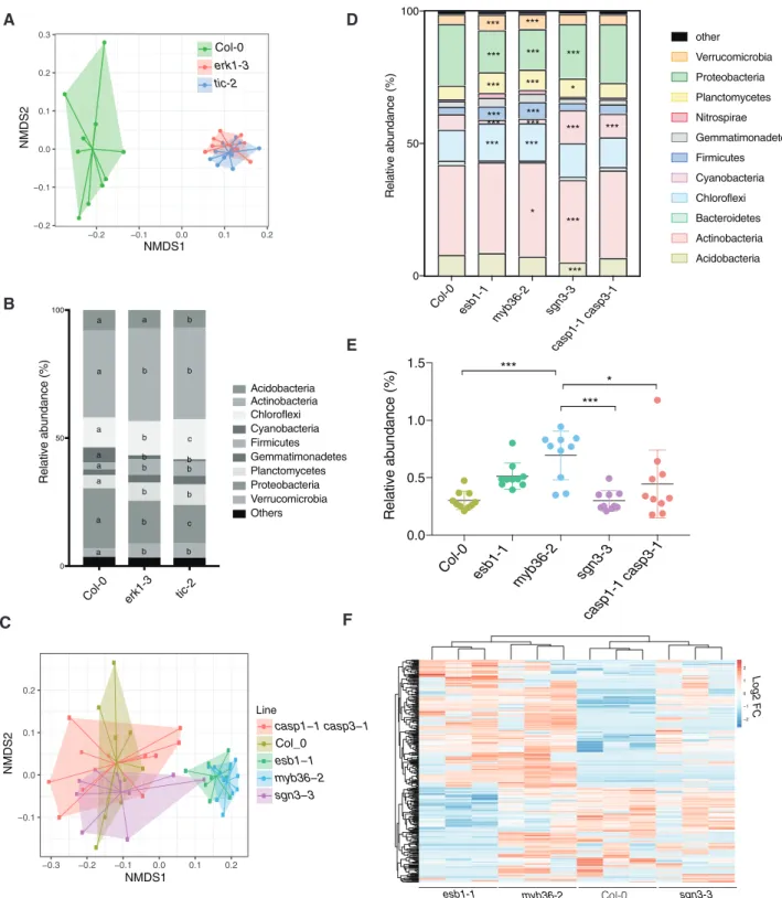
It has previously been shown that altered exudation from the plant root causes changes in the composition of the rhizosphere microbiome (Sasse et al. 2018). We therefore tested whether the unregulated leakage of components into and out oferk1-3and tic-2 mutants might also affect their respective root micro- biomes. To this end, we grew wild-type and single mutant plants in natural soils, and we analyzed the bacterial communities present in the root rhizosphere 4 weeks later. We found clear differences between the genotypes present in each subpopula- tion; the communities associated witherk1-3andtic-2mutants were most similar but differed significantly from wild-type plants (Fig. 4A). Analysis of similarities (ANOSIM) revealed sig- nificant variations in the microbial communities from roots of wild-type plants anderk1-3(ANOSIM, r¼0.921,P¼0.001) or tic-2plants (ANOSIM, r¼0.922,P¼0.001), whereas no signifi- cant differences were observed between the erk1-3 andtic-2 microbiomes (ANOSIM, r¼0.019,P¼0.558) (Fig. 4A).
We next carefully examined the different phyla of root-asso- ciated microbes and identified several groups that differed in abundance between the erk1-3 and tic-2-associated popula- tions. Notably, Chloroflexi and Proteobacteria populations dif- fered significantly among the three genotypes tested (Fig. 4B).
To independently validate these results, we inoculated seeds with a synthetic bacterial community (SynCom) isolated from an Arabidopsis root rhizosphere (Bai et al. 2015) and deter- mined the microbial communities present in mature roots and leaves. We found differences between the microbiomes present in leaves and roots of wild-type and the two mutants (Supplementary Fig. S7A). Most of the differences were associated with an abundance of Xanthomonadaceae (Proteobacteria) and Flavobacteriaceae (Bacteroidetes) (Supplementary Fig. S7B). To determine if the differences observed in the microbiome may be linked to defects in the apoplastic barrier, we analyzed the bacterial communities pre- sent in the root rhizosphere ofcasp1/3,esb1,sgn3andmyb36 mutants. We found that the bacterial communities ofmyb36 and esb1, both defective in lignin and suberin deposition in CS, were notably different from sgn3-3 or wild-type plants (Fig. 4C). Further analysis revealed that Chloroflexi, Firmicutes, Planctomycetes, Proteobacteria and Verrucomicrobiota populations differed significantly among myb36andesb1and the other genotypes tested (Fig. 4D). To independently validate thesefindings, we inoculated roots of these mutants with a GFP-tagged strain ofBacillus amilolique- faciens, a beneficial endophyte that promotes seedling growth.
We found that the abundance and level of colonization of this endophyte was greater inesb1andmyb36than in wild type and other CS mutants (Fig. 4E). To test if differences in root colon- ization may be linked to differences in the production of root metabolites, we performed a metabolome analysis of root exu- dates in mutants defective on endodermis function. We found thatesb1-1andmyb36-2exhibited notable similarities with the
metabolites exudated (72.2%,n¼372), which differed in abun- dance to the exudates found in wild-type and sgn3-3plants (Fig. 4F). These results suggest that the root colonization of microbes is strongly influenced by the secretion of specific root metabolites determined by the lignification and suberiza- tion status of the endodermis.
Discussion
The polarized deposition of cell wall material is crucial for the function and development of many root cell types (Roppolo and Geldner 2012). One of the most-studied examples of polar cell wall deposition is the root endodermis and its ring-like CS that prevents the inward and outward leakage of metabolites from the vasculature (Doblas et al. 2017a). A step in the separ- ation of the inner and outer facing plasma membrane domains is the proper localization of CASP proteins at the site of lignin deposition (Roppolo et al. 2011). Various CS-defective mutants have been described in recent years, the majority of which also show a mislocalization of CASP proteins at the site of lignifica- tion. Three different classes of CS mutants have been identified according to CASP1 expression and localization: i) those with no CASP1-GFP expression, such as in the loss-of-function myb36-3mutant; ii) mutants with defects in the continuity of the CASP domain, such assgn1,sgn3andesb1(Alassimone et al.
2016,Hosmani et al. 2013,Pfister et al. 2014); and iii) mutants with ectopic CASP deposition, such as lotr1/lcs2 or lotr2/
exo70a1(Kalmbach et al. 2017,Li et al. 2017).
In this study, we describe mutations in the cytoplasmic re- ceptor kinases ERK1 and RBK1, which lead to the mislocaliza- tion of CASP1-GFP and ectopic lignin deposition outside of the CS. Further, we show that double mutants lack a proper apo- plastic barrier but show increased lignin and suberin, which has been attributed to a compensatory mechanism (Doblas et al.
2017a) and likely dependent on the membrane-bound and cytoplasmic kinase module SNG3/SGN1 (Alassimone et al.
2016, Pfister et al. 2014). The mechanism by which the ERK/
RBK1 signaling module affects lignin deposition and suberiza- tion is currently unclear; however, evidence from the literature suggests that these proteins could be involved in membrane trafficking and the rearrangement of microtubules (Huesmann et al. 2012). For instance, ERK1 mutants exhibit increased trich- ome branch numbers (Reiner et al. 2015), likely due to incorrect microtubule rearrangements (Mathur et al. 1999). Similarly, a transient knockdown of the barley RBK1 homolog, HvRBK1, leads to defects in cortical microtubule stability in epidermal cells (Huesmann et al. 2012). Indeed, microtubules have also been shown to play an important role in the polar deposition of subcellular components and proteins that are involved in cell wall formation (Marchant 1979). EXO70A1, which is important for the polar localization of CASP proteins at the site of CS formation, together with other members of the exocyst com- plex, is also present at the site of secondary cell wall thickenings in tracheary elements, in a microtubule-dependent manner (Kalmbach et al. 2017,Vukasinovic et al. 2017).
Downloaded from https://academic.oup.com/pcp/article/62/2/248/6104897 by guest on 01 September 2021
Col-0 erk1-3 tic-2
0 50 100
Relative abundance (%)
Others Verrucomicrobia Proteobacteria Planctomycetes Gemmatimonadetes Firmicutes Cyanobacteria Chloroflexi Actinobacteria Acidobacteria
a a b
a b b
a
b c
a b b
a b b
a
b b
a b c
a b b
0.2 0.1 0.0 0.1 0.2 0.3
0.2 0.1 0.0 0.1 0.2
NMDS1
NMDS2
erk1-3 Col-0
tic-2
A
B
C
−0.1 0.0 0.1 0.2
−0.3 −0.2 −0.1 0.0 0.1 0.2
NMDS1
NMDS2
Line
casp1−1 casp3−1 Col_0
esb1−1 myb36−2 sgn3−3
Col-0 esb1-1
myb36-2 sgn3-3 casp1-1 casp3-1 0
50 100
Relative abundance (%)
Acidobacteria Actinobacteria Bacteroidetes Chloroflexi Cyanobacteria Firmicutes Gemmatimonadetes Nitrospirae Planctomycetes Proteobacteria Verrucomicrobia other
***
* ***
***
***
***
*** *** ***
*** ***
*** *** *
*** ***
***
*** ***
D
E
Col-0 esb1-1
myb36-2 sgn3-3 casp1-1 casp3-1 0.0
0.5 1.0
1.5 ***
*
***
Relative abundance (%)
F
esb1-1 myb36-2 Col-0 sgn3-3
−2
−1 0 1 2 Log2 FC
Fig. 4 Differences in rhizosphere microbial community present inerk1-3,tic-2and otherCSmutants. (A) Principal component analysis (PCA) of Bray- Curtis distances of bacterial communities present in roots of Col-0,erk1-3andtic-2plants grown in natural soils (n¼10). (B) Distribution of soil bacteria in roots of Col-0,erk1-3andtic-2plants (n¼10). Individual letter shows significant differences using Mann–Whitney test between the same zones (P<0.01). (C) PCA of Bray-Curtis distances of bacterial communities present in roots of Col-0,casp1-1; casp3-1, esb1-1, myb36-2andsgn3-3 plants grown in natural soils (n¼10). Asterisks shows significant differences using Mann–Whitney test between the same zones (P<0.01). (D) Distribution of soil bacteria in roots of Col-0,casp1-1; casp3-1, esb1-1, myb36-2andsgn3-3plants. Asterisks shows significant differences using Mann– Whitney test between the same zones (*P<0.01, ***P<0.0001). (E) Colonization assay withBacillus amilloliquefaciensin roots of Col-0,casp1-1;
casp3-1, esb1-1, myb36-2andsgn3-3plants. (F) Unsupervised hierarchical cluster heatmap analysis of metabolites exudated from roots of Col-0,esb1- 1, myb36-2 and sgn3-3plants. FC, fold change.
Downloaded from https://academic.oup.com/pcp/article/62/2/248/6104897 by guest on 01 September 2021
In addition, we identified TOL6 and TIC, two significant downstream components of the ERK1-mediated signaling path- way for CS formation. TOL6 is part of the plant Endosomal Sorting Complex Required for Transport (ESCRT) complex (Moulinier-Anzola et al. 2014, Sauer and Friml 2014) and a member of a gene family that acts redundantly to control plant morphogenesis (Korbei et al. 2013). In our study,tol6mutants did not show obvious CS apoplastic barrier defects; however, the quintuple mutant (tol2-1/tol3-1/tol5-1/tol6-1/tol9-1; tolQ) exhibited strong ectopic lignin deposition similar to erk1-3/
rbk1-1mutants. The ectopic lignification observed intolQdid not appear to interfere with apoplastic barrier function, sug- gesting that lignin deposition in this mutant is probably a sec- ondary effect of the disruption to vesicle transport. This idea is supported by the fact that together with microtubules, mem- brane vesicle transport plays a critical role in the localization of plant cell wall components (McFarlane et al. 2013,Miao and Liu 2010). TIC is one of the main regulators of the circadian clock (Davis and Millar 2001,Sanchez-Villarreal et al. 2013) and has been shown to be an integral component of iron homeostasis in roots (Duc et al. 2009,Sanchez-Villarreal et al. 2013). Our ana- lysis revealed that althoughtic1-2mutants show apoplastic bar- rier defects and ectopic deposition of lignin in the CS, this did not interfere with the polar localization of CASP1 in the endo- dermis. TIC is a major regulator of the circadian clock (Davis and Millar 2001,Sanchez-Villarreal et al. 2013); yet, the CS func- tion defects observed intic-2are not a direct cause of circadian rhythm defects. TIC has also been implicated in metabolic homeostasis, particularly in sugar production or its responses (Sanchez-Villarreal et al. 2013), both of which are critical factors in cell wall formation. The striking resistance to drought observed intic1-2 mutants (Sanchez-Villarreal et al. 2013) could be explained by an enhanced suberization of the endodermis that has also been shown in other CS mutants (Baxter et al.
2009). Our analysis has revealed that TIC plays an important role in the development of the root endodermis and that TIC function is likely regulated by phosphorylation. Indeed,TIChas also been found to interact genetically with SNF1 KINASE HOMOLOG 10 (AKIN10)/SNF1-RELATED PROTEIN KINASE 1.1 (SnRK1.1) to regulate circadian periodicity (Shin et al. 2017).
Collectively, these data indicate that the ERK1-TIC module provides a molecular link between metabolic homeostasis and nutrient uptake ability in plants.
Plant roots secrete sugars and other metabolites into the soil as a means of attracting beneficial microbes and defending themselves against pathogens, which ultimately shapes the mi- crobial communities present in the rhizosphere (Lanoue et al.
2010,Sasse et al. 2018). Root exudates are highly variable and defined by many parameters including plant accession (Monchgesang et al. 2016a,2016b), developmental stage, abi- otic stresses (Carvalhais et al. 2013,Chaparro et al. 2014), and more recently by the circadian clock (Hubbard et al. 2018).
Given that the CS and suberin lamellae are regulators of water and nutrient uptake in the root endodermis, it is plausible that perturbations in these structures could also shape the compos- ition of the root microbiome by altering exudate secretion, as observed in this study. Indeed, we showed thaterk1-3andtic-2
recruit similar microbial populations to their rhizosphere that differ markedly from the wild-type. Alternatively, modifications in the rhizosphere microbiome could be caused by differences in actively secreted compounds associated with suberization because our analysis has revealed that mutants defective in this pathway display similarities in the production of root exu- dates. Suberin lamellae are not thought to affect the apoplastic barrier directly but instead modulate transport through the endodermis (Andersen et al. 2015,Robbins et al. 2014), while some suberin components display antimicrobial properties (Buskila et al. 2011, Lulai and Corsini 1998,Ranathunge et al.
2008, Thomas et al. 2007). Thus, the increased suberization observed inerk1-3andtic-2could account for the specific mi- crobial communities present in these mutant roots (Fig. 5).
Taken together, our work provides evidence for a complex signaling cascade taking place at the root endodermis that is necessary for both the formation of a functional CS diffusion barrier and for correct accumulation of suberin and secretion of metabolites, which ultimately determines the microbial com- position of the rhizosphere.
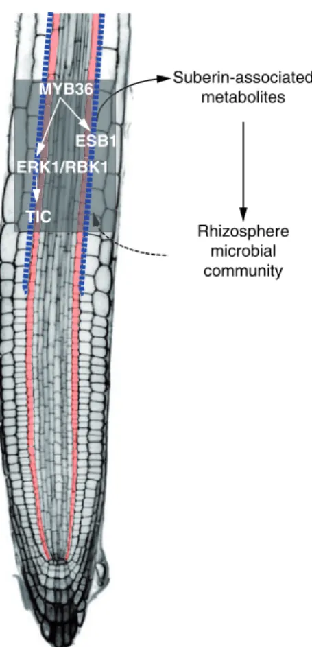
ERK1/RBK1 TIC
Suberin-associated metabolites
Rhizosphere microbial community MYB36
ESB1
Fig. 5 Hypothetical model for the role of the endodermal suberin path- way in rhizosphere microbial composition. The transcription factor MYB36 activatesESB1andERK1in the endodermis. Activation of the ERK1/RBK1 signaling cascade modulates TIC activity and influences suberin deposition at the CS. Suberin-associated metabolites are active- ly exudated and shape the microbial community composition of the rhizosphere. Blue dotted line, suberin deposition at CS; Orange cells, endodermis.
Downloaded from https://academic.oup.com/pcp/article/62/2/248/6104897 by guest on 01 September 2021
Materials and Methods
Plant lines and growth conditions
All plant material used in this study was derived from the wild-type Columbia (Col-0) or Wassilewskija (Ws) accession. T-DNA insertion alleles ofTIC(tic-2, SAIL_753_E03 and tic-3, SAIL_60_D02), ERK1 (erk1-1, rlck vi_a3-1:
SALK_148741; erk1-2, rlck vi_a3-2: SALK_010841 and erk1-3, rlck vi_a3-3:
SALK_060966) and RBK1 (rbk1-1: SALK_043441) were obtained from the Nottingham Arabidopsis Stock Centre (NASC). Mutant and wild-type (Col-0 or Ws) seeds were sown on soil (John Innes and Perlite mix), stratified for 2 d at 4C in the dark and grown at 20C under long photoperiod conditions (150lmol/m2/s and 16 h/8 h light/dark cycles) to induceflowering. Seeds were surface sterilized for 3 min in 70% ethanol, followed by a 2-min treatment in 1% NaOCl. Seeds were then washed six times in sterile H2O, dispersed in sterile 0.1% agarose and placed on half-strength MS medium (Murashige &
Skoog, Sigma), solidified with 0.8% Phytagar. Seedlings were grown vertically in growth chambers at 22C, under long days (16-hr light/8-hr dark), 100μE light, and were used at 5 d after shift to room temperature. For the evaluation of growth effect of excess iron, plants were grown on medium containing 5 mM KNO3, 2.5 mM KPO4, 3 mM MgSO4, 3 mM Ca(NO3)2, 70lM H3BO3, 14lM MnCl2, 0.5lM CuSO4, 1lM ZnSO4, 0.2lM Na2MoO4, 10lM NaCl, 0.01lM CoCl2and 50lM, 300lM or 500lM Fe-EDTA, solidified with 0.8% phytoagar.
Around 30 plants per line were grown on each plate. After 14 d, the fresh weight of all plants of one line per plate was measured. Relative fresh weight was calculated by normalization of the iron excess samples (300lM and 500lM Fe-EDTA) by the mock control (50lM Fe-EDTA).
Vector construction and plant transformation
For generation of expression constructs, Gateway Cloning Technology (Invitrogen) was used. pERK1::ERK1 was cloned using the genomic sequence of ERK1 including the endogenous promoter consisting of 1403 bp upstream of the ATG. pERK1::ERK1 was cloned into pGWB440 and into pFAST-R01 and transformed in Col-0 anderk1-3background, respectively. pRBK1::RBK1 was cloned using the genomic sequence of RBK1 including the endogenous pro- moter consisting of 1996 bp upstream of the ATG. pRBK1::RBK1 was cloned into pGWB440, pGWB553 and into pFAST-R07 and transformed in in Col-0 back- ground. Transgenic plants were generated by introduction of the plant expres- sion constructs into an Agrobacterium tumefaciens strain GV3101 and transformation was done byfloral dipping (Clough and Bent 1998).
Chromatin immunoprecipitation
Chromatin immunoprecipitation (ChIP) analysis was performed by following the protocol as described (Nakamichi et al. 2010) with modifications. Roots (100 mg fresh weight) from 11d-old plants were cross-linked by using 4 ml of the buffer (10 mM PBS, pH 7.0, 50 mM NaCl, 0.1 M sucrose and 1% formaldehyde) for 1 h at room temperature with the application of three cycles of vacuum infiltration (10 min under vacuum and 10 min of vacuum release). Glycine was added to afinal concentration of 0.1 M to stop the cross-linking reaction, and the samples were incubated for a further 10 min. After being washed with tap water, the samples were ground to afine powder by using a Multibeads Shocker (Yasui Kikai) at 1,500 rpm for 30 sec. The powder was suspended with 2 ml of Lysis buffer [50 mM TrisHCl, pH 7.5, 100 mM NaCl, 1% Triton X-100, 1 mM EDTA and EDTA-free Complete protease inhibitor (Roche)] and sonicated by using a Bioruptor UCD-250 (Cosmo Bio) with the following setting: mild inten- sity, 45 cycles (30 sec ON and 30 sec OFF) at 4C. A 100-μL sample of the chromatin sheared to between 200 and 1,500 bp was stored as the input frac- tion, and the rest (1.9 ml) was mixed with Dynabeads Protein G (Life Technologies) bound with anti-GFP antibody (ab290; Abcam) and incubated for 2 h at 4C. The beads were washed with Lysis buffer, twice with high-salt buffer [50 mM TrisHCl, pH 7.5, 400 mM NaCl, 1% Triton X-100, 1 mM EDTA and EDTA-free Complete protease inhibitor (Roche)], and then with Lysis buffer.
After Elution buffer (50 mM TrisHCl, pH 8.0, 10 mM EDTA and 1% SDS) and proteinase K (0.5 mg/ml) were added to the beads, the beads were incubated overnight at 65C. The DNA was purified with NucleoSpin Gel and PCR Clean- up (MachereyNagel) with Buffer NTB (Macherey-Nagel). Eluted solutions were used for qPCR. EIF4A (At3g13920) was used as a negative control
(Supplementary Table S2). Two independent experiments were performed with three biological replicates for each.
Permeability of the apoplastic barrier
For the visualization of the penetration of the apoplastic barrier by propidium iodide, seedlings were incubated in the dark for 10 min in a fresh solution of 15 mM (10 mg/ml) propidium IIodide (PI) and rinsed two times in water. The penetration of the apoplastic barrier was quantified by the number of cells from the‘onset of elongation’until the PI signal was blocked by the endodermis from entering the vasculature. The‘onset of elongation’was defined as the point where an endodermal cell in a median optical section was clearly more than twice its width (Alassimone et al. 2010).
Histological analysis
Tissue wasfixed and stained as described previously (Musielak et al. 2016). In brief, 5-day-old seedlings werefixed in 4% paraformaldehyde (PFA) and cell walls were stained with SCRI Renaissance 2200 (SR2200) (0.1% (v/v) SR2200, 1% (v/v) DMSO, 0.05% (v/v) Triton-X 100, 5% (v/v) glycerol and 4% (w/v) paraformalde- hyde in PBS buffer (pH 7.4)) in PBS for 15 min at room temperature with no vacuum applied. Fixed seedlings were washed twice with PBS (pH 7.4) and cleared with ClearSee (10% xylitol (w/v), 15% sodium deoxycholate (w/v) and 25% urea (w/v) in water) for 4 d at room temperature (Kurihara et al. 2015).
Cleared samples were washed twice with PBS (pH 7.4) and embedded in 4%
agarose. The agarose blocks were cut into 200lm sections with a VIBRATOMEVR Series 1000 Sectioning System.
Electron microscopy analysis
For light transmission electron microscopy (TEM), roots were dehydrated in an ethanol/propylene oxide series, embedded in Spurr’s resin (Premix Kit-Hard, TAAB Laboratory and Microscopy, Aldermaston, UK) and polymerized at 70C overnight. Longitudinal ultrathin sections (60 nm) were cut using an ultra- microtome (ultra-RMC Products), mounted on copper grids and contrasted with 1 % uranyl acetate in water for 25 min followed by lead citrate for 3 min.
Sections were examined in a transmission electron microscope (Spirit Biotwin 12 FEI Company). Measurements were carried out using the TEM Imaging Platform program.
Confocal microscopy
Confocal laser scanning microscopy experiments were performed using a Zeiss Axio Observer.Z1 inverted microscope equipped with a confocal laser-scanning module (Zeiss LSM 710 and Zeiss LSM 880, Warwick) or a Leica SP5 and SP8 (Nottingham).
Excitation and emission setting were used as follows: GFP 488 nm, 500– 550 nm; propidium iodide 516 nm, 560–700 nm; tagRFP 561 nm, 578–700 nm;
SR2200 405 nm, 410–550 nm; calcofluor white 405 nm, 425–475 nm; and basic fuchsin 561 nm, 570–650 nm. For examining CASP1 expression, basic fuchsin and calcofluor white M2R staining, 6d-old roots werefixed in paraformaldehyde and cleared in ClearSee as described (Ursache et al. 2018). Fluorol yellow staining for visualization of suberin was performed and quantified as described in Barberon et al. (2016)andNaseer et al. (2012), using afluorescent microscope Leica DM 5000. Confocal images were analyzed and contrast and brightness were adjusted with the Fiji package of ImageJ (http://fiji.sc/Fiji) and Adobe Photoshop Elements Editor.
The confocal images of the pERK1::ERK1-GFP reporter under different iron concentrations were taken with a Zeiss Axio Observer.Z1 inverted microscope equipped with a confocal laser-scanning module (Zeiss LSM 880). The exact same settings were used during one experiment. To be able to compare different roots, optical median sections were made. Theflorescent area was selected with FIJI (ImageJ) and the area, area-integrated intensity and mean gray value of each image were measured. Intensities were corrected with the mean intensity of areas without signal (background). The corrected totalfluorescence (CTF) was calculated by using this formula: CTF¼integrated density(selectedfluorescent areamean backgroundfluorescence).
Downloaded from https://academic.oup.com/pcp/article/62/2/248/6104897 by guest on 01 September 2021