GENE EXPRESSION STUDIES FOR THE EVALUATION OF MOLECULAR
INTERACTIONS BETWEEN ECSTASY AND ANTIDEPRESSANTS
PhD thesis
Peter Petschner
Doctoral School of Pharmaceutical Sciences Semmelweis University
Supervisors: Gyorgy Bagdy, DSc., Laszlo Tothfalusi, Ph.D.
Official reviewers: Tibor Zelles, Ph.D., Istvan Gacsalyi, Ph.D.
Head of the Final Examination Committee:
Krisztina Takacs-Novak, DSc.
Members of the Final Examination Committee:
Ildiko Miklya, Ph.D., Lucia Wittner, Ph.D.
Budapest, 2016
2
TABLE OF CONTENTS
TABLE OF CONTENTS ... 2
1. THE LIST OF ABBREVIATIONS ... 4
2. INTRODUCTION ... 8
2.1. MDMA ... 8
2.1.1. General aspects ... 8
2.1.2. Acute effects ... 8
2.1.3. Long-term serotonergic toxicity ... 10
2.1.4. The therapeutic potential of MDMA ... 11
2.2. Venlafaxine ... 12
2.2.1. General aspects ... 12
2.2.2. Synaptic theory of depression ... 13
2.2.3. The use of antidepressant medications in post stroke recovery... 14
2.2.4. Unresolved questions about venlafaxine’s effects... 15
2.3. MDMA and venlafaxine ... 16
2.3.1. Important interaction possibilities ... 16
3. OBJECTIVES ... 20
4. METHODS ... 21
4.1. Animals ... 21
4.2. Drug Administration and Experimental Design ... 21
4.3. RNA Extraction and Sample Preparation ... 23
4.4. Data Analysis ... 24
4.4.1. Pathway analysis of the MDMA treatment ... 25
4.4.2. Pathway analysis of the VLX treatment ... 27
4.4.3. Analysis of the combined treatment ... 27
4.5. PCR Validation ... 28
4.6. Availability of supporting data ... 29
5. RESULTS ... 30
5.1. MDMA ... 30
5.1.1. Differentially Expressed Genes after MDMA treatment ... 30
5.1.2. Gene Set Enrichment Analysis following the MDMA treatment ... 31
5.2. Venlafaxine ... 33 5.2.1. Differentially expressed genes following chronic venlafaxine treatment 33
3
5.2.2. Network analysis following chronic venlafaxine treatment ... 35
5.3. The double treatment ... 38
5.3.1. Results of the MDMA/VLX vs. MDMA/SHAM comparison ... 38
5.3.2. Results of the MDMA/VLX vs SAL/VLX comparison ... 43
5.3.3. The results of the MDMA/VLX vs. SAL/SHAM comparison ... 47
5.3.4. The results of linear models... 62
6. DISCUSSION ... 63
6.1. The MDMA/SAL vs. SAL/SHAM comparison ... 63
6.2. The SAL/VLX vs. SAL/SHAM comparison ... 67
6.2.1. Neurotransmitter release ... 67
6.2.2. Synaptogenesis, neuron migration ... 69
6.2.3. Synaptic plasticity ... 69
6.2.4. Behavior, learning and memory ... 71
6.2.5. Mitochondrial antioxidant activity ... 72
6.2.6. Insulin signaling ... 73
6.2.7. Other pathways ... 75
6.3. The double treatment ... 77
6.3.1. The MDMA/VLX vs. MDMA/SHAM comparison ... 77
6.3.2. The MDMA/VLX vs SAL/VLX comparison ... 80
6.3.3. The MDMA/VLX vs SAL/SHAM comparison ... 82
7. CONCLUSIONS ... 88
8. SUMMARY ... 90
9. ÖSSZEFOGLALÁS ... 91
10. BIBLIOGRAPHY ... 92
11. BIBLIOGRAPHY OF THE CANDIDATE’S PUBLICATIONS ... 122
11.1. Journal articles related to the thesis... 122
11.2. Articles unrelated to the thesis ... 123
12. ACKNOWLEDGEMENTS ... 124
4
1. THE LIST OF ABBREVIATIONS
5-HT serotonin, 5-hydroxytryptamine Ace angiotensin-converting enzyme
Alpl1 tissue-nonspecific alkaline phosphatase ANOVA analysis of variance
Ascl1 Achaete-scute complex like 1 Bcl2 B-cell CLL/lymphoma 2
BDNF brain-derived neurotrophic factor Ca2 carbonic anhydrase 2
Camk1g calcium/calmodulin-dependent protein kinase I gamma Camk2b calcium/calmodulin-dependent protein kinase II beta Camk2g calcium/calmodulin-dependent protein kinase II gamma Camk2n2 calcium/calmodulin-dependent protein kinase II inhibitor 2 cAMP cyclic adenosine monophosphate
Cd47 CD 47 antigen Cdh22 cadherin 22 Cdh7 cadherin 7, type 2 Clstn2 calsyntenin 2
Cnr1 cannabinoid receptor, type 1 Cntn2 contactin 2
Col27a1 procollagen, type XXVII, alpha 1 Col4a2 procollagen, type IV, alpha 2
Col4a3bp procollagen, type IV, alpha 3 (Goodpasture antigen) binding protein
Col5a1 collagen, type V, alpha 1 Cox17 copper chaperone
Cox4i1 cytochrome c oxidase subunit IV isoform 1 DA Dark Agouti
Dao1 D-amino acid oxidase Dpp4 dipeptidyl-peptidase 4 DR dorsal raphe nucleus
Eif4ebp1 eukaryotic translation initiation factor 4E binding protein 1 Enpp1 ectonucleotide pyrophosphatase/phosphodiesterase1
Epha5a ephrin receptor 5a
Erp29 endoplasmic retuclum protein 29 Faah fatty acid amid hydrolase
FC frontal cortex FDR false discovery rate FLX fluoxetine
fMRI functional magnetic resonance imaging FST forced swimming test
Gabrb2 gamma-aminobutyric acid receptor, subunit beta 2 Gabrb3 gamma-aminobutyric acid receptor, subunit beta 3
5 Gad2 glutamic acid decarboxilase 2 Galp galanin-like peptide
GalR1 galanin receptor 1 GalR2 galanin receptor 2 GalR3 galanin receptor 3
Gas2 growth arrest-specific protein 2 GEO gene expression omnibus Gfap glial fibrillary acidic protein Glp1r2 glucagone-like peptide 1 receptor
Gnao guanine nucleotide binding protein, alpha o Gnaq guanine nucleotide binding protein, q polypeptide GO gene ontology
Gpx1 glutathione peroxidase 1 Gria3 glutamate receptor, AMPA 3
Grin2a NMDA-type glutamate receptor, type 2A Grin2b NMDA-type glutamate receptor, type 2B GSEA gene set enrichment analysis
Hcn1 hyperpolarisation-activated cyclic nucleotide gated potassium channel 1
HPA hypothalamus-pituitary-adrenal axis Hsd11b hydroxysteroid 11-beta dehydrogenase 2 Hsf2 heat shock factor 2
HSP heat-shock protein Hspca heat-shock protein 1 Igf2 insulin like growth factor 2
Il1rapl1 interleukin 1 receptor accessory protein-like 1
Kcnc2 potassium voltage gated channel, Shaw-related subfamily, member 2
Kcnd2 potassium voltage gated channel, Shal-related family, member 2 Kif1b Kinesin family member 1b
Kif2b Kinesin family member 2b Kif5a Kinesin family member 5a Lphn1 latrophilin 1
MDD major depressive disorder
MDMA ±3,4-methylenedioxy-methamphetamine MinPplr minimum probability of positive log ratio Miz1 Msx-interacting-zinc finger
Mmp9 matrix metallopeptidase 9
Mrpl42 mitochondrial ribosomal protein L42 Mrpl48 mitochondrial ribosomal protein L48 MSigDB molecular signature database
Myo5a myosin 5a NA noradrenaline
Negr1 neuronal growth regulator 1 Nell2 nel-like 2 homolog
6 NES normalized enrichment score
Nr2f6 nuclear receptor subfamily 2, group F, member 6 Nr4a3 nuclear receptor subfamily 4, group A, member 3 NRG1 neuregulin 1
Ntrk2 neurotrophic tyrosine kinase, receptor type 2 Ntrk3 neurotrophic tyrosine kinase, receptor type 3 OD optical density
P2ry6 pyrimidinergic receptor P2Y, G-protein coupled, 6 Pcdh17 protocadherin 17
Pcdhac2 protocadherin alpha subfamily C, 2 PCR polymerase chain reaction
PDE phosphodiestherase
Pdpk1 3-phosphoinositide dependent protein kinase 1 Pex2 peroxisomal biogenesis factor 2
PFC prefrontal cortex
Pink1 PTEN induced putative kinase 1 Pla2g2c phospholipase A2, group 2C
Pou3f2 POU domain, class 3, transcription factor 2 Ppia cyclophilin A
Ppp3r1 calcineurin B Prdx1 peroxiredoxin 1
Psma6 proteasome (prosome, macropain) subunit, alpha type 6 Psme1 proteasome (prosome, macropain) 28 subunit, alpha PUMA propagating uncertainty in microarray experiments Pyy peptide yy
Rgs9 regulator of G-protein signaling 9 Rims1 RAB3 interacting molecule 1 Rora RAR-related orphan receptor alpha Rph3a rabphilin 3A
Rpl14 ribosomal protein L14 Rpl32 ribosomal protein L32 Rpl37 ribosomal protein L37 Rpl8 ribosomal protein L8 Rps23 ribosomal protein S23 Rps27a ribosomal protein S27a Rps3a ribosomal protein S3a S27a ribosomal protein S27a SAL saline
SEM standard error of the mean SERT serotonin transporter SHAM sham-surgery
Sipa1l1 signal-induced proliferation-associated 1 like 1 Slc1a3 high-affinity glial glutamate transporter
Slc2a4 facilitaded glucose transporter, GLUT4
7 Slc38a5 solute carrier family 38, member 5
Slco1a5 solute carrier organic anion transporter family, member 1a5 Slick sodium- and chloride-activated ATP-sensitive potassium channel SNRI selective serotonin and noradrenaline reuptake inhibitor
SSRI selective serotonin reuptake inhibitor
Stat3 signal transducer and activator of transcription 3 Sv2b synaptic vesicle glycoprotein 2b
Syn2 synapsin II Synj2 synaptojanin 2 Syt8 synaptotagmin 8
Tap1 transporter 1, ATP-binding cassette, sub-family B TCA tricyclic antidepressant
TPH tryptophan hydroxylase Txn1 thioredoxin 1
Ucp3 uncoupling protein 3 Unc13b unc-13 homolog B
Vamp1 synaptobrevin 1, vesicle associated membrane protein 1 Vdac1 voltage-dependent anion channel 1
VLX venlafaxine
VMAT vesicular monoamine transporter Zfp180 zinc finger protein 180
Zfp36l2 zinc finger protein 36, C3H type-like 2 Zfp462 zinc finger protein 462
Znf313 zinc finger protein 313
8
2. INTRODUCTION 2.1. MDMA
2.1.1. General aspects
Among the European population approximately 12.3 million users have already tried or will try the amphetamine derivate, ±3,4-methylenedioxy-methamphetamine (MDMA) in their lifetimes, mostly in the form of ecstasy tablets [1].
At the end of the last decade a transient drop of ecstasy use in the European population was observed as a result of the poor quality of the tablets, but recently usage patterns recovered, because of the substantially improved quality and the new administration forms, like the MDMA powder, which may be inhaled and the crystal form with definitely higher purity [1].
In Hungary the drop arrived earlier and usage remained continuously lower among teenagers [2]. An explanation may be that in 2006/2007 a book was published [3], aimed to teenagers, parents and teachers and promoted by the media about the symptoms of ecstasy abuse, the dangers of its use and the additional risks caused by contaminations and unknown active ingredients in the tablets.
In the US drug usage patterns mimic those in most European countries (except Hungary), where recently an increasing demand for the tablets is observable. A recent work of Maxwell analyzing drug use patterns among young individuals concluded that MDMA is currently returning to the market and again, ever more users are turning to the drug for its acute effects [4]. While MDMA is usually considered responsible only for a limited number of drug related deaths, new psychobiological and functional findings emphasize the long-term threats imposed by its use [3], a serious concern because of the recovering illegal markets in many countries.
2.1.2. Acute effects
The effects of ecstasy in rodents and humans are biphasic (for a review see [5]).
In the first phase MDMA causes a release of serotonin (5-hydroxytryptamine, 5-HT), noradrenaline (NA) and dopamine by reversing transmembrane transporter functions [6- 10]. In serotonergic terminals the serotonin transporter (SERT) is responsible for the
9
acute reuptake of 5-HT into the cytoplasm, which is normally released into the synaptic cleft as a result of neuronal activity [10]. From the cytoplasm 5-HT molecules will be transported by the vesicular monoamine transporter (VMAT) into the vesicles [10].
Both processes are reversed by MDMA, thus, the uptake of MDMA into the cells is paralleled by the acute release of 5-HT [9, 11-13]. Similar processes happen in dopaminergic and noradrenergic terminals [6, 8, 14]. The acute increase of the monoamine levels in the synaptic clefts is responsible for the initially observable so called “positive” effects.
Among these positive effects, euphoria is a consequence of the dopamine release in the reward circuitry [15], but it has to be noted that MDMA is able to further modulate its own effects within the mesolimbic dopaminergic system through different 5-HT receptors activated by the released 5-HT [16-18]. Additionally, differentially from other amphetamine derivates, MDMA is able to cause an entactogenic effect, a feeling of elevated sociability, which is a unique feature of this drug among similar compounds [19]. This elevated sociability is usually accompanied by elevated anxiety, decreased aggressiveness [16, 20-22] and increased locomotor activity [16, 23-25]. These acute effects, i.e. euphoria, elevated sociability and the tirelessness made MDMA a common party drug at rave and techno parties in the late twentieth century and are contributing to the current rise in its popularity [7].
At the same time, neurotoxicity may also occur even in this early phase. The autoregulation of the cerebral blood flow is normally responsible for the “use- dependent” supply of neurons with nutrients and oxygen. Administration of MDMA in rats results in the impairment of this mechanism and while blood flow within several brain regions is reduced, glucose demand is elevated [26]. These processes may result in an acute lack of nutrients and oxygen within neurons accompanied by a possible functional impairment. MDMA acutely also causes an elevation in body temperature [16, 25-29], which may further add to its neurotoxic properties [7]. The hot environment in “rave” and “techno” parties, the lack of proper hydration together with the elevated physical activity may further exacerbate the stress the neurons have to cope with in human users.
Besides the impact on the central nervous system, MDMA may cause adverse effects at the kidneys [30], in the cardiovascular system and at the heart [31, 32] and can
10
also induce hepatotoxicity [32]. The latter effects are usually responsible for the deaths involving MDMA [33], but acute serotonergic toxicity caused by the concomitant use of other 5-HT releasing agents may also play a decisive role [34, 35].
2.1.3. Long-term serotonergic toxicity
Beside of the acute neurotoxicity described above, in the long-run, both in experimental animals and in human users a decrease in serotonergic markers was reported, which was interpreted as a selective vulnerability of the 5-HT system for MDMA [36-38]. These serotonergic markers include SERT and tryptophan hydroxylase (TPH), the latter being the key enzyme in 5-HT synthesis. The decrease in TPH and SERT mRNAs in rats could be observed in the cortical regions both after 7 and 21 days following a single dose MDMA administration [37], paralleled by a decrease in paroxetine binding sites [22]. Anatomical innervation supports the involvement of the cortical regions in 5-HT impairments, since serotonergic projections originating from the dorsal raphe nucleus (DR) and to a lesser extent from the median raphe nuclei in the brainstem [39] innervate upper brain structures, e.g. the frontal cortical regions [40, 41].
The frontal lobe plays major roles in differential functions of the adult brain. It is involved in cognitive tasks, like risk evaluation [42], executive functions [42, 43], and memory [42-46] and impairments within these regions also strongly associate with neuropsychiatric diseases [42, 47]. As a probable consequence of the decreased serotonergic tone and nonspecific impairments, corresponding functional deficits were observed in humans and rodents following MDMA administration, e.g. sleep disturbances, increased anxiety and impulsivity levels, aggression, and foremost, impaired decision making, learning/memory deficits and depression [7, 37, 48-52].
Besides cognitive functions and the modulation of affective circuitries frontal cortex (FC) is also implicated in motor system functions. Accordingly, MDMA caused functional deficits in motor related tasks. In humans, elevated reaction time [53] and tremor during movements [54] were reported, while previous polydrug (incl. MDMA) users showed impairments in motor speed dexterity compared to controls, which could not be explained by the use of other substances [55]. On the other hand, no group differences were found in simple visual reaction time [56] or the finger tapping test [57] between MDMA users and controls. The parallel measurements of brain activation via functional magnetic resonance
11
imaging (fMRI) and performance in an event-related motor tapping test resulted in changes of brain activity in the basal ganglia-thalamocortical circuitry, however, no significant group differences in motor performance could be observed [58]. Furthermore, heavy users often complain about unconscious motor movements as a probable result of MDMA abuse (for a review see [51]). In rats, two weeks following a single dose MDMA administration elevated motor activity could be demonstrated [59]. In summary, all the latter changes suggest that MDMA may negatively influence motor functions.
Besides the serotonergic and accompanying functional impairments, neuroprotective mechanisms also may occur and recovery processes can begin. Heat-shock proteins (HSPs) are responsible for ameliorating the damage caused by cellular stress of different origin, including hyperthermia or ischemia [60]. Three days after a single dose MDMA administration elevated levels of HSP 27 have been demonstrated in the FC, while this elevation disappeared 7 days after treatment [36]. Another protein, brain-derived neurotrophic factor (BDNF), a member of neurotrophic factors, is implicated in dendritic arborization, synaptogenesis, etc. and may play a role in memory formation and other cognitive processes [61, 62]. Evaluating MDMA’s effects on BDNF mRNA expression an elevation was reported in FC up to 7 days following administration [63]. These results suggest a partial reinstatement of the cortical networks.
2.1.4. The therapeutic potential of MDMA
While the above results support a long-lasting serotonergic deficit with possible accompanying consequences, functional impairments following MDMA use are usually moderate. This has resulted in previous and recent criticism of reports underlining neurochemical impairments, arguing that in human users, serious, long-lasting consequences are rare. In a paper published in 2004, Green also discussed possible implications in drug- assisted psychotherapy, especially following serious psychic trauma [64]. This form of application is based on MDMA’s entactogenic effect, through which MDMA could raise the possibility of a more successful psychotherapy (despite or rather besides the fact that it may cause mood disorders on the long-run). The Multidisciplinary Association for Psychedelic Studies (www.maps.org) supports several ongoing studies investigating the role of MDMA in drug-assisted psychotherapy of war veterans and other patients suffering from post-traumatic stress disorder with the approval of the U.S. Food and Drug Administration. These studies are
12
the continuations of the report published in 2010 [65]. In the latter study Mithoefer et al.
demonstrated an 83% clinical response to psychotherapy in chronic post-traumatic stress disorder patients receiving MDMA compared with 21% in the control subjects. While the latter results are impressive, MDMA-caused serotonergic deficits, though region- and time dependent, seem to be obvious and subsequent molecular events, which may further characterize its functional impairments remained so far poorly examined leaving room for progress (for a review in humans see [66] and in animals see [5] and [52]).
The studies examining molecular events except the decrease in the serotonergic markers (described earlier) are scarce. The few reports, which evaluated alterations in mRNA levels of genes after MDMA administration mostly measured those which were thought to be related to MDMA-caused molecular alterations [37, 50, 67-69]. While these studies are valuable tools, they are only limitedly able to reveal so far unknown pathways, which may be altered by the drug on the long-run. If we assume that even smaller changes in neurotransmitter levels can alter the level of downward signaling molecules, the molecular pattern arising on the ground of the 5-HT damage may manifest in complex and hardly predictable changes. Thus, genome-wide gene expression analysis could provide new aspects about the chronic use of MDMA, besides, through the recent advance of network scale analysis, binding the caused functional deficits to intracellular changes in expression levels.
2.2. Venlafaxine
2.2.1. General aspects
Major depressive disorder (MDD) is a psychiatric disease, characterized by diminished interest, anhedonia, depressed mood and negative thoughts, sleeping problems and tiredness [70]. In the 1960’s, based on the observation of already effective antidepressants, Schildkraut proposed the so called “monoamine hypothesis” of mood disorders, namely, that affective disorders are inherently related with the imbalance of monoamines within the brain [71]. This concept formed the basis of MDD ever since and fostered the development of generations of antidepressants, the older tricyclic antidepressants (TCAs), acting at multiple targets, and the newer selective serotonin
13
reuptake inhibitors (SSRIs) and selective serotonin/noradrenaline reuptake inhibitors (SNRIs), among others.
These substances elevate the levels of monoamines acutely in the synapses by inhibiting their reuptake from the synaptic cleft. Because TCAs, with their dual 5-HT and NA reuptake inhibition, were more effective than SSRIs in the treatment of MDD [72, 73] and the combination of fluoxetine (FLX, an SSRI) and desipramine (basically a NA-reuptake inhibitor [NRI]) resulted in elevated antidepressant effects [74, 75], SNRIs were synthetized. These act both at serotonergic and noradrenergic neurotransmitter reuptake combining TCAs multiple mechanisms of action and greater efficacy, but limiting their side effects by acting exclusively at these targets. A prominent member of SNRIs is venlafaxine (VLX), which, indeed, seems to be superior to SSRIs in the treatment of MDD in terms of remission rates, earlier onset of action and economic costs [76, 77].
While all of these medications may be effective in the treatment of affective disorders, there is an ongoing debate about the exact role of monoamines in the disease.
The main argument of the sceptics is that all of the medications elevate monoamine levels acutely, still, their therapeutic efficacy is only obvious after weeks, which led to the hypothesis that adaptive changes within the 5-HT system are responsible for their therapeutic efficacy [78].
Another argument of the critics is that the response rate to these medications is unpredictable and widely varies. Approximately 30-40% of patients do not respond to current pharmacotherapeutic efforts suggesting that sole monoaminergic manipulations are insufficient in the treatment of the disease.
Furthermore, monoaminergic theory could also not provide an answer why the combination of psychological and pharmacological therapies provides better results than any of these alone.
2.2.2. Synaptic theory of depression
The above mentioned discrepancies have led to other theories trying to unravel the underlying molecular changes behind MDD. The role of BDNF was raised and supported by some [79-81] and opposed by other authors [82-85] causing the scientific community to conclude that the effects of BDNF are probably region dependent having
14
beneficial effects especially in the hippocampus [86, 87]. Other proposed mechanisms involved the immunological and neuroendocrine systems [suggesting a malfunction in hypothalamus-pituitary-adrenal axis (HPA)] or the epigenetic modifications of the DNA, through which changes in the expression levels of several genes may occur (for a review see [88]).
Recent findings from antidepressant treatments, studies in MDD patients and animal experiments suggested that neuronal network plasticity may be substantially changed during MDD and also, nonetheless reversely, in subsequent therapeutic efforts (for a review see [89]). Studies have demonstrated morphological abnormalities, i.e., differences in gray matter volume, neuronal organization, electrophysiological activity and receptor pharmacology in the circuitry connecting the medial prefrontal cortex (PFC), amygdala and hippocampus of depressed patients (for reviews see [90-92]). All these findings suggested structurally abnormal networks in the brains of MDD patients.
Consequently, the emerging synaptic theory (or network hypothesis) of depression also suggested that pharmacological treatment (primarily via manipulating monoaminergic neurotransmission) may result in the enhancement of synaptic plasticity and that under these circumstances positive environmental stimuli, like psychotherapy, could reinstate optimal network functions [89].
Thus, the theory was in-line with both clinical experiences in MDD patients and also explained so far unexplained spots in antidepressant treatments. At the same time, however, it also suggested other useful applications for antidepressants, e.g. in diseases, where a damage to neuronal networks is obvious.
2.2.3. The use of antidepressant medications in post stroke recovery
Some antidepressants were, indeed, recently successfully applied for post stroke motor recovery.
In a study, ten stroke patients received reboxetine, a NRI, in a double-blind placebo-controlled design. Reboxine induced improvements in motor functions, like tapping speed and grip strength, but left dexterity and thumb movements, evoked by transcranial magnetic stimulation, unchanged [93].
The beneficial effects of FLX were also demonstrated in human experiments.
Dam et al. demonstrated improvements in walking and daily activities in FLX-treated
15
severely disabled stroke patients [94] and Pariente et al. also reported FLX-induced improvements in motor skills in patients with subcortical motor stroke [95].
Additionally, in the FLAME clinical trial, the efficacy of FLX was tested in 113 ischemic stroke patients with hemiplegia and hemiparesis (patients with previously diagnosed MDD were strictly excluded). FLX was administered 3 months long starting within 5-10 days following the onset of stroke and all patients had physiotherapy beside the pharmacological intervention. The motor improvement and the number of independent patients at day 90 were significantly higher in the FLX group when compared to the placebo treatment [96]. In addition, meta-analyses both from human and animal subjects further supported the efficacy of SSRIs after stroke [97, 98].
But not only SSRIs or NRIs may be beneficial in post-stroke motor recovery. In a randomized, double-blind, crossover study 7 days long VLX treatment improved finger-tapping rate in a motor task compared to the placebo group and a positive correlation was reported with the activation of motor and sensory cortices [99].
Besides improving motor functions FLX was also able to reduce the number of post stroke depression cases in the FLAME trial [96]. Similarly, VLX could induce a statistically significant reduction in the symptoms of post stroke depression patients [100].
All of the latter results suggest that chronic treatments with 5-HT and/or NA reuptake inhibitor antidepressants are able to beneficially influence post-stroke motor recovery besides reductions in post-stroke depressive symptomatology.
2.2.4. Unresolved questions about venlafaxine’s effects
Studies investigating the molecular alterations underlying VLX’s effects are limited in number. Gene expression studies in animals, which could reveal important alterations at the molecular level, were usually investigating VLX’s mechanisms of action in one of the regions of the depressive circuitry (amygdala, hippocampus, PFC) [101-105]. However, these studies may provide only limited information, since it was demonstrated by human brain imaging and autopsy studies that several other brain areas including for example the FC, cingulate cortex, the striatal structures or thalamus may mediate the symptoms of depression [106].
16
Indeed, supporting the involvement of the FC region which participates in motor functions, a recent study found associations between depressed mood and altered locomotor patterns [107]. In addition, MDD also occurs in diseases affecting the FC, e.g. frontal lobe atrophy [108] or multiple sclerosis related depression [109] and FC is also involved in cognitive functions, which are impaired in MDD.
We have already discussed the connection between monoaminergic manipulations and motor recovery or the role of the serotonergic innervations of the FC in previous chapters (see 1.2.1). All these facts suggest that studies investigating antidepressant effects within this region may be of importance in understanding the consequences of the treatments.
Beside regional limitations, most of the studies investigating gene expression behind VLX’s effects were addressing acute effects, which are plausibly less relevant in the drug’s main therapeutic actions [76, 110].
Additionally, these studies were hypothesis-driven excluding the possibility of the discovery of new pathways and actions of the drug. A hypothesis-free approach would be especially important because following chronic treatment diverse and heterogeneous changes may occur, also suggested by the synaptic theory of depression.
2.3. MDMA and venlafaxine
2.3.1. Important interaction possibilities
As a releaser, MDMA, causes highly elevated levels of monoamines and through the vasoactive properties of the released substances induces disruption of local cerebral blood flow from glucose and oxygen demand and hyperthermia [16, 26, 36, 69]. Since it is transported into the neuron terminals it can also attenuate the function of mitochondria-attached monoamine oxidase B enzymes and via these mechanism elevates free radical production within the cells (for a review see [111]). The antidepressant VLX also releases NA and 5-HT, but in lesser extent, thus, it lacks the accompanying negative effects.
While there are undoubtedly similarities in the acute actions of the two drugs, they have markedly different effects on the long-term. While MDMA causes a selective 5-HT deficiency (as discussed in 1.1), TCAs, SSRIs and SNRIs all elevate
17
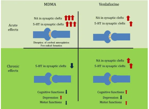
monoaminergic synaptic transmission, as a particular consequence of the desensitization of presynaptic autoreceptors, mainly 5-HT1A serotonergic- [112-114], but possibly also 5-HT1B serotonergic and α2 adrenergic receptors (for a review see [115]). Thus, MDMA and VLX act fundamentally different at the molecular level in the long-term (Fig 1).
The previously discussed alterations also suggest that MDMA and VLX may counteract each other functionally on the long-run, e.g. in motor- and cognitive functions. MDMA administration, as discussed in previous chapters, was usually related with negative effects on motor functions, like elevated reaction time [53], tremor or motor speed dexterity [55], while VLX treatment was associated with elevations in motor performance in healthy individuals [99].
Cognitive functions following MDMA use were also impaired, like decision making, learning and memory [7, 49, 52]. At the same time, pre-training administration of VLX showed heterogeneous effects in memory tasks, but post-training treatment improved performance in rats (for a review see [116]), while positive effects were also suggested on affective cognition (for a review see [117]).
Figure 1 The acute and chronic consequences of MDMA and venlafaxine administration in the serotonin and noradrenaline content of synaptic clefts and the possible subsequent functional alterations.
18
Acutely, both 3,4-methylenedioximethamphetamine (MDMA) and venlafaxine (VLX) enhance serotonin (5-HT) and noradrenaline (NA) levels, though in a different extent, in the synaptic clefts of frontal cortical neurons. On the long- term, they have markedly different effects. Even a single-dose MDMA administration causes a selective serotonergic neurotoxicity and thus, decreases in the 5-HT concentrations, while chronic VLX treatment, through the desensitization of 5-HT1A autoreceptors elevates synaptic 5-HT levels in the synaptic clefts. At the bottom of the figure some of the possible functional consequences of the altered serotonergic tone were listed. For references and further details see main text.
In addition, MDD in previous MDMA users is more common than among the general population [118, 119] thereby exposing them to antidepressant treatments later in life.
While it has been demonstrated by numerous studies that pretreatment with antidepressants may reduce MDMA-induced effects, probably by interfering with SERT (in the case of SSRIs) and NA transporter (in the case SNRIs and NRIs) [120-126], antidepressant administrations following MDMA use were only scarcely investigated.
Thompson et al. showed that 5-weeks long chronic FLX treatment was able to reverse MDMA-induced elevated anxiety and depression levels in the emergence test and in the forced swimming test (FST), respectively, but left reduced social interactions unchanged in rats [127]. At the same time, Durkin et al. showed reduced efficacy of chronic FLX treatment following previous MDMA administration [128]. Paroxetine binding sites were also altered in cortical regions as a response to a previous exposure of the drug [22]. Dark Agouti (DA) rats represent the human poor metabolizer phenotype. Since these animals have a less active variant of the CYP2D1 enzyme, corresponding to the human CYP2D6 responsible for MDMA metabolism, they are model animals for those human individuals, who are especially vulnerable to the neurotoxic effects of the drug [5, 28]. In DA rats, a challenge with FLX 6 months after an initial MDMA administration resulted in altered responses, namely, an elevated time of aggressive behavior, suggesting that SSRI antidepressant effects may be altered even after a single dose of MDMA and such a long time [129].
The above studies all impose the possibility that effects of antidepressants may be altered by previous MDMA use. The lack of further studies evaluating the interactions of previous MDMA and later antidepressant administration is surprising, since these aspects are not only important scientific questions, but may also influence therapeutic approaches in addicts. Antidepressants with a dual mechanism of action remained so far completely uninvestigated in similar experimental setups, despite the fact that they may be superior to SSRIs through their extended mechanism of action.
19
Because of the complex nature of possible alterations, and the mass of involved pathways and functions suggest a need again for a hypothesis-free approach. Thus, microarrays, able to measure whole-genome gene expression levels and create hypotheses following data acquisition instead before it, may have advantages over classical hypothesis-driven experimental setups for such purposes [130].
20
3. OBJECTIVES
1. Selective serotonergic toxicity and functional alterations on the long-run following a single-dose of MDMA were already described in the literature, but underlying chronic molecular changes remained so far uninvestigated in the FC region. In order to reveal which genes or pathways may be involved in the effects 21 days after MDMA administration, we performed a genome-wide microarray analysis in the FC of DA rats, the latter representing the vulnerable human individuals to the drug’s effects.
2. The real therapeutic efficacy of VLX treatment is only obvious after weeks suggesting adaptive processes on the molecular and network levels of cortical neurons, which remained poorly understood after chronic use. To fill this gap, we have performed a genome-wide microarray-analysis after a chronic 3-weeks long VLX treatment regimen in the FC of the DA rat strain to characterize the transcriptional background behind the drug’s therapeutic effects.
3. Following the analysis of MDMA’s and VLX’s effects individually (see 1. and 2.), we also investigated the genome-wide molecular changes after the combined treatment of a single-dose of MDMA and a subsequent 3-weeks-long VLX treatment in the FC of DA rats to reveal, whether a consecutive VLX treatment could compensate for the MDMA-induced changes, and to report, if and how a previous MDMA treatment may influence the effects of VLX on the molecular level.
21
4. METHODS 4.1. Animals
The animal experiments and housing conditions were carried out in accordance with the European Community Council Directive of 24 November 1986 (86/609/EEC), and the National Institutes of Health Principles of Laboratory Animal Care (NIH Publication 85-23, revised 1985) as well as special national laws (the Hungarian Governmental Regulation on animal studies, 31 December 1998 Act). The National Scientific Ethical Committee on Animal Experimentation and the Food Chain Safety and Animal Health Directorate of the Central Agricultural Office, Hungary (permission number: 22.1/3152/001/2007) approved the experiments.
Altogether 42 male DA rats (Harlan, Olac Ltd, Shaw’s Farm, Blackthorn, Bicester, Oxon, UK) were used, aged circa 8 weeks [126.71 ± 3.30 g (mean + SEM) at the beginning of the experiment]. The animals (four per cage) were kept in standard cages and under controlled environmental conditions (temperature 21 ± 1 °C, humidity: 40- 50%, 12 hour light-dark cycle starting at 6:00 a.m.). Food and drinking water were available for them ad libitum.
4.2. Drug Administration and Experimental Design
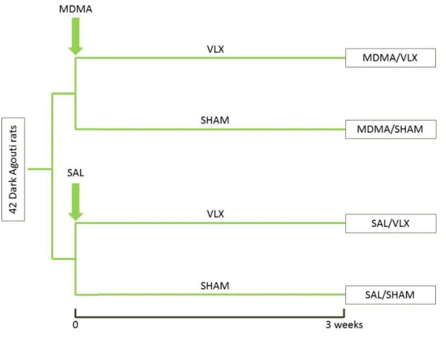
The animals were randomly assigned to four groups according to the treatment regimens (SAL/SHAM, MDMA/SHAM, SAL/VLX, MDMA/VLX, see. Fig. 2).
MDMA (Sanofi, Hungary, purity >99.5%), dissolved in 0.9% NaCl (SAL) at an equivalent dose of 15 mg/kg free base was administered intraperitoneally (i.p.) in a volume of 1 ml/kg to the MDMA treated animals. Control animals received SAL i.p. in equivalent volumes (1 ml/kg).
VLX (Egis Pharmaceuticals, Hungary) was dissolved similarly in 0.9% NaCl solution and Alzet 2001 osmotic minipumps (Durect Corp., CA, USA) were filled with the solution. In VLX treated groups these Alzet osmotic minipumps were inserted subcutaneously under the back skin of the animals half day after the initial injections to avoid acute serotonin-syndrome in previously MDMA treated rats. The pumps delivered 40 mg/kg VLX each day. All surgery was performed under halothane anesthesia, and all
22
efforts were made to minimize suffering of the animals. The control group underwent sham surgery/osmotic minipump insertion (containing saline) in a randomized manner (which will be abbreviated by SHAM to avoid confusion with the MDMA control group). The surgical procedures had to be repeated each week for 3 weeks, due to the limited volume of the osmotic pumps. After surgery, animals were always returned to their cages and were kept there until the next procedure.
During surgery two animals died and one has lost its pump, thus, altogether 39 animals went through the entirety of these procedures.
Figure 2 Experimental groups and setup.
Altogether 42 Dark Agouti (DA) rats were divided initially into two groups. One group of the animals received a single 3,4-methylenedioxy-methamphetamine (MDMA) injection (in a dose of 15 mg/kg), while the other saline.
Thereafter animals were further divided into two groups, receiving either chronic venlafaxine treatment (VLX, 40 mg/kg/die via osmotic minipumps) or undergoing sham surgery/implantation of saline filled minipumps. Thus, at the end of the experiments altogether 4 groups: SAL/SHAM, MDMA/SHAM, SAL/VLX and MDMA/VLX were created based on the different combinations of treatments. SAL – saline, SHAM – sham operation/treatment with saline filled minipumps.
23
4.3. RNA Extraction and Sample Preparation
The rats were sacrificed quickly by decapitation 3 weeks after the surgery. The brains were removed and 2 mm thick coronal sections were cut, and the FC regions were dissected according to Paxinos and Watson [131] (from bregma +1.7 to -0.3 mm) and stored at -80°C. TRIzol reagent (Ambion, TX, USA) in a volume of 1 ml was used for homogenization, according to the manufacturer’s instructions. Then the homogenized samples were centrifuged at 12000 x g at 4°C for 10 minutes, supernatant transferred to Eppendorf tubes and incubated at room temperature for 5 minutes.
Chloroform was added in a volume of 200 µl, the mixture was vortexed and incubated at room temperature for 2-3 minutes. After centrifugation at 12000 x g at 4°C for 15 minutes the upper aqueous phase was separated, mixed with 500 µl of isopropranol and incubated for another 10 minutes at room temperature. Following the centrifugation of the samples at 12000 x g at 4°C for 10 minutes the supernatant was removed and 1 ml 75% ethanol was added. The centrifugation was repeated at 7500 x g at 4°C for 5 minutes, the supernatant was removed, and again, 1 ml 75% ethanol was added.
Following the third centrifugation step (at 7500 x g at 4°C for 5 minutes), ethanol was removed and RNA pellets dried. Pellets, dissolved in 20 µl diethylpyrocarbonate treated-dH2O, were stored at -80°C until further processing.
Thereafter 1-2 µl of the solution were used for optical density (OD, 260/230 and 260/280 ratios) measurements to assess the quality and quantity of the samples. The OD ratios were measured for all samples, and in addition, randomly repeated to determine reliability of the quality measurements. No differences were observed. Samples with lowest RNA concentrations were excluded from further analysis in a way that finally all groups consisted of 8 animals. Two randomly selected samples from the same treatment group were pooled resulting in 4 pooled samples per treatment group. Thus, altogether 16 pooled samples (4 per treatment group) were sent for microarray analysis with the Illumina (San Diego, CA, USA) RatRef-12 v1 beadarray expression chip to Service XS (Leiden, Netherlands). At Service XS a purification process and quality control measurements with Agilent Bioanalyzer and Nanodrop spectrophotometer were made and one pooled sample from the MDMA/VLX treated group had to be excluded due to degradation.
24
4.4. Data Analysis
The raw data returned from Service XS were processed with beadarray [132], preprocessCore [133] and puma [134] Bioconductor [135] packages for R statistical programming language [136] as described in [137]. Beadarray package was used to analyze bead level data and to give the flexibility to use other options than the standard Illumina methods provided by Illumina’s BeadStudio software. The standard Illumina summarization method provides a point estimate for each expression level of each bead, whereas the uncertainty of such estimates remains excluded from further analysis. Since microarray experiments are associated with low precision probe-level measurements (called probe-level measurement error) relevant results may be biased by the resulting noise. To avoid such bias, we used the puma (Propagating Uncertainty in Microarray Analysis) package, which uses probe-level measurement error in subsequent analyses, however, has the prerequisite of special data formats.
According to our original aims, to reveal molecular alterations related to pharmacological manipulations we have chosen wider inclusion criteria and used “log = TRUE; n = 10” settings by creating the summary data for each bead via the createbeadsummaryData, allowing excluded outliers by standard Illumina method to be included in our downstream analyses, (similar settings were also used by others [137]).
For normalization, the quantile normalization method was used via the preprocessCore package. The backgroundCorrect method in the puma package used was set to
“minimum”, meaning that any intensity equal or less than zero after background subtraction was set equal to half the minimum of the positive corrected intensities for that array. Additionally, pumaComb, pumaDE, and write.rslts functions with default settings were used to create summary tables and statistical values used in the subsequent analyses. These calculations resulted in the so called minimum probability of positive log-ratio (MinPplr), a Bayesian-based value for significance [138], besides also providing the fold change values between different comparisons. MinPplr has been shown earlier to be superior to classical methods, as a result of the inclusion of probe- level errors in microarray experiments [138]. To further address the problems of multiple testing, which is the result of the high number of statistical hypothesis tests
25
(more than 22000 in our experiment) performed [139], we used a stricter criteria and changes were considered statistically significant only when the MinPplr was below 0.001 in the MDMA and double treated group, and 0.005 in the VLX group. This is a usual reduction used in microarray experiments. While several methods exist to correct for multiple hypothesis testing, like the Bonferroni-method or the family-wise error rate, which may compensate for the false positive results, they may be very restrictive because of the assumption that every hypothesis test is independent from the others (for example the Bonferroni-correction divides the significance criterion [usually 0.05] by the number of tests performed [139]). Thus, we considered them inappropriate for our original aims, namely, the discovery of so far undiscovered pathways in the effects of the pharmacological agents. In case of the individual genes we haven’t used correction for multiple testing, rather decreased the limit for significance to the mentioned levels.
In summary, we used 3 different methods in the analysis of individual genes to reduce the possible biases known to be attributed to microarray experiments. First, we used BeadArray chips, which were designed to reduce spatial artefacts on microarray plates because of the random localization of the beads on the plate. Second, we included probe-level errors in downstream analyses with the puma package to address the problem of single point estimations of expression values, thereby enhancing the accuracy of the results. This method also allowed us to calculate the MinPplr value, which is a better predictor of the probability of a different expression than conventional p-value. Third, we have reduced the significance criteria to lower levels to address the elevated possibility of false positive results resulting from multiple hypothesis testing.
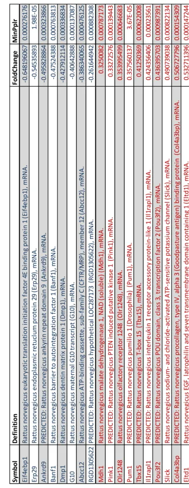
Please note, that throughout the dissertation genes will not be presented, which were only unconfirmed loci at the time of the analysis. We assumed that such genes may unnecessarily add to the complexity of the presented data and raise concerns about the reliability of results, since these genes were only annotated based on sequence homology or similarity with known proteins without any further validation.
4.4.1. Pathway analysis of the MDMA treatment
Besides calculating individual significance levels for each gene, gene set enrichment analysis (GSEA) was performed using GSEA version 3.1 from the Broad Institute at MIT (http://www.broadinstitute.org/gsea) [140, 141]. GSEA is a widely used
26
method for the discovery of altered molecular pathways instead of individual genes. The basis of GSEA is the use of the so called gene sets, which contain genes grouped together by similarities between them, like common regulatory patterns, biological functions, etc. Based on these similarities multiple gene set databases exist, called molecular signature databases (MSigDBs). After the definition, which gene set database to use, in the next step, GSEA creates a ranked list (based on the respective t-values of the genes after performing hypothesis tests for every transcript between two treatment groups,) to order all the genes on the microarray platform and thereby evaluating their relative expression compared to other transcripts. Thereafter a cumulative value for a given gene set is created; this is the so called nominal enrichment score (NES), which is a summary of the relative expression values of the individual genes belonging to a given gene set [139, 140]. The NES can, thus, be used to assess the magnitude of the alterations of genes grouped by certain criteria.
In the current analysis, gene identifiers used both in the array dataset and in the gene sets, were gene symbols. The entries in the data set were collapsed to gene symbols with the median expression value used for the probe sets. Gene sets were restricted to contain between 15 and 500 genes, since smaller or larger sets may result in statistical artefacts. T-test was used for ranking genes and the permutation type was the gene set, because of the small sample size per treatment group. Number of permutations was set to 1000 with the seed of permutation: 149. Other basic and advanced fields were left as default.
Gene sets (GMT format) from the MSigDB for C5 category (gene ontology [GO] gene sets) were used and in addition, neuronal function related gene sets were selected from the GO homepage (www.geneontology.org) manually.
Normalized enrichment score (NES), nominal p-value and false discovery rate (FDR) were calculated for the gene sets. The latter represents a correction for multiple testing (discussed in 3.4) and basically gives an estimation about how much of the significant results may be false positive [139, 142]. A gene set with a nominal p-value
<0.05 and FDR <0.25 was considered statistically significant.
Network visualization and analysis using enrichment results was done using Cytoscape 2.8.3. with its plugin “Enrichment Analyzer”. Following cut-off rates were
27
used: similarity coefficient cut-off 0.1, p-value cut-off 0.05 and FDR cut-off 0.25 [143, 144].
4.4.2. Pathway analysis of the VLX treatment
Following the evaluation of the effects of MDMA, in case of the VLX treated groups, besides of the above mentioned methodology, we also used textmining methods in NCBI’s medical databases to create individual gene sets related to VLX’s well- known positive effects in neuropathic pain and migraine [145, 146]. To estimate the relations between genes and the latter diseases the hits of Pubmed queries “<gene name> AND (pain OR neuropath* OR nocicept* OR migraine)” were counted. We wrote R scripts [136]with the genome wide annotation database for Rattus Norvegicus [147] and the GO annotation database [148] from Bioconductor [135] and reversely mapped the genes with hits into the GO hierarchy. In addition we also used the Pain Genes Database (http://www.jbldesign.com/jmogil/enter.html) to create two additional gene sets [149]. This database contains pain related results from knockout mice. We have created one gene set from the genes resulting in decreased nociception (PAIN_DB_DOWN) and one including those genes which knockout resulted in elevated nociception (PAIN_DB_UP) in mice. All the resulting gene sets (altogether 1781, among them 1454 from the MSigDB C5 and 327 individually selected ones) were included in the analysis widening the possibility of the discovery of new pathways.
4.4.3. Analysis of the combined treatment
Following the analyses of the individual treatments we addressed if and how MDMA and VLX may interact in the FC of DA rats. The GSEA results of the double treated animals with the above mentioned methodology resulted almost exclusively in seemingly additive effects. Therefore, we tried to identify those genes which could reflect interactions between the two treatments by ANOVA.
We have used the R package “lmdme: linear model decomposition for designed multivariate experiments” [150], which uses linear models to compare expression values between treatments and calculate interactions between them. The method was eligible for the use of ANOVA in unbalanced designs, the latter being a limitation of
28
several other methods [150]. The input table contained the normalized results of the experiment obtained following the preliminary filtering of the raw data according to the description in previous chapters. We used a significance criterion 0.001 to obtain statistically relevant results to reveal which genes (unconfirmed loci were excluded) may reflect interactions in the double treated group. The figure representing the result was created with the R package ggplot2 [151].
4.5. PCR Validation
Altogether 19 RNA (Table 1) products were validated on a Fluidigm GEx real-time polymerase chain reaction (PCR) 96x96 array (San Francisco, CA, USA).
Table 1 Validated RNA products from the polymerase chain reaction experiment as presented on the GEx array.
The table shows the validated genes for the polymerase chain reaction experiment on the plate. The mRNAs validated were selected based on the results of the microarray experiment of the single-dose 3,4-methylenedioxy- methamphetamine treatment and on the preliminary results for the venlafaxine treatment. Cyclophilin A (PPIA) was selected as internal control. Camk2b - calcium/calmodulin-dependent protein kinase II beta; Camk2g - calcium/calmodulin-dependent protein kinase II gamma; Camk2n2 - calcium/calmodulin-dependent protein kinase II inhibitor 2; Cnr1 - cannabinoid receptor type 1; Dao1 - D-amino acid oxidase; Gria3 - glutamate receptor, ionotropic, AMPA3; Grin2b - glutamate receptor, ionotropic NMDA2B; Hsd11b - hydroxysteroid 11-beta dehydrogenase 2; Igf2 - insulin like growth factor 2; NAC – non assay control, Nr2f6 - nuclear receptor subfamily 2, group F, member 6;
NRG1 - neuregulin 1; P2ry6 - pyrimidinergic receptor P2Y6, G-protein coupled, Pla2g2c - phospholipase A2, group 2C; Sipa1l1 - signal-induced proliferation-associated 1 like 1; Slc38a5 - solute carrier family 38, member 5; Slco1a5 - solute carrier organic anion transporter family, member 1a5; Syt8 - synaptotagmin 8; Tap1 - transporter 1, ATP- binding cassette, sub-family B.
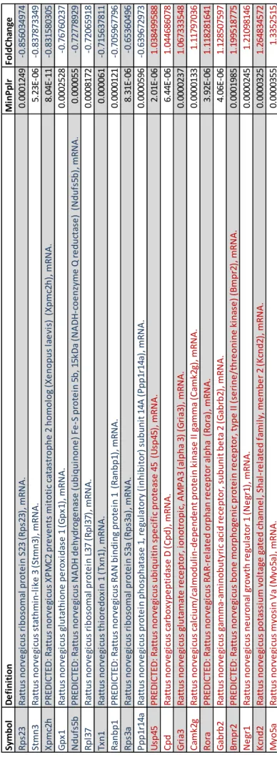
Table 2 Fold change values for selected genes presented in the thesis on the beadarray and PCR platforms on a logarithmic scale.
The table shows base 2 logarithmic values of fold changes when compared to the SAL/SHAM group for validated and discussed genes on the two different platforms, the Illumina RatRef-12 v1 beadarray expression chip and the Fluidigm GEx real-time polymerase chain reaction (PCR) 96x96 array, respectively. Camk2b - calcium/calmodulin- dependent protein kinase II beta; Camk2g - calcium/calmodulin-dependent protein kinase II gamma; Gria3 - glutamate receptor, ionotropic, AMPA3; Grin2b - glutamate receptor, ionotropic NMDA2B; MDMA - 3,4-
1 2 3 4
A NAC NAC NAC NAC
B CNR1 IGF2 PPIA TAP1
C SLCO1A5 P2RY6 NRG1 CAMK2N2
D GRIA3 CAMK2G CAMK2B SIPA1L1
E NAC NAC NAC NAC
F GRIN2B DAO1 HSD11B1 NR2F6
G SLC38A5 NAC NAC NAC
H SYT8 PLA2G2C NAC NAC
29
methylenedioximethamphetamine, single-dose, 15 mg/kg i.p.; PCR – polymerase chain reaction; VLX – venlafaxine, 3 weeks long, 40 mg/kg via osmotic minimpumps.
Genes were selected based on analyses of the MDMA and preliminary results of the VLX treatments. The Taqman Gene Expression assays for the appropriate RNAs were obtained from Applied Biosystems (Carlsbad, CA, USA) and directly sent to Service XS since the validation experiment was also performed by them. Each sample was used in duplo (200 ng and 500 ng) following quality control measurements (samples with already degraded or insufficient amount of RNA were excluded). Upon receiving the raw results individually written R scripts built around the cor.test function with default settings were used for the comparison of the fold change values between the microarray and PCR data.
The Pearson correlation coefficients were 0.384 (0.491) and 0.389 (0.465) in the MDMA-, 0.438 (0.552) and 0.421 (0.572) in the VLX-, 0.421 (0.589) and 0.441 (0.602) in the MDMA/VLX for the 200 ng and 500 ng samples, respectively (the Spearman rank correlation coefficients are given in parentheses). In all cases p-values for Pearson’s correlation coefficients were below 0.05 (and 0.005 for Spearman rank correlation). For genes discussed and explicitly presented in the thesis the fold change values on a logarithmic scale measured by the two different methods are given in Table 2.
4.6. Availability of supporting data
As a usual requirement of microarray experiments [152], data supporting the results have been deposited in NCBI's Gene Expression Omnibus (GEO) under the GEO Series accession number GSE47541 (http://www.ncbi.nlm.nih.gov/geo/query/
acc.cgi?acc=GSE47541).
MDMA VLX MDMA/VLX MDMA VLX MDMA/VLX
Camk2g 0.941 1.047 1.173 0.705 0.689 1.164
Grin2b 0.446 0.579 0.471 0.28 0.287 0.42
Camk2b 0.935 1.139 1.091 0.704 0.289 0.392
Gria3 0.492 0.856 1.001 0.327 0.51 0.941
log2 fold change on microarray (vs. SAL/SHAM) log2 fold change in PCR (vs. SAL/SHAM) Genes
30
5. RESULTS 5.1. MDMA
5.1.1. Differentially Expressed Genes after MDMA treatment
Three weeks following an MDMA administration 155 genes were differentially expressed, among them 66 were up- and 89 downregulated, respectively. After the correction for unconfirmed loci altogether 87 genes remained, 51 down- and 36 upregulated, respectively (MinPplr < 0.001). Significantly upregulated genes included some related to calcium signaling pathways (Camk2g and Camk1g) and an NMDA-type glutamate receptor, NMDA2B (Grin2b). At the same time, genes related to response to neuronal stress, like the alpha subunit of the heat-shock protein 1 (Hspca) and the heat- shock factor 2 (Hsf2) were downregulated, in addition to the high-affinity glial glutamate transporter (Slc1a3).
31
5.1.2. Gene Set Enrichment Analysis following the MDMA treatment According to the GSEA analysis 55 gene sets were enriched after the single-dose MDMA treatment, (including literature-based and Msig DB C5 gene sets). The upregulated gene sets were the “response to hyperoxia”, “positive regulation of synapse assembly”, “regulation of synaptic plasticity”, “growth factor activity” and “dendrite development” gene sets. Thus, dendrite and synapse development and growth factor activity were among the upregulated processes 3 weeks after MDMA administration [153].
At the same time, gene sets related to protein synthesis and localization (e.g.
“translation”, “macromolecule biosynthetic process” or “protein localization”), transmembrane transport (e.g. “organic acid transport”, “carboxylic acid transport”,
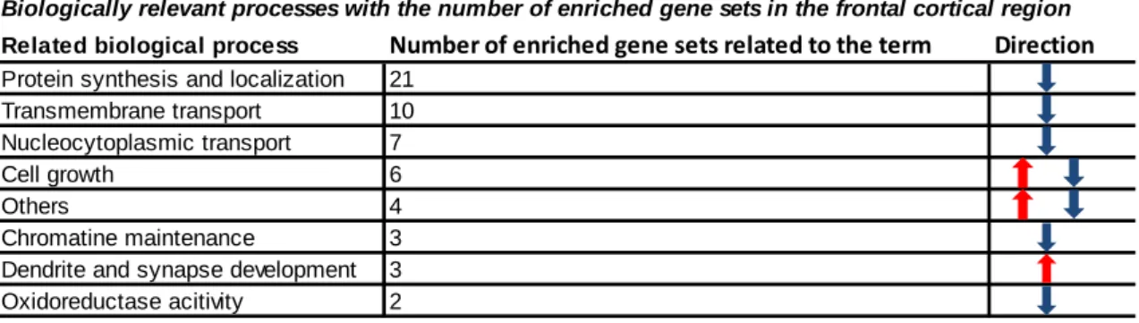
“amino acid transport”) nucleocytoplasmic transport (e.g. “nucleocytoplasmic transport”, “pattern specification process”, “protein import into nucleus”) were downregulated, along with those involved in chromatin maintenance and oxidoreductase activity (see Table 3 for changes with the highest and lowest NESs). To give a biological perspective to these alterations, the gene sets were grouped by biological and neuronal aspects (Table 4). Thus, it became clearly visible that the most prominent alterations were related to protein synthesis and protein localization (with 21 significantly dysregulated GO sets) and 10 gene sets were involved in transport processes [153].
(Please note, individually selected gene sets used in all analyses can be downloaded from the Supplementary table S2 from [153] or at http://www.ncbi.nlm.nih.gov/ pmc/articles/PMC3902429/)
32
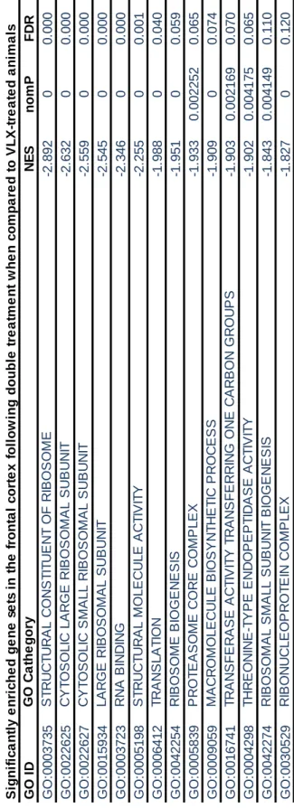
Table 3 The significantly enriched top gene sets in the frontal cortex of Dark Agouti rats 3 weeks after a single dose of MDMA earlier.
The table shows gene sets altered by a single dose of 3,4-methylenedioxi-methamphetamine (MDMA, 15 mg/kg) 3- weeks earlier. In the table Gene Ontology (GO) identifiers, their names, the nominal enrichment scores (NES), nominal p-values (nomP) and the false discovery rates (FDR) are presented. The NES gives an overall estimation about the magnitude of the dysregulation for given set. Adapted from [153].
Table 4 Biologically relevant processes according to the gene set enrichment analysis and individual considerations in MDMA treated animals.
Following gene set enrichment analysis (GSEA) gene sets were ordered into relevant biological processes to provide a more comprehensive overview about the effects of a single dose of 3,4 methylenedioxy-methamphetamine (MDMA) 3-weeks earlier, when compared to the control group. In the table biological processes, the number of related gene sets and the directions of alterations are presented (red arrow – upregulations, blue arrow – downregulations) Adapted from [153].
GO ID GO Cathegory NES nomP FDR
GO:0015849 ORGANIC ACID TRANSPORT -1.750 0 0.175
GO:0046942 CARBOXYLIC ACID TRANSPORT -1.737 0 0.122
GO:0006412 TRANSLATION -1.715 0 0.102
GO:0009059 MACROMOLECULE BIOSYNTHETIC PROCESS -1.702 0 0.081 GO:0007389 PATTERN SPECIFICATION PROCESS -1.650 0 0.147
GO:0003723 RNA BINDING -1.640 0 0.137
GO:0016836 HYDRO LYASE ACTIVITY -1.637 0 0.122
GO:0006886 INTRACELLULAR PROTEIN TRANSPORT -1.632 0 0.118 GO:0006325 ESTABLISHMENT AND OR MAINTENANCE OF
CHROMATIN ARCHITECTURE -1.624 0 0.116
GO:0016358 DENDRITE DEVELOPMENT 1.383 0.049 0.142
GO:0008083 GROWTH FACTOR ACTIVITY 1.387 0 0.165
GO:0048167 REGULATION OF SYNAPTIC PLASTICITY 1.480 0.025 0.088 GO:0051965 POSITIVE REGULATION OF SYNAPSE ASSEMBLY 1.521 0 0.073
GO:0055093 RESPONSE TO HYPEROXIA 1.547 0.034 0.097
Significantly enriched gene sets in the frontal cortex after single-dose MDMA administration
Related biological process Number of enriched gene sets related to the term Direction Protein synthesis and localization 21
Transmembrane transport 10 Nucleocytoplasmic transport 7
Cell growth 6
Others 4
Chromatine maintenance 3 Dendrite and synapse development 3 Oxidoreductase acitivity 2
Biologically relevant processes with the number of enriched gene sets in the frontal cortical region
33
5.2. Venlafaxine
5.2.1. Differentially expressed genes following chronic venlafaxine treatment
Comparison of the individual transcripts resulted in 381 differentially expressed genes, and 222 remained following the correction for unconfirmed loci in the VLX treated group compared to the SHAM controls (MinPplr < 0.005). Among them 23 were already experimentally linked to depression or its therapy. From these, Ace, Cox17, Faah, Gfap, Pyy, Vdac1 were downregulated, while Ascl1, Bcl2, Camk2b, Camk2g, Cd47, Gad2, Gnaq, Gria3, Grin2b, Hcn1, Negr1, Ntrk2, Ntrk3, Ppp3r1, Sv2b, Syn2, Synj2, Vamp1 were upregulated [154].
In addition, 23 genes could have been identified with a possible role in MDD based on the pathomechanism or current antidepressant therapies (candidate genes). The Cntn2, Dpp4 were downregulated while Cdh22, Clstn2, Enpp1, Epha5a, Gas2, Glp1r2, Grin2a, Kif1b, Kif2b, Kif5a, Lphn1, Mmp9, Myo5a, Pdpk1, Pex2, Prdx1, Rims1, Rph3a, Slc2a4, Ucp3, Unc13b [154]. See Table 5 for genes with the highest and lowest fold change values within significantly altered genes. Some prominent genes altered by other antidepressant treatments, like galanin and its receptors, or Galp remained unaltered in the FC of DA rats [155].