Allosteric Inhibitors
R. W . Miller
I. Introduction 2 II. Limiting Models for Allosteric Inhibition 3
A. Models Requiring Changes in Conformational State 9 B. Models Postulating Multiple Reaction Pathways or Time-Dependent
Enzyme Activity (Transient Forms) 10 III. Methods That Probe Mechanisms of Allosteric Inhibition 13
A. Kinetic Methods 13 B. Chemical Methods 14 C. Physical Probes 15 IV. Variation of Allosteric Properties among Source Organisms 16
V. Classes of Allosteric Inhibitors 17
A. Nucleotides 17 B. Amino Acids 22 C. Carbohydrates 22 D . Inorganic Ions and Miscellaneous Inhibitors 23
V I . Allosteric Inhibition of Specific Enzymes 23
A. Lactate Dehydrogenase 24 B. Malic Enzymes 25 C. Isocitrate Dehydrogenase 26
D . L-Glutamate Dehydrogenase 27 E. Aspartate Transcarbamylase 30 F. Glutamine Synthetase 34 G. Phosphorylase b 35 H. Phosphofructokinase 37 VII. Allosteric Inhibition in Enzyme Complexes and Membrane Systems 38
References 40 1
I. INTRODUCTION 1
Feedback inhibition has become a well-established concept in the search for metabolic control mechanisms that regulate the rate of metabolite synthesis. The discovery that end products of biosynthetic pathways may inhibit directly the initial reactions of the pathway provides an attractive explanation for the precise metabolic homeostasis that is essential in all organisms. However, direct in vivo observations
of enzymic feedback phenomena generally are not possible due to prob
lems of transport of metabolites across membranes and the presence of other regulatory processes such as enzyme repression. Therefore, the study of the effects of inhibitors on the quaternary structure, tertiary structure, catalytic activity, and affinity for substrates of purified enzymes has produced a great deal of information as to how inhibitors may modulate the rates of catalyzed processes.
A particularly significant type of enzyme inhibition has been examined intensively in recent years and has emerged as a plausible mechanism for the general hypothesis of feedback regulation of metabolic trans
formations. Enzymes that display anomalous order (higher than one) with respect to a subtrate or coenzyme produce sigmoidal plots of reac
tion velocity versus ligand concentrations. Inhibitors are often ob
served to increase the order of the reaction with respect to substrate or to give inhibition patterns that lead to a change in the VmRX, Km
(substrate concentration which gives half-maximal reaction velocity, referred to as S0.5), or other parameters that describe binding and rates of catalysis by enzymes. Inhibitors that bind at a site remote from the catalytic site and produce effects of this type are referred to as allosteric inhibitors or negative effectors as opposed to compounds that modify, sterically block, bind at, or otherwise interfere directly with the catalytic site. Depending on the type of chemical transformation inhibited (i.e., single- or multiple-substrate reactions), characteristic and diagnostic kinetic data are obtained which may lead to detailed physical and chemical studies. These combined methods have greatly increased knowledge of regulatory phenomena. In this area it is somewhat artificial to distinguish between allosteric inhibitors and allosteric activators since similar mechanisms have been proposed (1) for both. However, since
1
Standard abbreviations are used for purine and pyrimidine nucleotides. N A D and N A D P are used for nicotinamide adenine dinucleotide and nicotinamide adenine dinucleotide phosphate, respectively.
allosteric inhibitors account for many cases of feedback regulation this chapter will emphasize the former. Table I lists allosteric inhibitors and the enzymes they may control. It is apparent that by far the largest number of compounds represented are purine and pyrimidine nucleotides. Indeed, Atkinson (2, 3) has proposed that many enzymes respond to the relative concentrations of adenine nucleotides (AMP, ADP, ATP, cyclic 3',5'-AMP) rather than to the concentration of a single inhibitor or activator.
Many enzymes have more than one negative effector. While there are many well-known examples of feedback inhibition listed in Table I, there is no apparent limitation of allosteric inhibition to end products of metabolic pathways. Inorganic anions and cations, antibiotics, com
plex carbohydrates, and other seemingly unrelated compounds can bind at remote receptor sites and cause allosteric effects which, while they may represent control mechanisms in the general sense, are not associated with feedback control of metabolism. However, metabolic regulation is clearly an extremely complex and subtle process, and relationships between biosynthetic reactions and their allosteric inhibitors are not necessarily always as obvious as end-product inhibition.
II. LIMITING MODELS FOR ALLOSTERIC INHIBITION Several limiting mechanistic models have been proposed to explain the behavior of allosteric enzymes. The models attempt to account for the sigmoidal shape of plots of reaction velocity (or fractional binding- site saturation) against substrate or effector concentration. Data of this type have generally suggested cooperative interactions between subunits of an oligomeric protein where the binding of effector molecules at a regulatory site may change the binding constant for substrate (i£-type system) or may influence the activity of the catalytic site (V-type sys
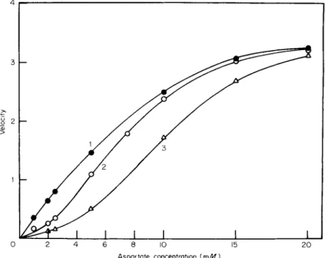
tem) or both. Curve 2 of Fig. 1 ( 4 ) shows cooperative behavior for a well-documented allosteric enzyme, aspartate transcarbamylase. This enzyme catalyzes the first step in pyrimidine biosynthesis and is in
hibited by the end product of that pathway, cytidine triphosphate.
Cytidine triphosphate is a true allosteric inhibitor; that is, it decreases the reaction rate at low substrate concentrations by increasing the dependence of the rate on the concentration of substrate molecules.
Control curve 2 of Fig. 1 shows a dependence on substrate concentra
tion that is anomalous since ordinary substrate dependence would
give a typical hyperbolic curve. An effector molecule could modify this cooperative effect so as to produce more or less cooperativity. The nega
tive effector CTP increases the order of the dependence of activity on substrate concentration in the case of aspartate transcarbamylase (curve 3 ) . The cooperative interactions associated with molecules of the same ligand are designated homotropic, while interactions involving the in
hibitor (CTP) or the activator (AMP) and the substrate are hetero
topic. The allosteric activator AMP changes the curve to a rectangular hyperbola which is the response expected in the absence of substrate- induced cooperative interactions. Heating the enzyme also eliminates cooperativity (Curve 1, Fig. 1 ) . With the native enzyme the rate, in the absence of ligands other than substrate, is slower than expected from a normal hyperbolic response. This effect suggests that the substrate must
4 r -
8 10 A s p a r t a t e concentration (m M )
2 0
FIG. 1. Dependence of reaction rate on aspartate concentration at p H 7.0.
Velocity expressed as units of activity per milligram of protein X 10
3
. Legend:
O , native enzyme; # , heated enzyme; and A , native enzyme in the presence of 0.1 m M C T P . Assay mixtures contained 0.05 M tris at p H 7.0, 3.3 m M carbamyl phosphate, asparatate as indicated, and 0.75 tig enzyme protein/ml. [Reprinted from Weitzman and Wilson (4) b y permission of the copyright owner. Copyright (1966) by the American Society of Biological Chemists.]
convert the enzyme to a more active form for maximum reaction veloc
ity to occur and is designated positive homotropic cooperativity. If in the presence of an activator the rate of reaction at low substrate con
centration is greater than that expected from a hyperbolic relation
ship, the implication is that increased substrate concentrations convert the enzyme to a less active form. This type of interaction is labeled negative homotropism.
Since both types of behavior are reported for some enzymes it is im
portant to diagnose the type of cooperativity present in a given system.
Also, some models allow only positive homotropic cooperativity and kinetic analysis enables the investigator to distinguish between models.
Allosteric inhibitors lead to more positive homotropic cooperativity. In this chapter inhibitors falling in the category of effectors that change cooperativity between substrate molecules by binding at a site remote from the catalytic center will be considered mainly. Inhibitors such as reaction products that bind or interfere directly at the catalytic site, some of which also may be described as feedback inhibitors, will not be considered. Also, noncompetitive inhibition (i.e., change of VmRX), which is often observed with multisubstrate enzymes, will not be dis
cussed unless evidence for interaction of the inhibitor at a separate allosteric site or for an effect on the cooperative behavior of one of the substrates is available. Noncompetitive inhibition can result from direct interaction with the active site in multisubstrate cases. Just as some allosteric activators may produce negative homotropic interactions for substrate molecules, allosteric inhibitor molecules may exhibit nega
tive or positive homotropic interactions depending on whether increasing the concentration of the inhibitor produces more or less drop in activity than expected from a normal hyperbolic inhibitor response. Again, diag
nosis of the type of cooperativity for the inhibitor being observed is important in ascribing kinetic phenomena to an appropriate mechanistic model.
Allosteric inhibition may be competitive, in the sense that it is com
pletely overcome at high substrate concentrations (pure X-type system), or partially noncompetitive. Lineweaver-Burk plots obtained with allo
steric enzymes may be straight lines but are generally curved [see Fig.
2 (5) ]. Thus, in the presence or absence of heterotropic ligands a hyper
bolic response of reaction velocity to substrate concentration yields a straight Lineweaver-Burk plot, while positive homotropic interactions give an upward curvature and negative homotropism causes a downward curvature. Mahler and Cordes (5a) and Farago and Denes (5b) give
further graphical information that is useful in identifying the different types of cooperative interactions.
1 1 1 L ' 1 • . 1 L _ 0 1 2 3 0 0.1 0 . 2 0 . 3 0 . 5 0 . 7 0 . 9
1 / [ N A D ] ( m / J ^ "
1
) 1 / [ P y r u v a t e K r r W "
1
FIG. 2. Inhibition of Pythiam D(—)-lactate dehydrogenase b y G T P . (a) Coopera)
tive binding of N A D H in the presence of G T P ; ( b ) noncompetitive inhibition by G T P with respect to N A D as varied ligand; (c) cooperative binding of lactate in the presence of G T P ; (d) noncompetitive inhibition by G T P with respect to pyruvate as varied ligand. The concentration of enzyme used in experiments (a) and (d) was 3 fig and in experiments (b) and ( c ) , 12 fig. Concentration of G T P as specified in the figures. [Reprinted from LeJohn (5) by permission of the copyright owner. Copyright (1971) b y the American Society of Biological Chemists.]
The cooperative binding of ligands to a protein such as hemoglobin is described by the empirical Hill equation, which relates the fractional saturation of binding sites to the ligand concentration and the Hill coefficient n. A commonly used method for assessing cooperativity from enzyme kinetic data employs a modified form of the Hill equation where the reaction velocity v is given by
v = Vma*S"/K +S» (1)
log (v/Vm^ - v) = n log (S) - log (K) (2a) log fa/Fmax - Vi) = log {K') ~ 7l' log ( / ) (2b)
Here, Fm ax is the maximum reaction velocity at infinite substrate concen
tration in the absence of inhibitors, and S is the substrate concentration.
Reaction order is designated by the Hill coefficient n while K is a con
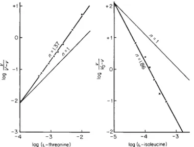
stant. Many sigmoidal curves yield straight lines when replotted by this method [Eq. (2a) ] . The coefficient n is the slope of the line obtained when the log of the kinetic parameter is plotted as shown in Fig. 3 (5c, 6).
Under certain conditions n could be interpreted as being equal to the number of cooperative binding sites. However, this assumption is theo
retically sound only when the total number of effector sites on an enzyme molecule is large relative to the number that actually bind effectors under a given set of conditions (7). Since this prerequisite is seldom fulfilled, n is often not as large as the number of binding sites. In the case of aspartate transcarbamylase the number of substrate binding sub- units was estimated to be four while later evidence obtained by complete dissociation (8, 9) showed that there were six.
The Hill equation may also be used to plot data obtained with negative effectors when substrate concentration is held constant at a saturating level and inhibitor concentration (/) is varied. In this case negative slopes are obtained [Eq. (2b)], but the magnitude of n remains greater than one for systems in which there is positive cooperativity between
FIG. 3 . L-Threonine deaminase activity as a function of the concentration of substrate (left) and allosteric inhibitor (right). [Reprinted from M o n o d et al.
(6c) b y permission of the copyright owner. Copyright 1963 b y Academic Press.]
inhibitor molecules. Values of n less than one again would indicate nega
tive homotropic cooperativity for the inhibitor.
Ligand binding functions may also be described by equations of the form
1 c + dS + eS
2
aS + bS* (3)
where y is the fractional saturation of binding sites and S is the free- ligand concentration (10). Reciprocal plots of 1/y against 1/S are curved upward in cases where positive homotropic cooperativity is present. The shape of the reciprocal plot is given by the sign of the second derivative
(W),
+ ( 4 )
where F = a 2
e + b 2
c - abd
which is determined by the sign of the factor F, a combination of con
stants. If F is negative, downward curvature of plots is present and negative homotropic effects are indicated. Some site interaction hypothe
ses are symmetrical and allow negative values of the second derivative while others do not. An analysis of this type was applied to glutamate dehydrogenase where the coenzyme NAD gives downward-curved recip
rocal saturation plots, indicating that the degree of saturation at low ligand concentration is higher than would be expected from a normal hyperbolic saturation phenomenon (10). Another interpretation of this negative homotropic process would be an apparent activation of the enzyme at high cofactor concentration. However, the rate of change of saturation or reaction velocity may be higher than expected throughout the entire concentration range.
In one type of model system the binding of effector molecules at specific regulatory sites is postulated to alter the observed rate of the catalyzed reaction through concerted or stepwise changes in three-dimen
sional structure and transitions between conformational states. Such changes may or may not be reflected in the state of aggregation of the subunits. The models generally are applied to enzymes that exhibit cooperative interactions between the subunit protomers. Ligands thus alter the interactions between the subunit peptide chains of an oligomeric enzyme. A second type of mechanism accounts for apparent cooperative effects through combinations of different reaction pathways (11) or as time dependence of enzyme activity (12). In theory these effects may be observed even with single-site enzymes.
A. Models Requiring Changes in Conformational State
Monod et at. (1) have proposed that data indicating cooperative effects fit a model that requires an existing equilibrium between two conforma
tional states of an enzyme. This equilibrium may be shifted by binding of allosteric ligands to either form. The R state may represent the reactive form of the enzyme. A ligand that binds preferentially to the T state thus inhibits the catalyzed reaction by shifting this equilibrium to the right.
A positive effector (substrate or activator) shifts the equilibrium to the active form by preferentially binding to the R state,
R ^ ± T
The protomers of the oligomeric enzyme may be identical and bind only substrates, or they may be nonidentical and have specific binding sites for substrate and negative or positive effectors. When a bound ligand molecule enhances the affinity of binding of further molecules of the same ligand, cooperativity is said to be positive homotropic, as mentioned previously. When a nonidentical ligand (effector) changes the affinity of binding of another ligand, the interaction is said to be hetero
topic and may be positive or negative. Heterotropic interactions (syner
gistic effects) may be observed between two different ligands which are both inhibitors. Molecules of a single allosteric inhibitor would be ex
pected to show homotropic interactions in bringing about a physical change in the quaternary structure of the enzyme. Some instances of product inhibition appear to be due to an allosteric effect. In this case the phenomenon still would be termed a heterotropic negative interaction, since the inhibitor is nonidentical with the starting substrates.
In the model of Monod et al. the transitions between the R and T forms are required to be concerted; that is, for a given enzyme molecule, the binding of an effector molecule encourages the transition in all of the protomers to take place without the formation of stable hybrid states containing mixtures of protomers in the R and T states. The concerted model does not allow negative homotropic interactions. The effectors may exert their influence through weak or noncovalent forces such as electronic forces or conceivably could even bind covalently where the binding reaction is enzyme catalyzed and equilibration is therefore rapid.
Binding may cause small physical displacements in the positions of key amino acid residues which shift the equilibrium between the conforma
tional states. Actual details of structural transitions are known in only a very few cases, although allosteric interactions often correlate with
alterations in bonding between the subunit protomers. In the original theory, the authors of this model considered only exclusive binding of effectors (1) in discussing heterotropic effects including negative interac
tions. That is, the effector ligand would bind to only one of the states.
General nonexclusive binding (binding of heterotropic effectors to both states with unequal affinities) was later considered (13). This modifica
tion limits the extent to which the equilibrium may be shifted by effectors.
The equilibrium would be pulled to completion in the case of exclusive binding in the presence of excess effector. Nonexclusive binding of effectors considerably extends the range of kinetic data that may be accommo
dated by this hypothesis. The postulation of a single conformational equilibrium is, of course, a limiting case. The presence of several concerted transitional equilibria in an enzyme system would be more complex but probably would be expected in most allosteric enzymes.
Koshland et al. (14) have proposed a different binding model to ac
count for allosteric effects. According to the induced-fit hypothesis, effectors (negative or positive) may induce stepwise conformational changes in the enzyme protein which can in turn cause higher- or lower- affinity binding for further molecules of substrate. This model allows nonexclusive binding as well as hybrid, i.e. partially changed, states and also allows negative homotropic cooperativity (Ha). The essential differences between these related models are illustrated in Fig. 4 (15).
B. Models Postulating Multiple Reaction Pathways or Time-Dependent Enzyme Activity (Transient Forms)
It has been shown that model mechanisms requiring a single, inde
pendent active site can account for nonhyperbolic kinetic data if there are multiple pathways leading to the binding of substrate molecules to the single active site (11), as shown in Fig. 5A. Therefore, multiple binding sites or subunit interactions are not required in some cases in order to explain anomalous kinetic order.
A general model for a two-substrate (G, P ) , two-product (G', P') enzyme-catalyzed reaction with an effector A allows multiple pathways to several enzyme-substrate complexes if association of the enzyme with substrates and products is random (not ordered) and multiple pathways are operating as shown in Fig. 5B (16). This scheme was applied to phosphorylase a. Rapid equilibration of substrates in the bimolecular associations with the enzyme was established by isotope exchange experi-
Simple sequential model
FIG. 4 . T w o models for allosteric inhibition that require changes in conforma
tional states (represented b y circles and boxes). [Reprinted from Hammes and W u (15) by permission of the copyright owner. Copyright June, 1 9 7 1 , b y the American Association for the Advancement of Science.]
merits. An agent that perturbed the rapid equilibration by favoring one set of complexes could produce curved Lineweaver-Burk plots even in the absence of cooperative interactions. However, in the case of phosphorylase, this does not occur and therefore cooperativity must be invoked. In the
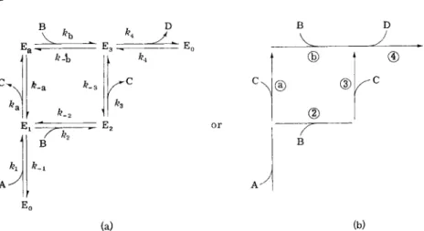
(a) (b) FIG. 5A. Multiple reaction pathways for an enzyme-catalyzed reaction of the
type A - f B ^± C + D . (a) Written in a completely reversible form, where E0 is
the free form of enzyme and Ei, E2, E3, and Ea are enzyme-substrate complexes or other forms of the enzymes, ( b ) A less complex representation with steps a, 3, and 4 considered to be irreversible because products ( C and D ) are omitted and initial velocities are measured. [Reprinted from Sweeney and Fisher (11) b y permission of the copyright owner. Copyright 1968 b y the American Chemical Society.]
FIG. 5B. Proposed rapid-equilibrium, random kinetic mechanism for rabbit muscle phosphorylase a: A, E, G, and P are, respectively, 5 - A M P , enzyme, glycogen, and P i ; G ' is glycogen with one less terminal glucose while P' is glucose 1-phosphate.
The 24 dissociation constants and four rate constants for enzyme in the presence or absence of A M P are indicated. [Reproduced from Engers et al. (16) b y permis
sion of the National Research Council of Canada from the Canadian Journal of Biochemistry.]
presence of the multiple equilibria which exist in multisubstrate systems, the relative magnitude of the rate constants must be determined in order to assess the importance of cooperativity in accounting for kinetic results.
The example of phosphorylase shows that the presence of alternative pathways of interconversion between enzyme intermediates does not make cooperative interactions any less likely to be important than in simpler systems. Indeed, some evidence has been obtained that would indicate that oligomeric proteins are not required for true cooperative interactions between ligands. The binding of certain molecules to single chain bovine serum albumin appears to be cooperative when the data are plotted ac
cording to the Hill equation (16b).
Another model system that may have some application in explaining nonhyperbolic kinetic data involves changes in catalytic activity of an enzyme as a function of time in the presence of modifiers (12). Such a situation would arise if the rate of binding of a modifier were of the same order of magnitude or slower relative to the chemical trans
formation of the substrates.
III. METHODS THAT PROBE MECHANISMS OF ALLOSTERIC INHIBITION
A. Kinetic Methods
It is clear that each enzyme system must be carefully investigated kinetically to determine if cooperativity is indicated. If negative or posi
tive interactions are indicated, kinetic, chemical, and physical criteria may be applied to establish the type of model that most closely fits the experimental data. It is possible to distinguish between the allosteric models for cooperativity and the purely kinetic models by comparing experimental data with curves predicted from derived saturation func
tions (1, Ha). For example, in an enzyme system having n protomers, n molecules of a ligand (M) may be bound to either of the R and T states with unequal affinity. The fraction of the binding sites occupied by M is generally denoted by YM. Expressions for YM have been derived for cases in which one or two ligands interact with separate binding sites (1). These equations lead to a mathematical description of a funda
mental property of the models, i.e., that a heterotropic allosteric ligand should modify the homotropic interactions of the other ligand. Since steady-state or initial reaction velocities are usually observed more readily than actual binding data, the model of Monod et al. (1) also
has been evaluated in terms of steady-state reaction rates (17).
The sequential model for substrate binding has led to the deriva-
tion of characteristic saturation functions which, if negative homotropism is present, may show stepwise sigmoidal behavior when the log of ligand concentration is plotted against fractional saturation (5b, 14)- Plots of reaction velocity against substrate concentration which show more than one inflection point are compatible with this scheme.
A test for distinguishing between random and nonrandom pathway models often is readily carried out (18). In multiple-substrate systems, nonrandom pathways leading to ternary complexes may account for co
operative effects. The test for effector-induced changes in reaction pathway involves measuring the kinetics of exchange of isotopically labeled sub
strates in the absence and in the presence of the effector. No change in the rate of isotope exchange indicates that rapid, random equilibration is maintained. This method circumvents the difficulties of direct binding measurement in some cases.
B. Chemical Methods
Additional weight has been given to subunit interaction mechanisms by chemical studies which may dissociate an oligomeric enzyme into subunits having different functions or which eliminate cooperativity.
Treatment of certain allosteric enzymes with mercurials produces these effects and can give information about the protomer composition of the oligomeric system. Somewhat less complete information is often ob
tained by other changes in the chemical environment of an allosteric system. For example, variation of pH, ionic strength, or the introduction of surface-active agents may cause a loss of allosteric interactions.
In some cases where positive homotropic cooperativity or nega
tive inhibitor interaction is present, such treatments may lead to a sub
stantial increase in catalytic activity so long as the structural integrity of the active site is preserved.
Examples of alteration in the effectiveness of an allosteric inhibitor with this type of chemical treatment may be found throughout the litera
ture of heterotropic allosteric interactions. For instance, Gerhart and Pardee (19, 20) reported that titration of sulfhydryl groups of E. coli aspartate transcarbamylase with p-chloromercuribenzoate leads to a loss of allosteric properties including allosteric inhibition by CTP. The enzyme dissociates into two types of subunits, as determined by sedi
mentation velocity measurements (8). The catalytic subunit has a molecular weight of 96,000 and is not subject to any heterotropic effects.
The other subunit binds the heterotropic effectors ATP and CTP and
has a lower molecular weight of around 34,000. Weber (9) showed that the regulatory subunits break down further in the presence of sodium dodecylsulfate into two peptide chains of 17,000 molecular weight, while the catalytic subunit yields three fragments of 33,000 molecular weight.
The effect of the mercurial is to dissociate the native oligomer into two catalytic trimers and three regulatory dimers.
Other chemical agents and pH also may alter the allosteric interactions of aspartate transcarbamylase. The native enzyme exhibits pH optima at pH 8.5 and 10.2. At the latter pH, allosteric inhibition by CTP is lost (4). Treatment with 1 M urea also modifies CTP inhibition and renders it noncompetitive.
C. Physical Probes
The most commonly employed method for detecting gross changes in physical dimensions of an allosteric enzyme is ultracentrifugal analysis of sedimentation velocities. However, more sensitive probes of the struc
ture have been applied and these methods will be described briefly.
Ultraviolet and X irradiation have produced differential inactivation of the regulatory properties of allosteric enzymes in several instances [21-23). Such treatments may destroy sulfhydryl groups or aromatic residues and provide insight into the role of these amino acids in quater
nary and tertiary structure changes. In the case of a homoserine dehydro- genase-aspartokinase complex from E. colt, an allosteric inhibitor of the aspartokinase moiety, L-threonine, provided protection against X irradia
tion (21).
Perhaps the simplest physical treatment for altering multisite allosteric behavior employs variations in temperature. Since allosteric equilibria may involve physical rearrangements of quaternary structure with ap
preciable free-energy changes, the equilibrium positions of R —> T transi
tions may be expected to be shifted markedly by lowering or raising temperature to values different from the in vivo environment. Indeed, many allosteric interactions are cold or heat sensitive. Heat treatment may cause different alterations than chemical blocking of sulfhydryl or other functional groups, and it is thus a useful tool for determining whether the homotropic and heterotropic interactions of an enzyme sys
tem have the same structural basis.
Changes in the absorbance, fluorescence, or phosphorescence spectra of proteins may accompany binding of allosteric ligands due to a change in the environment of the aromatic amino acid residues {24). Electronic
spectra must be regarded as a powerful tool for probing the tertiary structure of proteins. Chromophores or spin-labeled compounds having unpaired electrons may also be incorporated to reflect overall conforma
tional changes, although in this case some structural perturbation may accompany incorporation of the probe.
A powerful thermal method of analyzing confomational transitions has been successfully applied (15, 25, 26). It combines displacement of confor
mational equilibria by a very rapid change in temperature followed by spectroscopic observation of the relaxation of the enzyme system to new equilibrium concentrations. Relaxation spectra obtained by the tempera
ture jump method resolve the overall changes (which may be associated with either catalysis or control) into discrete steps on a time basis.
For example, it was found that the simple two-state model of Monod et al. (1) could not account for the presence of two distinct relaxation processes in aspartate transcarbamylase when the allosteric inhibitor bromocytidine triphosphate and an aspartate analog (succinate) reacted with the enzyme in the presence of a saturating concentration of carbamyl phosphate. It appears that the two processes correspond to two distinct conformational transitions, one of which is due to the in
hibitor and one to the substrate analog. This example will be discussed further below.
Studies on the effects of physical and chemical treatments on allosteric properties together with examination of the binding properties of dis
sociated protomers may provide evidence for cooperative mechanisms of action that are only partially consistent with the overall proposals of Monod et al. (1) or of Koshland et al. (14). These methods provide considerably more insight into the allosteric interactions than do kinetic data alone and are capable of discerning subtle changes in homotropic and heterotropic interactions which will undoubtedly require new hy
potheses for a more complete explanation. The actual physical basis of allosteric transitions must await determination of the structure of the native and the altered forms of enzymes by more powerful physical methods such as X-ray diffraction (27).
IV. VARIATION OF ALLOSTERIC PROPERTIES AMONG SOURCE ORGANISMS
The isolation of an enzyme subject to allosteric inhibition from a single source organism does not necessarily imply that observed allosteric
control of the catalyzed metabolic step is a general phenomenon in all organisms. The same enzyme isolated from different sources may often show different allosteric behavior, be susceptible to different feedback inhibitors, and be more or less liable to dissociation with loss of allosteric properties. These phenomena are consistent with a general hypothesis that allosteric control is a genetic design feature of a given enzyme system which meets the metabolic requirements of a specific organism and its environment.
V. CLASSES OF ALLOSTERIC INHIBITORS
A. Nucleotides
Purine nucleotides, especially AMP, ADP, and ATP, are important effectors (negative and positive) of dehydrogenases of the citric acid cycle (energy-producing oxidations) and the enzymes of pyrimidine bio
synthesis as well as of fatty acid and carbohydrate metabolism. While in certain cases of feedback inhibition the products of a metabolic path
way may regulate their own biosynthesis (6), the role of the nucleotides in allosteric interactions appears to be much more complex, as indicated in Table I (2, 28-118). It is possibly the overall effects of the purine and pyrimidine nucleotides on these several metabolic pathways which largely determine the fine balance of in vivo reaction rates that are the founda
tion of metabolic homeostasis. Divalent Cations, especially Mg 2 +
, are im
portant modulators of the negative allosteric interactions of the nucleo
tides. In many cases, the magnesium salt of the nucleotide is much more effective than the metal-free compound. It is generally assumed that the metal is involved in binding the effector to the regulatory site.
Nicotinamide dinucleotides are recurring allosteric inhibitors and often produce noncompetitive substrate or product inhibition with N A D ( P ) - linked oxidoreductases, some of which are of importance in releasing reducing equivalents for energy-coupled phosphorylation. The pyrimidine nucleotides CTP and UTP are well-known negative effectors of the initial steps of the biosynthesis of the pyrimidine ring including the synthesis of carbamyl phosphate and carbamyl L-aspartate. Uridine nucleotides have also been found to modulate enzymes of carbohydrate metabolism such as sucrose phosphate synthetase (47).
Halogenated derivatives of pyrimidine nucleotides have been used to replace the naturally occurring allosteric ligands in some studies because
E C T y p e of Ref- Inhibitor E n z y m e Systematic name number e v i d e n c e
0
erence Nucleotides
G T P , N A D D-Lactate dehydrogenase D-Lactate: N A D oxido- 1. 1. 1. 28 k 5 G T P , N A D
reductase
A M P , A D P , A T P , N A D H Malate dehydrogenase L-Malate: N A D oxidoreductase 1. 1. 1. 37 k 28, 29 A M P , A D P , A T P , N A D H
(decarboxylating)
A T P , N A D H , N A D P H Malate dehydrogenase L-Malate : N A D P oxido 1. 1. 1. .40 k 30, 31
(K+ acetyl-CoA) reductase (decarboxylating)
A T P , M g A T P Isocitrate dehydrogenase Z/ireo-D-Isocitrate: N A D P 1. 1. 1. .42 c, k 32 A T P , M g A T P
oxidoreductase A T P , N A D H Glucose-6-phosphate dehy
drogenase
D-Glucose-6-phosphate: N A D P oxidoreductase
1. 1. 1 .49 k 33-35 C T P , U T P D-Glycerate dehydrogenase D-Glycerate: N A D (P) oxido 1. 1. 1 .60 k 36 C T P , U T P
reductase
N A D P H , N A D H , (anthra- Tryptophan pyrrolase L-Tryptophan: oxygen oxido 1. 1 3 . 1 . 1 2 k 37, 38
nilic acid) reductase
G T P , G D P , inosine tri Glutamate dehydrogenase L-Gluiamate: N A D (P) oxido 1. 4 . 1 .3 c, k, p 39-43
phosphate reductase
k, p, c 4, 15, 19, C T P , U T P , G T P Aspartate transcarbamylase Carbamoyl phosphate: L-aspar- 2. 1. 3 .2 k, p, c 4, 15, 19, C T P , U T P , G T P
tate carbamoyltransferase 20, 44
A T P , C l - Glycogen synthetase U D P - G l u c o s e : glycogen a-4-glucosyltransf erase
2 .3. .1 .11 k 45,46 U T P , U D P , A T P , (citrate) Sucrose phosphate synthetase U D P - G l u c o s e : D-fructose- 2. .3. 1 .14 k 47 U T P , U D P , A T P , (citrate)
6-phosphate 2-glucosyl- transferase
A M P Phosphoribosyl pyrophos
phate amidotransferase
Ribosylamine-5-phosphate:
phosphoribosyltransferase
2 .3. .2 .14 k 48
UDP-N-Acetyl-D-glucos- amine
A D P
A T P , alanine, phenyl
alanine A T P U M P A M P
A T P , U T P , C T P C M P
d U T P , d T T P M g A T P
2
" , N A D H , N A D P H , P—Pi N A D H ( a - K G A )
CMF-N-Acetyl neuraminic acid
UDP-N-Acetylglucosamine A M P (tryptophan) A M P (fructose-6-P)
Hexose phosphate amino
transferase Hexokinase Pyruvate kinase Acetokinase
Carbamyl phosphate synthetase
Hexosediphosphatase 5'-Nucleotidase Cytidine deaminase Deoxycytidylate deaminase Inorganic pyrophosphatase Citrate synthetase
UDP-A^-Acetylglucosamine 2-epimerase
N-Acetylglucosamine kinase Glutamine synthetase Glutamokinase
L-Glutamine: D-fructose 2 6 . 1 . .16 k 52 aminotransferase
A T P : D-hexose-6-phospho- 2 .7. . 1 . .1 k 53 transf erase
A T P : pyruvate phospho 2 .7. . 1 . ,40 k 54-56 transferase
A T P : acetate phosphotrans 2, .7. 2. 1 k 57 ferase
A T P : carbamate phosphotrans 2. .7. 2. 5 c, P, k 58,59 ferase (dephosphorylating)
D-Fructose-1,6-diphosphate 3 .1 .3. .11 P, k, c 21, 60 1-phosphohydrolase
5'-Ribonucleotide phospho- 3. 1. 3. 5 k 61,62 hydrolase
Cytidine aminohydrolase 3. 4 . 4 . 5 k 63 Deoxycytidylate aminohy 3. 5. 4 . 12 k 64, 65
drolase
Pyrophosphate phosphohy- 3. 6. 1. 1 k 66 drolase
Citrate oxaloacetate lyase 4. 1. 3. 7 k 68, 69 (CoA acetylating)
UDP-2-Acetamido-2-deoxy-D- 5. 1. 3. 7 k 70 glucose 4-epimerase
k 71 L-Glutamate: ammonia ligase 6 . 3 . 1 . 2 k, p , c 72 L-Glutamate: A T P phospho- k 73
transferase
E C T y p e of Ref- Inhibitor E n z y m e Systematic name number e v i d e n c e
0
erence
U T P , C T P Uridine-cytidine kinase k 74
U M P , U D P Diphosphogalactose UDP-Galactose k 75
TDP-Glucose 4-epimerase 4-epimerase
mino acids and energy- linked metabolites
Threonine, isoleucine, D L - « - Homoserine dehydrogenase L-Homoserine: N A D oxido 1. .1 . 1, .3 k , p 76-78
amino-/3-hydroxyvaleric reductase
acid
a-Ketoglutarate, NHJ", Glutamate dehydrogenase L-Glutamate: N A D oxido 1. .4. .1 .2 k 79, 80
D-glutamate reductase
Phenylalanine, alanine Pyruvate kinase A T P : pyruvate phosphotrans 2. ,7. 1. .40 k 81,82 ferase
Threonine, lysine, A M P Aspartokinase A T P : L-aspartate 4-phospho- 2. .7. 2. .4 k 83-86 transferase
Arginine N-Acetyl glutamate 5-phos- A T P : N-acetyl-L-glutamate k 5b, 87
photransf erase phosphotransferase
Aspartate, malate, citrate Phosphoenolpyruvate Orthophosphate: oxaloacetate 4. 1. 1. .31 k 88-90 carboxylase carboxylase (phosphorylat-
ing)
L-Isoleucine, L-valine Threonine deaminase L-Threonine hydrolase 4. ,2. . 1 . .16 k, c 6,1-93 (deaminating)
Prephenate, tyrosine, 3-Deoxy-D-arabinoheptulo- 7-Phospho-2-oxo-3-deoxy-D- 4. 1. ,2. .15 k, p 94-96 phenylalanine, tryptophan sonate-7-phosphate arabinoheptonate D-erythase
synthetase 4-phosphate lyase (pyruvate
phosphorylating)
Phenylalanine, tyrosine Chorismate mutase k 97
L-Aspartate Pyruvate carboxylase Pyruvate: carbon dioxide ligase 6. 4. 1. 1 k 98, 99
Carbohydrates and other metabolites
Fructose-1,6-diphosphate, Malate dehydrogenase L-Malate: N A D oxidoreductase 1 .1 .1 .39 k 101 3-phosphoglycerate
Glucose, glucose-6-P, Phosphorylase b <*-l,4-Glucan: orthophosphate 2 .4 .1 .1 k, c, p 102-105
adenine, adenosine glucosyltransferase
P E P ( A T P ) Phosphofructokinase (E. coli) A T P : D-fructose-6-phosphate 1-phosphotransferase
2 .7, .1 .11 k 106-108 P—Pi, butyryl-CoA Acetyl-CoA carboxylase A c e t y l - C o A : carbon dioxide
ligase ( A D P )
6. .4. ,1 .2 k 109
Polydextran sulfate ( P D S ) Nuclear exoribonuclease k 110
Miscellaneous inhibitors
3,5-Dihydroxy-4-methoxy- Catechol methyltransferase #-Adenosylmethionine: 2. .1. 1. .6 k 111
benzoate catechol o-methyltransferase
s - A T P sulfurylase ATP-Sulfate adenylyltrans 2 . 7 . 7 . 4 k 112
ferase
Neuromuscular blocking Acetylcholine hydrolase 3. .1. 1. ,7 k 113,114
agents 113,114
Allantoin Guanine deaminase
(isoenzyme A )
Guanine aminohydrolase 3. 5. 4. 3 k 115 Ouabain ( N a
+
- f K + ) A T P a s e ( N a
+
and K + ) A T P phosphohydrolase 3. 6. 1. 3 k U, 116 Antimycin A Succinate-cytochrome
c reductase P 117
H P 04 2
~ Methylglyoxal synthase k 118
0
Abbreviations: k, kinetic; c, chemical; p , physical.
the affinity for these derivatives is often more favorable or because spec
troscopic changes accompanying binding are displaced to longer wave
lengths (15). This effect eliminates interference by the protein absorb- ance of the enzyme in the ultraviolet.
B. Amino Acids
The biosynthesis of the aliphatic amino acids is generally under allosteric feedback control at the steps catalyzed by aspartokinase and threonine deaminase. The synthesis of a key intermediate in this path
way, 2-ketobutyrate, from homoserine is also suppressed by threonine and isoleucine. Thus, the pathway of isoleucine synthesis appears to be a classical case of feedback inhibition through allosteric interactions
(76-78).
Another example of feedback inhibition is the allosteric regulation of 3-deoxy-D-arabinoheptulosonate 7-phosphate synthetase by the end products of aromatic amino acid synthesis: prephenate, tyrosine, phenyl
alanine, and tryptophan. In general, allosteric inhibition of amino acid biosynthesis seems to follow more clearly discernible patterns than does inhibition involving the nucleotides. This may be so because the nucleo
tides are coenzymes in many metabolic transformations as well as pre
cursors of the nucleic acids.
Two citric acid cycle intermediates, malate and citrate, as well as aspartate, are reportedly allosteric modulators of the phosphorylating decarboxylation of oxaloacetate (88-90). Phosphoenolpyruvate is a cen
tral intermediate in the interconversion of these metabolites as shown in Fig. 6.
C. Carbohydrates
Phosphorylase was one of the earliest allosteric enzymes to be studied.
Glucose and glucose-6-P, products of the breakdown of glycogen, as well as AMP are important allosteric ligands of this enzyme, which will be discussed in more detail in Section VI,G. It is interesting that fructose 1,6-diphosphate and 3-phospho-D-glycerate, intermediates in the utiliza
tion of glycogen as an energy source, allosterically inhibit a step of the citric acid cycle, malate dehydrogenase. This illustrates how far from the site of inhibition an allosteric ligand may originate.
Aspartate "* OAA Citrate FIG. 6. Schematic diagram of the feedback systems affecting malic enzyme and related enzymes utilizing phosphoenolpyruvate ( P E P ) and pyruvate in E. coli when carbohydrates are the energy source: ( - f ) and ( — ) represent activation or inhibition, respectively; O A A , oxaloacetate; A c C o A , acetyl coenzyme A. [ R e printed from Sanwal and Smando (144) b y permission of the copyright owner.
Copyright 1969 b y the American Society of Biological Chemists.]
D. Inorganic Ions and Miscellaneous Inhibitors
Many negative allosteric effectors bear no relationship to metabolic products. This does not preclude the possibility that separate specific binding sites for this type of effector are present in the structure of certain allosteric enzymes. Chloride ion (45) inhibits glycogen synthetase while ammonium ion (79) produces anomalous product inhibition of glutamate dehydrogenase of certain microorganisms. As mentioned previ
ously divalent cations may drastically alter the inhibition picture ob
served with nucleotides. It is clear that the ionic environment is an important factor in determining the actual allosteric control mechanisms of many enzymes.
Antibiotics such as antimycin A (117) and the plant product, ouabain (44> H6), may also inhibit enzymes of energy transport and oxidative phosphorylation. Although this type of inhibition may not appear to be designed into the enzyme systems, it is important to determine the nature of the actual site of binding and mechanism of inhibition since other physiological effectors may act in similar ways.
VI. ALLOSTERIC INHIBITION OF SPECIFIC ENZYMES The purpose of this section is to examine various enzyme systems for which allosteric inhibitors have been identified in order to determine how such inhibitors help establish the mechanisms of cooperativity and
regulation of activity. Of special interest is the chemical and physical evidence for allosteric inhibition, since purely kinetic results often can be explained by alternative mechanisms. Conventional steady-state or initial rate kinetic evidence has rarely established that a proposed reac
tion mechanism uniquely accounts for the available data. However, kinetic analysis often indicates which enzymes should be examined by chemical and physical techniques for further evidence of cooperativity.
A. Lactate Dehydrogenase
D(—) -Lactate dehydrogenase (EC 1.1.1.28) isolated from certain fungi is allosterically inhibited by GTP in contrast to the L-lactate de
hydrogenases of mammalian, bacterial, and fungal sources (5). These fungi form lactate as the sole product of glycolysis and hence the subse
quent accumulation of pyruvate may be controlled by GTP levels.
This step may therefore be an important site of control of gluconeo- genesis, since pyruvate is a major precursor of the hexoses. It was re
ported that GTP also inhibits the first step of gluconeogenesis catalyzed by pyruvate carboxylase (5) and acts as a cofactor in the energy-requir
ing conversion of oxaloacetate to phosphoenolpyruvate. Because of the central role of GTP in the balance between glycolysis and gluconeo
genesis, the allosteric effect of the nucleotide on lactate dehydrogenase was studied in detail. The catalyzed reaction was shown to follow a sequential, ordered binary reaction path with first NAD and then lactate associating with the enzyme to form the ternary complex (5).
Catalysis of lactate oxidation is followed by ordered dissociation of pyruvate and then NADH from the active site. Under these circum
stances an allosteric inhibitor would be expected to yield upward-curved Lineweaver-Burk plots due to increased cooperativity between substrate molecules. Figure 2 graphically illustrates the behavior of GTP toward binding of NADH and D(-)-lactate. Inhibition of the binding of the products, NAD and pyruvate, by GTP is "linear" noncompetitive. This result indicates a lack of cooperative binding of the products in the presence of GTP. In this study (5), rates of the catalyzed reactions were used as an indicator of substrate binding effects. One of the products, NAD, does produce cooperative inhibition of the enzyme (5) with respect to NADH. The kinetic results are consistent with the ordered binary association mechanism and indeed the product inhibition results were predictable from the steady-state rate equation of Cleland [5, 119).
Guanosine triphosphate does not change the Fm ax with respect to
NADH indicating that inhibition is caused solely by an increase in the apparent Km for the substrate (i.e., an increase in binding cooperativity).
This result is noteworthy because it indicates that the allosteric inhibitor reduces the affinity of the catalytic site for the substrate but does not
affect the catalytic conversion of the ternary complex.
Substrate concentrations are of great importance in determining the effectiveness of the control process. Since the mechanism of reaction (ordered or random) is of considerable importance in interpreting the results of inhibitor experiments with multisubstrate enzymes, this factor should be conclusively determined by kinetic and other means, includ
ing isotope exchange experiments, where possible. Many N A D - and NADP-dependent dehydrogenases have a compulsory binding order with the coenzyme binding first and dissociating last.
B. Malic Enzymes
L-Malate dehydrogenases are widely distributed, and enzymes iso
lated from a single source organism may be linked to either N A D (EC 1.1.1.37, EC 1.1.1.39) or NADP (EC 1.1.1.40). The function of the NAD-linked mitochondrial enzyme is the energy-producing oxidation of malate to oxaloacetate or pyruvate through decarboxylation of oxalo- acetate. The NADP-linked enzyme is thought to provide a source of NADPH and pyruvate for lipogenesis. Malic enzymes that have different coenzyme specificity are found in the same cell. The reactions are linked together through the action of transhydrogenases that distribute reducing equivalents between NADH and NADPH. The NADP-linked oxidation of malate (EC 1.1.1.40) has been found to be under allosteric control by adenine nucleotides and by coenzyme A and derivatives (29) while the product, NADH, inhibits the NAD-linked enzyme (28). As with most dehydrogenases linked to pyridine nucleotides, the association of the enzyme with substrates is ordered, with coenzyme binding being required for malate binding.
Both the N A D - and NADP-linked malic enzymes appear from kinetic evidence to have an allosteric site that binds nucleotide derivatives of adenine. The reduced coenzymes also change the catalytic properties by binding at this same site. There may be a second regulatory site for another ligand, fructose 1,6-diphosphate, in the case of the N A D - specific enzyme of Streptococcus faecalis (101). Both the catalytic rate of breakdown of the ternary complex ( Fm a x) and the affinity of the enzyme for dicarboxylic substrates and products (S0.5) are altered by
adenine nucleotides. The data are consistent with a two-state model.
However, except for desensitization experiments utilizing high pH, glycine (31), or thermal inactivation (120), the evidence for allosteric inhibition of these important enzymes is based purely on interpretations of kinetic data. Physical evidence for conformational alterations are lacking.
The NADP-specific malic dehydrogenases consistently show allosteric inhibition by acetyl-CoA. The effector accentuates the sigmoidal kinetic behavior with respect to malate by diminishing the affinity of the enzyme for this substrate while the Vmax value remains unchanged. Thus, acetyl-CoA may regulate the amount of pyruvate formed from malate, pyruvate being a precursor of acetyl-CoA. The interactions of oxaloace- tate, pyruvate, malate, and acetyl-CoA are clearly complicated, as indicated in Fig. 6. Adenine and pyridine nucleotides together with acetyl-CoA control the interconversion of these metabolites in an ex
tremely complex manner, especially since AMP is also an activator of the interconversion of pyruvate and acetyl-CoA while the latter com
pound activates phosphoenolpyruvate carboxylase in some organisms.
C. Isocitrate Dehydrogenase
The pyridine-nucleotide-dependent oxidation of isocitrate to oxaloace- tate is of interest because, while the NAD-linked enzyme has allosteric properties with respect to isocitrate, the identified effectors activate the enzyme and no allosteric inhibitors are known. The NADP-linked en
zyme, on the other hand, is allosterically inhibited by ATP.
Adenosine monophosphate is an activator of NAD-specific isocitrate dehydrogenase (EC 1.1.1.41) of Neurospora crassa (121). This activation is of special interest because it is postulated to be due to a change in reaction pathway from random association of the substrates with the enzyme to an ordered association with the pyridine nucleotide co
enzyme binding first (122). The enzyme shows a sigmoidal response to isocitrate concentration (121) but no negative allosteric effectors have been identified. However, this enzyme as isolated from yeast does respond to changes in the ratio of AMP, ADP, and ATP (3) and to changes in the ratio of NAD to NADH in higher plants (123). Since NADH produces product inhibition of this allosteric enzyme, the concept of control by nucleotide ratios may be of importance in determining activity under in vivo conditions. However, the molecular basis for this type of inhibition is not clear. Determination of the order of the reaction
from Hill plots indicates (124) that there are four binding sites for isocitrate and two for each effector (NAD, Mg
2 +
, and AMP) /giving a total of ten specific sites. Magnesium ions are required for the reaction. There
fore, two of the isocitrate binding sites may be noncatalytic positive effector sites. The observed effects of isocitrate on the activity of the enzyme are consistent with a sequential modification model in which enzyme molecules bearing one to four molecules of isocitrate may have different catalytic activities or affinities for the other reactants.
By comparison, the NADP-specific enzyme of microbial origin (EC 1.1.1.42) is allosterically inhibited by ATP and binds 2 moles of the negative effector per mole of enzyme. The binding (32) of the inhibitor was calculated to be accompanied by a substantial free-energy and entropy change, as might be expected for a concerted conformational transition. Intermediates of the Krebs and glyoxalate (oxaloacetate and glyoxalate) cycles inhibit this enzyme synergistically. However, the mechanism of this interaction is unclear.
The isocitrate dehydrogenases are examples of key regulatory enzymes that exhibit different coenzyme requirements, different regulatory prop
erties, and different homotropic and heterotropic interactions, depending on the source. Indeed the isoenzymes from the same source may be quite different in all of these respects. Determination of the chemical, structural, and evolutionary basis of these differences would be of great significance in the area of metabolic regulation.
D. L-Glutamate Dehydrogenase
L-Glutamate dehydrogenase (EC 1.4.1.3) (GDH) catalyzes the assim
ilation of ammonia and the formation of glutamate from a-ketoglutarate.
Reducing equivalents are provided by.NADH or NADPH. The coenzyme associates with the enzyme prior to reaction with oxoglutarate and am
monium ions. The oligomeric enzyme of bovine liver is allosterically inhibited by heterotropic effectors, GTP, GDP, and inosine triphosphate
(ITP). The subunit molecular weight is around 50,000. This enzyme has been the subject of many studies employing chemical modification of specific amino acid residues. Data of this type have yielded informa
tion as to which amino acids are involved in the allosteric interactions of the enzyme. Table II lists some of the modifying reagents and the effects on the negative interaction with GTP (39-42, 125-129). It is clear that specific lysyl and tyrosyl side chains are of importance for both the catalytic activity and the allosteric interactions of the enzyme,
'Reagent Reaction Modification to G D H
Change in negative Ref- allosteric interactions erence Pyridoxal phosphate
Acetic anhydride 4- (Iodoacetamido)salicylic
acid
Schiff base formation, lysine
Acetylation (five amino groups per M W 50,000) Alkylates adjacent to active
site
Dimethyl sulfoxide solvent Replaces bound water
[
1 4
ClMethylmercuric iodide
Tetranitromethane l-Fluoro-2,4-dinitro-
benzene
Trinitrobenzene sulfonate
N-Acetylimidazole
Mercaptide formation
Nitration
Dinitrophenylation
Reacts with 3-amino groups of lysine
O-Acetylation
Inactivates, prevents aggre
gation to high molecular weight forms
Inactivates, disaggregates Incompletely inactivates
enzyme
Facilitates conversion of ac
tive monomer to inactive monomer
Blocks one specific — S H group per M W 54,500 Tyrosine no. 412 specifically
nitrated
Six lysyl and six tyrosyl resi
dues dinitrophenylated Disaggregates enzyme after
one amino group per M W 53,500 is modified
Modifies one tyrosine residue Lost
Lost
125,129
125a Diminishes G T P inhibition 126,127
faster than catalytic ac
tivity is lost
Enhances inhibition b y G T P 128
Releases cooperativity and desensitizes for G T P inhibition
Desensitizes to G T P inhibi
tion
G T P inhibition lost; G T P specifically protects against loss
G T P inhibition released
G T P inhibition diminished while physical properties and activity of enzyme unaffected
41
40
181
while a specific sulfhydryl group in each subunit is involved in control of the catalytically active conformation of the enzyme. Moreover, some modifications can be shown to selectively inactivate binding sites and thereby establish the specificity of separate areas for ADP (a positive
effector) and GTP and for the substrates. For example, the effectors protect against dinitrophenylation of the occupied site (130). It has been suggested that, during unprotected dinitrophenylation, three sep
arate but identical sets of amino acids, each containing six lysyl and three tyrosyl residues, are modified since all site reactivity is lost only when six lysyl and three tyrosyl residues are modified per site per mole of catalytically active oligomer (130).
The conformational change that accompanies binding of GTP has been followed (131) through the use of a covalently bound fluorescent probe, 1-anilinonaphthalene 8-sulfonate (ANS). The fluorescence efficiency of the probe depends largely on the non-polar nature of the immedi
ate chemical environment. Guanosine triphosphate greatly enhances the fluorescence of GDH-bound ANS, indicating that the region where the dye is bound is changing conformation to surround the probe with a less polar environment. Nitrated GDH that has partially lost the nega
tive GTP interaction shows a much less pronounced enhancement of ANS fluorescence when treated with the negative effector, confirming that GTP acts through a conformational change.
Reaction of 4-(iodoacetamide) salicylic acid specifically at or near the catalytic site of GDH (126, 127) on only one subunit of the oligomer allowed an assessment of the effect of the inhomogeneous oligomer on the effectiveness of inhibition due to GTP binding at the remaining active negative effector sites. It was concluded (126) that, while each subunit of the unmodified enzyme is equally inhibited by GTP, modifica
tion of one subunit decreases the inhibition produced by GTP in the remaining (unmodified) subunits. Modification of nine subunits further decreases inhibition of the remaining subunits. This conclusion would tend to support a sequential model (14) for allosteric interaction with GTP since if only two (R and T) states of the enzyme are allowed, modification of one subunit should eliminate the negative effector inter
action altogether according to the concerted model (1, 126).
The existence of a distinct form of the enzyme having enhanced alanine dehydrogenase activity and diminished glutamate activity is supported by the report that diethylstilbestrol favors the alanine activ
ity. Apparently this compound and its bromoacetyl derivative bind re- versibly at an estrogen binding site. In the presence of NADH, the effects are irreversible and a covalent link with the ligand is formed.
This chemical modification desensitizes the enzyme to the effects of the positive and negative effectors (131a).
Certain other kinetic results are not compatible with a simple two- state model. Double reciprocal plots of reaction rate and coenzyme con
centration often show an unexpected downward curve, especially in the presence of inorganic phosphate (130). This effect has been interpreted by Dalziel (10) as being a result of a negative homotropic interaction affecting the subunits of the enzyme. That is, at high cofactor concentra
tions, cooperativity is diminished and the enzyme is activated. The concerted model predicts only positive homotropic interactions.
The physical probes (fluorescence efficiency, sedimentation velocity) and chemical modification studies have elucidated many facets of the allosteric interactions and regulatory mechanisms of mammalian GDH.
A full understanding of the significance of the various chemically modified forms of the enzyme may require complete structural analysis.
Although modifications of this type may perturb the native conforma
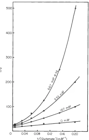
tions, they remain useful probes of major importance where enzyme stability permits their use. NAD-Linked glutamate dehydrogenase (EC 1.4.1.2) is, in contrast to the beef liver enzyme, not inhibited by GTP.
In fact, both guanylates and adenylates activate the enzyme, which was isolated from mitochondria of a fungus (79). The enzyme is a tetramer with subunits of around 50,000 molecular weight. A completely different set of negative effectors, including D-glutamate, a-ketoglutarate, and ammonium ions, has been identified in fungi (79, 80). «-Keto- glutarate causes double reciprocal plots of reaction velocity and L-gluta- mate concentration to become curved upward, as shown in Fig. 7. The inhibition is partially noncompetitive since the Vmax is diminished. A plot of Fm ax against effector concentration yielded a straight line as expected from a reaction pathway requiring binary association of NAD and enzyme followed by ternary association of NAD-enzyme, ammonium ion, and a-ketoglutarate (79). These results are compatible with the con
certed two-state allosteric transition model.
E. Aspartate Transcarbamylase
As mentioned previously, carbamoyl phosphate: L-aspartate carba- moyltransferase (EC 2.1.3.2) provides a classic example of designed-in feedback inhibition which operates through conformational transitions.
This enzyme is unique in that the allosteric inhibitor CTP has been shown to bind at a specific site located on a subunit different from the catalytic