Identification of transcriptional and phosphatase regulators as interaction partners of human ADA3, a component of histone acetyltransferase
complexes
Sevil ZENCIR*, Adam SIKE†, Melanie J. DOBSON‡, Ferhan AYAYDIN§, Imre BOROS†1 and Zeki TOPCU¶1
*Department of Medical Biology, Faculty of Medicine, Pamukkale University, Denizli 20070, Turkey,†Department of Biochemistry and Molecular Biology, Szeged University, Szeged H-6726, Hungary,‡Department of Biochemistry and Molecular Biology, Dalhousie University, Halifax, NS, Canada B3H 4R2,§Cellular Imaging Laboratory, Biological Research Center, Hungarian Academy of Sciences, H-6726, Szeged, Temesv´ari krt. 62, Hungary, and ¶Department of Pharmaceutical Biotechnology, Faculty of Pharmacy, Ege University, Izmir 35100, Turkey
ADA (alteration/deficiency in activation) 3 is a conserved component of several transcriptional adaptor and HAT(histone acetyltransferase) complexes that regulate RNA polymerase II- mediated gene expression. Within the HAT complexes ADA3 is associated with ADA2 and the HAT GCN5 (general control non-repressed 5). ADA3 plays roles in diverse cellular processes and also in malignancies by modulating GCN5 catalytic activity and/or by interactions with other regulators. To gain a better understanding of ADA3 function, we used a yeast two-hybrid approach to screen a human fetal cDNA library for proteins that interacted with hADA3 (human ADA3). We identified three novel hADA3-interacting partners, a transcriptional regulator, AATF (apoptosis-antagonizing transcription factor), and regulatory subunits of the PP1 (protein phosphatase 1) and PP2A (protein phosphatase 2A) [PPP1R7 (PP1 regulatory subunit 7) and PPP2R5D (PP2A 56 kDa regulatory subunit δ isoform) respectively]. Analysis of truncated versions of
hADA3 indicated that the C-terminal ADA2-interacting domain was not required for these interactions. Fluorescent microscopy analysis and co-immunoprecipitation provided support for the co-localization and interaction of hADA3 with these proteins in human cells. Expression of the interacting proteins altered expression of an hADA3-regulated reporter gene, suggesting functional consequences for the interactions. The detected interactions of hADA3 might extend the spectrum of mechanisms by which ADA3 can contribute to the regulation of gene expression and shed light on processes mediated by these newly identified ADA3 partners.
Key words: alteration/deficiency in activation 3 (ADA3), histone acetyltransferase, protein–protein interaction, transcrip- tional regulation, yeast two-hybrid technology, Spt/Ada/Gcn5/
acetyltransferase (SAGA).
INTRODUCTION
In eukaryotes, the basic building block of chromatin is the nucleosome, a core of eight histone proteins wrapped by 146 bp of DNA. Bothin vivoandin vitrostudies indicated that the nucle- osomal organization represses transcription by limiting access of transcription factors to their target sites in the nucleosome-bound DNA [1]. How transcription factors contend with the inhibitory effects of chromatin structure remains a central question in the process of gene expression regulation. The repressive nature of chromatin can be relieved in the cell by chromatin remodelling and the histone-modifying activities that alter chromatin structure making it accessible to and recognized by the components of the RNA polymerase II transcriptional machinery (see [1,2] for reviews). Acetylation of nucleosomal histone N-terminal tails by HATs (histone acetyltransferases) has been established as a major mechanism leading to gene activation, whereas HDACs (histone deacetylases) contribute to transcriptional repression [1].
ADA (alteration/deficiency in activation) 3 is a conserved component of several HAT complexes, including SAGA [Spt/
Ada/Gcn5 (general control non-repressed 5)/acetyltransferase]
in yeast, the related STAGA (SPT3/TAFII31/GCN5L/acetylase)/
TFTC (TATA-binding protein-free TAF-containing complex) [3,4] in humans, and as well the metazoa-specific ATAC (Ada2A- containing) complex [5–12]. Each of these complexes contain the GCN5 acetyltransferase. GCN5 and the ADA2 and ADA3 adaptor proteins form a heterotrimer within HAT complexes. This HAT module, in which a fourth factor SGF29 (SAGA-associated factor 29) has recently been recognized [13], seems to be conserved in the yeast and human SAGA-type complexes. Interactions among its constituents modulate GCN5 acetyltransferase activity and target it to nucleosomal core histones. Additionally, the GCN5–ADA2–ADA3 triad plays both structural and functional roles by stabilizing the complex and bridging interactions with sequence-specific transcription factors and members of the basal transcription machinery [5,6,8,10,11,14–17]. However,
Abbreviations used: AATF, apoptosis-antagonizing transcription factor; AD, activation domain; ADA, alteration/deficiency in activation; ADE2, adenine- requiring 2; ANCO, ankyrin repeat domain-containing protein; ATAC, Ada2A-containing;β-Gal,β-galactosidase; Co-IP, co-immunoprecipitation; CFP, cyan fluorescent protein; CREB, cAMP-response-element-binding protein; DMEM, Dulbecco’s modified Eagle’s medium; ERK, extracellular-signal- regulated kinase; Gal4, galactose metabolism 4; GFP, green fluorescent protein; hADA2A, human ADA2A; hADA3, human ADA3; HAT, histone acetyltransferase; HDAC, histone deacetylase; hGIP, human glutaminase-interacting protein; HIS3, histidine-requiring 3; HPV, human papillomavirus;
LacZ,β-galactosidase; MAPK, mitogen-activated protein kinase; MAP3K12, MAPK kinase kinase 12; MBIP, MAP3K12-binding inhibitory protein 1;
MEK, MAPK/ERK kinase; MEL1,α-galactosidase; ORF, open reading frame; NA, numerical aperture; PHF21A, PHD finger protein 21A; PP1, protein phosphatase 1; PP2A, protein phosphatase 2A; PPP1R7, protein phosphatase 1 regulatory subunit 7; PPP2R5D, PP2A 56 kDa regulatory subunitδ isoform; SAGA, Spt/Ada/Gcn5/acetyltransferase; SD/−Trp, SD medium lacking tryptophan; TACC, transforming acidic coiled-coil-containing protein;
TRP1, tryptophan-requiring 1; X-α-Gal, 5-bromo-4-chloroindol-3-ylα-D-galactopyranoside; SD/−Ade/−His/−Trp/X-α-Gal, SD medium lacking adenine, histidine and tryptophan and containing X-α-Gal; YFP, yellow fluorescent protein.
1 Correspondence may be addressed to either of these authors (email borosi@bio.u-szeged.hu or zeki.topcu@ege.edu.tr).
Biochemical Journal www.biochemj.org
the role of ADA3 within the complex has yet to be fully elucidated. ADA3 has been found to interact with two ankryin- repeat-containing transcriptional regulators [ANCO (ankyrin repeat domain-containing protein) 1 and 2] [18]. The ANCO proteins can function either as co-activators or, by recruiting HDACs, as co-repressors [19]; highlighting the complexity of transcription regulatory mechanisms ADA-type adaptor proteins may participate in through protein–protein interactions.
To understand how ADA3-containing complexes regulate the expression of specific genes, some of which are known to play direct roles in oncogenesis, and identify novel pathways affected by ADA3, we aimed to identify new interaction partners of hADA3 (human ADA3).
Among the various methods available for the investigation of protein–protein interactions, the yeast two-hybrid genetic selection system is a powerful technique [20]. This method involves the expression within the yeastSaccharomyces cerevisiae cell nucleus of two proteins being assessed for interaction [21].
Each protein is expressed as a chimaera, fused to one domain of the yeast Gal4 (galactose metabolism 4) transcription factor.
Interaction of the two fusion proteins brings the two domains of Gal4 into close enough proximity to restore transcription factor function, detected by activation of Gal4-responsive reporter genes. We used the yeast two-hybrid system to screen a human fetal brain cDNA library.
We report in the present study the identification of three novel ADA3 interaction partners. Direct interaction of ADA3 with these proteins has not previously been shown. The known functions of the newly identified ADA3 partners as regulators of transcription, chromatin structure, apoptosis and protein phosphorylation suggest many possible links to the various functions in which ADA3 has been implicated. We verified hADA3 interaction with its newly identified partners in mammalian cells by Co- IP (co-immunoprecipitation) and confocal microscopy analyses.
Moreover, we demonstrate that expression of these partner proteins affects activation of a reporter gene, suggesting that their ability to interact with ADA3 may have functional consequences for ADA3-mediated transcriptional regulation.
EXPERIMENTAL Yeast strains and media
The two-hybrid reporter yeast strains used in this study were AH109 (MATa, trp1-901, leu2-3, 112, ura3-52, his3- 200, gal4,gal80,LYS2::GAL1UAS GAL1TATA-HIS3,GAL2UAS- GAL2TATA-ADE2, URA3::MEL1UAS-MEL1TATA-lacZ, MEL1) and Y187 (MATα, ura3-52,his3-200, ade2-101,trp1-901, leu2-3, 112, gal4, met, gal80, MEL1, URA3::GAL1UAS -GAL1TATA- lacZ) (Clontech). Yeast cells were cultured at 30◦C in YEPD medium and adenine (1%yeast extract/2%bactopeptone/2% dextrose and 1% adenine) or SD medium (0.67% yeast nitrogen base without amino acids, 2%dextrose, supplemented with appropriate amino acids and bases) [22]. The medium containing 40μg/ml X-α-Gal (5-bromo-4-chloroindol-3-ylα-D- galactopyranoside) were solidified with 2.0% agar. All media reagents were obtained from either DIFCO Laboratories or Sigma.
Yeast two-hybrid screen
To create a yeast expression plasmid encoding hADA3 fused to the C-terminus of the yeast Gal4 transcription factor DNA- binding domain (Gal4BD–hADA3), the 1.3-kbp hADA3 (isoform 1) ORF (open reading frame) was amplified by RT (reverse transcription)–PCR from human osteosarcoma cells (U2OS)
using the primers 5-cggccatatgcctgggccatgagtgagtt-3 and 5- catagtcgacgctacccatccagcagcttc-3with an initial denaturation at 94◦C for 4 min, followed by 35 cycles of denaturation at 94◦C for 30 s, annealing at 64◦C for 40 s and polymerization at 72◦C for 2 min. The PCR product was cloned into the vector TOPO-TA (Life Technologies) to create plasmid TOPOTA–hADA3 and the insert was verified by sequencing. The hADA3-encoding region was excised from TOPOTA–hADA3 as an NdeI/SalI fragment and inserted into the yeast two-hybrid vector pGBKT7 (Clontech) digested with the same restriction enzymes to generate the plasmid pGBKT7-hADA3.
The toxicity of Gal4BD–hADA3 fusion protein expression was assessed by comparing colony size and phenotype of yeast transformed with pGBKT7 or pGBKT7-hADA3 on SD/−Trp [SD medium lacking tryptophan; selects for the TRP1 (tryptophan-requiring 1) gene-marked plasmid] and for self-activation of the Gal4-responsive reporter genes by plating yeast transformed with pGBKT7–hADA3 on SD/−Ade/−His/−Trp/X-α-Gal (SD medium lacking adenine, histidine and tryptophan and containing X-α-Gal) and SD medium lacking tryptophan and containing X-α-Gal.
The yeast two-hybrid screen was performed as described previously [23]. Briefly, yeast strain Y187, pre-transformed with a human fetal brain cDNA library in the Gal4 AD (activation domain) vector pGADT7-Rec (Clontech) were mated with yeast strain AH109 transformed with pGBKT7-hADA3 and the diploids were screened for hADA3-interacting clones using standard methods [21]. A total of 1×10⁷cells were screened with a mating efficiency of 5.8%in the library having 8×10⁷cells.
Clones capable of activating integrated reporter genes, namelyHIS3 (histidine-requiring 3), ADE2 (adenine-requiring 2), MEL1 (α-galactosidase 1) and LacZ (β-galactosidase) under the control of the Gal4-responsive UAS (upstream activating sequence) were identified by plating the transformants on SD/−Ade/−His/−Leu/−Trp/X-α-gal [SD solid medium lacking leucine, tryptophan, adenine and histidine and containing X-α-gal; to select for the presence of pGBKT7-hADA3 and theLEU2(leucine-requiring 2) gene-containing library plasmid;
Clontech]. The transformants capable of growth and giving blue colonies due to conversion of X-α-gal into a blue precipitate by theα-galactosidase expressed by the MEL1 reporter gene, were subsequently checked forβ-galactosidase expression from thelacZreporter gene [24]. Clones failing to grow under more stringent conditions (in the presence of 25 mM 3-aminotriazole, a competitive inhibitor of theHIS3gene product) were discarded.
Plasmids were isolated from positive yeast clones and used to transformEscherichia colito ampicillin resistance. Plasmids from bacterial transformants were tested in yeast to confirm their ability to activate the reporter genes when co-transformed with pGBKT7- hADA3. hADA3 bait-dependent reporter gene expression was assessed by transforming two-hybrid reporter yeast with plasmids expressing the putative hADA3 interactors in the absence of other fusion proteins and with other unrelated fusion proteins (results not shown). For positive clones, cDNA inserts were analysed by restriction enzyme digestion and by DNA sequencing (Robarts Research Institute, London, ON, Canada). A second independent cDNA library was used for confirmation experiments (Clontech).
To map the region of hADA3 needed for interaction with the potential partner proteins, we made deletions in the pGBKT7- ADA3 plasmid with a set of restriction enzyme digestions. We employed the PvuII/SalI, SacI/SalI, EcoRI/SalI and SmaI/SalI enzymes corresponding to C-terminal-truncated versions of the hADA3 ORF. For the N-terminal-truncated constructs, we cloned into the NcoI/SalI-digested pGBKT7 vector a series of PCR products of hADA3 amplified from pGBKT7-hADA3
using the primers 5-cggcccatgggcctcatggggccactgaccgaa-3, 5- cggcccatggcccctatggaggatt-3, 5-cggcccatggtgcgcatggctgacaa-3 (forward) and 5-cataaagcttgctacccatccagcagcttc-3(reverse).
β-Gal (β-galactosidase) liquid assay
Aβ-Gal liquid assay was subsequently performed for quantitative estimation of protein–protein interactions of the positive clones with full length and truncated hADA3 constructs as described previously [24,25].
Western blotting analyses
Expression of full-length hADA3 and hADA3 deletion derivatives as Myc-epitope tagged Gal4BD fusions from pGBKT7-based plasmids was confirmed by Western blotting using mouse monoclonal antibodies specific against Gal4BD (Clontech) and c-Myc (Cell Signaling Technology).
For expression of FLAG epitope-tagged hADA3 in mammalian cells, the hADA3-encoding region was excised from pGBKT7- hADA3 by digestion with NcoI/SalI and ligated with the EcoRV/SalI-digested mammalian expression vector pCMV3- Tag2a, to create pCMV3-Tag2a/hADA3. Plasmids capable of directing expression in mammalian cells of FLAG-tagged versions of the proteins identified as potential hADA3-interacting partners were obtained from a commercial supplier (Origene).
Expression of fusion proteins from the plasmids was confirmed by transfecting CHO (Chinese-hamster ovary) cells with the plasmids and analysing the protein extracted from the cells by SDS/PAGE (12%gel) and Western blotting with an anti-FLAG mouse monoclonal antibody (Sigma).
Co-localization analyses
For confocal microscopy analyses, a plasmid expressing hADA3 as an CFP (cyan fluorescent protein) fusion protein was created by PCR amplification of the hADA3-encoding region from the pGBKT7-hADA3 construct using the primers 5-cggcagatctcctgggccatgagtgagtt-3 (forward) and 5- cataaagcttgctacccatccagcagcttc-3 (reverse). The PCR product was digested with BglII and HindIII and ligated with the plasmid pECFP-C2 digested with the same enzymes to create the plasmid pECFP-hADA3 (CFP-ADA3). Plasmids expressing the candidate hADA3-interacting proteins fused to YFP (yellow fluorescent protein) (pEYFP-ADA3 interacting proteins) were similarly constructed by amplifying the coding regions from the respective mammalian expression plasmids, digesting the PCR products with EcoRI/SmaI or EcoRV/SmaI and ligating with the vector pEYFP-C3 digested with the same restriction enzymes. As controls, coding regions for proteins known to interact [hADA2A (human ADA2A)] and not interact [hGIP (human glutaminase- interacting protein)] with hADA3 were excised from pGBKT7- based plasmids using appropriate restriction enzymes and cloned into pEYFP-C3 [13,23]. The pECFP-C2 and pEYFP-C3 plasmids were derived from the pEGFP-C2, pEGFP-C3, pECFP-C1 and pEYFP-C1 plasmids (Clontech) using the AgeI/EcoRI restriction enzymes. The plasmids were verified by restriction endonuclease digestions and sequencing.
Human U2OS cells were seeded in an 8-well chamber slide (Lab-Tek) in complete DMEM (Dulbecco’s modified Eagle’s medium) and transfected with 10μg of the CFP and YFP fusion protein-expression plasmids pECFP-hADA3, pEYFP-PPP1R7, pEYFP-PPP2R5D, pEYFP-PHF21A and pEYFP-AATF DNA the next day by using ExGen 500 transfection reagent (Fermentas) according to the manufacturer’s instructions. Transfected cells
were incubated in a 5%CO2incubator at 37◦C for 24 h before being analysed using a confocal laser-scanning microscope.
Confocal laser-scanning microscopy was performed using Olympus Fluoview FV1000 confocal laser-scanning microscope (Olympus Life Science Europa). The microscope configuration was: objective lens UPLFLN ×40 [water, NA (numerical aperture) 0.8] and×60 (oil, NA 1.3); sampling speed, 8μs/pixel;
scanning mode, sequential unidirectional; excitation, 458 nm (CFP) and 515 nm (YFP); laser transmissivity, 30%and 10% were used for CFP and YFP respectively; main dichroic beamsplitter, DM458/515 and intermediate dichroic beamsplitter, SDM 510; and CFP was detected between 470 and 520 nm and YFP was detected between 520 and 570 nm. DIC (differential interference contrast) or standard transmission images were captured with a 515 nm laser line.
Co-IP assay
U2OS cells were transfected as described above with 10μg of pCDNA3-Ada3-FLAG, pEYFP-PPP1R7, pEYFP-PPP2R5D, pEYFP-PHF21A and pEYFP-AATF DNA. The medium was removed and the cells were washed twice following either 12 h (pEYFP-transfected cells) or 24 h (pCDNA3-transfected cells) incubations in a 5%CO2 incubator at 37◦C. Cells were harvested by centrifugation at 2000gfor 5 min. The supernatant was removed and the pellet was resuspended in 1×lysis buffer (Cell Culture Lysis Reagent 5×, Promega) supplemented with 1x PIC (5 mg/ml aprotinin, 10 mg/ml leupeptin, 5 mg/ml pepstatin and 5 mg/ml PMSF) and incubated on ice for 1 h. Samples were then centrifuged at 7000gat 4◦C for 5 min, supernatants were removed and stored at −80◦C. Cell extracts were mixed by overnight rotation at 4◦C then pre-cleared for 2 h at 4◦C.
The anti-FLAG M2 agarose beads (Sigma) were washed twice with Nonidet P40 buffer [150 mM NaCl, 1% Triton X-100 and 50 mM Tris/HCl (pH 7.5)] and incubated with cell extracts.
The precipitates were collected by centrifugation at 1000gfor 2 min at 4◦C and washed four times with Nonidet P40 buffer.
The final precipitates were dissolved in 2× SDS-loading dye [0.125 M Tris/HCl (pH 6.8), 4% SDS, 21% glycerol, 2 mg of Bromophenol Blue and 10% 2-mercaptoethanol], resolved by SDS/PAGE (10% gel) and transferred on to a nitrocellulose membrane (200 mA for 2 h) for Western blot analyses using the enhanced chemiluminescence detection system (ECL®, Amersham Biosciences). Enhanced YFP proteins were detected using an anti-GFP (green fluorescent protein) polyclonal antibody that recognizes the GFP variant enhanced YFP.
Luciferase reporter gene assay
To monitor the effect of putative hADA3-interacting proteins on transcription in mammalian cells, human HeLa and U2OS cells were seeded in six-well plates at 2.5×10⁶ cells per ml in DMEM, supplemented with 10%FBS (fetal bovine serum) and cultured in a 5%CO2incubator at 37◦C for 24 h until they were approximately 70–80% confluent. Cells were transfected with 0.2μg of anMDM2promoter luciferase gene plasmid (pGL2- MDM2-Luc) using ExGene 500 transfection reagent (Fermentas).
We then co-transfected U2OS cells having theMDM2promoter with the pCMV3-Tag2a/hADA3 construct (0.2μg) alone or together with a plasmid directing expression of one of the hADA3- interacting partners (0.2μg) according to the manufacturer’s instructions. At 48 h after transfection, cells were harvested and 20μl of the lysates were analysed using the Luciferase Assay system (Berthold Detection System Orion L/MPL4) according to the manufacturer’s guidelines.
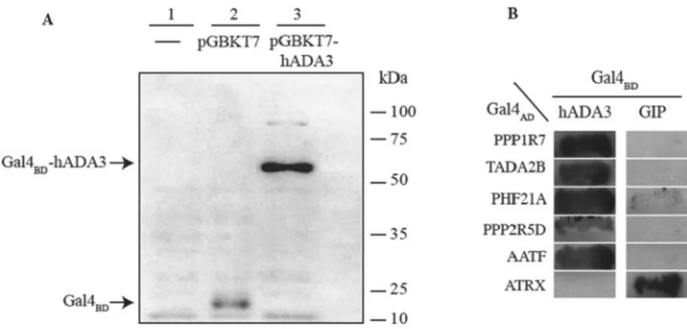
Figure 1 Expression of hADA3 protein in yeast and reporterα-galactosidase activity resulting from the interaction of ADA3–Gal4BDand Gal4ADfusion proteins (A) Expression of hADA3 as a Gal4BD-hybrid protein in yeast. Protein was extracted from untransformed yeast (lane 1) as well as the yeast transformed with pGBKT7 (lane 2) or with pGBKT7-ADA3 (lane 3, Gal4BD-hADA3 has an expected molecular mass of 70 kDa). Total yeast extracts were separated by SDS/PAGE (10 % gel) and analysed by Western blotting using Gal4BD-specific primary antibodies. Molecular mass is on the right-hand side in kDa. (B) Detection of reporterα-galactosidase activity resulting from the interaction of hADA3–Gal4BDand Gal4ADfusion proteins. The two-hybrid reporter yeast strain AH109 was co-transformed with plasmids expressing hADA3 (left-hand panel) or hGIP (right-hand panel) as Gal4BD-fusion proteins together with a plasmid expressing the Gal4AD-fusion protein indicated on the left-hand side. Co-transformants were streaked on solid selective medium (SD/−Ade/−His/−Trp/−Leu/X-α-Gal) and incubated for 1 week at 28◦C.
Growth and expression of theα-galactosidase reporter gene (indicated by a dark colour) shows interaction of the two hybrid proteins. A known hGIP-interacting protein, ATRX (αthalassaemia/mental retardation syndrome X-linked), was used as a negative control for the hADA3 interaction.
RESULTS
Yeast two-hybrid screen identifies novel hADA3-interacting partners in a human fetal brain cDNA library
In order to use hADA3 in yeast two-hybrid library screens as bait, we first constructed a plasmid (pGBKT7-hADA3) that would express hADA3 in yeast as a Gal4 DNA-binding domain (Gal4BD) fusion protein. The expression of the Gal4BD–hADA3 fusion protein inS. cerevisiaetransformed with the plasmid pGBKT7- hADA3 was confirmed by Western blotting (Figure 1A). hADA3 expression was not toxic in yeast cells as indicated by the observation that the number and size of the colonies between the yeast populations transformed with the pGBKT7 vector or pGBKT7-hADA3 did not significantly differ. Transformation of pGBKT7-hADA3 into the yeast two-hybrid reporter strain AH109 resulted in colonies on SD/−Trp owing to the presence of theTRP1gene on the plasmid, but did not result in colonies when either adenine or histidine was omitted, indicating that neither of the Gal4-responsive reporter genes ADE2 or HIS3 were activated by the Gal4BD–ADA3 fusion protein alone. This observation proved that in our experimental system ADA3 did not activate transcription in the absence of an interacting partner protein expressing an AD.
Next we identified interacting partners of ADA3 by co- expressing Gal4BD–ADA3 with fusion proteins obtained by ligating the GAL4AD-encoding region to human fetal brain cDNAs. In our screen of 1×10⁷Gal4BD–ADA3-expressing yeast cells, which were transformed with plasmids expressing human fetal brain cDNA–Gal4ADfusion proteins, we recovered several co-transformants that were able to form colonies under the conditions that required Gal4-responsive reporter gene activation.
The ability of these colonies to grow in the absence of adenine and histidine suggested that they contained a library plasmid ex- pressing an hADA3-interacting protein. The specificity of hADA3 interactions with prospective partners was tested by a second line of yeast two-hybrid experiments. For this pGBKT7-hADA3 and another bait expressing plasmid carrying Gal4BDfused to the hGIP (pGBKT7-GIP) [23] were introduced independently into host cells in order to compare their interaction with the newly identified putative ADA3 partners (Figure 1B). The results of
these tests indicated that five of the GAL4 AD-human fetal brain cDNA plasmids were capable of activating the reporter genes together with pGBKT7-hADA3, but not with pGBKT7-GIP. The plasmids from these five yeast transformants were isolated and the cDNA insertions were characterized by restriction mapping and sequencing. Sequence analyses of the five cDNA insertions and comparisons with database sequences using BLAST searches [26] revealed that the isolated cDNAs correspond to five proteins, four of which have not been previously identified as interacting partners of hADA3. These were AATF (apoptosis- antagonizing transcription factor, PHF21A{PHD finger protein 21A; also known as BHC80 (BRAF35 [BRCA2 (breast cancer 2, early onset)-associated factor 35]–HDAC complex protein)}, serine/threonine PPP2R5D [PP2A (protein phosphatase 2A) 56 kDa regulatory subunitδisoform] and PPP1R7 [PP1 (protein phosphatase 1) regulatory subunit 7]. The fifth cDNA encoded ADA2B, one of the two human ADA2 transcriptional adaptor proteins, which has been demonstrated by several laboratories independently to interact with ADA3 [6]. The recovery of ADA2B in our screen validated the ability of this approach to detect authentic interactions. Our subsequent analyses focused on the four novel hADA3 partners with the aim of characterizing their interaction with hADA3 and demonstrating the interactions and their functional significance in mammalian cells.
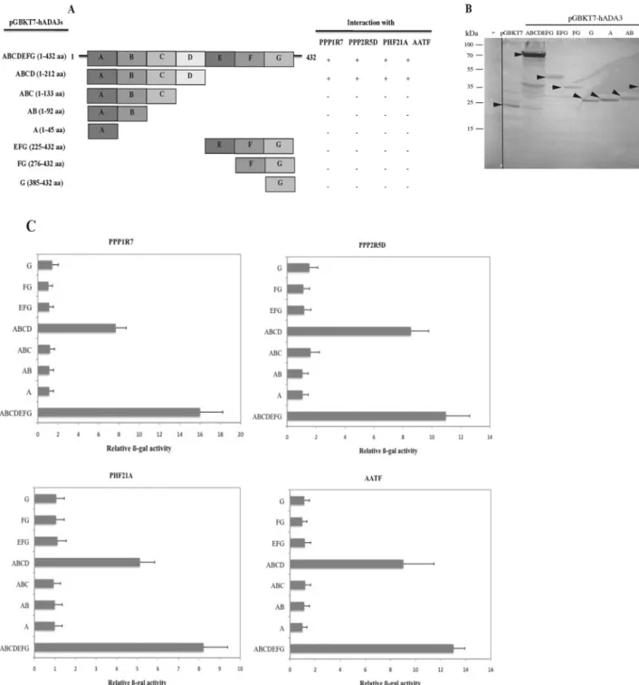
hADA3 participates in the detected novel protein–protein interactions with its N-terminal region
To identify the hADA3 domain required for interactions with the four identified novel partners, a series of plasmids encoding different portions of hADA3 as GAL4BD fusion proteins were created and tested for their ability to activate the Gal4-responsive reporter genes when co-transformed into yeast strain AH109 with the positive library plasmids (Figure 2A). Only full-length hADA3 and a truncation retaining hADA3 residues 1–212 were sufficient for interaction with all four proteins. Western blotting analysis confirmed that all hADA3 truncated Gal4BDfusion proteins were expressed at levels sufficient to detect the interaction (Figure 2B).
The lack of interaction of the hADA3 fusion protein containing residues 1–133 of hADA3 indicated that residues between 133 and 212 were required for the interaction (Figure 2C).
Figure 2 Mapping the regions of hADA3 required for interaction with partner proteins
(A) The regions of hADA3 shown on the left [regions A (1–45), B (46–92), C (93–133), D (134–212), E (225–275), F (276–384) and G (385–432)] fused to the Gal4BD-encoding region were assessed for their two-hybrid interactions with different partner proteins expressed as Gal4ADfusion proteins. (+and−represent interaction and no interaction respectively). (B) AH109 yeast, untransformed (−) or transformed with the pGBKT7, full-length pGBKT7-ADA3 and truncated hADA3 as Myc-epitope tagged GalBDfusion proteins, were grown in selective media (SD/−Trp).
Proteins were extracted from yeast and analysed by Western blotting using an anti-Myc mouse monoclonal antibody. Molecular mass is on the left-hand side in kDa. (C) A quantitative estimate of the two-hybrid interaction of the truncated versions of hADA3 with the different partner proteins shown in (A) was obtained by performing a liquid assay forβ-Gal activity. Activity was expressed as the percentage of that obtained for interaction of the protein with full-length hADA3. Results are the means+−S.D. obtained from three independentβ-Gal assays.
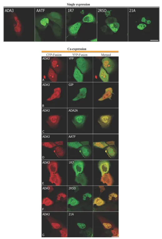
Co-localization studies verified hADA3 interaction with its newly identified partners in mammalian cells
Next we investigated co-localization of hADA3 with the proteins identified as its potential partners by yeast two-hybrid screening by confocal microscopy in human cells. For this we constructed plasmids that would express hADA3 as a CFP fusion protein
(CFP–hADA3) and interacting partner proteins as YFP fusion proteins (YFP–PPP1R7, –AATF, –PPP2R5D and –PHF21A). The plasmids were individually used to transfect U2OS cells to allow localization of the proteins in the absence of the interacting partner proteins to be determined (Figure 3, upper panel). Vectors ex- pressing CFP or YFP were used as negative controls. U2OS cells transfected with CFP–hADA3 displayed fluorescence signal both
Figure 3 Cellular localization and co-localization of hADA3 and its interacting partners in mammalian cells
Upper panel, U2OS cells were transfected with a plasmid expressing CFP–hADA3 or with a plasmid expressing an hADA3-interacting partner, AATF, PPP1R7 (1R7), PPP2R5D (2R5D) and PHF21A (21A), as a YFP fusion protein. Images were captured by confocal microscopy. The signal from CFP–hADA3 (ADA3) is pseudo-coloured red, whereas YFP signals are shown in green. Scale bar is 10μm. Lower panel, U2OS cells were co-transfected with a plasmid expressing hADA3 fused to CFP (ADA3) and a second plasmid expressing YFP (A), GIP (B), ADA2A (C), AATF (D), PPP1R7 (E) PPP2R5D (F) or PHF21A (G) fused to YFP. Co-localization was analysed by confocal laser-scanning microscopy. CFP–hADA3 signals are shown in red in the left-hand column and YFP signals in green for the same cells in the middle column. Yellow in the merged image in the right-hand column indicates co-localization. The arrows in (B) show GIP-specific cytoplasmic spots which are devoid of CFP–ADA3 protein. Scale bar is 10μm.
in the nucleus and also in the cytoplasm. In the cytoplasm hADA3 was observed surrounding the nucleus and in the nucleus it was excluded from the nucleolus, the position of which was judged by bright field images. Intensely stained spots were also frequently observed in cells expressing CFP–hADA3 and might represent aggregations (Figure 3, upper panel). A similar distribution of ADA3 within mammalian cells has been observed by others [18].
Of the four proteins identified as hADA3-interacting partners, PPP1R7 localized mostly in the cytoplasm, although in some cells fluorescence was also observed in the nucleus. The primary localization site of PPP2R5D was also cytoplasmic and intense spots were frequently observed near the nuclear periphery. In contrast with these, AATF showed primarily nuclear residency.
In some cells very strong nuclear accumulation of AATF was detectable. Finally, we detected PHF21A exclusively in the
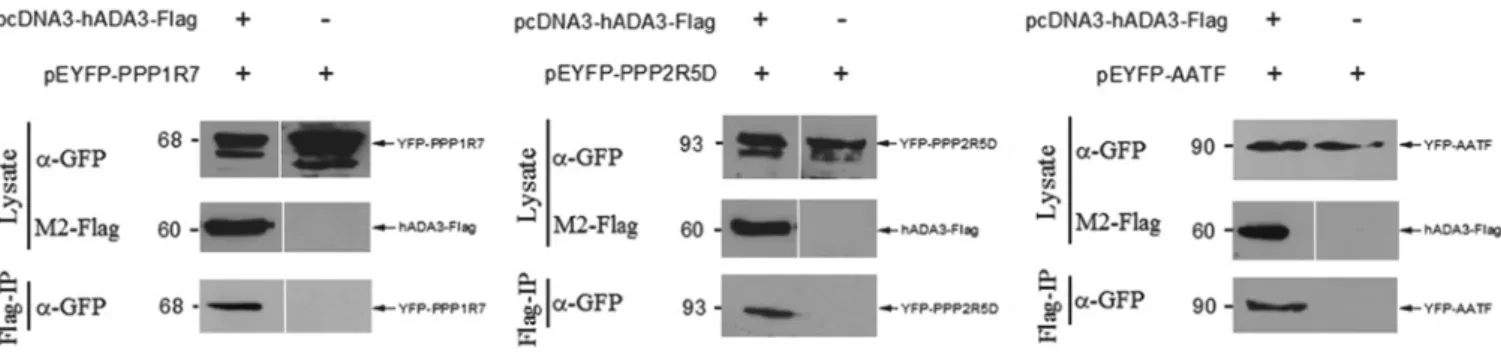
Figure 4 Co-IP of PPP1R7, PPP2R5D and AATF with hADA3 from mammalian cells
Co-IPs were carried out using U2OS cells transfected with a plasmid expressing an hADA3 partner protein fused to YFP (pEYFP-PPP1R7, pEYFP-PPP2R5D and pEYFP-AATF), and also when co-transfected with a plasmid expressing FLAG-tagged hADA3 (pcDNA3-hADA3-FLAG). Cell lysates were prepared and immunoprecipitated with a monoclonal anti-FLAG antibody followed by Western blotting with a polyclonal anti-GFP antibody (α-GFP) to detect the YFP fusion proteins. Molecular masses are given in kDa on the left-hand side of each gel.
nucleus. As was observed for hADA3, PHF21A was excluded from the nucleolus (Figure 3, upper panel).
We next investigated co-localization of hADA3 with each of the potential interacting partner proteins (Figure 3, lower panel).
As negative controls, CFP-tagged hADA3 was co-expressed with either YFP (Figure 3, lower panel A) or with a YFP-tagged hGIP, a protein with which it does not interact [23] (Figure 3, lower panel B). As a positive control, CFP-tagged hADA3 was co-expressed with a known interactor, hADA2A, fused to YFP (Figure 3, lower panel C). As expected, the fluorescence signal for hADA2 and hADA3 showed almost complete co-localization. When CFP-tagged hADA3 was co-expressed with the potential novel interactor proteins expressed as YFP fusions, co-localization was observed in the merged images, consistent with potential interaction, although the extent of overlap and the cellular compartment in which signals co-localized differed between different partner proteins (Figure 3, lower panels D–G). AATF and hADA3 displayed distributions similar to that shown when each protein was expressed independently with co-localization of the signals mostly in the nucleus. AATF was observed in some subnuclear regions resembling nucleoli from which hADA3 seemed to be excluded (Figure 3, lower panel D). In some cells, AATF and hADA3 signals also co-localized in a brightly staining compartment near the cell periphery (Figure 3, lower panel D).
When hADA3 was co-expressed with PPP1R7, the nuclear localization of hADA3 was significantly reduced, with both proteins residing mostly outside of the nucleus (Figure 3, lower panel E).
When hADA3 was co-expressed with PPP2R5D, the two proteins showed almost identical distribution, with an overlapping signal observed in the nucleus and in the cytoplasm in the merged image (Figure 3, lower panel F). PHF21A retained its predominantly nuclear localization in the presence of hADA3.
Within the nucleus the two proteins displayed an overlapping distribution (Figure 3, lower panel G).
Taken together, the microscopy analysis demonstrates hADA3 co-localization with each of the newly identified partners in human cells, consistent with potential physical interaction between the proteins. Combined with the results of the yeast two-hybrid protein-interaction assay, the two independent methods suggest these may be biologically relevant interactions.
Co-IP of hADA3 with its interacting partners
To determine whether the hADA3 interactions detected in yeast also occur in mammalian cells, Co-IP experiments were performed. For this, hADA3 tagged with a FLAG epitope
and the potential partner proteins fused to YFP, YFP–PPP1R7, YFP–PPP2R5D, YFP–PHF21A and YFP–AATF, were expressed in U2OS cells. The whole-cell lysates prepared from these transfected cells were mixed and hADA3–FLAG precipitated using anti-FLAG antibody-conjugated agarose beads. We showed that YFP–PPP1R7, YFP–PPP2R5D and YFP–AATF co-immuno- precipitated with hADA3–FLAG (Figure 4). Co-IP was not observed for YFP–PHF21A. Although we could not verify PHF21A as an authentic hADA3-interacting partner, we did not exclude this protein from our subsequent analyses as the lack of Co-IP may have been due to the low expression level of this fusion protein (results not shown).
The newly identified hADA3-interacting partners have differential effects on reporter gene expression
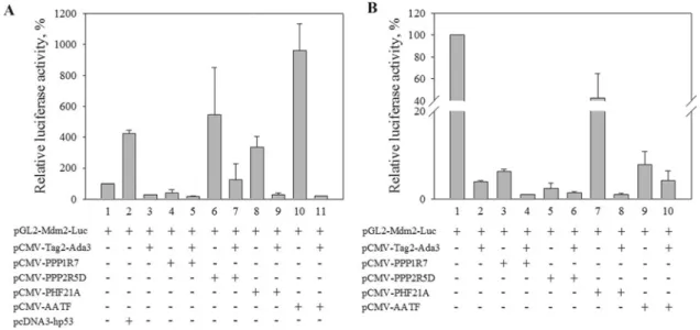
After demonstrating the existence of physical interactions between hADA3 and PPP1R7, AATF and PPP2R5D in mammalian cells, we wanted to test whether these interactions might have functional consequences. Given the role of hADA3- containing complexes in transcriptional regulation, we addressed this by assessing the transcriptional regulatory potential of each of the partner proteins. For this purpose, plasmids directing the expression of one of the identified hADA3-interacting partners were transiently transfected into HeLa cells and U2OS cells together with, or in the absence of, a second plasmid driving hADA3 expression. The reporter plasmid used in these experiments contained the luciferase gene under the control of theMDM2gene promoter. Expression of hADA3 has previously been reported to effect p53-mediated transcription of theMDM2 gene [9]. Assays were conducted in both a p53-negative cell line HeLa and in p53-expressing U2OS cells to examine the effects of expression of the newly identified hADA3 interactors both in the presence and absence of the transcriptional transactivator tumour suppressor p53. The luciferase activities determined 2 days after transfections demonstrated that PPP1R7, PPP2R5D, PHF21A and AATF had differential effects on the MDM2 promoter- driven reporter gene expression. In the absence of p53, PPP1R7 alone had a weak negative effect on reporter gene expression from the MDM2promoter (Figure 5A). In contrast, PPP2R5D, PHF21A and AATF each acted as activators (Figure 5A). In fact, under the conditions tested, in HeLa cells PPP2R5D and AATF activated the MDM2promoter as well or better than observed for p53 expression (Figure 5A). Co-transfection of hADA3 had a negative effect on expression, significantly reducing the level observed when each partner was expressed without hADA3 (Figure 5A). The extent of negative regulation, however, in the
Figure 5 Effect of expression of hADA3-interacting proteins and hADA3 on activation of a luciferase reporter gene under the control of theMDM2promoter HeLa cells (p53 negative) (A) and U2OS cells (p53 positive) (B) were transfected with a reporter plasmid (pGL2-Mdm2-Luc) encoding a luciferase gene under the control of an ADA3-dependent p53-activated promoter (MDM2) or co-transfected with the reporter plasmid and the indicated plasmids (0.2μg) expressing hADA3 (p-CMV-Tag2-Ada3) or one of the interacting partner proteins [pCMV-PPP1R7, pCMV-PPP2R5D, pCMV-PHF21A, pCMV-AAAIF or human p53 (pcDNA-hp53)] as indicated. Luciferase activity was measured 48 h post-transfection. Results are means+−S.D.
from three independent assays.
different protein combinations differed significantly, excluding the likelihood that the reduction was merely due to an unspecific toxic effect of the hADA3 protein. In p53-expressing U2OS cells, expression directed by the MDM2 reporter was very strong, consistent with the previously reported activation of this promoter by p53 [9]. The three verified hADA3 interactor proteins and PHF21A had a negative effect on this activation, with hADA3 expression resulting in a further reduction in reporter gene expression (Figure 5B). Curiously, in this experimental arrangement, expression of hADA3 alone in the p53-positive cells in the absence of the partner proteins had a negative effect on transcription directed by the MDM2 promoter. This was somewhat surprising since hADA3 and p53 were expected to act as co-activators, but the results of the present study might reflect the effect of overproducing hADA3, as only one subunit of a multi-protein complex, in cells with endogenous, rather than overexpressed, levels of p53.
DISCUSSION
We report in the present study identification of novel interacting partners of hADA3, a factor implicated in regulation of gene expression by several mechanisms. ADA3 is a constituent of several HAT complexes, among them SAGA and ATAC, which share a common GCN5 catalytic subunit [27]. Although hADA3 itself has no catalytic activity, it is required for the structural integrity of these complexes, and is believed to make physical contacts with GCN5 and also with transcriptional activators. In addition, involvement of hADA3 is suggested in CBP [CREB (cAMP-response-element-binding protein)-binding protein]/p300 mediated acetylation and nuclear receptor-mediated gene regulation [28]. Through these complexes, or by other interactions, hADA3 is also involved in p53 and cyclin A modi- fications, HPV (human papillomavirus) E6-mediated cell trans- formation, steroid synthesis regulation andβ-catenin-mediated transcription activation [9,29,30]. As the role of p53 in different types of cancers, and similarly, the role of HPV E6 in ovarian
cancer, β-catenin signalling in colorectal adenocarcinomas, retinoid signalling through RAR (retinoic acid receptor) in acute promyelocytic leukaemia and squamous cell cancers is well doc- umented, exploring the possible contribution of hADA3 to these malignancies is strongly warranted. One step in this direction is uncovering the protein network in which hADA3 fulfils its roles.
We first identified the novel hADA3 interacting partners by yeast two-hybrid library screening and then analysed their co- localization with hADA3 in human cells using fluorescence microscopy and their effects on transcriptional activation of a p53-regulated reporter gene.
Taken together with co-IP of three of the novel partners with hADA3 from human cell extracts, the analyses support the identified interactions of hADA3 as being biologically relevant and important for the further study of hADA3 function. Additional support comes from an overview of the known functions of the newly identified hADA3 interactors. Despite the incomplete knowledge of their specific functions, their known activities and their identified other partners make them in many aspects highly probable candidates for participation in hADA3-mediated processes.
Among the identified hADA3 partners, AATF is a 63 kDa protein with nuclear and nucleolar localization [31–34].
Expression of AATF was reported in a number of tissues, including the heart, kidney, placenta and thymus. It was first characterized as a physiological partner of apoptosis-inducing kinase MAP3K12 [MAPK (mitogen-activated protein kinase) kinase kinase 12] [33,34]. MAP3K12 is a member of the serine/threonine kinase family that forms heterodimers with CREB and Myc through its leucine-zipper domain [35], which is one of the well characterized protein–protein interaction motifs [24]. Furthermore, MBIP (MAP3K12-binding inhibitory protein 1), a reported physiological partner of MAP3K12, plays important roles in acetylation of histone H3 and H4 that are known components of an ADA-containing HAT complex (ATAC) [12].
AATF, upon phosphorylation [34], also activates transcription of p53, a known interacting partner of hADA3 [7,9]. Overexpression of ADA3 elevated cellular p53 levels and increased transcriptional
activity of the genes that are the targets of p53 [9]. Therefore identifying AATF as a partner of ADA3 suggests another link between the ATAC complex and p53 transcription.
Although PHF21A, a protein with 78 kDa molecular mass, was identified as a potential hADA3-interacting protein in our yeast two-hybrid screen and displayed a nuclear localization pattern remarkably similar to hADA3, we were unable to verify it as a bona fidehADA3 partner protein by co-IP. PHF21A is expressed in a variety of human tissues with a high rate of expression in the brain [36–38]. This protein, also called BHC80, contains a PHD zinc finger domain and is a component of BRAF35/HDAC complex (BHC). It also has C- and N-terminal leucine zippers, one of which has high homology with the leucine zipper contained in the transcriptional activator c-Myc [36]. BHC, in association with HDAC, suppresses the expression of neuronal genes in non- neuronal cells. The BHC co-repressor complex bears MHG and zinc finger domains, which were reported to be important in transcriptional suppression [36,38]. BHC functions as a chromatin remodeller, mediating specific modifications in histones. The PHF21A protein also inhibits demethylation of Lys4 in histone H3. Therefore, being a component of HDAC, PHF21A would be another potentially important partner of hADA3 protein, although this remains to be established.
Reversible protein phosphorylation is one of the most widely utilized mechanisms to regulate cellular processes. The activity of many cellular enzymes is determined by the net protein phosphorylation level, which is, in turn, determined by the balance of specific protein kinase and protein phosphatase activities [39].
We found regulatory subunits (PPP2R5D and PPP1R7/SDS22) of two different phosphatases as two further partners of hADA3.
The 70 kDa PPP2R5D protein is a subunit of the PP2A family. PP2A is a major intracellular phosphatase that regulates diverse cellular processes such as DNA replication, transcription, signal transduction, inflammation, cell growth and differentiation [40,41]. It is known that PP2A or PP2A-containing holoenzymes control ERK (extracellular-signal-regulated kinase) 1/2 activation via their interaction with and dephosphorylation of the upstream kinase Raf1 (v-raf-1 murine leukaemia viral oncogene homologue 1), leading to sequential phosphorylation/activation of MEK (MAPK/ERK kinase) 1/2 and ERK1/2. MEK1 is as an essential component of the MAPK signal transduction pathway, which is involved in many cellular processes such as proliferation, differentiation, transcription regulation and development. It activates the ERK1 and ERK2 MAPKs [42].
PPP1R7, also known as SDS22, is a regulatory subunit of PP1, a member of one of the major classes of serine/threonine protein phosphatases. PP1 regulates numerous cellular processes including mitosis by dephosphorylation of key proteins [43–45].
PPP1R7 is a conserved leucine-rich repeat protein required for the completion of mitosis in yeast [46]. PPP1R7 is essential for chromosome dysjunction during the metaphase/anaphase transition. PPP1R7 was also suggested to facilitate PP1 mediated dephosphorylation by interaction with PP1 [43,47].
It is a known interaction partner of Aurora B, a mitotic serine/threonine kinase that contributes to the regulation of cell- cycle progression. PPP1R7 dephosphorylates phospho-Aurora B and, thus, indirectly functions to stabilize kinetochore–
microtubule interactions [48,49]. Other important interaction partners of Aurora are CPEB1 (cytoplasmic polyadenylation element binding protein 1), JTB (jumping translocation breakpoint), TACC (transforming acidic coiled-coil-containing protein) 1, TPX2, PPP2CA, the PP1 isoforms PPP1CA, PPP1CB and PPP1CC, and p53/TP53 [48–52]. The interaction of Aurora B with TACC1 and TACC3 may be significant for TACC1 involvement in the promotion of cell division prior to the
formation of differentiated tissue. Aurora B also acts as a key regulatory component of the p53/TP53 pathway, critical for oncogenic transformation of cells [52]. Further connections between the pathways are evident with hADA3-dependent acetylation being required for increases in p53 stability and target- gene induction [9].
PPP1R7 is also known to interact with the oncogenic transcription factor YEATS4, a component of the NuA4 HAT acetyltransferase complex, which is involved in transcriptional activation of several genes principally by acetylation of nucleosomal histones H4 and H2A and also in numerous complexes with chromatin remodelling activity [53,54]. YEATS2 is a scaffolding subunit of the ADA2A-containing (ATAC) HAT complex, comprising of CSRP2BP (cysteine-rich protein 2-binding protein), KAT2A, TADA2L (transcriptional adapter 2α), TADA3L (transcriptional adapter 3α), ZZ3, MBIP, WDR5 (WD repeat-containing protein 5), YEATS2, CCDC101 and DR1 [11]. TACC3 plays a role in the microtubule-dependent coupling of the nucleus and the centrosome. Both of these proteins interact with hADA3-containing complexes GCN5L2 and PCAF (p300/CREB-binding protein associated factor) [50].
Taken together, we have identified three new interacting partners for hADA3 and provide here evidence to support these interactions occurring in human cells. The existence of a potential fourth partner was implicated on the basis of a yeast two-hybrid screen. Further experimentation is needed to elucidate the biological consequences of the newly identified hADA3 interactions. The known physiological roles of the new proteins, however, suggest relevance for the function of hADA3 in transcriptional regulation. Further functional analysis of these interactions may identify new pathways regulated by ADA3- containing complexes.
AUTHOR CONTRIBUTION
Sevil Zencir carried out cloning, screening, verified the interactions and analysed data;
Adam Sike helped to verify the interactions; Melanie Dobson interpreted data and edited the paper; Ferhan Ayaydin performed confocal analyses; Imre Boros and Zeki Topcu designed the experiments, interpretated the data and wrote the paper.
FUNDING
This work was supported by the TUBITAK (Scientific and Technological Research Council of Turkey) [grant number 108T945 (to Z.T.)], the NSERC (Natural Sciences and Engineering Research Council of Canada) [grant number 155268 (to M.J.D.)], the European Union via the European Social Fund, the T´arsadalmi Meg´ujulas Operative Program (project numbers T´AMOP-4.2.2/B-10/1-2010-0012 and T´AMOP-4.2.1./B-09/1/KONV- 2010-0005) the Hungarian State Science Fund and the Turkish–Hungarian Bilateral Project.
REFERENCES
1 Li, B., Carey, M. and Workman, J. L. (2007) The role of chromatin during transcription.
Cell128, 707–719
2 Suganuma, T. and Workman, J. L. (2011) Signals and combinatorial functions of histone modifications. Annu. Rev. Biochem.80, 473–499
3 Martinez, E., Kundu, T. K., Fu, J. and Roeder, R. G. (1998) A human SPT3–TAFII31–
GCN5-L acetylase complex distinct from transcription factor IID. J. Biol. Chem.273, 23781–23785
4 Wieczorek, E., Brand, M., Jacq, X. and Tora, L. (1998) Function of TAF(II)-containing complex without TBP in transcription by RNA polymerase II. Nature393, 187–191 5 Ogryzko, V. V., Kotani, T., Zhang, X., Schiltz, R. L., Howard, T., Yang, X. J., Howard, B. H.,
Qin, J. and Nakatani, Y. (1998) Histone-like TAFs within the PCAF histone acetylase complex. Cell94, 35–44
6 Eberharter, A., Sterner, D. E., Schieltz, D., Hassan, A., Yates, III, J. R., Berger, S. L. and Workman, J. L. (1999) The ADA complex is a distinct histone acetyl transferase complex inSaccharomyces cerevisiae. Mol. Cell. Biol.19, 6621–6631
7 Wang, T., Kobayashi, T., Takimoto, R., Denes, A. E., Snyder, E. L., El-Deiry, W. S. and Brachmann, R. K. (2001) hADA3 is required for p53 activity. EMBO J.20, 6404–6413
8 Balasubramanian, R., Pray-Grant, M. G., Selleck, W., Grant, P. A. and Tan, S. (2002) Role of the Ada2 and Ada3 transcriptional coactivators in histone acetylation. J. Biol. Chem.
277, 7989–7995
9 Nag, A., Germaniuk-Kurowska, A., Dimri, M., Sassack, M. A., Gurumurthy, C. B., Gao, Q., Dimri, G., Band, H. and Band, V. (2007) An essential role of human Ada3 in p53 acetylation. J. Biol. Chem.282, 8812–8820
10 Grau, B., Popescu, C., Torroja, L., Ortu˜no-Sahag´un, D., Boros, I. and Ferr´us, A. (2008) Transcriptional adaptor ADA3 ofDrosophila melanogaster is required for histone modification, position effect variegation, and transcription. Mol. Cell. Biol.28, 376–385
11 Wang, Y. L., Faiola, F., Xu, M., Pan, S. and Martinez, E. (2008) Human ATAC is a GCN5/PCAF-containing acetylase complex with a novel NC2-like histone fold module that interacts with the TATA-binding protein. J. Biol. Chem.283, 33808–33815 12 Guelman, S., Kozuka, K., Mao, Y., Pham, V., Solloway, M. J., Wang, J., Wu, J., Lill, J. R
and Zha, J. (2009) The double-histone-acetyltransferase complex ATAC is essential for mammalian development. Mol. Cell. Biol.29, 1176–1188
13 Lee, K. K., Sardiu, M. E., Swanson, S. K., Gilmore, J. M., Torok, M., Grant, P. A., Florens, L., Workman, J. L. and Washburn, M. P. (2011) Combinatorial depletion analysis to assemble the network architecture of the SAGA and ADA chromatin remodeling complexes. Mol. Syst. Biol.7, 503
14 Horiuchi, J., Silverman, N., Marcus, G. A. and Guarente, L. (1995) ADA3, a putative transcriptional adaptor, consists of two separable domains and interacts with ADA2 and GCN5 in a trimeric complex. Mol. Cell. Biol.15, 1203–1209
15 Muratoglu, S., Georgieva, S., P´apai, G., Scheer, E., En¨unl¨u, I., Komonyi, O., Cserp´an, I., Lebedeva, L., Nabirochkina, E., Udvardy, A. et al. (2003) Two differentDrosophila ADA2 homologues are present in distinct GCN5 histone acetyltransferase-containing complexes. Mol. Cell. Biol.23, 306–321
16 Pankotai, T., Komonyi, O., Bodai, L., Ujfaludi, Z., Muratoglu, S., Ciurciu, A., Tora, L., Szabad, J. and Boros, I. (2005) The homologousDrosophila transcriptional adaptors ADA2a and ADA2b are both required for normal development but have different functions.
Mol. Cell. Biol.25, 8215–8227
17 Gamper, A. M., Kim, J. and Roeder, R. G. (2009) The STAGA subunit ADA2b is an important regulator of human GCN5 catalysis. Mol. Cell. Biol.29, 266–280
18 Li, C. W., Dinh, G. K., Zhang, A. and Chen, J. D. (2008) Ankyrin repeats cofactors interact with ADA3 and modulate its coactivator function. Biochem. J.413, 349–357 19 Zhang, A., Yeung, P. L., Li, C. W., Tsai, S. C., Dinh, G. K., Wu, X., Li, H. and Chen, J. D.
(2004) Identification of a novel family of ankyrin repeats containing cofactors for p160 nuclear receptor coactivators. J. Biol. Chem.279, 33799–33805
20 Topcu, Z. and Borden, K. L. (2000) The yeast two-hybrid system and its pharmaceutical significance. Pharm. Res.17, 1049–1055
21 Fields, S. and Song, O. (1989) A novel genetic system to detect protein-protein interactions. Nature340, 245–246
22 Rose, M. D., Winston, F. M. and Hieter, P. (1990) Methods in Yeast Genetics: a Laboratory Course Manual, Cold Spring Harbor Laboratory Press, New York 23 Zencir, S., Ovee, M., Dobson, M, Mohanty, S. and Topcu, Z. (2011) Identification of
brain-specific angiogioenesis inhibitor 2 as an interaction partner of glutaminase interacting protein. Biochem. Biophys. Res. Commun.411, 792–797 24 Topcu, Z., Mack, D. L., Hromas, R. A. and Borden, K. L. (1999) The promyelocytic
leukemia protein PML interacts with the proline-rich homeodomain protein PRH: a RING may link hematopoiesis and growth control. Oncogene18, 7091–7100
25 MacDonald, P. N. (2001) Two-hybrid Systems. Humana Press, Totowa
26 Zhang, Z., Schwartz, S., Wagner, L. and Miller, W. (2000) A greedy algorithm for aligning DNA sequences. J. Comput. Biol.7, 203–214
27 Lee, K. K. and Workman, J. L. (2007) Histone acetyltransferase complexes: one size doesn’t fit all. Nat. Rev. Mol. Cell Biol.8, 284–295
28 Li, C. W., Ai, N., Dinh, G. K., Welsh, W. J. and Chen, J. D. (2010) Human ADA3 regulates RARalpha transcriptional activity through direct contact between LxxLL motifs and the receptor coactivator pocket. Nucleic Acids Res.38, 5291–5303
29 Germaniuk-Kurowska, A., Nag, A., Zhao, X., Dimri, M., Band, H. and Band, V. (2007) Ada3 requirement for HAT recruitment to estrogen receptors and estrogen-dependent breast cancer cell proliferation. Cancer Res.167, 11789–11797
30 Kumar, A., Zhao, Y., Meng, G., Zeng, M., Srinivasan, S., Delmolino, L. M., Gao, Q., Dimri, G., Weber, G. F., Wazer, D. E. et al. (2002) Human papillomavirus oncoprotein E6 inactivates the transcriptional coactivator human ADA3. Mol. Cell. Biol.22, 5801–5812 31 Di Padova, M., Bruno, T., De Nicola, F., Iezzi, S., D’Angelo, C., Gallo, R., Nicosia, D.,
Corbi, N., Biroccio, A., Floridi, A. et al. (2003) Che-1 arrests human colon carcinoma cell proliferation by displacing HDAC1 from the p21WAF1/CIP1 promoter. J. Biol. Chem.
278, 36496–36504
32 Fanciulli, M., Bruno, T., Di Padova, M., De Angelis, R., Iezzi, S., Iacobini, C., Floridi, A.
and Passananti, C. (2000) Identification of a novel partner of RNA polymerase II subunit 11, Che-1, which interacts with and affects the growth suppression function of Rb. FASEB J.14, 904–912
33 Bruno, T., De Angelis, R., De Nicola, F., Barbato, C., Di Padova, M., Corbi, N., Libri, V., Benassi, B., Mattei, E., Chersi, A. et al. (2002) Che-1 affects cell growth by interfering with the recruitment of HDAC1 by Rb. Cancer Cell2, 387–399
34 Bruno, T., De Nicola, F., Iezzi, S., Lecis, D., D’Angelo, C., Di Padova, M., Corbi, N., Dimiziani, L., Zannini, L., Jekimovs, C. et al. (2006) Che-1 phosphorylation by ATM/ATR and chk2 kinases activates p53 transcription and the G2/M checkpoint. Cancer Cell6, 473–486
35 Fukuyama, K., Yoshida, M., Yamashita, A., Deyama, T., Baba, M., Suzuki, A., Mohri, H., Ikezawa, Z., Nakajima, H., Hirai, S. and Ohno, S. (2000) MAPK upstream kinase (MUK)-binding inhibitory protein, a negative regulator of muk/dual leucine zipper-bearing kinase/leucine zipper protein kinase. J. Biol. Chem.275, 21247–21254
36 Hakimi, M., Bochar, D. A., Chenoweth, J., Lane, W. S., Mandel, G. and Shiekhattar, R.
(2002) A core-BRAF35 complex containing histone deacetylase mediates repression of neuronal-specific genes. Proc. Natl. Acad. Sci. U.S.A.99, 7420–7425
37 Lan, F., Collins, R. E., De Cegli, R., Alpatov, R., Horton, J. R., Shi, X., Gozani, O., Cheng, X. and Shi, Y. (2007) Recognition of unmethylated histone H3 lysine 4 links BHC80 to LSD1-mediated gene repression. Nature448, 718–722
38 Klajn, A., Ferrai, C., Stucchi, L., Prada, I., Podini, P., Baba, T., Rocchi, M., Meldolesi, J.
and D’Alessandro, R. (2009) The rest repression of the neurosecretory phenotype is negatively modulated by BHC80, a protein of the BRAF/HDAC Complex. J. Neurosci.29, 6296–6307
39 Hunter, T. (1994) 1001 protein kinases redux towards 2000. Semin. Cell Biol.5, 367–376 40 Mccright, B. and Virshup, D. M. (1995) Identification of a new family of protein
phosphatase 2a regulatory subunits. J. Biol. Chem.270, 26123–26128
41 Yu, U. Y. and Ahn, J. H. (2010) Phosphorylation on the PPP2R5DB regulatory subunit modulates the biochemical properties of protein phosphatase 2A. BMB Rep.43, 263–267 42 Adams, D. G., Coffee, Jr, R. L., Zhang, H., Pelech, S., Strack, S. and Wadzinski, B. E.
(2005) Positive regulation of Raf1-MEK1/2- ERK1/2 signaling by protein serine/threonine phosphatase 2A holoenzymes. J. Biol. Chem.280, 42644–42654
43 Ceulemans, H., Vulsteke, V., De Maeyer, M., Tatchell, K. and Stalmans, W. (2002) Binding of the concave surface of the Sds22 superhelix to theα4/α5/α6-triangle of protein phosphatase-1. J. Biol. Chem.277, 47331–47337
44 Jiang, Y., Scott, K. L., Kwak, S. J., Chen, R. and Mardon, G. (2011) Sds22/PP1 links epithelial integrity and tumor suppression via regulation of myosin II and JNK signaling.
Oncogene30, 3248–3260
45 Ceulemans, H., Van Eynde, A., PeˆArez-Callej´on, E., Beullens, M., Stalmans, W. and Bollen, M. (1999) Structure and splice products of the human gene encoding sds22, a putative mitotic regulator of protein phosphatase-1. Eur. J. Biochem.262, 36–42 46 Ohkura, H. and Yanagida, M. (1991)S. pombe gene sds22+essential for a midmitotic
transition encodes a leucine-rich repeat protein that positively modulates protein phosphatase-1. Cell64, 149–157
47 Renouf, S., Beullens, M., Wera, S., Van Eynde, A., Sikela, J., Stalmans, W. and Bollen, M.
(1995) Molecular cloning of a human polypeptide related to yeast sds22, a regulator of protein phosphatase-1. FEBS Lett.375, 75–78
48 Cheetham, G. M., Knegtel, R. M., Coll, J. T., Renwick, S. B., Swenson, L., Weber, P., Lippke, J. A. and Austen, D. A. (2002) Crystal structure of aurora-2, an oncogenic serine/threonine kinase. J. Biol. Chem.277, 42419–42422
49 Bayliss, R., Sardon, T., Ebert, J., Lindner, D., Vernos, I and Conti, E. (2004) Determinants for Aurora-A activation and Aurora-B discrimination by TPX2. Cell Cycle3, 404–407 50 Gangisetty, O., Lauffart, B., Sondarva, G. V., Chelsea, D. M. and Still, I. H. (2004) The transforming acidic coiled coil proteins interact with nuclear histone acetyltransferases.
Oncogene23, 2559–2563
51 Eyers, P. A., Churchill, M. E. and Maller, J. L. (2005) The Aurora A and Aurora B protein kinases: a single amino acid difference controls intrinsic activity and activation by TPX2.
Cell Cycle4, 784–789
52 Kim, H. J., Cho, J. H., Quan, H. and Kim, J. R. (2011) Down-regulation of aurora B kinase induces cellular senescence in human fibroblasts and endothelial cells through a p53-dependent pathway. FEBS Lett.16, 3569–3576
53 Delaval, B., Ferrand, A., Conte, N., Larroque, C., Hernandez-Verdun, D., Prigent, C. and Birnbaum, D. (2004) Aurora B–TACC1 protein complex in cytokinesis. Oncogene23, 4516–4522
54 Doyon, Y., Selleck, W., Lane, W. S., Tan, S. and Cote, J. (2004) Structural and functional conservation of the NuA4 histone acetyltransferase complex from yeast to humans. Mol.
Cell. Biol.24, 1884–1896 Received 16 March 2012/14 November 2012; accepted 20 November 2012
Published as BJ Immediate Publication 20 November 2012, doi:10.1042/BJ20120452