MLL-fusion-driven leukemia requires SETD2 to safeguard genomic integrity
Anna Skucha
1,2, Jessica Ebner
2, Johannes Schmöllerl
2, Mareike Roth
3, Thomas Eder
2, Adrián César-Razquin
1, Alexey Stukalov
1, Sarah Vittori
1, Matthias Muhar
3, Bin Lu
4, Martin Aichinger
3, Julian Jude
3, André C. Müller
1, Balázs Gy ő rffy
5, Christopher R. Vakoc
4, Peter Valent
6, Keiryn L. Bennett
1, Johannes Zuber
3,
Giulio Superti-Furga
1,7& Florian Grebien
2,8MLL-fusions represent a large group of leukemia drivers, whose diversity originates from the vast molecular heterogeneity of C-terminal fusion partners of MLL. While studies of selected MLL-fusions have revealed critical molecular pathways, unifying mechanisms across all MLL-fusions remain poorly understood. We present the fi rst comprehensive survey of protein – protein interactions of seven distantly related MLL-fusion proteins. Functional investigation of 128 conserved MLL-fusion-interactors identi fi es a speci fi c role for the lysine methyltransferase SETD2 in MLL-leukemia. SETD2 loss causes growth arrest and differentiation of AML cells, and leads to increased DNA damage. In addition to its role in H3K36 tri-methylation, SETD2 is required to maintain high H3K79 di-methylation and MLL-AF9-binding to critical target genes, such as Hoxa9 . SETD2 loss synergizes with pharmacologic inhibition of the H3K79 methyltransferase DOT1L to induce DNA damage, growth arrest, differentiation, and apoptosis. These results uncover a dependency for SETD2 during MLL-leukemogenesis, revealing a novel actionable vulnerability in this disease.
DOI: 10.1038/s41467-018-04329-y
OPEN
1CeMM Research Center for Molecular Medicine of the Austrian Academy of Sciences, Vienna 1090, Austria.2Ludwig Boltzmann Institute for Cancer Research, Vienna 1090, Austria.3Research Institute of Molecular Pathology, Vienna 1030, Austria.4Cold Spring Harbor Larboratory, Cold Spring Harbor, 11724 NY, USA.5MTA TTK Lendület Cancer Biomarker Research Group, Institute of Enzymology, Second Department of Pediatrics, Semmelweis University, Budapest 1094, Hungary.6Department of Internal Medicine I. Division of Hematology and Hemostaseology, Ludwig Boltzmann Cluster Oncology, Medical University of Vienna, Vienna 1090, Austria.7Center for Physiology and Pharmacology, Medical University of Vienna, Vienna 1090, Austria.8Institute for Medical Biochemistry, University of Veterinary Medicine, Vienna 1210, Austria. These authors contributed equally: Johannes Zuber, Giulio Superti-Furga, Florian Grebien. Correspondence and requests for materials should be addressed to F.G. (email:florian.grebien@vetmeduni.ac.at)
1234567890():,;
L eukemia-associated fusion proteins serve as a paradigm for modern cancer research, as the molecular machineries that provide their functional cellular context have emerged as amenable to targeted molecular approaches1,2. Families of related leukemia fusion proteins that share genomic and biological properties represent unique opportunities to study how the combination of distinct functional protein modules can drive oncogenic transformation. The largest family of “multi-partner translocations” in acute leukemia comprises fusions involving the product of the KMT2A (MLL) gene. MLL-fusion proteins are found in acute lymphoblastic leukemia (ALL) and acute myeloid leukemia (AML) and are often associated with adverse prognosis, particularly in pediatric patients
3. Expression of MLL-fusions enhances proliferation and blocks myeloid differentiation of hematopoietic progenitor cells, leading to their pathological accumulation. In line, many MLL-fusions can act as potent oncogenes in cell line models and animal models of leukemia
4.
In leukemia, the MLL N-terminus takes part in >120 different translocations, resulting in the generation of MLL-fusion proteins encompassing more than 75 different partner genes
5. It has therefore been proposed that the oncogenic activity of MLL- fusion proteins depends on chromatin targeting functions exerted by the MLL N-terminus in combination with other functional properties encoded by the fusion partners
6. Several regions in the MLL N-terminus are critical for the activity of MLL-fusions. For instance, the CxxC-domain is essential for DNA binding of MLL- fusion proteins
7. Furthermore, the MLL-interacting protein Menin links MLL-fusion proteins with LEDGF, and the H3K36me3-binding PWWP domain of LEDGF is critical for the function of MLL-fusions
8. In fact, a direct fusion of the LEDGF PWWP domain to MLL was able to replace Menin altogether
9.
Numerous studies have established strong links between the molecular function of the fusion partner and the mechanistic basis of oncogenic transformation in MLL-fusion-induced leu- kemogenesis
4. Pioneering biochemical experiments have shown that several fusion partners of MLL, such as AF4, AF9, and ENL are members of the DOT1L complex (DotCom) and the super- elongation complex (SEC)
10–13, which are both involved in transcriptional control. As the SEC can regulate the transcrip- tional activity of RNA polymerase II, it was hypothesized that these MLL-fusions induce aberrant regulation of transcriptional elongation on MLL-target genes
14.
A large number of factors was shown to influence the onco- genic properties of MLL-fusions, including signaling proteins
15–17, epigenetic modulators
18–21, and transcription fac- tors
22–24, as well as the wild-type MLL protein
25. However, it is unclear whether these molecular mechanisms pertain to the entire family of MLL-fusions or if they specifically affect the leukemo- genicity of isolated MLL-fusion proteins. In fact, specific mole- cular mechanisms of oncogenic transformation were postulated to prevail for selected MLL-fusions. For instance, inhibition of the arginine methyltransferase PRMT1 was shown to reduce the leukemic potential of several oncogenic fusion proteins, including MLL-EEN and MLL-GAS7, but not MLL-AF9, MLL-AF10, or MLL-ENL
26,27. Furthermore, the enzymatic activity of CBP was shown to be required for leukemogenic activity of fusions of MLL with the histone acetyltransferase CREBBP
28,29. Finally, dimer- ization might play an important role in nuclear translocation and oncogenic transformation in fusions of MLL to the cytoplasmic partner proteins GAS7 and AF1p, yet the underlying molecular mechanism is unclear
30,31.
Here, we set out to survey the molecular composition of a diverse subset of distantly related MLL-fusion protein complexes to characterize their unique and common properties, and to reveal possible actionable vulnerabilities that are based on specific molecular mechanisms shared by MLL-fusions. We identify the
methyltransferase SETD2 as an interactor of all MLL-fusion proteins. shRNA-mediated and CRISPR/Cas9-mediated loss of SETD2 leads to growth arrest and differentiation of MLL-fusion- expressing cells in vitro and in vivo. Moreover, we show that loss of SETD2 is associated with increased DNA damage. SETD2 loss disrupts a H3K36me3-H3K79me2 signature on MLL-target genes and sensitizes MLL-AML cells to pharmacologic inhibition of the known MLL-fusion protein effector DOT1L. In summary, we describe a novel dependency for SETD2 in the initiation and maintenance of MLL-rearranged leukemia, highlighting a novel vulnerability in this disease.
Results
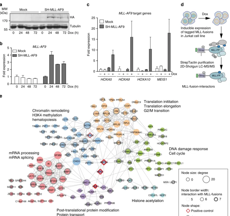
Functional proteomic survey of MLL-fusion proteins. Reason- ing that critical effectors might be enriched among the physical interaction partners of distantly related MLL-fusion proteins, we undertook an unbiased survey of the protein–protein interactions of MLL-fusion proteins in leukemia cells. Using FRT/Flp-medi- ated locus-specific cassette exchange, we generated isogenic Jurkat leukemia cell lines allowing for Doxycycline (Dox)-inducible, single-copy expression of affinity-tagged variants of seven MLL- fusions that were previously proposed to employ different molecular mechanisms of oncogenic transformation (MLL-AF1p, MLL-AF4, MLL-AF9, MLL-CBP, MLL-EEN, MLL-ENL, MLL- GAS7, Fig. 1a, b and Supplementary Fig. 1a-c). Subcellular frac- tionation revealed that all selected MLL-fusion proteins localized to the nucleus (Supplementary Fig. 1d) and were capable of inducing expression of the MLL-fusion-target genes HOXA5, HOXA9, HOXA10, and MEIS1 (Fig. 1c).
Protein complexes around MLL-fusion proteins were purified from nuclear lysates of cell lines expressing seven distinct MLL- fusions (Fig. 1d and Supplementary Fig. 2a). Purifications were analyzed by LC-MS/MS using both one-dimensional and two- dimensional gel-free proteomic approaches, recovering 4600 proteins in total, engaging in 15,094 putative interactions (Fig. 1e and Supplementary Fig. 2b)
32–34. p-value-based filtering for the 300 most significant interactions per MLL- fusion resulted in a network of 960 high-confidence cellular proteins (Supplementary Fig. 2b). Validation of the network confirmed previously reported interactions of MLL-fusions with protein complexes important for transcriptional control and epigenetic regulation, including the PAF complex, the SWI/
SNF complex, and Polycomb Repressor Complex 1 (Supple- mentary Fig. 2c)
12,35–37. The network also revealed abundant unique interaction partners of all MLL-fusion proteins, indicating that distinct MLL-fusions can engage specific molecular pathways. 406 proteins in the network (42.3%) co- purified with more than one MLL-fusion protein, while 128 proteins (13.3%) interacted with at least five of the seven MLL- fusions (Supplementary Fig. 2b), indicating a strong degree of topological conservation within the MLL-interaction network.
Further analysis of the 128 conserved partners of MLL-fusion proteins revealed six distinct protein communities (p < 0.01;
Supplementary Table 1), whose annotation retrieved molecular functions that are highly relevant to the biology of MLL-fusion proteins, including chromatin remodeling, transcriptional elongation, and hematopoiesis (Fig. 1e). Interestingly, protein families that had not been reported to interact with MLL-fusion proteins before were also identified, such as factors involved in DNA-repair, RNA splicing, and RNA transport.
In summary, our comprehensive analysis and validation of the
cellular interaction networks shows that distinct MLL-fusion
proteins engage in a high number of direct, as well as indirect
protein–protein interactions. Structurally different MLL-fusion
proteins share 128 conserved interaction partners, which are
enriched in six functional communities that are highly relevant for the biology of MLL-fusion proteins.
shRNA screen identi fi es SETD2 as an effector of MLL-fusions.
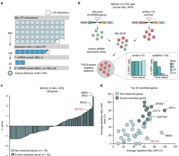
As our primary validation reduced the number of potential cri- tical effectors in the network of MLL-fusion protein-interactors from 960 to 128, we next aimed to further narrow down the circle of candidate proteins using sequential functional genomic approaches (Fig. 2a). To systematically investigate the functional contribution of the conserved 128 MLL-interaction partners to MLL-fusion-dependent leukemia, we devised a shRNA screen in the human MLL-AF9-expressing AML cell line MOLM-13. In the
system used by us, transcriptional coupling of fluorescent reporter proteins to shRNA expression upon Dox-induction allows for dynamic monitoring of competing growth kinetics in mixed cell populations expressing experimental shRNAs (GFP) vs. non- targeting control shRNAs (shRen.713, dsRed, Fig. 2b). While expression of a control shRNA did not differentially affect cell proliferation in mixed populations over time, strong shRNA- induced negative selection of GFP-positive cells was observed upon targeting of MEN1, an interaction partner of all seven investigated MLL-fusion proteins with a well-known function in MLL-fusion-induced leukemogenesis
38(Fig. 2b, bottom). We used this setup to systematically test the effects of 128 shRNA- pools targeting conserved MLL-fusion interaction partners on
ANP32B HDGFRP2
LUZP1 ZC3H18 PRRC2C
MSN NIFK
STIP1 CSNK2A2
PSME1 RPL5
DCTN1 PPHLN1
NOLC1 EIF4G1 VIM
DNAJC9 TCOF1
RPL22 EIF3I
LUC7L2 LYAR GNL2
EIF3L TPM4
RBM4 RPL26
PPP2R1A
CSNK2A1 LASP1 HSP90AA1
MAPRE1
PSMA3
MAP7D1 ZFP91 CHAMP1 RFC2 RFC1
MEN1 KMT2A
SMC1A ZMYM3 RUNX1
FUBP1
ZMYM2 PBRM1
DPY30 SMARCB1
WDR18
SMARCC1 RPL18
PA2G4 ACTB
BOD1L1 PRKAR1A
PML HOOK3 ANAPC7
CSNK2B PSMA7
CTCF
NUP50 NXF1
PSIP1HCFC1 NUMA1
RAE1 TPR
KPNA2 PPP1R9B SMC3 SND1
CIR1 SUPT6H AKAP8L C14orf166
RRP12 ARL6IP4
THOC7
SRSF11CSTF2 PRPF4B
SPEN
SF3A3 DDX46
KAT7
JADE2
CDCA7L NOP2
JADE3 MCM3
WDR33
SCAF11 SON MCM2 TBL3 GPATCH4
ZC3H4 CPSF4
XAB2 IK DHX16 CHERP
DNAJC8 LUC7L3
ZNF512 ZNF22 COPB2 VEZF1
ZNF131 TIMM50
TCEB3 SETD2
TRIM28 CCT7
PAICS TBL1XR1 CCT4
CHD3 CCT3
GATAD2B CHD4
CCT5
UBAP2L
WBP11 SF3B2 IKZF1
NUDT21
Translation inititation Translation elongation G2/M transition
DNA damage response Cell cycle
Histone acetylation Post-translational protein modification
Protein transport mRNA processing
mRNA splicing
Chromatin remodeling H3K4 methylation hematopoiesis
0 20
Node size: degree
Positive control Other Node shape:
Node border width:
interaction with MLL-fusions
5 6 7
a
MLL-AF9 target genes
0 10 15 20 25
Fold expression
5
– + – + – + – + – + – + – + – + Dox HOXA5 HOXA9 HOXA10 MEIS1
Mock SH-MLL-AF9
StrepTactin purification 2D-Shotgun LC-MS/MS
MLL-FP Strep
MLL-fusion-interactors
Strep MLL-FP
0 1 2 3 4 5
Fold expression
0 24 48 72 0 24 48 72 Dox (h) Mock
SH-MLL-AF9 MLL-AF9
Inducible expression of tagged MLL-fusions in Jurkat cell line
Dox
b
c d
e
HA Tubulin 0 24 48 72 0 24 48 72
SH-MLL-AF9 Mock
Dox (h) 55
170 MW (kDa)
Fig. 1Functional proteomic survey of the MLL-fusion interactome. Cells expressing Strep-HA (SH)-tagged MLL-AF9 or mock-transfected cells were treated with Dox for the indicated time points and transgene expression was monitored by immunoblotting (a) and qPCR (b) (means ± s.d.n=3).cSH-MLL-AF9- expressing cells were treated with Dox for 24 h and the expression of indicated MLL-target genes was measured by qPCR (mean ± s.d.n=3).dSchematic illustration of the strategy of affinity purification of protein complexes associated with MLL-fusion proteins from nuclear lysates of cell lines expressing affinity-tagged MLL-fusion proteins.eGene ontology (GO) enrichment of six distinct protein communities among the core 128 interactors shared by at least 5 of 7 MLL-fusion proteins
AML cell growth. Relative depletion of all shRNA-pools was normalized to a negative-control shRNA (shRen.713) and to a strong growth inhibitory positive-control shRNA (shMyb.670)
22. As the read-out of this screen is inhibition of proliferation, we would expect that essential genes would be enriched among the strongest hits. Indeed, scoring of shRNA-induced effects upon knockdown of all 128 MLL-fusion interactors revealed a strong positive correlation between growth inhibition and reported gene essentiality (Fig. 2c)
39–42. However, as we intended to identify proteins with MLL-fusion-specific roles in the network, we rea- soned that their loss-of-function might preferably affect the via- bility of MLL-fusion-expressing leukemia cells. Thus, we re-
screened the 40 candidate genes with the highest confidence in MLL-AF9-expressing MOLM-13 cells and in the MLL-wild-type leukemia cell lines K562 and HL-60. As expected, knockdown of MLL-interactors with known essential functions, such as RFC1, SF3A3, or CCT3, led to growth inhibition in both MLL-fusion cells and MLL-wild-type leukemia cells (Fig. 2d). In contrast, depletion of the known MLL-interaction partner MEN1 selec- tively inhibited growth of MLL-fusion cells while sparing MLL- wild-type cells, proving the validity of the chosen strategy.
Knockdown of the gene encoding the H3K36me3-specific methyltransferase SETD2 showed a strong bias towards inhibition of proliferation of MLL-fusion-expressing cells, while causing
a b
c d
–3 –2 –1 0 1 2 3
Non-essential genes (n = 76) Known essential genes (n = 52)
MEN1 RFC1
SETD2 RFC2 MOLM-13 (MLL-AF9)
128 genes
0 20 40 60 80 100 120
0 20 40 60 80 100 120
Average depletion MLL-AF9 (%) Average depletion MLL-wild type (%)
Non-essential genes Known essential genes
RFC1
MEN1 SETD2
SF3A3 CCT3
SUPT6H Top 40 candidate genes
TRE3G tGFP PGK Neo
128 pools (6 shRNAs/gene)
Mix 50:50
Induce shRNA expression (Dox)
FACS-based negative selection
dsRed PGK Neo
shRen.713 (control) MOLM-13 rtTA3 cells
(human MLL-AF9)
shMEN1.1105
% GFP+ cells
Time (days) shRen.713
% GFP+ cells
Time (days)
TRE3G
Days 6 10 2
14 40
960
1° shRNA screen (MLL-r) 128
Interaction with ≥ 5 MLL-FP
2° shRNA screen (MLL-r vs. MLL-wt) Critical effectors of MLL-FPs MLL-FP interactome
= 20 interactors
z-score
Fig. 2shRNA screen identifies SETD2 as a critical effector of MLL-fusions.aSchematic representation of thefiltering strategy. Affinity purification coupled to mass spectrometry identified 960 candidate genes (top 300 interactors per bait, ranked byp-value) to interact with at least one of seven selected MLL- fusion proteins. 128 proteins interacted with≥5 of seven MLL-fusions. 40 candidate genes were screened in MLL-rearranged vs. MLL-wild-type cell lines.
Each square corresponds to 20 interactors.bSchematic outline of retroviral vectors and experimental design of the FACS-based negative selection RNAi screen. Competitive proliferation assays were set up by mixing cells in a 50:50 ratio (experimental-GFP vs. control-dsRed) and cultivation in the presence of Dox. The relative ratio of GFP-positive vs. dsRed-positive cells was monitored byflow cytometry over 14 days. Bar graphs (bottom) represent the performance of positive (shMEN1.1105) and negative (shRen.713) controls shown as percentage of GFP+cells over time.cSummary of RNAi screening data in the MOLM-13 cell line. Positive and negativez-scores correspond to candidate genes with stronger and weaker depletion phenotypes. Gene essentiality was assigned based on published datasets.dAverage depletion values obtained from two subsequent RNAi screens performed in the MLL- AF9-expressing MOLM-13 cell line are plotted against mean depletion values from counterscreens performed in two MLL-wild-type cell lines (K562 and HL-60)
negligible cell depletion in K562 and HL-60 cells, suggesting an MLL-fusion-specific function (Fig. 2d, Supplementary Fig. 3a).
SETD2 was one of 42 core proteins that interacted with all seven MLL-fusions, as it co-purified with MLL-fusion proteins in all affinity-purification experiments with significant peptide coverage (Supplementary Fig. 3b). Consistently, co-immunoprecipitation experiments showed that this interaction involved the N-terminal part of MLL, which is conserved in all MLL-fusion proteins studied, and the C-terminus of SETD2, which encompasses all annotated functional domains of the SETD2 protein (Supplementary Fig. 3c).
SETD2 expression was higher in AML samples than in normal hematopoietic stem and progenitor cell types and mature myeloid cells
43(Supplementary Fig. 3d). SETD2 expression was highest in patients with 11q23 aberrations featuring MLL-translocations, as compared to samples with normal karyotype AML or myelodys- plastic syndrome (Supplementary Fig. 3e).
Thus, we identified the methyltransferase SETD2 as a selective effector of MLL-AF9 AML cells through functional genomic investi- gation of conserved interaction partners of MLL-fusion proteins.
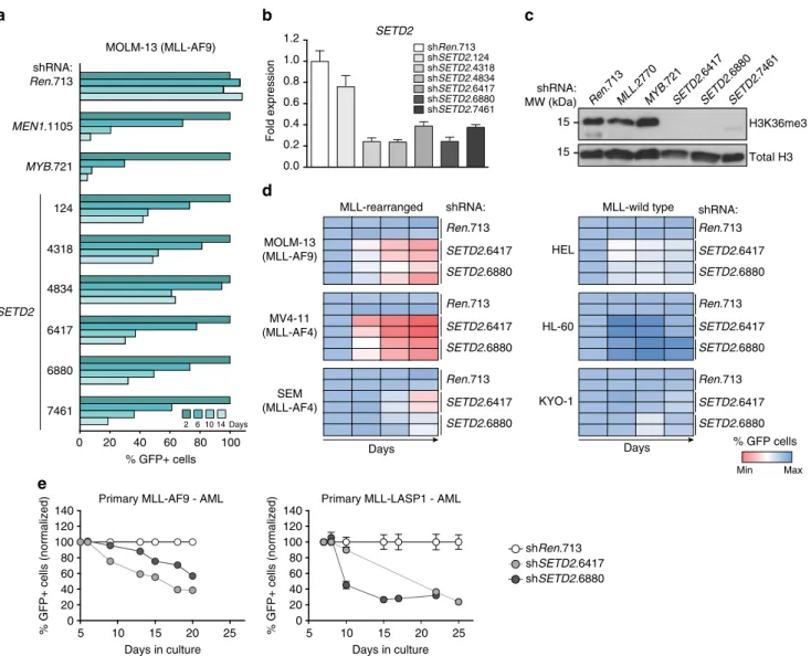
SETD2 is essential for MLL-fusion-expressing cells. We next aimed at validating the shRNA screen results at the level of individual shRNAs. Expression of all six SETD2-targeting shRNAs induced strong growth inhibition in MOLM-13 cells, in line with significant downregulation of SETD2 mRNA (Fig. 3a, b).
As SETD2 is the only protein known to mediate tri-methylation of H3K36
44, we investigated the effect of SETD2 downregulation on total cellular H3K36me3 levels. The three strongest SETD2- targeting shRNAs caused near-complete clearance of global H3K36me3 signals (Fig. 3c). Importantly, growth inhibition was not generally associated with H3K36me3 loss, as we did not observe changes in global H3K36me3 levels upon downregulation of MLL and MYB, which strongly affected proliferation of MLL- AF9 AML cells (Fig. 3c).
As our screening data indicate that SETD2 knockdown selectively inhibits the proliferation of MLL-fusion-expressing cells, we sought to extend this observation to a larger panel of human leukemia cell lines. In addition to MOLM-13 cells also the MLL-AF4-expressing cell lines MV4-11 and SEM showed
5 10 15 20 25
0 20 40 60 80 100 120 140
Days in culture
% GFP+ cells (normalized)
Primary MLL-AF9 - AML
5 10 15 20 25
0 20 40 60 80 100 120 140
Days in culture
% GFP+ cells (normalized)
Primary MLL-LASP1 - AML
shSETD2.6417 shRen.713 shSETD2.6880 6417
6880 SETD2
7461 shRNA:
Ren.713
MEN1.1105
MYB.721
MOLM-13 (MLL-AF9)
% GFP+ cells 20 40 60 80 100 0
Days 6 10
2 14
MLL-rearranged MLL-wild type
MOLM-13 (MLL-AF9)
MV4-11 (MLL-AF4)
SEM (MLL-AF4)
shRNA:
Ren.713 SETD2.6417 SETD2.6880
Ren.713 SETD2.6417 SETD2.6880
Ren.713 SETD2.6417 SETD2.6880
HEL
HL-60
KYO-1
shRNA:
Ren.713 SETD2.6417 SETD2.6880
Ren.713 SETD2.6417 SETD2.6880
Ren.713 SETD2.6417 SETD2.6880
Min Max
% GFP cells Days
124
4318
4834
SETD2
0.0 0.2 0.4 0.6 0.8 1.0 1.2
Fold expression
shRen.713 shSETD2.124 shSETD2.4318 shSETD2.4834
shSETD2.7461 shSETD2.6880 shSETD2.6417
Days shRNA:
MW (kDa)
H3K36me3 Total H3 15
15
SETD2 .7461 SETD2
.6880 SETD2
.6417 MYB
.721 MLL
.2770 Ren
.713
a b c
d
e
Fig. 3SETD2 is required for proliferation of MLL-leukemia cells.aResults of FACS-based competitive proliferation assay shown as the percentage of GFP- positive MOLM-13 cells expressing individualSETD2-targeting shRNAs in the presence of Dox over 14 days. One representative experiment of four is shown.bqPCR analysis ofSETD2mRNA levels in MOLM-13 cells expressing indicated shRNAs after 48 h of Dox treatment (mean ± s.d.n=3).cWestern blot analysis of H3K36me3 levels in MOLM-13 cells expressing indicated shRNAs after 72 h of Dox treatment.dHeatmap representation of competitive proliferation assays performed in human cell lines harboring MLL rearrangements (left) vs. MLL-wild-type cells (right) expressing indicated shRNAs targetingSETD2as described ina. Representative results of two out of three experiments are shown.eTime course of GFP expression of primary human AML cells from patients expressingMLL-AF9andMLL-LASP1fusion genes expressing indicated shRNAs (mean ± s.d.n=3)
significant anti-proliferative responses and induction of apoptosis upon SETD2 knockdown (Fig. 3d, left, and Supplementary Fig. 4a-c). In contrast, SETD2 downregulation in the MLL-wild- type cell lines HEL, HL-60, and KYO-1 only marginally affected proliferation (Fig. 3d, right, Supplementary Fig. 4c). SETD2 knockdown resulted in a strong proliferative disadvantage in primary human AML cells from patients expressing MLL-AF9 and MLL-LASP1 fusion genes (Fig. 3e).
Taken together, downregulation of SETD2 caused a strong anti-proliferative response in primary human AML cells and cell lines expressing various MLL-fusion genes, suggesting a requirement for SETD2 in the oncogenic context of MLL-fusion proteins.
MLL-target genes exhibit high H3K36me3 levels. To investigate the relationship between SETD2 and MLL-fusions we profiled the global distribution of the SETD2-dependent H3K36me3 mark in a
mouse AML cell line expressing MLL-AF9 and activated Nras (G12D)
22using ChIP-Rx
45. As expected, H3K36me3 was present on gene bodies of expressed genes. We found that MLL-AF9 target genes
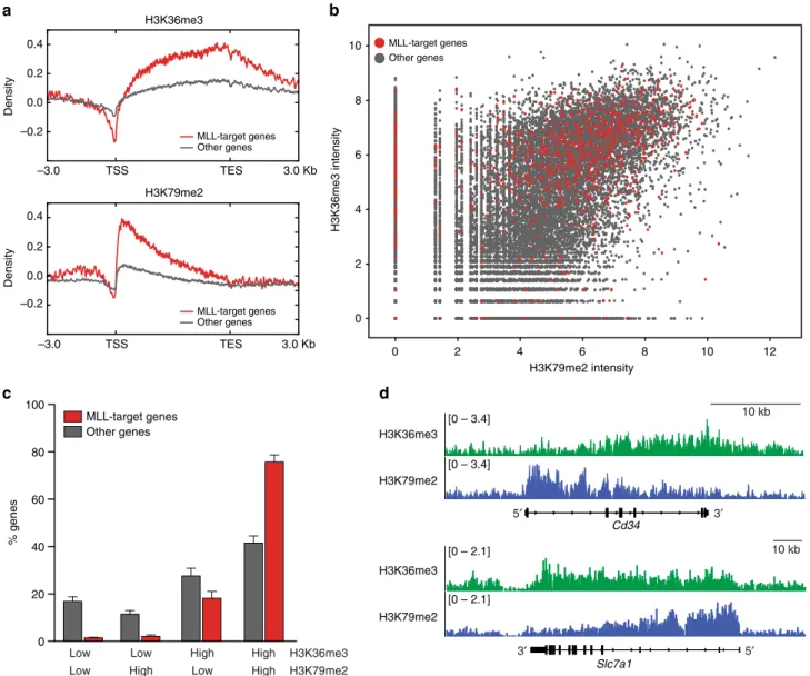
22displayed significantly higher H3K36me3 levels than non- MLL-target genes (Fig. 4a, top). In line with previous data, MLL- fusion target genes were also highly positive for the DOT1L- dependent H3K79me2 mark
21(Fig. 4a, bottom), and the global levels of H3K36me3 and H3K79me2 modifications showed a strong positive correlation in mouse MLL-AF9/NrasG12D cells (Fig. 4b). However, while only 42% of non-MLL-target genes were highly positive for both marks, 76% of MLL-target genes displayed a combined H3K36me3/H3K79me2-high signature (Fig. 4c, d, and Supplementary Fig. 5). As MLL-fusion-binding was shown to correlate with H3K79me2 on MLL-target genes
21and depend on recognition of H3K36me3 marks
9, these data suggest that the SETD2-dependent H3K36me3 modification is part of an epige- netic signature that marks target genes of MLL-fusion proteins together with H3K79me2.
–3.0 TSS TES 3.0 Kb
–0.2 0.0 0.2 0.4
–3.0 TSS TES 3.0 Kb
–0.2 0.0 0.2 0.4
Other genes MLL-target genes
H3K79me2 H3K36me3
Other genes MLL-target genes
0 20 40 60 80 100
% genes
Other genes MLL-target genes
High
Low H3K36me3
H3K79me2 Low
Low Low
High High High
0 2 4 6 8 10
0 2 4 6 8 10 12
H3K79me2 intensity
H3K36me3 intensity
Other genes MLL-target genes
Cd34
Slc7a1 H3K79me2
H3K36me3
H3K79me2 H3K36me3
10 kb
10 kb
DensityDensity
5′
3′
5′ 3′
[0 – 2.1]
[0 – 2.1]
[0 – 3.4]
[0 – 3.4]
a b
d c
Fig. 4MLL-target genes are marked by a H3K36me3-H3K79me2 signature.aMetagene plots of ChIP-Rx data for H3K36me3 (top) and H3K79me2 (bottom) for MLL-target genes (red) or non-MLL-target genes (gray) in mouseMLL-AF9/NrasG12D AML cells.bDot plot of normalized ChIP-Rx signal intensities for H3K36me3 vs. H3K79me2 marks on MLL-target genes (red) or non-MLL-target genes (gray) in the mouse genome (PearsonR=0.44,p<
2.2 × 10⁻¹⁶).cBar graph showing percentages of genes among MLL-target genes and non-MLL-target genes associated with the indicated histone marks;
low: not exceeding input counts per gene, high: exceeding input counts per gene (mean ± s.d.n=2).dH3K36me3 (green) and H3K79me2 profiles (blue) of selected MLL-AF9-target genes
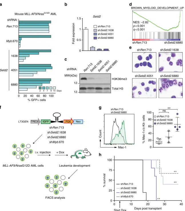
Loss of SETD2 induces myeloid differentiation and DNA damage. Next we sought to characterize global changes in gene expression upon SETD2 ablation. Dox-inducible knockdown of Setd2 caused a strong growth disadvantage in mouse MLL-AF9/
NrasG12D cells (Fig. 5a, b, Supplementary Fig. 6a). Setd2 downregulation led to almost complete loss of cellular H3K36me3 signals (Fig. 5c). RNA-seq analysis showed that 868 genes were differentially expressed upon Setd2 knockdown in
Setd2
0 0.5 1.5
1
Fold expression
shRen.713
shSetd2.6880 shSetd2.4051 shSetd2.1638
BROWN_MYELOID_DEVELOPMENT_UP
shRen.713 shSetd2.6880 NES: –2.82
p< 0.001 q< 0.001
LT3GEN TRE3G tGFP PGK Neo
MLL-AF9/NrasG12D AML cells Leukemia development
FACS analysis i.v. injection + Dox 0 20 40 60 80 100
% GFP+ cells shRNA:
Ren.713
Myb.670
Mouse MLL-AF9/NrasG12D AML
Days 4 7 9
2 14
H3K36me3 Total H3 Ren
.713
Setd2 .6880 Setd2
.4051 Setd2
.1638 shRNA:
MW(kDa) 12
shRen.713
shSetd2.6880 shSetd2.4051
shSetd2.1638 1638
4051 Setd2
6880
0 10 20 30 40
0 25 50 75 100
Days post transplant
% survival shRen.713
shSetd2.6880 shSetd2.1638
shMyb.670
Start Dox
**
**
**
0 20 40 60 80 100
% Mac-1+/c-Kit- cells
shRen.713 shSetd2.1638 shSetd2.6880 ns **
shSetd2.6880 shRen.713
Mac-1
Count
12
shMyb.670 shSetd2.6880 shSetd2.1638 shRen.713
a b d
c
e
f g
h
Fig. 5SETD2 loss induces myeloid differentiation of MLL-leukemia cells.aResults of competitive proliferation assay shown as the percentage of GFP- positive mouseMLL-AF9/NrasG12D AML cells expressing indicated shRNAs in the presence of Dox over 14 days. One representative experiment out of three is shown.bqPCR analysis ofSetd2mRNA levels inMLL-AF9/NrasG12D cells expressing indicated shRNAs after 48 h of Dox treatment (mean ± s.d.n
=3).cWestern blot analysis of H3K36me3 levels inMLL-AF9/NrasG12D cells expressing indicated shRNAs after 72 h of Dox treatment.dGene Set Enrichment Analysis indicating myeloid differentiation ofMLL-AF9/NrasG12D AML cell line upon knockdown ofSetd2. NES, Normalized Enrichment Score.
eMicrographs of cytospin preparations ofMLL-AF9/NrasG12D AML cells after expression of indicated shRNAs.fSchematic representation of the in vivo transplantation assay.MLL-AF9/NrasG12D AML cells expressing indicated shRNAs were transplanted into sub-lethally irradiated C57BL/6 Ly5.1 mice. Dox was administrated to the drinking water starting from day 3 (arrow inh) and disease progression was monitored by bioluminescence imaging. Terminally sick mice were sacrificed and analyzed.gFlow cytometric analysis of Mac-1 onMLL-AF9/NrasG12D AML cells upon shRNA-mediatedSetd2knockdown in vivo (mean ± s.d.n≥3).hKaplan–Meier survival curves of C57BL/6 Ly5.1 mice transplanted withMLL-AF9/NrasG12D AML cells expressingSetd2- targeting shRNAs. Survival curves of mice transplanted with cells expressingSetd2-targeting shRNAs were compared to cells expressing control shRNAs using a Log-rank test. ns, not significant, **p< 0.01 (t-test)
MLL-AF9/NrasG12D AML cells. While 458 genes were upregu- lated, 410 genes were downregulated in shSetd2-cells, (padj < 0.01, Supplementary Fig. 6b). Consistent with a role of SETD2 in the DNA damage response
46, MLL-AF9/NrasG12D AML cells expressing two different Setd2-targeting shRNAs showed upre- gulation of DNA damage-associated gene expression (Supple- mentary Fig. 6c). Indeed, Setd2 downregulation resulted in significantly higher levels of DNA damage in the absence of genotoxic agents, as measured by alkaline comet assay and phosphorylated histone H2AX (γ-H2AX, Supplementary Fig. 7a- c). Knockdown of Setd2 led to induction of p21, reduced cell cycle progression, and induction of apoptosis of MLL-AF9/NrasG12D AML cells (Supplementary Fig 7c-e).
Gene Set Enrichment Analysis revealed that Setd2 down- regulation induced gene expression programs associated with myeloid differentiation (Fig. 5d). Indeed, Setd2-deficient cells displayed clear signs of terminal myeloid maturation, including nuclear segmentation and increased granularity (Fig. 5e), as well as downregulation of the progenitor marker c-Kit and upregula- tion of the mature myeloid marker Mac-1 (Supplementary Fig. 8a). Similarly, SETD2 downregulation in the human MLL- AF4-expressing cell line MV4-11 and in MLL-AF9-expressing MOLM-13 cells induced increased cell surface levels of the differentiation marker CD36 together with macroscopic changes characteristic of myeloid maturation (Supplementary Fig. 8b, c).
To test whether loss of SETD2 could overcome the MLL-AF9- dependent differentiation block in myeloid progenitors in vivo, we transplanted MLL-AF9/NrasG12D AML cells expressing Setd2- targeting or control shRNAs into recipient mice. shRNA expression was induced by Dox-administration and the immuno-phenotype of the developing leukemia was analyzed by flow cytometry (Fig. 5f).
Knockdown of Setd2 induced strong downregulation of c-Kit concomitant with upregulation of Mac-1 in leukemic cells in vivo, resulting in a significant increase in disease latency (Fig. 5g, h and Supplementary Fig. 8d). This is consistent with recent results showing that knockout of Setd2 greatly increased the latency of MLL-AF9-induced AML
47. While most leukemia cells isolated from moribund recipients of control AML cells showed robust shRNA expression (as measured by GFP levels), shRNA-expressing cells were strongly outcompeted by shRNA-negative cells in case of Setd2 knockdown in vivo (Supplementary Fig. 8e).
In summary, shRNA-mediated downregulation of SETD2 caused growth arrest, induction of apoptosis, and increased DNA damage. Furthermore, SETD2 loss induced terminal myeloid differentiation in MLL-fusion-expressing mouse and human AML cells in vitro and in vivo, indicating that the MLL- fusion-induced differentiation block is SETD2-dependent.
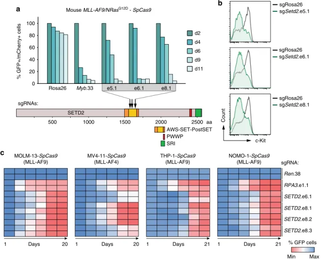
The SETD2 SET domain is required for AML growth. To interrogate the translational potential of our findings, we next
500 1000 1500 2000 2500
AWS-SET-PostSET PWWP
SRI
aa SETD2
sgRNAs:
0 20 40 60 80 100
d2 d4 d6 d9 d11
% GFP+/mCherry+ cells
Mouse MLL-AF9/NRasG12D - SpCas9
Rosa26 Myb.33 e5.1 e6.1 e8.1
a b
c
c-Kit
Count
sgSetd2.e5.1 sgRosa26
sgSetd2.e6.1 sgRosa26
sgSetd2.e8.1 sgRosa26
RPA3.e1.1 sgRNA:
Ren.38
SETD2.e6.1 SETD2.e8.1 SETD2.e8.2 SETD2.e8.3
Min Max
% GFP cells NOMO-1-SpCas9
(MLL-AF9)
Days 21
1 THP-1-SpCas9
(MLL-AF9)
Days 21
1 MV4-11-SpCas9
(MLL-AF4)
Days 20
1 MOLM-13-SpCas9
(MLL-AF9)
Days 20
1
Fig. 6The SETD2 SET domain is required for proliferation of MLL-leukemia cells.aResults of competitive proliferation assays shown as percentages of mCherry+/GFP+mouseMLL-AF9/NrasG12D-SpCas9AML cells expressing indicated sgRNAs (top). Data from one representative experiment of two are shown. Schematic representation of the domain structure ofSETD2(bottom).bFlow cytometric analysis of surface expression of c-Kit inMLL-AF9/
NrasG12D AML-SpCas9 cells upon CRISPR/Cas9-mediated mutagenesis ofSETD2.cHeatmap representation of competitive proliferation assays shown as the percentage of GFP-positive human AML cell lines stably expressingSpCas9cells upon CRISPR/Cas9-mediated mutagenesis of theSETD2SET domain
wanted to establish whether the methyltransferase activity of SETD2 is necessary for the observed effects. Direction of SpCas9- cleavage to functional protein domains was shown to greatly increase the read-out in competitive proliferation assays
48. We employed CRISPR/Cas9-mediated mutagenesis of the enzymatic SET domain to investigate whether catalytic activity of SETD2 was required for the oncogenicity of MLL-fusion proteins.
Introduction of three sgRNAs targeting the Setd2 SET domain in SpCas9-expressing MLL-AF9/NrasG12D AML cells led to a strong depletion of transduced cells over time, as shown before
48(Fig. 6a). Notably, CRISPR/Cas9-mediated mutagenesis of the Setd2 SET domain was sufficient to induce myeloid differentia- tion of MLL-AF9/NrasG12D cells, as measured by down- regulation of c-Kit together with upregulation of Mac-1 (Fig. 6b, Supplementary Fig. 9a). In line, we found strong anti-proliferative effects, induction of myeloid differentiation, and apoptosis upon mutagenesis of the SETD2 SET domain in the human MLL- rearranged AML cell lines MOLM-13 and MV4-11, THP-1, and NOMO-1 (Fig. 6c; Supplementary Fig. 9b-f, Supplementary Fig. 10).
These data show that the SET domain of SETD2 is required to sustain the proliferative capacity and differentiation block of MLL-fusion protein-expressing AML cells. In addition, these results imply a functional involvement of the H3K36me3 mark in the maintenance of MLL-fusion-dependent leukemia and offer a plausible route for future pharmacological intervention.
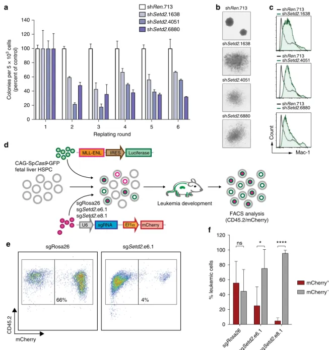
Ef fi cient MLL-fusion-mediated transformation requires SETD2. All our data show a strong functional requirement for the expression and activity of SETD2 in the progression of MLL- leukemia. As it is possible that alternative molecular mechanisms pertain during initiation of MLL-rearranged leukemia, we tested the involvement of SETD2 in this process. Setd2 knockdown resulted in a significant reduction in MLL-AF9-induced serial re- plating capacity of mouse hematopoietic stem/progenitor cells (HSPC), indicating that Setd2 expression is required to unleash the full oncogenic potential of MLL-AF9 (Fig. 7a). Setd2 ablation induced loss of compact colony morphology characteristic of blast-like cells and induced the formation of large, dispersed colonies reminiscent of mature myeloid clusters (Fig. 7b). Flow cytometry confirmed that Setd2-deficient colonies expressed high levels of the mature myeloid marker Mac-1 (Fig. 7c). To inves- tigate the effect of SETD2 on oncogenic transformation in vivo, we co-transduced fetal-liver-derived HSPC expressing a SpCas9 transgene
49with retroviral vectors encoding MLL-ENL and Setd2-targeting or control sgRNAs. The contribution of cells carrying sgRNA-induced mutations in the Setd2 SET domain to leukemia development was investigated by flow cytometric ana- lysis of mCherry expression upon transplantation (Fig. 7d). While cells expressing a control sgRNA showed robust contribution to MLL-ENL-induced leukemia in vivo (56%), cells carrying Setd2- mutations induced by two different sgRNAs were clearly under- represented in the leukemic population (5–25%, Fig. 7e, f).
Thus, both downregulation and mutagenesis of SETD2 was incompatible with efficient oncogenic transformation by MLL- fusion oncoproteins in vitro and in vivo. These results indicate that SETD2 expression is required for leukemogenesis and establish SETD2 as a novel actionable target in MLL-rearranged leukemia.
SETD2 loss sensitizes MLL-AML cells to DOT1L inhibition.
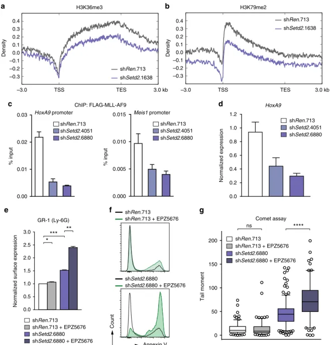
Finally, we aimed to obtain insight into the molecular mechanism that functionally connects SETD2 activity with MLL-fusion- induced leukemia. ChIP-Rx showed that Setd2 downregulation led to a concomitant reduction of both H3K36me3 and
H3K79me2 levels on MLL-target genes (Fig. 8a, b and Supple- mentary Fig 11a), while it did not alter H3K4me3 density (Sup- plementary Fig. 11b). As chromatin binding of MLL-fusion proteins was shown to depend on H3K36me3 recognition via the conserved interaction partner LEDGF
9, we hypothesized that reduction of H3K36me3 levels upon Setd2 loss might impair chromatin binding of MLL-fusions. Indeed, knockdown of Setd2 caused reduced binding of MLL-AF9 to the promoters of the canonical MLL-target genes Hoxa9 and Meis1 (Fig. 8c), leading to reduced Hoxa9 expression (Fig. 8d).
Given the dependence of the dual H3K36me3-H3K79me2 signature across MLL-target genes on SETD2 and the strong requirement of MLL-leukemia for the H3K79 methyltransferase DOT1L
21, we reasoned that SETD2 loss might cooperate with pharmacologic inhibition of DOT1L. Treatment of mouse MLL- AF9/NrasG12D and human MLL-AF4-expressing MV4-11 cells with the clinical DOT1L inhibitor EPZ5676
50potentiated the effects of SETD2 downregulation, including growth inhibition, induction of apoptosis, and onset of myeloid differentiation. Importantly, none of these parameters were altered in SETD2-proficient cells in the presence of the same concentrations of the inhibitor (Fig. 8e, f, Supplementary Fig. 11c-f). Finally, and consistent with a role of DOT1L in DNA repair
51, we found that combination of SETD2 loss and DOT1L inhibition synergized in the induction of DNA damage (Fig. 8g and Supplementary Fig. 11g).
These data show that loss of SETD2 expression in MLL-fusion AML cells interferes with the H3K36me3-H3K79me2-signature on MLL-target genes and impairs chromatin binding of MLL- fusion proteins. In consequence, SETD2 loss led to hyper- sensitization of MLL-leukemia cells to small-molecule-mediated DOT1L inhibition, which provides a rationale for potential future combination therapies in AML.
Discussion
Here, we provide the first comprehensive protein–protein inter- action network of MLL-fusion proteins in leukemia cells. We show that functional annotation of conserved MLL-interaction partners by loss-of-function screening enables the identification of conserved actionable nodes among the molecular network of MLL-fusions. As exemplified by our discovery of the histone methyltransferase SETD2 as an essential factor in MLL- rearranged leukemia, this approach can reveal novel genetic dependencies and yield new entry points for targeting of the entire group of MLL-rearranged leukemia, comprising over 75 different MLL-fusion partners.
Our results show that MLL-fusion proteins engage a large number of distinct protein–protein interactions. This could be explained by the modular nature of wild-type core MLL com- plexes
52,53and by the specific architecture of their leukemic counterparts
4. Our analysis of protein–protein interactions of selected, molecularly distinct MLL-fusion proteins greatly expands the cellular catalog of MLL-interacting proteins. In addition, it also provides novel insights into the topologies of MLL-fusion proteins that transform cells via unknown mechan- isms. For instance, interactome analysis of the MLL-GAS7-fusion protein showed that it specifically interacts with components of the CTLH complex, which is involved in microtubule dynamics and chromosome segregation
54.
A core set of 128 proteins constitutes the conserved inter-
actome of MLL-fusion proteins. In addition to known inter-
action partners of the wild-type MLL protein, such as MEN1,
DPY30, and LEDGF, it also contains several proteins whose
link to AML biology have only recently been established. For
instance, the protein SON interacts with MEN1 to regulate the
expression of leukemia-specific genes in a MLL-dependent
manner
55. Thus, functional annotation of the core network of MLL-fusion interactors will contribute to establish novel links between MLL-fusion proteins and important cellular processes that had previously not been associated with the biology of MLL-fusion proteins, such as mRNA splicing or protein transport. Given the involvement of these molecular pathways in basic cellular physiology, it is not surprising that more than one third of proteins in the network of conserved MLL-fusion protein interaction partners were identified as essential in recent genome-wide screens
39–42.
To discern MLL-fusion-associated genetic dependencies from essential genes we employed a subtractive shRNA screening approach. Strikingly, the gene encoding the methyltransferase SETD2 was identified as an MLL-fusion-specific hit from this screen with high confidence. shRNA-mediated knockdown as well as CRISPR/Cas9-induced mutagenesis of SETD2 caused proliferation arrest and myeloid differentiation of MLL-fusion- expressing primary and transformed human, and mouse AML cells in vitro and in vivo. This is surprising, because SETD2 has been implied to have tumor suppressor activity in various
sgRosa26 sgSetd2.e6.1
66% 4%
mCherry
CD45.2
CAG-SpCas9-GFP fetal liver HSPC
Luciferase MLL-ENL IRES
FACS analysis (CD45.2/mCherry) U6 sgRNA Ef1α mCherry
sgRosa26 sgSetd2.e6.1 sgSetd2.e8.1
Leukemia development
a b c
d
f
shRen.713
shSetd2.6880 shSetd2.4051 shSetd2.1638
Colonies per 5 × 10³ cells (percent of control)
Replating round 0
20 40 60 80 100 120 140
1 2 3 4 5 6
shRen.713
shSetd2.6880 shSetd2.4051 shSetd2.1638
sgRosa26 sgSetd2
.e6.1 sgSetd2
.e8.1 0
20 40 60 80 100 120
mCherry+ mCherry–
% leukemic cells
****
*
ns
Mac-1
Count
shSetd2.1638 shRen.713
shSetd2.4051 shRen.713
shSetd2.6880 shRen.713
e
Fig. 7SETD2 is required for oncogenic transformation by MLL-fusions.aSerial replating assay of primary MLL-AF9-transformed fetal liver cells upon shRNA-mediated knockdown ofSetd2. Colony numbers were normalized to cells expressing shRen.713 (mean ± s.d.n=3).bMorphology of colonies of MLL-AF9-transformed fetal liver cells upon shRNA-mediated knockdown ofSetd2.cFlow cytometric analysis of Mac-1 expression of MLL-AF9- transformed fetal liver cells upon shRNA-mediated knockdown ofSetd2.dSchematic representation of the in vivo transformation assay. Fetal liver cells fromSpCas9-transgenic mice were co-transduced with retroviral vectors expressing MLL-ENL andSetd2-targeting or control sgRNAs. Cells were transplanted into lethally irradiated C57BL/6 recipient mice. Terminally sick mice were sacrificed and bone marrow cells were analyzed.eRepresentative flow cytometry plots of donor-derived bone marrow cells from mice transplanted with MLL-ENL and a control sgRNA (sgRosa.26, left) or a sgRNA targeting the SET domain ofSetd2(sgSetd2e6.1, right). Live cells were gated.fQuantification offlow cytometry analysis of donor-derived bone marrow cells as shown ine(mean ± s.d.n≥4). ns, not significant, *p< 0.05, ****p< 0.0001 (t-test)
malignancies, including leukemia
56–58. SETD2 knockdown was reported to cause a driver-independent proliferative advantage of leukemia cells in vitro and in vivo
58. In contrast, another report showed that mutational disruption of the SETD2 SET domain was incompatible with MLL-AF9 AML cell growth
48. Furthermore, several genome-wide CRISPR/Cas9 screens identified SETD2 as an essential gene in leukemia cell lines
39–42. Finally, a recent report showed that while homozygous Setd2 deletion in the mouse strongly delayed leukemogenesis, heterozygous Setd2 deletion accelerated MLL-AF9-induced leukemia and caused chemoresistance
47. This is consistent with increased frequencies of SETD2 mutations in high-risk leukemia patients that show
increased genomic complexity and chromothripsis
57, and often exhibit therapy resistance and relapse
56. Therefore, as the majority of cancer patients carry heterozygous SETD2 mutations, SETD2 might act as a haplo-insufficient tumor suppressor. In contrast complete loss of SETD2 strongly impedes leukemia development.
The most prominent cellular function of SETD2 is its non- redundant H3K36 tri-methylation activity
44. The H3K36me3 mark is enriched on gene bodies of actively transcribed genes
59. We found that MLL-target genes displayed high H3K36me3 levels, validating our proteomic identification of SETD2 as an interactor of MLL-fusion proteins at the genomic level. Given the
HoxA9 promoter
0.00 0.01 0.02 0.03
% input
0.000 0.005 0.010 0.015
% input
Meis1 promoter ChIP: FLAG-MLL-AF9
shRen.713 shSetd2.6880 shSetd2.4051
0.0 0.2 0.4 0.6 0.8 1.0 1.2
shRen.713 shSetd2.6880 shSetd2.4051 HoxA9
Normalized expression
0.0 0.5 1.0 1.5 2.0 2.5 3.0
Normalized surface expression
GR-1 (Ly-6G) shRen.713 shSetd2.6880 shSetd2.4051
shRen.713 shSetd2.6880 shRen.713 + EPZ5676 shSetd2.6880 + EPZ5676
–3.0 TSS TES 3.0 kb
0.4 0.3 0.2 0.1 0.0 –0.1 –0.2 –0.3
shRen.713 shSetd2.1638 H3K36me3
Density
0.4 0.3 0.2 0.1 0.0 –0.1 –0.2 –0.3
–3.0 TSS TES 3.0 kb
shRen.713 shSetd2.1638 H3K79me2
Density
shRen.713 shSetd2.6880 shRen.713 + EPZ5676
0 50 100 150 200
shSetd2.6880 + EPZ5676
ns ****
Comet assay
Tail moment
*
*** **
Annexin V
Count
shRen.713
shRen.713 + EPZ5676
shSetd2.6880
shSetd2.6880 + EPZ5676
a b
c d
e f g
Fig. 8SETD2 loss sensitizes AML cells to DOT1L inhibition. Metagene plots of ChIP-Rx data for H3K36me3 (a) and H3K79me2 (b) after shRNA-mediated knockdown ofSetd2in mouseMLL-AF9/NrasG12D AML cells.cqPCR analysis of enrichment onHoxA9andMeis1promoter regions after MLL-AF9-FLAG- ChIP upon shRNA-mediated knockdown ofSetd2(mean ± s.d.n=3).dqPCR analysis ofHoxA9mRNA levels in MLL-AF9-FLAG cells expressing indicated shRNAs (mean ± s.d.n=3).eQuantification of surface expression of Gr-1 (Ly-6G) after shRNA-mediated knockdown ofSetd2in mouseMLL-AF9/
NrasG12D AML cells treated with EPZ5676 (500 nM) (mean ± s.d.n=2).fFlow cytometric analysis of Annexin V-positive cells in MV4-11 cells treated with EPZ5676 (50 nM) after shRNA-mediated knockdown ofSETD2.gQuantification of tail moments in an alkaline comet assay performed after shRNA- mediated knockdown ofSetd2in mouseMLL-AF9/NrasG12D AML cells treated with EPZ5676 (500 nM). Quantification of >150 cells is shown. ns, not significant, *p< 0.05, **p< 0.01, ***p< 0.001, ****p> 0.0001 (t-test)
large number of proteins harboring a H3K36me3-recognizing PWWP motif
60, several key cellular processes were postulated to be influenced by this epigenetic mark, including transcriptional elongation, splicing, and epigenetic control of gene expression
61. Consistent with a role for the PWWP motif of the MLL-interactor LEDGF in chromatin binding of MLL-fusion proteins
9, SETD2 loss caused reduced binding of MLL-AF9 to target promoters.
Thus, the interaction between SETD2 and MLL-fusion proteins could be required to ensure efficient chromatin binding of MLL- fusions through the maintenance of high H3K36me3 levels on MLL-target genes.
Our results clearly show that SETD2 is involved in the control of the DNA damage response in MLL-fusion-expressing cells.
SETD2-deficient cells exhibited high amounts of DNA damage and increased γ-H2AX levels, even in the absence of exogenous genotoxic stress. This is in line with a recent study showing that SETD2 mutations in leukemia impair the DNA damage response, thereby leading to chemotherapy resistance
47. This defect is attributed to loss of H3K36me3-dependent recruitment of repair proteins to sites of DNA damage. It was shown that MLL-AF9- transformed cells require an intact DNA damage response for full oncogenicity, as experimental induction of DNA damage led to differentiation of leukemia cells
62. In line with this, down- regulation of SETD2 was sufficient to induce myeloid differ- entiation of AML blasts. Therefore, a physical and functional interaction between MLL-fusion proteins and SETD2 could be required to guarantee efficient, H3K36me3-dependent repair of DNA lesions that continuously occur during MLL-fusion-induced oncogenic transcription. However, given the accumulation of DNA damage upon SETD2 loss, continuous SETD2 inhibition might result in increased formation of chemoresistant AML subclones.
We found that SETD2 was required for the maintenance of a specific dual H3K36me3-H3K79me2 signature on target genes of MLL-fusions. The H3K79me2 mark is catalyzed by the histone methyltransferase DOT1L, which is critical for the establishment and maintenance of MLL-rearranged leukemia
21. SETD2 down- regulation rendered MLL-fusion-expressing AML cells hyper- sensitive to the pharmacological DOT1L-inhibitor EPZ5676 (Pinometostat), which is currently in clinical development. It will be interesting to test whether this synergy can be exploited to efficiently target chemoresistant AML cells that are carrying SETD2 mutations.
In summary, our combined proteomic-functional genomic analysis of MLL-fusion protein interactors enabled us to reveal the molecular logic of how modular protein–protein interactions can influence the oncogenicity of MLL-fusion proteins. Our studies provide novel insights into the biology of MLL-fusion proteins and identify an unexpected dependency of MLL-fusion- expressing leukemia cells on the methyltransferase SETD2 during leukemia initiation and maintenance, validating SETD2 as an actionable target MLL-rearranged leukemia.
Methods
Constructs. MLL-fusion genes were assembled by fusing the cDNA of theMLLN- terminus (amino acids 1-1396) to C-terminal parts of AF1p(EPS15), AF4(AFF1), AF9(MLLT3), CBP (CREBBP), EEN (SH3GL1), ENL (MLLT1), and GAS7 (GAS7) and cloned into pcDNA5/FRT/TO/SH/GW. Generation of the miR-E shRNA vectors RT3GEN and RT3REN was previously described63,64. The SEM cell line was infected with SGEN64. A pMSCV-MLL-AF9-IRES-Venus construct was used for the in vitro re-plating assay, while a pMSCV-MLL-ENL-IRES-Luc2 construct was used for the in vivo transformation assay. A V5-tagged version of the N- terminal part of MLL (amino acids 1-1396) was cloned into a vector containing a Doxycycline-inducible promoter. The C-terminal fragment of SETD2 (amino acids 950-2570) was cloned with a N-terminal 6×-Myc tag. The library of 768 shRNAs was designed to target 128 conserved interaction partners of≥5 selected MLL- fusions with six shRNAs per gene. 97-mer oligomers (Integrated DNA Technol- ogies) were reconstituted in H2O and stored at−80 °C. Mini-pools of six shRNAs
targeting the same candidate gene were amplified in parallel PCR reactions using Pfx DNA polymerase (Invitrogen) as described64. Reactions were pooled and purified using PCR Clean-up kit (Qiagen). PCR products were digested with EcoRI and XhoI (New England Biolabs) and ligated with retro- or lentiviral vectors allowing for inducible or constitutive shRNA expression together with selection markers. After dialysis, ligations were introduced into Mega X DH10ß T1 electro- competent cells (Invitrogen) by electroporation (2 kV, 200Ω, 25 µF) using a MicroPulser Electroporator (Bio-Rad). The library was purified using Midi Prep Kit (Qiagen). The presence of shRNA cassettes was verified by Sanger sequencing.
For CRISPR/Cas9-mediated mutagenesis, sgRNAs were cloned into lentiviral vectors allowing for constitutive sgRNA expression together with GFP or mCherry as previously described65. Sequences of sgRNAs used in the study are listed in Supplementary Table2.
Cell culture. All standard human leukemia cell lines such as: MOLM13, MV4-11, HEL, HL-60, KYO-1, were obtained from DSMZ (Deutsche Sammlung von Mik- roorganismen und Zellkulturen GmbH (DSMZ,www.dsmz.de)) or the American Type Culture Collection (ATCC,www.atcc.org) and modified to express the eco- tropic receptor and rtTA3. The murine Tet-OnMLL-AF9/NrasG12D AML cell line (RN2) was previously described66. All cell lines were cultured in RPMI 1640 (Gibco) supplemented with 10%FBS, 100 U/ml penicillin, and 100 µg/ml strepto- mycin. Platinum-E cells were maintained in DMEM (Gibco) supplemented with 10% FBS, 100 U/ml penicillin, and 100 µg/ml streptomycin.SpCas9-expressing variants of MOLM-13 andMLL-AF9/NrasG12D cells were generated by lentiviral transduction followed by selection with Blasticidin (10 µg/ml). TheSpCas9- expressing subclone of MV4-11 was a gift from G. Winter (Dana Farber Cancer Institute, Harvard University). TheSpCas9-expressing THP-1 and NOMO-1 cell lines were previously described67. MLL-AF9-FLAG cells were previously descri- bed22. For proliferation curves, cells were seeded at low densities in triplicates and cell numbers were determined using a multi-channel electronic cell counter (CASY-I; Omni Life Science) in regular intervals. The DOT1L inhibitor EPZ5676 was obtained from BPS Bioscience. Human leukemic blast cells from heparinized samples of AML patients (n=3) were isolated on Ficoll-Hypaque gradients and stored in liquid nitrogen. After thawing, cells were cultured in RPMI 1640 medium containing 10% BIT 9500 Serum Substitute, 100 ng/ml SCF, 50 ng/ml Flt3L, 20 ng/
ml IL-3, 20 ng/ml G-CSF (all PeproTech), 10−4M ß-mercaptoethanol, 50 µg/ml gentamicin, and 10 µg/ml ciprofloxacin plus 500 nM SR1 and 1 µm UM72968. This protocol typically leads to sustained proliferation of primary human AML cells over 20 days, yielding a >10-fold expansion in vitro. All patients gave written informed consent before blood or bone marrow was obtained. The study was approved by the Institutional Review Board of the Medical University of Vienna.
Personal data from AML patients were used according to ethics approvals of clinical partners for collection of clinical and genetic data upon informed consent.
All cell lines have been tested for mycoplasma contamination. Cell lines used in this study were not listed in the database of commonly misidentified cell lines main- tained by ICLAC.
Viral transduction. For retroviral transductions, Platinum-E cells were transiently transfected with pGAG-POL and retroviral expression vectors using the calcium- phosphate method in the presence of Chloroquine (25 µm, Sigma-Aldrich). Virus- containing supernatant was harvested,filtered (0.45 µm), and supplemented with polybrene (5 µg/ml). Target cells were spinoculated at 1300×gfor 90 min. For lentiviral transductions, HEK293T cells were transiently transfected with psPAX2, pMD2.G, and lentiviral expression vectors. Virus-containing supernatant was harvested,filtered (0.45 µm), and supplemented with polybrene (5 µg/ml). Target cells were spinoculated at 1300×gfor 90 min. Human primary AML cells were transduced with concentrated lentiviral supernatants via centrifugation (1200×g, 90 min) at a multiplicity of infection of 20.
Generation of Flp-In cell lines. Jurkat Flp-In cells (Invitrogen) were transduced with pLenti6/TR (Thermo) and a clone expressing high levels of the tetracycline repressor (TR) was isolated. Cells were transfected with targeting constructs (in pcDNA5/FRT/TO) together with pCAAGS-Flp-E by nucleofection using program X-001 (Amaxa). Targeted cells were selected in Clonacell TCS medium (Stem Cell Technologies) supplemented with 600 µg/ml Hygromycin B. Clones were isolated and expanded in liquid medium in the presence of Hygromycin B. Expression of MLL-fusions was tested by qRT-PCR after induction of transgene expression by addition of 1 µg/ml Doxycycline for 24 h.
Affinity purification of protein complexes. Nuclear extracts from transgene- expressing Jurkat cells were prepared and single-step STREP-Tactin purifications of MLL-fusion proteins were performed as described33. All purifications of MLL- fusion proteins were performed from 1 × 10⁹freshly harvested cells. After being washed with PBS, cells were incubated in buffer N (300 mM sucrose, 10 mM HEPES pH 7.9, 10 mM KCl, 0.1 mM EDTA, 0.1 mM EGTA, 0.1 mM DTT, 0.75 mM spermidine, 0.15 mM spermine, 0.1% Nonidet P-40, 50 mM NaF, 1 mM Na3VO4, protease inhibitors) for 5 min on ice. Nuclei were collected by cen- trifugation (500×gfor 5 min), and the supernatant was removed. The nuclear pellet was washed with buffer N. For the extraction of nuclear proteins, nuclei were