www.impactjournals.com/oncotarget/ Oncotarget, 2017, Vol. 8, (No. 46), pp: 80278-80285
UCP2 and PRMT1 are key prognostic markers for lung carcinoma patients
Corina T. Madreiter-Sokolowski1, Balázs Győrffy3,4, Christiane Klec1, Armin A.
Sokolowski2, Rene Rost1, Markus Waldeck-Weiermair1, Roland Malli1 and Wolfgang F. Graier1
1 Institute of Molecular Biology and Biochemistry, Medical University of Graz, Graz, Austria 2 Dentistry and Maxillofacial Surgery, Medical University of Graz, Graz, Austria
3 MTA TTK Lendület Cancer Biomarker Research Group, Institute of Enzymology, Budapest, Hungary 4 2nd Department of Pediatrics, Semmelweis University, Budapest, Hungary
Correspondence to: Wolfgang F. Graier, email: wolfgang.graier@medunigraz.at
Keywords: lung cancer cell proliferation, mitochondria, Ca2+ controls respiration, protein arginine methyltransferase 1, uncoupling protein 2
Received: July 14, 2017 Accepted: August 15, 2017 Published: August 28, 2017
Copyright: Sokolowski et al. This is an open-access article distributed under the terms of the Creative Commons Attribution License 3.0 (CC BY 3.0), which permits unrestricted use, distribution, and reproduction in any medium, provided the original author and source are credited.
ABSTRACT
Cancer cells have developed unique strategies to meet their high energy demand.
Therefore, they have established a setting of Ca2+-triggered high mitochondrial activity.
But mitochondrial Ca2+ uptake has to be strictly controlled to avoid mitochondrial Ca2+ overload that would cause apoptotic cell death. Methylation by protein arginine methyl transferase 1 (PRMT1) desensitizes the mitochondrial Ca2+ uptake machinery and reduces mitochondrial Ca2+ accumulation in cancer cells. In case of PRMT1- driven methylation, proper mitochondrial Ca2+ uptake is reestablished by increased activity of uncoupling protein 2 (UCP2), pointing to an importance of these proteins for cancer cell survival and activity. Accordingly, in this study we investigated the impact of UCP2 and PRMT1 on the fate of human lung cancer cells (A549, Calu-3 and H1299) as well as on patients suffering from lung carcinoma. We show that combined overexpression of UCP2 and PRMT1 significantly enhances viability, proliferation as well as mitochondrial respiration. In line with these findings, the overall survival probability of lung carcinoma patients with high mRNA expression levels of UCP2 and PRMT1 is strongly reduced. Furthermore, analysis via The Cancer Genome Atlas (TCGA) reveals upregulation of both proteins, UCP2 and PRMT1, as common feature of various cancer types. These findings suggest that proper mitochondrial Ca2+ uptake is essential for devastating tumor growth, and highlight the importance of a tightly controlled mitochondrial Ca2+ uptake to ensure proper ATP biosynthesis while avoiding dangerous mitochondrial Ca2+ overload. By that, the study unveils proteins of the mitochondrial Ca2+ uptake as potential targets for cancer treatment.
INTRODUCTION
Although more than 100 distinct types of cancer are known, cancer cells share similar molecular characteristics, including loss of cell cycle control, resulting in nearly unlimited replication potential, tissue invasion and metastases [1]. Uncontrolled proliferation of
cancer cells comes along with an increased energy demand [2]. For a long time, glycolysis was assumed to be the main source of energy in cancer cells [3]. However, a less active form of pyruvate kinase isoenzyme M2 (PKM2) seems to be the bottleneck in glycolysis of cancer cells [4]. By the reduced conversion of phosphoenolpyruvate to pyruvate, less active PKM2 limits the production of Research Paper
adenosine triphosphate (ATP) from glycolysis, but boosts the accumulation of intermediate products like nucleic acids, phospholipids and serines as essential “building blocks” for cancer cells [4]. Cancer cells with less active PKM2 have to rely on ATP production via oxidative phosphorylation (OXPHOS) in mitochondria. This non- glycolytic metabolic pathway for energy supply is fueled by amino acids as well as by fatty acids that are entering the citrate cycle and firing mitochondrial respiration [4].
Notably, mitochondria’s highly efficient ATP production is outranging anaerobic glycolysis and is especially attractive for cells with elevated energy demands [5]. The activity of the mitochondrial respiration chain is strongly reliant on Ca2+-dependent dehydrogenases of the citrate cycle in the mitochondrial matrix [6]. In line with that, constitutive Ca2+ flux from the ER to mitochondria was demonstrated to be essential to maintain viability of cancer cells with high proliferation activity [7]. The importance of the ER- mitochondrial Ca2+ transfer in cancer cells was further highlighted by the finding that several tumor suppressors, including p53, manipulate contact sites between these two organelles in regions of mitochondria-associated-ER membranes (MAMs), where ER and mitochondria are in close proximity to each other to ensure Ca2+ and lipid flux [8, 9]. Recently, we showed that common cancer cell lines exhibit a stronger tethering between mitochondria and ER in comparison to non-cancerous cells, which makes them more vulnerable for mitochondrial Ca2+ overload, induced by, for instance, the polyphenol resveratrol [10].
However, cancer cells seem to have a unique strategy to protect themselves against lethal mitochondrial Ca2+
accumulation. Methylation of the mitochondrial Ca2+
uptake 1 (MICU1) [11], a protein controlling the activity of the mitochondrial Ca2+ uniporter (MCU) [12], by protein arginine methyl transferase 1 (PRMT1) causes a strong sensitivity loss of the mitochondrial Ca2+ uptake machinery in cancer cells and counteracts the risk of mitochondrial Ca2+ overload [13]. Nevertheless, to ensure sufficient mitochondrial Ca2+ uptake, the uncoupling protein 2 (UCP2) is able to normalize mitochondrial Ca2+
uptake in case of PRMT1-driven methylation of MICU1 in cancer cells [13]. Accordingly, we hypothesize that cancer cells develop an exclusive strategy for balancing mitochondrial Ca2+ uptake through the expression pattern of UCP2 and PRMT1 in order to meet the Ca2+ demand of the dehydrogenases to fuel OXPHOS, while protecting themselves from lethal mitochondrial Ca2+ accumulation.
In line with our hypothesis, recent studies have already linked the expression of either UCP2 or PRMT1 to tumor growth, state of metastasis and chemo resistance [14- 16]. In this study, we considered the recent reports on the functional interaction of UCP2 and PRMT1 [13] and investigated the importance of this relation on cancer cell’s viability, proliferation and patient’s survival.
RESULTS
Combined overexpression of UCP2 and PRMT1 increases cell viability and proliferation of human lung cancer cells
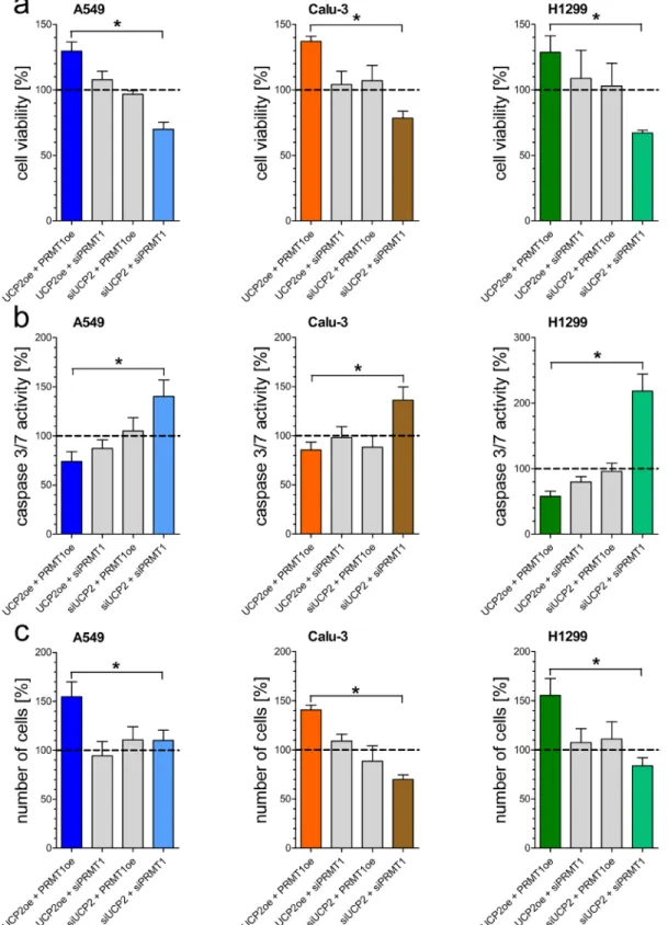
Since mitochondrial Ca2+ uptake is essential for mitochondrial metabolic activity, we manipulated the expression of PRMT1 and UCP2, proteins known to affect the mitochondrial Ca2+ uptake machinery, to investigate their effect on human non-small-cell lung cancer cell lines A549, Calu-3 and H1299. Origin of each cell lines was validated by Sanger sequencing [Supplementary Figure 1A]. Knockdown or overexpression, verified by qRT-PCR [Supplementary Figure 1B], of either UCP2 or PRMT1 alone did not affect cell viability and apoptotic caspase 3/7 activity. Combined depletion of UCP2 and PRMT1 by specific siRNAs strongly decreased cell viability [Figure 1A], whereas activity of apoptotic caspases 3/7 was significantly increased [Figure 1B]. In contrast, overexpression of both proteins improved cell viability and diminished apoptotic caspase 3/7 activity. In line with these data, cancer cell proliferation was significantly enhanced by combined overexpression of UCP2 and PRMT1 in comparison to cells with double knockdown [Figure 1C].
Combined overexpression of UCP2 and PRMT1 results in enhanced mitochondrial respiration activity
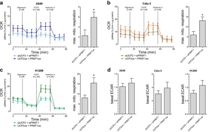
Based on our previous work [13], we assume that this positive impact of UCP2 and PRMT1 on cancer cell viability and proliferation is due to a more efficient mitochondrial respiration. Therefore, OCR was determined after addition of metabolic modulators. Indeed, A549 [Figure 2A], Calu-3 [Figure 2B] as well as H1299 [Figure 2C] cells with combined overexpression of UCP2 and PRMT1 had an enhanced maximal mitochondrial respiration, representing an increased capacity to create ATP via mitochondrial respiration, in comparison to cells with transient knockdown of UCP2 and PRMT1, whereas the basal rate of glycolysis (ECAR) was not altered [Figure 2D].
Survival rate of lung carcinoma patients is influenced by expression of UCP2 and PRMT1
Since viability as well as proliferation of lung cancer cells were strongly affected by expression levels of UCP2 and PRMT1, we analyzed the survival of lung carcinoma patients with different mRNA expression patterns of UCP2 and PRMT1. The median value of mRNA expression was
Figure 1: Impact of UCP2 and PRMT1 on cell viability, caspase activity and proliferation of lung carcinoma cell lines. Cell viability of A549 (left panel), Calu-3 (middle panel) and H1299 (right panel) cells with knockdown of UCP2 or PRMT1 and/
or overexpression of UCP2 and PRMT1 was measured via Celltiter-Blue assay and calculated as percentage of viable cells normalized to control condition a. Caspase activity of A549 (left panel), Calu-3 (middle panel) and H1299 (right panel) cells, treated with siRNA against UCP2 or PRMT1 and/or overexpressing UCP2 and PRMT1, was measured by Caspase 3/7-Glo assay and normalized to control conditions b. Cellular proliferation of A549 (left panel), Calu-3 (middle panel) and H1299 (right panel) cells diminished of UCP2 and PRMT1 and/or overexpressing these proteins was determined by counting cells 48 h after seeding and cell number was normalized to control condition c.
Bar graphs represent mean +/- SEM (n=3).
used as cutoff value and the patient group was divided into four cohorts (PRMT1-high/UCP2-high; PRMT1- low/UCP2-low; PRMT1-high/UCP2-low; PRMT1-low/
UCP2-high), whose overall survival time was analyzed.
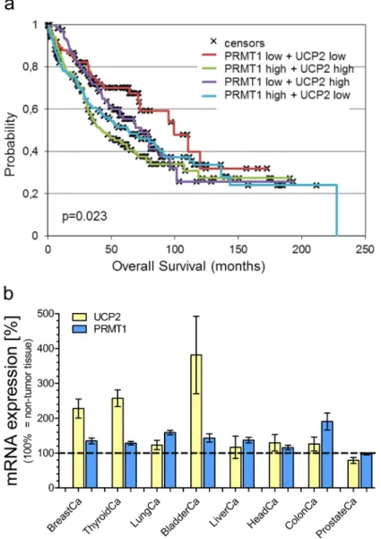
The overall survival probability [Figure 3A] was strongly decreased in the cohort with mRNA expression levels of both, PRMT1 and UCP2, above the median value in comparison to the group with low expression levels of both genes (p = 0.023). High mRNA expression of one of these genes already negatively affected overall survival (PRMT1-high/UCP2-low vs. PRMT1-low/UCP2- low: p=0.0406; PRMT1-low/UCP2-high vs. PRMT1- low/UCP2-low: p=0.16), but not in such an extent as combined high mRNA expression of PRMT1 and UCP2.
In particular, 43% of patients with simultaneously high PRMT1 and UCP2 expression, while 70% of patients with simultaneously low PRMT1 and UCP2 expression were alive at 5 years follow-up.
Combined upregulation of UCP2 and PRMT1 as a common feature of numerous cancer types
Our results from cell viability experiments with human lung carcinoma cells as well as analysis of lung
carcinoma patient’s overall survival reveal a positive impact of UCP2 and PRMT1 on cancer cells. Therefore, we investigated whether high mRNA expression levels of UCP2 and PRMT1 are a common feature in cancerous tissues. Analysis of The Cancer Genome Atlas (TCGA) database revealed an increase in UCP2 as well as in PRMT1 mRNA expression in several tumor tissues in comparison to adjacent normal tissue [Figure 3B]. Only prostate adenocarcinoma was found to have low levels of PRMT1 mRNA expression. Notably, in this particular type of cancer also UCP2 expression was lower than in the adjacent normal tissue, possibly demonstrating that the mRNA expression of UCP2 and PRMT1 is dependent on the respective other protein [Figure 3B].
DISCUSSION
Recently, we have shown that PRMT1-driven methylation of MICU1 desensitizes the mitochondrial Ca2+
uptake machinery and, therefore, reduces mitochondrial Ca2+ accumulation in cancer cells [13]. Hence, under these conditions proper mitochondrial Ca2+ uptake is reestablished by UCP2 that normalizes the sensitivity of MICU1 and, thus, reestablishes normal mitochondrial
Figure 2: Mitochondrial respiration and basal rate of glycolysis in different lung carcinoma cell lines. Oxygen consumption rate (OCR) of A549 (n=6) a., Calu-3 (n=5) b. and H1299 (n=6) c. cells with either combined overexpression or knockdown of UCP2 and PRMT1. 2 µM Oligomycin, 0.3 µM FCCP and 2.5 µM Antimycin A were injected as indicated. Bar graphs (mean +/- SEM) represent maximal mitochondrial respiration (max. mito. respiration) of the different cells. Rate of basal extracellular acidification (ECAR) of A549 (left panel, n=6), Calu-3 (middle panel, n=5) and H1299 (right panel, n=6) cells was presented as bar graphs (mean +/- SEM ) d.
Ca2+ uptake [13]. Since an increased tethering between mitochondria and the biggest internal Ca2+ store, the ER, was found in cancer cells [10], we hypothesize that cancer cells with high levels of PRMT1-driven methylation control their mitochondrial Ca2+ uptake via UCP2 to ensure suitable mitochondrial ATP production by stimulating dehydrogenases with a proper amount of Ca2+. Elevated levels of mitochondrial respiration in lung carcinoma cells with combined overexpression of UCP2 and PRMT1 are in line with this suggestion. The finding that viability and proliferation of lung carcinoma cells was strongly increased in case of upregulated PRMT1
and UCP2 expression further supports a link between the expression of these two proteins and cancer cells’
perilousness. In line with this assumption, cancer cells depleted from PRMT1 and UCP2 showed an increased vulnerability and exhibited a largely decreased cell viability and proliferation. The remarkably high levels of apoptotic caspase activity in these cells are likely a result of PRMT1-uncontrolled mitochondrial Ca2+ uptake and cell death by mitochondrial Ca2+ overload due to the enhanced tethering between the ER and mitochondria in these cells [10]. Notably, unaltered cell viability, apoptotic caspase activity and proliferation in cells depleted of
Figure 3: Survival analysis of lung carcinoma patients and mRNA expression analysis. Overall survival probability of lung carcinoma patients was analyzed in regard to the individual mRNA expression of UCP2 and PRMT1 (UCP2-high/PRMT1-high vs. UCP2-high/PRMT1-low vs. UCP2-low/PRMT1-high vs. UCP2-low/PRMT1-low) and presented as Kaplan-Meier survival plot a.
mRNA expression levels of UCP2 and PRMT1 in different cancer types were determined via TCGA analysis and presented as bar graphs, normalized to mRNA expression levels of UCP2 and PRMT1 in adjacent normal tissue (BreastCa/breast invasive carcinoma: n=114;
ThyroidCa/thyroid carcinoma: n=59; LungCa/lung carcinoma: n=109; BladderCa/bladder urothelial carcinoma: n=19; LiverCa/liver hepatocellular carcinoma: n=50; HeadCa/head and neck squamous cell carcinoma: n=43; ColonCa/colorectal adenocarcinoma: n=32;
ProstateCa/prostate cancer: n=52) b.
either UCP2 or PRMT1, might point to a still functional interrelation between both proteins as recently described [13]. These results obtained from cell culture experiments are in line with our overall survival analyses of patients suffering from lung cancer, which has already been linked to a strongly altered Ca2+ homeostasis [17]. In particular, the negative correlation of survival probability of patients with the expression of UCP2 and PRMT1 supports our hypothesis about the impact of proper mitochondrial Ca2+
uptake on the metabolic efficiency of cancer cells that affects cancer abrasiveness and patient survival. On the other hand, the present data suggest that cancer cells in the patient cohort with low levels of UCP2 and PRMT1 might be metabolically less active, yielding attenuated cancer cell proliferation and, thus, increased patient survival. The overall survival of patients with high mRNA expression levels of PRMT1 alone was already significantly reduced in comparison to those patients with low mRNA expression levels of PRMT1 and UCP2. Therefore, we conclude that protection from mitochondrial Ca2+ overload by a PRMT1-controlled mitochondrial Ca2+ uptake is already a major advantage for cancer cells. Improper mitochondrial Ca2+ accumulation as Achilles’ heel of cancer cells has been already reported: i, in prostate cancer cells, mitochondrial Ca2+ overload was proven to strongly enhance apoptosis, induced by TRAIL (TNFα related apoptosis inducing ligand) [18]. Notably, prostate cancer was the only cancer type found in our TCGA analysis with low levels of PRMT1, which makes this specific cancer probably more vulnerable for mitochondrial Ca2+ overload.
ii, the expression levels of MICU1 was shown to correlate with the overall survival of ovarian cancer patients as well as cancer cell chemoresistance against cisplatin [19].
iii, in triple-negative breast cancer, the most aggressive type of breast cancer, upregulation of the channel forming protein responsible for mitochondrial Ca2+ uptake, MCU, was demonstrated to positively correlate with tumor size and lymph node infiltration [20]. One might argue, that overexpression of MCU might lead to mitochondrial Ca2+ overload and apoptosis as in prostate cancer cells, but, breast cancer is well known for a strong upregulation of PRMT1 [21], also confirmed by our TCGA analysis, which might protect cancer cells from Ca2+ overload via MICU1 methylation. iv, strongly elevated UCP2 levels have been linked to poorly differentiated breast cancer, indicating that similar as MCU also UCP2 helps breast cancer cells to proliferate, migrate and invade [14].
Therefore, a combined upregulation of PRMT1 and UCP2 as crucial risk in various cancer types, as found in our TCGA analysis, seems to be reasonable.
All together, these findings suggest that proper mitochondrial Ca2+ uptake might be essential for devastating tumor growth, and highlight the importance of a tightly-controlled mitochondrial Ca2+ uptake to prevent mitochondrial Ca2+ overload and, ultimately, cell death.
Our study reveals a major role of PRMT1 and UCP2 in cancer cell viability and proliferation as well as in patients’
overall survival probability, indicating that numerous different cancer types utilize upgraded mitochondrial Ca2+
uptake to meet their greatly enhanced energy demand.
Accordingly, targeting mitochondrial Ca2+ uptake proteins by newly developed compounds against distinct members of the mitochondrial Ca2+ uptake machinery would allow a personalized therapy that considers individual expression pattern in the very tumor, in order to efficiently evoke cancer cell injury and increased susceptibility against therapeutics.
MATERIALS AND METHODS
Cell culture, sanger sequencing and transfection H1299 cells were grown in Dulbecco’s Modified Eagle Medium (DMEM) from Sigma Aldrich (Vienna, Austria), A549 and Calu-3 cells in a 1:1 mixture of Ham‘s F12 and DMEM. Media were supplemented with 10% fetal bovine serum, 100 U/ml penicillin, 100 µg/
ml streptomycin and 1.25 µg/ml amphotericin B (Gibco, Lifetechnologies; Vienna, Austria). To verify the origin of the various cell lines, cell lysates were sent to Microsynth (Balgach, Switzerland) for Sanger sequencing. Cells were transiently transfected at a confluence of 60 - 80% with 1.5 µg plasmid DNA encoding UCP2 or PRMT1 as well as with 100 µM siRNA UCP2 or PRMT1 using 2.5 µl of TransFastTM transfection reagent (Promega; Madison, WI, US) in 1 ml of serum- and antibiotic-free medium.
Transfection mix was replaced by full culture medium after 24 hours. All experiments were performed 48 hours after transfection. siRNAs were obtained from Microsynth, and their sequences (5′-3′) were as follows: UCP2
5’-CACTGTCGACGCCTACAAGACCATC-3’,
‘5’-GTCATAGGTCACCA GCTCAGCACAG-3’; PRMT1:
5’-TGCTCAACACCGTGCTCTATGC-3’, 5’-TCCTCGA
TGGCCGTCACATACA-3’.
mRNA isolation and qRT-PCR
Total RNA from cells was isolated using the PEQLAB total RNA isolation kit (Erlangen, Germany) and reverse transcription was performed using a cDNA synthesis kit (Applied Biosystems; Foster City, CA).
qRT-PCR was performed using QuantiFast SYBR Green RT-PCR kit (Qiagen; Hilden, Germany) as described previously [13]. Primers for qRT-PCR were obtained from Invitrogen (Vienna, Austria), and their sequences (5′-3′) were: UCP2 forward, TCCTGAAAGCCAACCTCATG;
UCP2 reverse, GGCAGAGTTCATGTATCTCGTC;
PRMT1 forward, TGCTCAACACCGTGCTTATGC;
PRMT1 reverse, TCCTCGATGGCCGTCACATACA
Cell viability and apoptosis assay
For cell viability and apoptosis assays, cells were plated 24 h after transfection on 96-well plates at a density of 5,000 cells/well. Cell viability was measured 48 h after transfection using CellTiter-Blue assay and apoptotic caspase activity was determined by using the Caspase- Glo® 3/7 assay (Promega; Madison, WI, US) as reported previously [10].
Proliferation assays
For proliferation determination, cells were plated 24 h after transfection on 6-well plates at a density of 8,000 cells/well. Number of cells was counted 48 h after transfection and presented as percentage of control cell proliferation.
Measurement of mitochondrial respiration
24 h after transfection, A546, Calu-3 and H1299 cells were plated on Cell-Tak™ coated XF96 polystyrene cell culture microplates (Seahorse Bioscience®, Agilent;
California, US) at a density of 50,000 cells per well. After 24 h cells were washed and pre-incubated in XF assay medium supplemented with 1 mM sodium pyruvate, 2 mM glutamine and 5.5 mM D-glucose. Oxygen consumption rate (OCR) and extracellular acidification rate (ECAR) were subsequently measured every 7 min using an XF96 extracellular flux analyzer. After 15 min basal measurement, Oligomycin [2 µM], FCCP [0.3 µM] and Antimycin A [2.5 µM] were injected. Oxygen consumption was normalized to protein content (pmol O2/ (min×μg protein)).
Clinical data acquisition
According to mRNA expression of PRMT1 and UCP2, Kaplan-Meier survival plots presenting the survival of lung carcinoma patients were calculated using data from Gene Expression Omnibus (GEO). Patient samples were split into four groups regarding expression of the indicated genes (median as cutoff value). These four patient cohorts were compared by a Kaplan-Meier survival plot and logrank P values were calculated.
Data acquisition via Xena
The mRNA expression levels of the genes coding for PRMT1 and UCP2 in various tumor tissues were obtained as RNA SeqV2 RSEM values through UCSC Xena (https://xenabrowser.net/) as The Cancer Genome Atlas (Provisional, TCGA) datasets in April 2017. The selected
genomic profile was ‘gene expression RNAseq (polyA+
IlluminaHiSeq)’ and the entered gene set was a user- defined list. Expression levels of normal and tumor tissue samples with the same sample ID were matched [22]. The mRNA expression levels of tumor tissues were normalized to the mRNA expression levels of corresponding adjacent healthy tissue samples from the same patient (100%
value). Patients with missing expression levels of normal or tumor tissues were excluded.
Statistical analysis
The statistical analysis was performed with GraphPad Prism 5.0 using the unpaired Student’s t-test, and p<0.05 was considered to be significant.
Abbreviations
adenosine triphosphate (ATP), Dulbecco’s Modified Eagle Medium (DMEM), extracellular acidification rate (ECAR), Gene Expression Omnibus (GEO), mitochondria-associated-ER membranes (MAMs), mitochondrial Ca2+ uniporter (MCU), mitochondrial Ca2+
uptake 1 (MICU1), oxidative phosphorylation (OXPHOS), oxygen consumption rate (OCR), protein arginine methyl transferase 1 (PRMT1), pyruvate kinase isoenzyme M2 (PKM2), The Cancer Genome Atlas (TCGA), uncoupling protein 2 (UCP2).
ACKNOWLEDGMENTS
We thank A. Schreilechner for excellent technical assistance. B.G. was supported by the NVKP_16-1-2016- 0037 grant of the National Research, Development and Innovation Office, Hungary. Christiane Klec is a fellow of the Doctoral College “Metabolic and Cardiovascular Disease” at the Medical University of Graz and was funded by the FWF (W 1226-B18, DKplus Metabolic and Cardiovascular Disease; P P 28529-B27) and by Nikon Austria within the Nikon-Center of Excellence, Graz. Microscopic equipment is part of the Nikon-Center of Excellence, Graz that is supported by the Austrian infrastructure program 2013/2014, Nikon Austria Inc., and BioTechMed, Graz.
CONFLICTS OF INTEREST
The authors have no conflicts of interest to declare.
REFERENCES
1. Hanahan D, Weinberg RA. The hallmarks of cancer. Cell.
2000; 100: 57-70.
2. Zheng J. Energy metabolism of cancer: Glycolysis versus
oxidative phosphorylation (Review). Oncol Lett. 2012; 4:
1151-7. doi: 10.3892/ol.2012.928.
3. Liberti MV, Locasale JW. The Warburg Effect: How Does it Benefit Cancer Cells? Trends Biochem Sci. 2016; 41:
211-8. doi: 10.1016/j.tibs.2015.12.001.
4. Mazurek S. Pyruvate kinase type M2: a key regulator of the metabolic budget system in tumor cells. Int J Biochem Cell Biol. 2011; 43: 969-80. doi: 10.1016/j.biocel.2010.02.005.
5. Vazquez A, Liu J, Zhou Y, Oltvai ZN. Catabolic efficiency of aerobic glycolysis: the Warburg effect revisited. BMC Syst Biol. 2010; 4: 58. doi: 10.1186/1752-0509-4-58.
6. Denton RM. Regulation of mitochondrial dehydrogenases by calcium ions. Biochim Biophys Acta. 2009; 1787: 1309- 16. doi: 10.1016/j.bbabio.2009.01.005.
7. Cardenas C, Muller M, McNeal A, Lovy A, Jana F, Bustos G, Urra F, Smith N, Molgo J, Diehl JA, Ridky TW, Foskett JK. Selective Vulnerability of Cancer Cells by Inhibition of Ca2+ transfer from endoplasmic reticulum to mitochondria. Cell Reports. 2016; 14: 2313-24. doi:
10.1016/j.celrep.2016.02.030.
8. Bittremieux M, Parys JB, Pinton P, Bultynck G. ER functions of oncogenes and tumor suppressors: Modulators of intracellular Ca2+ signaling. Biochim Biophys Acta.
2016; 1863: 1364-78. doi: 10.1016/j.bbamcr.2016.01.002.
9. Doghman-Bouguerra M, Granatiero V, Sbiera S, Sbiera I, Lacas-Gervais S, Brau F, Fassnacht M, Rizzuto R, Lalli E.
FATE1 antagonizes calcium- and drug-induced apoptosis by uncoupling ER and mitochondria. EMBO Rep. 2016; 17:
1264-80. doi: 10.15252/embr.201541504.
10. Madreiter-Sokolowski CT, Gottschalk B, Parichatikanond W, Eroglu E, Klec C, Waldeck-Weiermair M, Malli R, Graier WF. Resveratrol specifically kills cancer cells by a devastating increase in the Ca2+ coupling between the greatly tethered endoplasmic reticulum and mitochondria.
Cell Physiol Biochem. 2016; 39: 1404-20. doi:
10.1159/000447844.
11. Perocchi F, Gohil VM, Girgis HS, Bao XR, McCombs JE, Palmer AE, Mootha VK. MICU1 encodes a mitochondrial EF hand protein required for Ca2+ uptake. Nature. 2010;
467: 291-6. doi: 10.1038/nature09358.
12. De Stefani D, Raffaello A, Teardo E, Szabo I, Rizzuto R.
A forty-kilodalton protein of the inner membrane is the mitochondrial calcium uniporter. Nature. 2011; 476: 336- 40. doi: 10.1038/nature10230.
13. Madreiter-Sokolowski CT, Klec C, Parichatikanond W, Stryeck S, Gottschalk B, Pulido S, Rost R, Eroglu E, Hofmann NA, Bondarenko AI, Madl T, Waldeck-Weiermair M, Malli R, et al. PRMT1-mediated methylation of MICU1 determines the UCP2/3 dependency of mitochondrial Ca2+
uptake in immortalized cells. Nat Commun. 2016; 7: 12897.
doi: 10.1038/ncomms12897.
14. Sayeed A, Meng Z, Luciani G, Chen LC, Bennington JL, Dairkee SH. Negative regulation of UCP2 by TGFbeta signaling characterizes low and intermediate-grade primary breast cancer. Cell Death Dis. 2010; 1: e53. doi: 10.1038/
cddis.2010.30.
15. Pons DG, Nadal-Serrano M, Torrens-Mas M, Valle A, Oliver J, Roca P. UCP2 inhibition sensitizes breast cancer cells to therapeutic agents by increasing oxidative stress.
Free Radic Biol Med. 2015; 86: 67-77. doi: 10.1016/j.
freeradbiomed.2015.04.032.
16. Yang Y, Bedford MT. Protein arginine methyltransferases and cancer. Nat Rev Cancer. 2013; 13: 37-50. doi: 10.1038/
nrc3409.
17. Arbabian A, Brouland JP, Apati A, Paszty K, Hegedus L, Enyedi A, Chomienne C, Papp B. Modulation of endoplasmic reticulum calcium pump expression during lung cancer cell differentiation. FEBS J. 2013; 280: 5408- 18. doi: 10.1111/febs.12064.
18. Kaddour-Djebbar I, Lakshmikanthan V, Shirley RB, Ma Y, Lewis RW, Kumar MV. Therapeutic advantage of combining calcium channel blockers and TRAIL in prostate cancer. Mol Cancer Ther. 2006; 5: 1958-66. doi:
10.1158/1535-7163.MCT-06-0011.
19. Chakraborty PK, Mustafi SB, Xiong X, Dwivedi SK, Nesin V, Saha S, Zhang M, Dhanasekaran D, Jayaraman M, Mannel R, Moore K, McMeekin S, Yang D, et al. MICU1 drives glycolysis and chemoresistance in ovarian cancer.
Nat Commun. 2017; 8:14634. doi: 10.1038/ncomms14634.
20. Tosatto A, Sommaggio R, Kummerow C, Bentham RB, Blacker TS, Berecz T, Duchen MR, Rosato A, Bogeski I, Szabadkai G, Rizzuto R, Mammucari C. The mitochondrial calcium uniporter regulates breast cancer progression via HIF-1alpha. EMBO Mol Med. 2016; 8: 569-85. doi:
10.15252/emmm.201606255.
21. Mathioudaki K, Scorilas A, Ardavanis A, Lymberi P, Tsiambas E, Devetzi M, Apostolaki A, Talieri M. Clinical evaluation of PRMT1 gene expression in breast cancer.
Tumour Biol. 2011; 32: 575-82. doi: 10.1007/s13277-010- 0153-2.
22. Kammerer S, Sokolowski A, Hackl H, Platzer D, Jahn SW, El-Heliebi A, Schwarzenbacher D, Stiegelbauer V, Pichler M, Rezania S, Fiegl H, Peintinger F, Regitnig P, et al.
KCNJ3 is a new independent prognostic marker for estrogen receptor positive breast cancer patients. Oncotarget. 2016;
7: 84705-17. doi: 10.18632/oncotarget.13224.