The role of complement in spinal cord injury
PhD dissertation
Fei Qiao
Doctoral School of Basic Medicine Semmelweis University
Mentor: Stephen Tomlinson, PhD, Professor and Vice Chair of the Department of Microbiology/Immunology of the Medical University
of South Carolina, USA
Supervisor: Béla Merkely, MD, PhD, DSc, Professor and director of the Department of Heart Center at the Semmelweis University
Official reviewers:
László Kőhidai, MD, Ph.D Krisztián Papp, MD, Ph.D
Head of the Final Examination Committee:
Ferenc Horkay, MD, PhD, DSc
Members of the Final Examination Committee:
Zoltán Prohászka, MD, PhD, DSc Zoltán Járai, MD, PhD
Table of Contents
LIST OF ABBREVIATIONS ……….5
1. INTRODUCTION ………....7
1.1. BACKGROUND AND SIGNIFICANCE ...7
1.1.1. Spinal cord injury ………...7
1.1.2. Complement system and neuroinflammation ………...….7
1.2. OVERVIEW OF THE COMPLEMENT SYSTEM AND EFFECTOR MECHANISMS ………..8
1.3. THE COMPLEMENT SYSTEM IN NEURODEGENERATION ……….9
1.4. WHY IT IS IMPORTANT TO STUDY THE ROLE OF COMPLEMENT IN SPINAL CORD INJURY? ………...10
1.4.1. Complement activation in the classical and the alternative pathway...12
1.4.2. Complement activation in the lectin pathway………...14
1.4.3. The membrane attack pathway (MAP), the terminal pathway …………...14
1.5. COMPLEMENT INHIBITORS AS THERAPEUTIC AGENTS ………15
2. OBJECTIVES ……….17
2.1. TO DEVELOP A NEUROPROTECTIVE STRATEGY BASED ON ATTENUATING COMPLEMENT-DEPENDENT SECONDARY DAMAGE AFTER SCI ……….18
2.2. TO INVESTIGATE THE NEUROPROTECTIVE EFFECT OF A NOVEL TARGETED COMPLEMENT INHIBITOR. ………... 18
3. METHODS AND MATERIALS ………....20
3.1. MICE ………...20
3.2. COMPLEMENT INHIBITOR- CR2-Crry ...20
3.3. ANTI-fB ANTI-BODY...21
3.4. SPINAL CORD INJURY SURGERY AND ANTIBODY TREATMENT ...21
3.4.1. Treatment ...21
3.4.2. SCI model ……… 22
3.4.2a. Instruments………...22
3.4.2b. Anaesthesia ……… 23
3.4.2c. Surgery and care ………23
3.5. LOCOMOTOR FUNCTION ANALYSIS ………23
3.5.1. The Basso, Beattie and Bresnahan (BBB) rating scale ………...24
3.5.2. The Basso Mouse Scale (BMS)..………...27
3.6. HISTOPATHOLOGICAL ANALYSIS ………...29
3.7. NEUTROPHIL AND MACROPHAGE INFILTRATION ………...30
3.8. COMPLEMENT DEPOSITION ………....30
3.9. STATISTICAL ANALYSIS ……….31
4. RESULTS ……….……….…32
4.1. RECOVERY OF LOCOMOTOR FUNCTION POST-TRAUMATIC INJURY …...32
4.1.1 Effect of C3 deficiency and of complement inhibition on locomotor recovery following SCI……….32
4.1.2 Effect of fB, CD59 deficiency and alternative complement pathway inhibition on locomotor recovery following SCI ………34
4.2. EFFECT OF COMPLEMENT DEFICIENCY AND COMPLEMENT INHIBITION ON TISSUE DAMAGE AND DEMYELINATION AFTER SCI...36
4.2.1. Macroscopic images of spinal cords isolated at 72 hours postinjury …..36
4.2.2. Effect of C3 deficiency and of complement inhibition on the extent of tissue destruction following SCI ………....38
4.2.3. Effect of C3 deficiency and of complement inhibition on the extent of tissue demyelination following SCI ……….40
4.2.4. Histopathology of spinal cord sections after SCI ………..……..42
4.2.5. Quantitative assessment of histopathological inflammation and injury..44
4.2.6. Morphometric analysis of tissue sparing 3 days after injury ………..….44
4.3. EXPRESSION OF NEUTROPHILS AND MACROPHAGE INFILTRATION AFTER SPINAL CORD INJURY ………..…….46
4.3.1a. Effect of C3 deficiency and of complement inhibition on the neutrophil infiltration following SCI………..46
4.3.1b. Effect of fB and CD59 deficiency and of alternative pathway complement
inhibition on the neutrophil infiltration following SCI …………..…48
4.3.2. Expression of macrophage infiltration after spinal cord injury ……..49
4.4. TARGETING AND BIODISTRIBUTION OF THERAPEUTICALLY ADMINISTERED CR2- Crry ……….……….50
4.5. DEPOSITION OF COMPLEMENT ACTIVATION ……….53
4.5.1. Time course of complement activation 1, 24 and 72 hours after SCI ...53
4.5.2. Complement deposition at epicenter of injury 24 h post-SCI ………….54
4.5.3. Quantitative assessment of complement deposition post-SCI …………56
4.6. HISTOLOGICAL ANALYSIS OF RECOVERY ………..……58
5. DISCUSSION ………...61
5.1 ROLE OF C3 AND COMPLEMENT INHIBITORY PROTEIN CR2-CRRY DURING SCI………62
5.2 ROLE OF FB THROUGH ALTERNATIVE PATHWAY DURING SCI …….65
5.3 ROLE OF CD59 THROUGH TERMINAL COMPLEMENT PATHWAY DURING SCI ………...66
6. SUMMARY AND CONCLUSIONS………..…..68
7. ABSTRACT………..……...70
8. ABSTRACT IN HUNGARIAN (ÖSSZEFOGLALÁS)………...72
9. BIBLIOGRAPHY………..73
10. PUBLICATIONS OF PH.D. CANDIDATE ...90
1. LIST OF PUBLICATION RELATED TO THESIS...90
Peer reviewed articles ………...90
2. OTHER PUBLICATIONS………..91
Peer reviewed articles………...91
11. ACKNOWLEDGEMENT………...94
List of abbreviations
SCI, spinal cord injury CP, classical pathway AP, alternative pathway MBL, mannose-binding lectin MAP, membrane attack pathway CNS, central nervous system
MAC, membrane attack complex fB, factor B
CR1, complement receptor 1 DAF, decay-accelerating factor MCP, membrane cofactor protein TBI, traumatic brain injury AD, Alzheimer’s disease Wt, Wild-type
fB-/-, fB-deficient CD59-/-, CD59-deficient PBS, phosphatebuffered saline AfB, anti-fB antibody
SCR, short consensus repeat
BBB, Basso,Beattie,Bresnahan rating scale BMS, Basso Mouse Scale
1. INTRODUCTION
1.1. Background and significance
1.1.1. Spinal cord injury
Spinal cord injury (SCI) is characterized by an initial traumatic injury phase, followed closely by secondary events that result in edema, ischemia, excitotoxicity, and inflammation(1). The mechanisms of secondary injury are not well defined, but it is clear that inflammatory processes play a significant role in functional recovery(2, 3). While the initial traumatic injury is difficult to guard against, the subsequent inflammatory cascade represents a therapeutic target for SCI. The only clinical therapy accepted currently for acute SCI is methylprednisolone, a therapy that has yielded disappointing results, with the data from clinical trails being contradictory and inconclusive(4-6).
1.1.2. Complement system and neuroinflammation
Complement is important for host defense against pathogens and is an effector mechanism for both innate and adaptive immune responses. Complement is also involved in immune homeostasis mechanisms including the catabolism of immune complexes and clearance of apoptotic cells. Under certain disease conditions in which inappropriate or excessive complement activation occurs, complement control mechanisms breakdown or are overcome and complement causes tissue injury. Complement play a key role in the pathogenesis of many inflammatory and ischemic disease conditions. Following primary injury to the spinal cord, a complex cascade of pathophysiologic processes occur that result in secondary injury and progressive degenerative effects that can determine the extent of recovery. The mechanisms involved in secondary tissue injury are not well defined, but inflammation and ischemia are considered to be key components. Evidence indicates an important, albeit not well defined, role for complement in the
pathophysiological processes that occur following both traumatic brain and spinal cord injury (2, 6-10)(2, 6-10)(2, 6-10)[2, 6-10].
1.2. Overview of the complement system and effector mechanisms
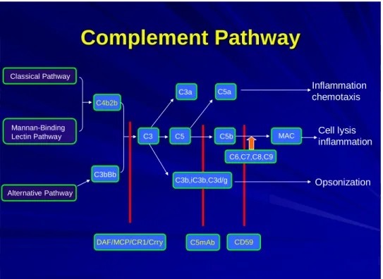
In order to appreciate the role of complement in spinal cord injury, it is essential to first understand the basics of the system. The complement system (Fig. 1), a major component of the innate immune response, is activated in many neurodegenerative diseases (11, 12).
The complement system is a self amplifying cascade of proteases(13). Activation results in 1) attraction and activation of phagocytes(14, 15); 2) opsonisation; and 3) formation of the membrane attack complex (MAC or C5b-9)(16). The complement system consists of nearly 30 proteins involved in non-specific immune response and the role of various complement components in the pathophysiology of several disorders has been well studied in the past two decades. The complement system is activated in several disorders, particularly in response to foreign proteins, polysaccharides, cell surface components of microbial origin, or toxic proteins associated with the pathogenesis of several disorders.
These foreign proteins activate the complement system by various pathways. The complement system is activated by the three major pathways, the classical pathway (CP), alternative pathway (AP), and lectin-mediated pathway. Activation of one of the pathways may lead to activation of the other pathway. In the central nervous system (CNS), the two main pathways found to be activated are the classical and the alternative pathway(17, 18). Some complement factors, including C1q (initiator of the classical complement pathway), C1r, C1s, C3 and C4 are locally produced in the CNS (19-22), whereas others derive from the blood. Activated complement can trigger an adaptive immune response, involving antigen presenting cells, T-cells and B-cells. The classical pathway is normally antibody dependent whereas the alternative pathway is usually antibody independent. Activation of complement by any pathway leads to the formation of a C3 convertase that cleaves C3 into C3a and C3b, with the latter binding covalently to the activating surface and participating in the formation of additional C3 convertase complexes (amplification loop). C3 convertases also participate in the formation of C5 convertase, which cleaves C5 to yield C5a and C5b. Formation of C5b initiates the
proteins C6, C7, C8 and (C9)n to form the cytolytic membrane attack complex (MAC or C5b-9). Cleavage fragments of C3 covalently bound to activating surfaces act as ligands for receptors on immune effector cells. C3a and C5a are chemotactic and can activate leukocytes. Formation of the MAC on an activating cell surface can cause direct cell lysis.
Host cells are normally protected from complement by membrane-bound inhibitors of the complement activation (inhibit C3 convertase formation) and of the terminal complement pathway (inhibit MAC formation). Membrane inhibitors of complement activation are complement receptor 1 (CR1), decay-accelerating factor (DAF) and membrane cofactor protein (MCP). Rodents express an additional inhibitor of complement activation termed Crry that is a structural and functional homolog of human CR1 (23, 24). Control of the terminal pathway and MAC formation in cell membranes occurs through the activity of CD59 that binds to C8 and C9 in the assembling MAC. The complement anaphylatoxins are responsible for the various proinflammatory events in the CNS and chemotaxis of the immunocompetent cells (25).
1.3. The complement system in neurodegeneration
The complement system is known to perform a wide range of functions in the human body. It forms an essential component of the host immune system and is associated with the clearance of molecules of foreign origin as well as the elimination of invading pathogens from the body. It plays an important role in adaptive immunity(26). However, it is nonspecific in action and unable to distinguish between self and non-self. Under normal conditions, it is strictly regulated by complement regulatory molecules. However, in neuroinflammatory disorders, the complement regulatory molecules fail to control the activated complement components. These activated complement components then act as a double-edged sword and are responsible for the degeneration of neurons(27). The devastating roles of the complement components in neurodegenerative disorders are well documented. Spinal cord injury (SCI )(6), traumatic brain injury (TBI)(28), Alzheimer’s disease (AD)(29), multiple sclerosis (MS)(30, 31), myasthenia gravis(32) and Parkinson’s disease (PD)(33) are examples of a few disorders in which activated complement components play an important role. Not only do these disorders arise out of
dysfunction in the normal metabolic machinery, but also neuroinflammatory disorders associated with microbial infections show involvement of complement components.
1.4. Why it is important to study the role of complement in spinal cord injury?
An impact to the spinal cord results in a primary injury, but the impact also triggers a series of downstream events that lead to secondary injury of tissue and the progressive degeneration of the spinal cord. Therapeutic interventions that minimize secondary injury will improve functional recovery after traumatic injury. Inflammation plays a key role in secondary injury following spinal cord injury (SCI). Inflammation is a rapid immune response that can be initiated by infection or tissue damage. An important component of an inflammatory response is the complement system, a collection of blood proteins that form part of the immune system and that can be activated by injured cells and tissues.
Activation of complement amplifies the inflammatory response and produces molecules that can be directly toxic or that can recruit and activate cells of the immune system to produce toxic molecules. Inhibiting the complement system has been shown to be an effective therapy for inflammatory disease in various animal models, and some complement inhibitors are in clinical trials.
We have shown that complement deficient mice and normal mice treated with a complement inhibitor are protected from neuronal injury following SCI, and that they have significantly improved functional scores over time compared to control mice. These data indicate an important role for complement in secondary injury, and indicate complement inhibition will reduce inflammation and provide neuroprotection following spinal cord injury. However, complement-dependent mechanisms involved in secondary injury following SCI are not known, and there remain concerns regarding the clinical application of the currently available systemic (body-wide) complement inhibitors with regard to their safety and efficacy. Complement activation products are important for host defense and immune maintenance mechanisms, and systemic complement inhibition can compromise the protective and beneficial roles of complement. This will not be optimal
in patients at risk of infection, and urinary tract infection is a frequent complication during initial and ongoing medical rehabilitation after SCI.
We propose to develop a neuroprotective strategy based on attenuating complement-dependent secondary damage after SCI using a novel and validated approach. The strategy involves the targeting of complement inhibitors to sites of complement activation and injury, and we have shown the approach to be highly effective in vitro and in mouse models of SCI and other inflammatory disease conditions. The targeting strategy enhanced the activity and protective effect of complement inhibitors by 10-20 fold compared to untargeted counterparts. Furthermore, untargeted, but not targeted complement inhibitors systemically inhibited complement and increased susceptibility to infection in a mouse model.
We propose to fully characterize our targeted complement inhibitors in a mouse model of SCI. In addition to therapeutic endpoints, we will use different types of complement inhibitor to investigate complement-dependent disease mechanisms. We will determine relationships between the generation of different complement activation products and other molecules associated with inflammation in order to gain a better understanding of the mechanisms of secondary tissue injury following SCI. These preclinical studies will establish the guidelines necessary for the translation of this therapy to the clinic using recombinant human proteins.
Complement
Complement Pathway Pathway
Classical Pathway
C3 MAC
C6,C7,C8,C9
C3a C5a Inflammation
chemotaxis
Cell lysis inflammation
C3b,iC3b,C3d/g Opsonization
Alternative Pathway Mannan-Binding Lectin Pathway
C4b2b
C3bBb
DAF/MCP/CR1/Crry
C5 C5b
C5mAb CD59
1.4.1. Complement activation in the classical pathway and the alternative pathway
The classical pathway of complement activation is found to be a major factor in the etiology of these disorders(33-37). Recent evidence indicates the involvement of the alternate pathway of complement activation in the disorders of the central nervous system(38). This emphasizes the importance of both pathways in the pathogenesis of neurodegenerative disorders. Classical pathway activation is usually antibody-dependent and is initiated when C1q binds to an immune complex. Activated C1 cleaves both C4 and C2 to generate C4a and C2a fragments, which combine to form the classical pathway C3 convertase, involved in the cleavage of component C3 to form C3a and C3b. C3a is
Fig. 1. The complement system The constituent pathways of the Complement system and the component proteins are shown.
combines with C4a and C2a to form C5 convertase, which leads to the formation of C5a and C5b. The former is an anaphylatoxin and the latter is involved in the production of terminal complement components, C6 to C9. C5b, with these terminal complement components, results in the formation of MAC(39, 40). The alternate pathway also results in the formation of complement anaphylatoxins and MAC, but activated through C3 component, requires factor B to form C3 convertase and C5 convertase(40).
The alternative pathway is activated on surfaces of pathogens that have neutral or positive charge characteristics and do not express or contain complement inhibitors.
This is due to a process termed “tickover” of C3 that occurs spontaneously, involves the interaction of conformationally altered C3 with factor B, and results in the fixation
of active C3b on pathogens or other surfaces reviewed in(38, 39), and activated by spontaneous hydrolysis of C3 to a cleavage product (C3b analog) that binds factor B (fB), leading to formation of the alternative pathway C3 convertase. The alternative pathway also provides an amplification loop for the classical and lectin pathways. The alternative pathway does not utilize C1q or MBL as the molecules that recognize antibody-marked targets, but relies on a postulated “tick-over” deposition of activated C3b on all cell membranes. What determines whether complement is activated or not on a particular cell surface, are the perturbations in the interaction between the deposited C3b and regulatory molecules. C3b in the circulation undergoes conversion, at a slow rate, to an active but uncleaved C3b-like form, C3i or C3(H2O). This C3b can bind to nucleophilic targets on cell surfaces and form a complex with plasma factor B that is then cleaved by factor D to form the C3 convertase, C3bBb (see fig. 1). In addition, properdin binds to and stabilizes the alternative pathway C3 convertase, extending the lifetime of the active convertase three- or four- fold(41). The C3 convertase catalyzes further cleavage of C3 into C3b.
The fact that each newly produced C3b molecule, even those that arise from the classical pathway, has the potential to form the convertase and thus to produce more C3b provides a means of amplification.
Both the classical path way and the alternative pathway C3 convertases participate
covalently bind C4b in the classical pathway C3 convertase, C4b2a, to form C4b2a3b, the classical pathway C5 convertase. Similarly, the addition of activated C3b to the alternative pathway C3 convertase, C3bBb, forms C3bBbC3b, the alternative pathway C5 convertase. Both C5 convertases can cleave C5 to yield C5a and C5b. C5a has powerful proimflammatory and chemotactic properties(42).
1.4.2. Complement activation in the lectin pathway
The lectin pathway is activated when mannose binding protein (MBL) or ficolins bind to conserved carbohydrate structures.
1.4.3. The membrane attack pathway (MAP), the terminal pathway
The terminal complement components formed by the cleavage of C5 either by the classical or the alternate pathway result in the formation of membrane attack complex (MAC). The MAC formation is regulated by complement defense protein CD59, deficiency of which results in neurodegenerative diseases(43). The neurotoxic effects of MAC on neuronal cells are time and concentration dependent. Mechanisms like free radical generation, cytokines, eicasonoids, and increased permeability to Na+ and Ca2+
ions induced by MAC might be responsible for the observed effect(44). Apart from their lytic properties, recent findings suggest that both cytolytically active and inactive forms of terminal complement complexes are involved in the accumulation of leukocytes into the cerebrospinal fluid and endothelial cell activation. At sublytic concentration, it not only protects host cells but also stimulates protein biosynthesis. The role of MAC, at sublytic and lytic concentration, is outlined by Wurzner(45). This explains the concentration-dependent dual role of MAC. At lytic concentration, it acts as a neurodegenerative agent and at sublytic concentration it acts as a neuroprotective agent.
Marked neurodegeneration in CNS disorders, even in the presence of the MAC, indicates the failure of regulation of MAC components and the need to restrict its activity to
sublytic concentration by proper regulation of the pathways leading to MAC formation.
Thus, regulating the complement pathways leading to its generation can control formation of MAC(46).
All pathways converge at C3 activation with the subsequent cleavage of C5. During this process, the anaphylatoxins C3a and C5a are generated, and C5 cleavage initiates the terminal complement pathway that culminates in the formation of the membrane attack complex (MAC). The MAC can be directly cytolytic and can stimulate the production of proinflammatory molecules when deposited in cell membranes at sublytic concentrations (for a review of the complement system, see Ref (47)).Cleavage of C5 is the final enzymatic step of the complement system. C5b bound to the convertase sequentially binds C6 and C7. The C5b67 complex is released from the convertase and associates with adjacent membrane via a labile hydrophobic binding site in the C6 component. The C5b67 complex stably associates with the membrane, then binds C8, and finally, multiple copies (up to 12) of C9. MAC induces concentration-dependent neuronal cell death and changes in membrane permeability to Na+, K+ and Ca2+, release of cytokines, eicosanoids, and reactive-free radicals. These changes occur at sublytic concentrations of MAC(44). MAC is also responsible for the demyelination of neurons in demyelinated forms of certain disorders(48). Upon binding to C5b-8, C9 unfolds and inserts in the membrane. The recruitment of additional C9 molecules, membrane attack complexes, MACs, form a functional pore in the cell membrane through which ions and small molecules can pass, bringing about osmotic lysis of the cell.
1.5. Complement inhibitors as therapeutic agents
Although, inflammation plays an important role in CNS disorders, no currently available anti-inflammatory agent offers significant neuroprotection in such disorders(40). Various types of complement inhibitory proteins are currently under investigation for therapy of inflammatory and ischemic disease (reviewed in (49-52)(49-52)(49-52)[49-52]).
Interventional studies with soluble complement inhibitors in rodent models of traumatic brain injury, and studies using transgenic mice with astrocyte-targeted expression soluble
damage (28, 53, 54)(28, 53, 54)(28, 53, 54)[28, 53, 54]. Clinical studies have shown elevated levels of complement proteins in cerebrospinal fluid from patients with traumatic brain injury, and a recent study demonstrated that a viral complement control protein modulates inflammation following spinal cord injury in rats (2, 55, 56)(2, 55, 56)(2, 55, 56)[2, 55, 56].
Virtually all complement inhibitory strategies reported to date depend on systemic complement inhibition. We have shown that the targeting of complement inhibitors to sites of complement activation and disease significantly improves their efficacy and obviates the need for systemic inhibition. One strategy to target complement inhibitors is to link them to antibody fragments, and we have shown that antibody-linked complement inhibitors (DAF, Crry and CD59) are significantly more effective than untargeted counterparts in vitro and in an animal model of tubulointerstitial disease(57-59)(57- 59)(57-59)[57-59]. Significantly, for CD59 to function effectively, targeting to the site of complement activation is a requirement (57)(57)(57)[57]. Due to considerations of species selective activity, Crry is a relevant C3 inhibitor for studies in mice.
In this study we propose to use a targeting strategy based on a fragment of complement receptor 2 (CR2) to target complement inhibitors specifically to sites of complement activation. Natural ligands for CR2 are iC3b, C3dg and C3d, cell-bound breakdown fragments of C3 that are deposited on activating surfaces(60, 61)(60, 61)(60, 61)[60, 61]. These C3 ligands are relatively long lived and are present in high concentrations at sites of complement activation. We have shown that human CR2-DAF and CR2-CD59 bind to C3-coated targets and are at least 20-fold more potent than their untargeted counterparts at providing protection from complement (58)(58)(58)[58] (see attached manuscript). We recently characterized CR2-Crry in a mouse model of intestinal ischemia/reperfusion injury (IRI) and demonstrated that CR2-Crry was 10-fold more effective than Crry-Ig, an untargeted systemic counterpart. Furthermore, CR2-Crry, unlike Crry-Ig, was therapeutically effective at a dose that did not result in systemic complement inhibition and that did not increase susceptibility to infection.
2. Objectives
Traumatic spinal cord injury initiates a cascade of pathophysiological events that cause secondary injury and determine the extent of functional recovery. Although the processes that occur following SCI are complex, inflammation is considered to play a key role in the progressive degenerative events that take place, especially within the lesion penumbra.
The complement system plays a key role in the pathogenesis of many inflammatory and ischemic conditions, and recent evidence indicates it also plays an important role in secondary SCI. Various systemic complement inhibitors are currently under therapeutic investigation as anti-inflammatory agents, but there remain concerns regarding their efficacy and safety. Complement activation products are important for host defense and immune homeostasis mechanisms, and systemic complement inhibition can compromise the protective and immunomodulatory roles of complement. In this context, CNS injury has been shown to be immunosuppressive, and further immune suppression by systemic complement inhibition may not be optimal in patients at risk of infection (urinary tract infection is a frequent complication during initial and ongoing medical rehabilitation after SCI).
The goals of the present study were (1) To investigate the dynamics of complement activation and its role in the development of SCI in mice. (2) To determine in vivo relationships between the generation of different complement activation products with cytokine production, adhesion molecule expression, leukocyte infiltration and activation, and injury. (3) To investigate the neuroprotective effect of a novel targeted complement inhibitor and develop a neuroprotective strategy based on attenuating complement-dependent secondary damage after SCI.
2.1. To develop a neuroprotective strategy based on attenuating complement-dependent secondary damage after SCI
We have developed a strategy to target complement inhibitors to sites of complement activation and injury, and have shown that targeted complement inhibitors are 10-20 fold more effective in vitro and in an experimental models of ischemia and reperfusion injury compared to conventional systemic approaches to inhibit complement. We hypothesize that our novel strategy to target complement inhibitors to the site of SCI will improve bioavailability, obviate the need to systemically inhibit complement, and provide a safe and highly efficacious therapy. We propose to investigate our hypothesis in a mouse model of SCI. Targeted complement inhibition will be achieved by the use of soluble recombinant chimeric molecules consisting of a targeting moiety linked to a complement inhibitor. Complement inhibitors will be mouse Crry (inhibits early in the complement pathway) or CD59 (inhibits late in pathway). The targeting moiety will be a fragment of complement receptor 2 (CR2) that binds to long lived degradation products of C3 that are deposited at sites of complement activation. In addition to therapeutic endpoints, we will utilize the targeted complement inhibitors that function at different points in the complement cascade to investigate disease mechanisms in a clinical setting.
2.2. To investigate the neuroprotective effect of a novel targeted complement inhibitor.
To investigate the neuroprotective effect of two complement inhibitory strategies, a novel targeted complement inhibitor (CR2-Crry) which targets specifically to sites of complement activation and does not require systemic complement inhibition and an anti- fB antibody that suppresses the alternative pathway of complement activation. For the therapeutic studies we used mouse CR2-Crry, a complement inhibitory fusion protein that functions at the C3 level. The complement inhibitor, Crry, is targeted specifically to sites of complement activation by means of the complement receptor 2 (CR2)-targeting moiety(62). CR2 is a member of the C3-binding protein family, and its natural ligands are cleavage fragments of C3 that become deposited at sites of complement activation.
systemic (untargeted) inhibition in terms of efficacy and host susceptibility to infection(63). The use of mouse Crry is appropriate when studying the effects of complement inhibition in mice, because complement inhibitors display different degrees of species selectivity. Crry is a structural and direct functional analog of human complement receptor 1 (CR1), and data obtained with Crry in rodents will likely translate in functional terms to the use of CR1 in humans.
As discussed in section 5.2, in the present study, we show that fB-deficient mice have an improved outcome after SCI. The level of protection afforded to fB-deficient mice was similar to that seen in our previous study using C3-deficient mice(64). These data indicate a dependence on the alternative pathway for complement-mediated SCI.
Together with the previous findings described above using B cell and C1q-deficient mice, it is likely that the alternative pathway plays a critical role in amplifying antibody- mediated classical pathway initiated complement activation. In this context, reduced pathology and improved locomotor recovery in fB-deficient mice was associated with significantly reduced C3 and MAC deposition compared to wt control mice, even though the classical (and lectin) pathway is intact in fB-deficient mice. A similar dependence on both antibody-mediated lectin pathway activation and alternative pathway activation has been described in models of intestine ischemia and reperfusion injury(65-67). In a more clinically relevant paradigm, we demonstrated that specific inhibition of the alternative pathway with anti-fB mAb was also protective against SCI, and to a similar level as fB deficiency.
3. METHODS AND MATERIALS
3.1. Mice
Female wild-type C57BL/6 and C57Bl/6 C3-deficient (C3-/-) mice were obtained from Jackson Laboratories (Bar Harbor, ME). Breeding pairs of fB-deficient (fB-/-) mice on C57BL/6 background were generated as described (68) and provided by Dr. J. Thurman (University of Colorado Health Sciences Center, Denver, CO) and a breeding colony established. In mice, there are two genes encoding CD59; CD59a is widely expressed and is the primary regulator of the MAC in mice, whereas CD59b expression is limited to the testis and, at very low levels, bone marrow (69). In this study, CD59a-deficient mice on C57BL/6 background were used and were generated as described (70). fB and CD59 deficiency was confirmed by genotyping. Mice weighing between 18 22 g (6 8 weeks old) were used in experiments. All mice were fed on standard laboratory food and given tap water ad libitum with a light dark cycle of 12 hours.
3.2. Complement Inhibitor-CR2-Crry
The fusion protein CR2-Crry was produced and purified as described previously (71). In brief, a cDNA construct of the recombinant fusion protein was prepared by joining the mouse CR2 sequence encoding the four N-terminal short consensus repeat (SCR) units (residues 1–257 of mature protein, National Center for Biotechnology Information Gen-Bank, accession number M35684) to sequences encoding extracellular regions of mouse Crry. The Crry sequence used encoded residues 1–319 of the mature protein (National Center for Biotechnology Information GenBank, accession number NM013499). To join CR2 to Crry, linking sequences encoding (GGGGS)2 were used.
The recombinant protein was expressed in NSO cells and purified by anti-Crry affinity chromatography as described (71). CR2-Crry has a circulatory half-life in C57BL/6 mice of 8 hours (71).
3.3. Anti-fB anti-body
The isolation and characterization of anti-fB mAb 1379 used in these studies was described previously, and the mAb effectively inhibits the mouse alternative pathway(72).
The mAb was generously provided by Drs. V. M. Holers, J. M. Thurman (University of Colorado Health Sciences Center, Denver, CO), and G. S. Gilkeson (Medical University of South Carolina).
3.4. Spinal cord injury surgery and antibody treatment
3.4.1. Treatment
Wild-type (wt) mice were randomized into sham (laminectomy, no SCI damage), vehicle control (phosphatebuffered saline [PBS]), and CR2-Crry treatment and anti-fB mAb treatment groups. Other groups consisted of C3-deficient (C3-/-), fB-deficient (fB-/-) and CD59a-deficient (CD59-/-) mice. For CR2-Crry treatment were administered a single dose of 0.25 mg of CR2-Crry by tail vein injection. All other animals received intravenous injections of phosphate buffered saline. For anti-fB mAb treatment, wt mice were randomized into four groups, with mice in each group receiving an intravenous (tail vein) injection of 100 µl PBS vehicle control or 2 mg anti-fB mAb in 100µl PBS at 1 and 12 hours after surgery, 12 and 24 hours after surgery, or 24 and 36 hours after surgery.
The 2mg dose used was based on previous studies characterizing the therapeutic effect of this Ab in mouse models of inflammation (72-74).
3.4.2. SCI model
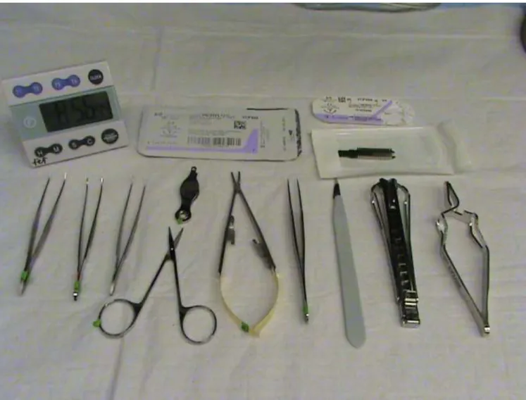
3.4.2a. Instruments
Fig. 2. Instruments of the spinal cord injury surgery.
3.4.2b. Anaesthesia
Mice were anesthetized with ketamine/xylazine anesthesia (80 mg/kg/10 mg/kg, i.p.) Animals were breathing spontaneously, and body temperature was maintained using a heat mat for the duration of the experiment.
3.4.2c. SCI Surgery
Mice subjected to laminectomy at the level of the 12th thoracic vertebra (T12), followed by contusion-induced SCI using the NYU weight drop impactor as described previously(64). After injury, the muscles and the subcutaneous tissue were closed in layers, the skin was closed with metal wound clips (World precision instruments), and mice were given 1 ml of saline per day for 3 days to compensate for loss of blood and dehydration. Bladders were expressed before testing, and each mouse was evaluated for 4 5 minutes on the day before surgery, immediately after surgery, and then manual bladder expression was performed twice daily until full bladder function was observed. Sham mice received a dorsal laminectomy without impact injury. There was less than 5%
surgical mortality, and zero mortality of mice that recovered from surgery. Groups of mice were sacrificed at 1, 3, 7 and 21 days after injury, and spinal cords were isolated for analysis.
3.5. Locomotor Function Analysis
Open-field observations of locomotor function recovery were independently scored by two observers blinded to experimental groups using the Basso, Beattie and Bresnahan (BBB) rating scale developed for rats(75), but later adapted by others for mice(76-79) or Basso Mouse Scale (BMS)(80).
3.5.1. The Basso, Beattie and Bresnahan (BBB) rating scale
TABLE 1
Basso, Beattie, and Bresnahan Locomotor Rating Scale 0 No observable hindlimb (HL) movement
1 Slight movement of one or two joints, usually the hip and/or knee
2 Extensive movement of one joint or extensive movement of one joint and slight movement of one other joint
3 Extensive movement of two joints
4 Slight movement of all three joints of the HL
5 Slight movement of two joints and extensive movement of the third 6 Extensive movement of two joints and slight movement of the third 7 Extensive movement of all three joints of the HL
8 Sweeping with no weight support or plantar placement of the paw with no weight support
9 Plantar placement of the paw with weight support in stance only (i.e., when stationary) or occasional, frequent, or consistent weight-supported dorsal stepping and no plantar stepping
10 Occasional weight-supported plantar steps; no FL–HL coordination
11 Frequent to consistent weight-supported plantar steps and no FL–HL coordination 12 Frequent to consistent weight-supported plantar steps and occasional FL–HL
coordination
13 Frequent to consistent weight-supported plantar steps and frequent FL–HL coordination
14 Consistent weight-supported plantar steps, consistent FL–HL coordination, and predominant paw position during locomotion is rotated (internally or externally) when it makes initial contact with the surface as well as just before it is lifted off at the end of stance; or frequent plantar stepping, consistent FL–HL coordination, and occasional dorsal stepping
15 Consistent plantar stepping and consistent FL–HL coordination and no toe clearance or occasional toe clearance during forward limb advancement; predominant paw position is parallel to the body at initial contact
16 Consistent plantar stepping and consistent FL–HL coordination during gait and toe clearance occurs frequently during forward limb advancement; predominant paw position is parallel at initial contact and rotated at lift off
17 Consistent plantar stepping and consistent FL–HL coordination during gait and toe clearance occurs frequently during forward limb advancement; predominant paw position is parallel at initial contact and lift off
18 Consistent plantar stepping and consistent FL–HL coordination during gait and toe clearance occurs consistently during forward limb advancement; predominant paw position is parallel at initial contact and rotated at lift off
19 Consistent plantar stepping and consistent FL–HL coordination during gait, toe clearance occurs consistently during forward limb advancement, predominant paw position is parallel at initial contact and lift off, and tail is down part or all of the time
20 Consistent plantar stepping and consistent coordinated gait, consistent toe clearance, predominant paw position is parallel at initial contact and lift off, and trunk
instability; tail consistently up
21 Consistent plantar stepping and coordinated gait, consistent toe clearance, predominant paw position is parallel throughout stance, and consistent trunk stability; tail consistently up
Note.
Slight: Partial joint movement through less than half the range of joint motion.
Extensive: Movement through more than half of the range of joint motion.
Sweeping: Rhythmic movement of HL in which all three joints are extended and then fully flex and extend again; animal is usually sidelying and plantar surface of paw may or may not contact the ground; no weight support across the HL is evident.
No weight support: No contraction of the extensor muscles of the HL during plantar placement of the paw; or no elevation of the hindquarter.
Weight support: Contraction of the extensor muscles of the HL during plantar placement of the paw; or, elevation of the hindquarter.
Plantar stepping: The paw is in plantar contact with weight support and then the HL is advanced forward and plantar contact with weight support is reestablished.
Dorsal stepping: Weight is supported through the dorsal surface of the paw at some point in the step cycle.
FL–HL coordination: For every FL step a HL step is taken and the HLs alternate.
Occasional: Less than or equal to half; #50%.
Frequent: More than half but not always; 51–94%. Consistent: Nearly always or always;
95–100%. Trunk instability: Lateral weight shifts which cause waddling from side to side or a partial collapse of the trunk.
The BBB locomotor rating scale is an open-field 21 point evaluation and is rated according to categories describing the quality of joint movements, the trunk, abdomen, and paw placement, stepping, trunk stability, and tail position. Animals were assessed preoperatively, on the day of surgery, and then daily postoperatively by an observer blinded to animal treatment. Changes in BBB score during spinal cord injury between vehicle control, C3-deficient, CR2-Crry-treated, and control animals were determined by analysis of variance with repeated measures using Scheff’s test for posthoc comparisons.
A P value of less than 0.05 was considered statistically significant. All data were subjected to statistical analysis using Statview Analysis Software (version 5; SAS Institute Inc., Cary, NC).
3.5.2. The Basso Mouse Scale (BMS)
TABLE 2
BASSO MOUSE SCALE (BMS) [80 0 No ankle movement
1 Slight ankle movement 2 Extensive ankle movement
3 Plantar placing of the paw with or without weight support -OROccasional, frequent or consistent dorsal stepping but no plantar stepping
4 Occasional plantar stepping
5 Frequent or consistent plantar stepping, no coordination -ORFrequent
or consistent plantar stepping, some coordination, paws rotated at initial contact and lift off (R/R)
6 Frequent or consistent plantar stepping, some coordination, paws parallel at initial contact (P/R, P/P) -ORFrequent or consistent plantar stepping, mostly coordinated, paws rotated at initial contact and lift off (R/R)
7 Frequent or consistent plantar stepping, mostly coordinated, paws parallel at initial contact and rotated at lift off (P/R)-ORFrequent or consistent plantar stepping, mostly coordinated, paws parallel at initial contact and lift off (P/P), and severe trunk instability
8 Frequent or consistent plantar stepping, mostly coordinated, paws parallel at initial contact and lift off (P/P), and mild trunk instability -ORFrequent or consistent
plantar stepping, mostly coordinated, paws parallel at initial contact and lift off (P/P), and normal trunk stability and tail down or up & down
9 Frequent or consistent plantar stepping, mostly coordinated, paws parallel at initial contact and lift off (P/P), and normal trunk stability and tail always up.
Slight: Moves less than half of the ankle joint excursion.
Extensive: Moves more than half of the ankle joint excursion.
Plantar placing: Paw is actively placed with both the thumb and the last toe of the paw touching the ground.
Weight support: (dorsal or plantar): The hindquarters must be elevated enough that the hind end near the base of the tail is raised off of the surface and the knees do not touch the ground during the step cycle.
Stepping: (dorsal or plantar): Weight support at lift off, forward limb advancement and re-establishment of weight support at initial contact.
Occasional: Stepping less than or equal to half of the time moving forward.
Frequent: Stepping more than half the time moving forward.
Consistent: Plantar stepping all of the time moving forward with less than 5 missed steps (due to medial placement at initial contact, butt down, knee down, skiing, scoliosis, spasms or dragging) or dorsal steps.
Coordination: For every forelimb step a hindlimb step is taken and the hindlimbs alternate during an assessable pass. For a pass to be assessable, a mouse must move at a consistent speed and a distance of at least 3 body lengths. Short or halting bouts are not assessable for coordination. At least 3 assessable passes must occur in order to evaluate coordination. If less than 3 passes occur then the mouse is scored as having no coordination.
Some coordination: Of all assessable passes (a minimum of 3), most of them are not coordinated.
Most coordination: Of all assessable passes (a minimum of 3), most of them are coordinated.
Paw position: Digits of the paw are parallel to the body (P), turned out away from the body (external rotation: E) or turned inward toward midline (internal rotation; I).
Severe trunk instability: Severe trunk instability occurs in two ways.
(1) The hindquarters show severe postural deficits such as extreme lean, pronounced waddle and/or near collapse of the hindquarters predominantly during the test. Or (2) Five or more of any of the following events stop stepping of one or both hindlimbs
• Haunch hit: the side of hindquarters rapidly contacts the ground
• Spasms: sustained muscle contraction of the hindlimb which appears to immobilize the limb in a flexed or extended position
• Scoliosis: lateral deviation of the spinal column to appear “C” shaped instead of straight
Mild trunk instability: Less than 5 events listed above and some sway in the hindquarters. Mild trunk instability is scored when the pelvis and haunches predominantly dip, rock, or tilt from side-to-side (tilt). If the tail is up, the swaying of the pelvis and/or haunches produces side-to-side movements of the distal third of the tail which also indicates mild trunk instability (side tail).
Normal trunk stability: No lean or sway of the trunk, and the distal third of the tail is steady and unwavering during locomotion. No severe postural deficits or events and less than 5 instances of mild instability.
3.6. Histopathological Analysis
Spinal cords were removed at 1, 3, 7 and 21 days postinjury for histological analysis.
Immediately after sacrifice, mice were perfused transcardially with PBS followed by 4%
paraformaldehyde in PBS. Spinal cords were then removed, placed into 4%
paraformaldehyde/PBS, and either cryoprotected in 30% sucrose for 48 hours before storing at -80°C or placed in formalin and processed to paraffin for histological analysis.
For histological assessment, sections of spinal cord were stained with hematoxylin and eosin (H&E) or luxol fast blue (LFB) as previously described (Clark G: Staining Procedures. Baltimore, MD: Williams and Wilkins, 1981, pp 111–129) and (81, 82) Histopathological damage was assessed quantitatively by an independent reviewer blinded to the experimental groups. H&E and LFB sections were scored from 0 to 3 for the presence and intensity of inflammatory cell infiltration, neuronal vacuolation, and
cumulative score of 0–9. When assessing recovery at 21 days after injury, the extent of demyelination, graded between 0–3 (with 0 being no evidence of demyelination) was added for a cumulative score of 0–12. To further quantify spinal cord injury, morphometric analyses was conducted to determine the degree of tissue sparing after injury. Transverse sections of spinal cord were stained with H&E, and the cross-sectioned area of spinal cord was measured at 150 μm increments extending 2mm either side of the injury epicenter using Zeiss Axiovision image analysis software (Carl Zeiss, Oberkochen, Germany). Measurements were averaged for animals in each group at each time point as previously described (83). All assessments were performed in a blinded fashion.
3.7. Neutrophil and Macrophage Infiltration
The presence of infiltrating neutrophils and macrophages was assessed using immunohistochemistry on frozen spinal cord sections. Standard immunohistochemical methods were used as previously described(71). Neutrophils and macrophages were identified by anti-mouse Gr-1 and Mac-3 (BD Biosciences), respectively. Neutrophils and macrophages were quantified at the spinal cord injury epicenter, defined as the section exhibiting maximal tissue damage. The total number of neutrophils and macrophages were quantified using computerized image analysis methods, as previously described(84, 85). Results are expressed as the number of neutrophils/macrophages per mm2. Specificity of immunostaining was confirmed by both the use of isotype control antibody and by the omission of primary antibody.
3.8. Complement Deposition
Spinal cord cryosections were fixed in cold acetone for 5 minutes and then washed in running water followed by PBS. Sections were then incubated for 1 hour at room temperature with either anti-mouse C3 fluorescein isothiocyanate (FITC) (Dako, Ely, UK), mouse anti-mouse fB mAb 1379 (see above), rabbit anti rat C9 that is cross reactive with mouse C9(86), or rat anti-mouse CD59 mAb 7A6(87). For C9 and CD59
For fB visualization, we used a mouse on mouse staining kit and protocol from Vector laboratories (Burlingame, CA). Sections were then counterstained with DAPI (Thermo Scientific, Rockford, IL) for nuclear detail. Sections were then coverslipped and analyzed for fluorescence intensity using a Zeiss LSM5 Confocal microscope. Fluorescence intensity for each complement component was scored on a scale of 0 3, where 0 is no staining, 1, mild, 2, moderate, and 3, intense. All observations were made by an observer blinded to group identities.
3.9. Statistical Analysis
All data are presented as Mean±SEM or mean±SD as indicated. Analyses were performed using Statview Analysis Software (version 5; SAS Institute Inc., Cary, NC) or SPSS 13.0 for Windows. Statistical significance between groups was determined by two- way analysis of variance with Bonferroni/ Dunn’s corrected post hoc t-tests. For Locomotor functional analysis, repeated measures of analysis of variance was used to determine differences between the groups. P <0.05 was considered statistically significant.
4. RESULTS
4.1. Recovery of Locomotor Function Post-Traumatic Injury
4.1.1 Effect of C3 Deficiency and of Complement Inhibition on Locomotor Recovery following SCI
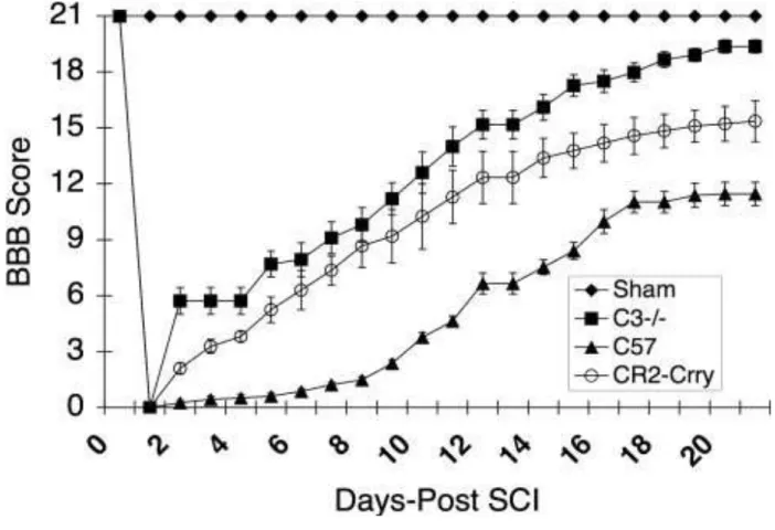
To investigate the role of complement in SCI, we induced contusion injury to the spinal cord in wt mice and in mice deficient in C3, a central protein of the complement system and common for all pathways of activation. Following injury, locomotor recovery was assessed using the modification of the BBB rating scale(75). All animals had a BBB score of 21 pre-injury and a score of 0 immediately after injury, with bilateral hindlimb paralysis (Fig. 3). Two days after injury, and every day thereafter through the termination of the study at day 21, the C3-deficient mice had a significantly improved BBB score compared to the wild-type controls (P < 0.001) (Fig. 3). By day 21 after injury, the C3- deficient mice showed a near normal BBB score of 19.6 ± 1.2 (P< 0.001), whereas the BBB score for wild-type mice was only 11.5 ± 2.14 which was significantly lower than that of C3-deficient mice (P< 0.001). These data indicate that C3 plays an important role in the posttraumatic events that affect functional recovery. Next, we determined whether C3 blockade, using an intravenously administered inhibitor previously shown to target to sites of complement activation, is a feasible posttraumatic therapeutic approach for improving functional recovery. Using the same spinal cord paradigm, a group of mice were treated with a single intravenous injection of 0.25 mg CR2-Crry at 1 hour after SCI.
As with the C3-deficient mice, the CR2-Crry-treated mice had a significantly improved BBB score compared to shamoperated controls at all time points from day 2 following traumatic injury (P<0.001) (Figure 3). The C3-deficient mice appeared to have a better outcome than the CR2-Crry-treated mice, but the difference was not significant.
Fig. 3 Combined BBB locomotor scores post-SCI within sham, vehicle control, C3-deficient, and CR2-Crry groups. Note significant improvement in BBB score at day 3, 7, and 21 in both the C3-deficient and CR2-Crry groups when compared to vehicle controls (P = 0.001) (n = 12).
The values are expressed as mean ± SE.
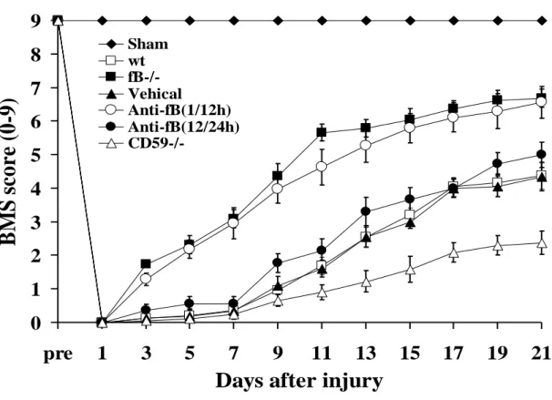
4.1.2 Effect of fB and CD59 Deficiency and of alternative complement pathway inhibition on Locomotor Recovery following SCI
Contusion injury to the spinal cord was induced in mice deficient in fB or CD59, in wt untreated mice, and in wt mice subsequently treated with anti-fB mAb or PBS (vehicle).
Locomotor function was assessed by the BMS scale, and all mice subjected to contusion injury exhibited a BMS score of 0 immediately after injury. Over the course of 21 days, locomotor function significantly improved in fB-/- mice and wt mice treated with anti-fB mAb at 1 and 12 hours after SCI compared to wt and PBS treated controls (Fig. 4). There were no significant differences in BMS scores between fB-/- mice and antifB–treated (1/12 hours) mice over the course of the experiment. When anti-fB mAb was administered at 12 and 24 hours after SCI, there was a trend toward improved locomotor function when compared to controls, but the difference did not reach significance (Fig. 4).
We also administered anti-fB mAb at 24 and 36 hours post-SCI, but there was no difference in functional recovery compared to PBS treated mice (data not shown). Unless otherwise stated, all data shown below for anti-fB treated mice was obtained using a 1 and 12 hour post-SCI treatment schedule. In contrast to the improvement seen in fB-/- mice and anti-fB (1/12 hours) treated mice, recovery of locomotor function was significantly impaired in CD59-/-mice compared to wt and vehicle treated controls. There was no difference in functional recovery between PBS (vehicle) treated mice and untreated wt mice after SCI.
0 1 2 3 4 5 6 7 8 9
pre 1 3 5 7 9 11 13 15 17 19 21
Days after injury
BM S s co re (0 -9 )
Sham wt fB-/- Vehical Anti-fB(1/12h) Anti-fB(12/24h) CD59-/-
Fig. 4. Locomotor recovery after SCI in complement deficient and inhibited mice.
Open-field 10-point BMS scores were recorded for 21 days after contusion-induced SCI in the indicated groups of mice. Anti-fB mAb treatment group received an i.v. injection of 2 mg at 1 and 12 or 12 and 24 hours after injury. BMS scores are significantly higher for fB-/- mice and anti-fB mAb (1/12 hours)-treated mice compared to wt or vehicle (PBS)- treated mice from day 3 postinjury. No significant difference between vehicle controls and anti-fB mAb (12/24 hours) treated mice. BMS scores are significantly lower in CD59-/- mice compared to wt and vehicle control mice from day 11 after injury (P < 0.
01). Mean ± SE, n = 8 - 10.
4.2. Effect of Complement Deficiency and Complement Inhibition on Tissue Damage And Demyelination after SCI
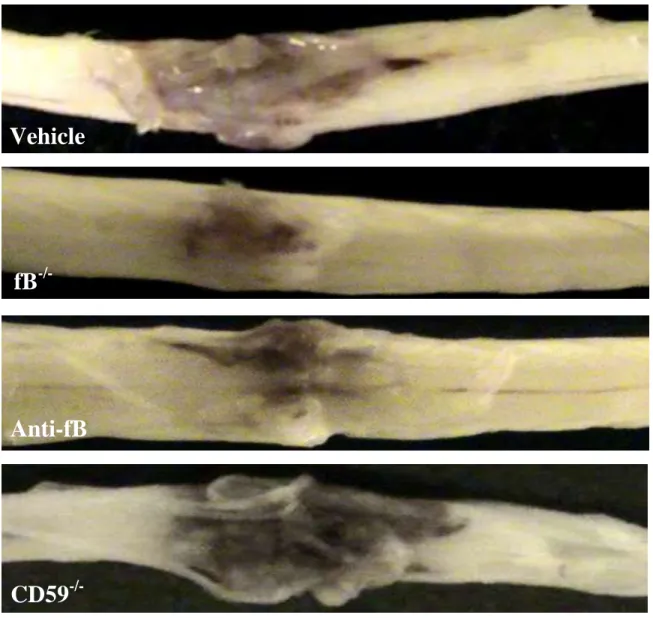
4.2.1. Macroscopic images of spinal cords isolated at 72 hours postinjury.
Spinal cord contusion results in a primary hemorrhage, inflammation, and loss/damage of neurons. We determined the effect of fB deficiency, fB inhibition (1/12 hours), or CD59 deficiency on spinal cord tissue injury by macroscopic examination of spinal cords and by assessment of histological changes within the spinal cords postinjury. At 72 hours after injury, macroscopic examination of control spinal cords demonstrated marked indentation at the injury site with evidence of hemorrhage. These features were markedly reduced in fB-/- and anti-fB mAb treated animals, but appeared exacerbated in spinal cords from CD59-/- mice (Fig. 5).
Vehicle
fB
-/-Anti-fB
Fig. 5. Macroscopic images of spinal cords isolated at 72 hours postinjury.
Mice received anti-fB mAb or PBS (vehicle) at 1 and 12 hours after injury.
Representative images shown, n = 6 8 per group.
CD59
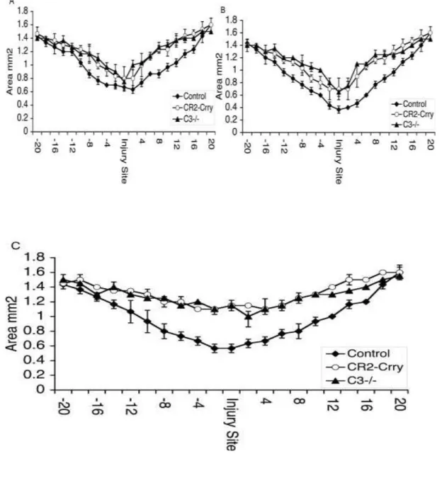
-/-4.2.2 Effect of C3 Deficiency and of Complement Inhibition on the Extent of Tissue Destruction following SCI
To determine whether C3 deficiency or complement inhibition with CR2-Crry attenuated overall spinal cord tissue damage, we determined the cross-sectioned area of spinal cords at 100 μm increments extending 2 mm either side of the initial injury impact site.
Measurements were made using spinal cords isolated from C3-deficient mice and mice treated with CR2-Crry or vehicle control (PBS). At 24 hours after injury, the profile of tissue damage was similar in both C3-deficient and CR2-Crrytreated groups (Fig. 6A). In the control group, there was a clear trend toward increased injury compared to the C3- deficient/inhibited groups, but by 24 hours after SCI the difference did not reach statistical significance at the injury site or on either side of the injury site. Comparable relative profiles were obtained for the three groups of animals at 72 hours after SCI (Fig.
6B). Seven days after injury, however, there was significantly more tissue sparing at and around the injury site in C3-deficient mice and in mice treated with CR2-Crry compared to vehicle control mice (Fig. 6C). There was no difference in tissue sparing between C3- deficient and CR2-Crrytreated mice at 7 days after SCI.
4.2.3. Effect of C3 deficiency and of complement inhibition on the extent of tissue demyelination following SCI
Fig. 6. Tissue sparing as assessed by analyzing the cross-sectional area of spinal cords removed from vehicle controls, C3-deficient, and CR2-Crrytreated animals at 24 hours (A), 72 hours (B), and 7 days after injury (C). Measurements were made from histological sections taken at 100-μm increments extending 2 mm either side of the injury site. No significant difference in tissue sparing was evident at 24 and 72 hrs. Significant tissue sparing was noted in CR2-Crry and C3-/- animals compared to vehicle controls at day 7 (P = 0.002). Mean ± SD, n = 4.
4.2.3. Effect of C3 deficiency and of complement inhibition on the extent of tissue demyelination following SCI
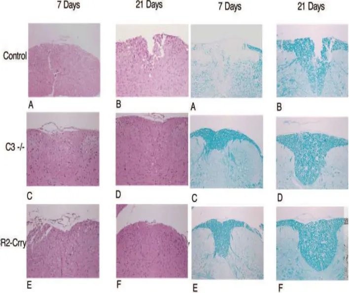
We also analyzed the extent of necrosis and demyelination in cords isolated from the different groups of animals 7 days and 21 days after SCI. In the vehicle control group, H&E staining of cord sections (centered around the injury site) revealed marked areas of necrosis with vacuolization of cells at day 7, with necrosis being somewhat less evident at day 21 (Fig. 7a, A and B). In contrast, the white matter beneath the injury site in cords isolated from C3-deficient mice appeared grossly intact at days 7 and 21 (Fig. 7a, C and D). Cords from CR2- Crry-treated mice also exhibited significant attenuation of injury when compared with vehicle controls, although there appeared to be more vacuolization in the cells within the white matter compared to the C3-deficient animals (Fig. 7a, E and F).
Luxol fast blue staining of cord sections from the control group revealed obvious demylineation in the central core of the white matter beneath the impact site (Fig. 7b, A and B). By comparison, there was markedly less demyelination in cords from C3- deficient mice and CR2-Crry-treated mice at 7 and 21 days after SCI (Fig. 7b, C–F).
There was no apparent difference in the extent of demyelination between the C3-deficient and complement-inhibited groups of animals.
Fig. 7a. H&E-stained sections of spinal cord centered on the injury site at days 7 and 21 after injury. A B: vehicle control. C D: C3- deficient animals. E F: CR2-Crry-treated animals. Original magnification, ×100.
Fig. 7b. Luxol fast blue stained section of spinal cord centered on the injury site at days 7 and 21 after injury. A B: vehicle control.
C D: C3-deficient animals. E F: CR2-Crry- treated animals. Original magnification,
×100.
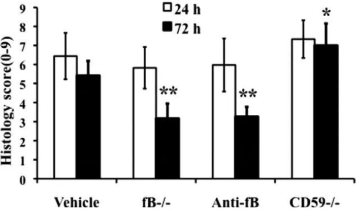
4.2.4. Histopathology of spinal cord sections after SCI
We next analyzed spinal cords microscopically at the injury epicenter using H&E stain to assess the extent of inflammatory cell infiltrate, neuronal vacuolation and hemorrhage.
Sections were prepared from cords isolated at 24 and 72 hours postinjury, and the sections were graded for a total cumulative score of 0–9 (see Materials and Methods).
Injury to spinal cords was evident in all groups at both 24 and 72 hours (Fig. 8).
Fig. 8. Histopathology of spinal cord sections after SCI. A-H: Transverse sections from the epicenter of injury were prepared from spinal cords isolated at 24 and 72 hours after injury and stained with H&E. Mice received anti-fB mAb or PBS (vehicle) at 1 and 12 hours after injury. Representative images shown, n = 6 per group.
No significant differences were noted at 24 hours postinjury, but at 72 hours there was significantly less damage seen in fB-/- mice and anti-fB mAb - treated mice compared to control mice. In contrast, damage was significantly exacerbated in CD59-/- mice compared to controls 72 hours after injury.
4.2.5. Quantitative assessment of histopathological inflammation and injury
Representative images of spinal cord sections postinjury are shown in Fig. 8, with quantification of data shown in Fig. 9. Histologically, control and CD59-/- spinal cord sections demonstrated evidence of hemorrhage, pronounced inflammation, neuronal cell vacuolation, and demyelination,and while these features existed in fB-/-mice and anti-fB mAb - treated mice, they were markedly reduced.
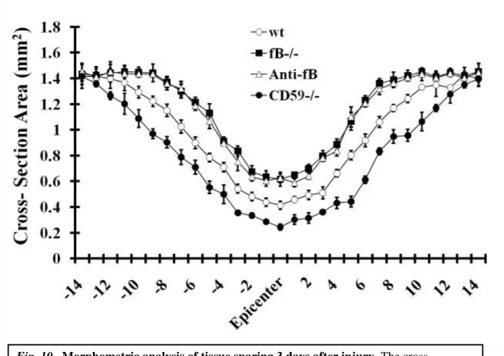
4.2.6. Morphometric analysis of tissue sparing 3 days after injury
Fig. 9. Quantitative assessment of histopathological inflammation and injury. H&E- stained sections from the epicenter of injury were scored from 0 to 3 for the presence and intensity of inflammatory cell infiltration, neuronal vacuolation, and hemorrhage (0 = no evidence, 3 = severe). Scores were then expressed as a cumulative score of 0-9. Assessments were made from spinal cords isolated at 24 and 72 hours postinjury. Mice received anti-fB mAb or PBS (vehicle) at 1 and 12 hours after injury.
To further quantify the impact of complement deficiency or inhibition on SCI, we analyzed tissue destruction by determining the cross-sectional area of spinal cords at 150µm increments extending 2 mm either side of the initial injury impact site. In accord with our subjective histological assessments, there was significantly increased tissue sparing in fB-deficient and fB-inhibited mice, and significantly less tissue sparing in CD59-/- mice, when compared to control mice (Fig. 10).
Fig. 10. Morphometric analysis of tissue sparing 3 days after injury. The cross- sectional area of H&E stained spinal cord sections was measured at 150 µm increments extending 2 mm either side of the injury site. There was a significant level of tissue sparing in fB-/- mice and anti-fB-treated mice compared with vehicle control (PBS)-treated mice, and a significant decrease in tissue sparing in CD59-/- mice compared with wt (at injury site and out to 1.2 mm each side of the injury site, P < 0.01). Mice received anti-fB mAb or PBS at 1 and 12 hours after injury. Mean ± SD n = 6 per group.