. . . .
. . . .
Usefulness of electroanatomical mapping
during transseptal endocardial left ventricular lead implantation
Valentina Kutyifa
1, Be´la Merkely
1, Szabolcs Szila´gyi
1, Endre Zima
1, Attila Ro ´ ka
2, A ´ kos Kira´ly
1, Istva´n Osztheimer
1, Levente Molna´r
1, Ga´bor Sze´plaki
1,
and La´szlo´ Gelle´r
1*
1Semmelweis University, Heart Center, Va´rosmajor utca 68, Budapest H-1122, Hungary; and2Hospital of St. Raphael, New Haven, CT, USA Received 14 August 2011; accepted after revision 14 October 2011; online publish-ahead-of-print 22 December 2011
Aim Failure rate to implant left ventricular (LV) lead transvenously is 4 – 8% in cardiac resynchronization therapy (CRT) patients. Epicardial lead placement is an alternative method and if not applicable case reports and small series showed the feasibility of endocardial LV lead implantation. Electroanatomical mapping might be a useful tool to guide this procedure.
Methods and results
Four patients had undergone endocardial LV lead implantation after unsuccessful transvenous implantation or epicar- dial LV lead dysfunction using the transseptal approach. Electroanatomical mapping was used to mark the location of the transseptal puncture. This location point guided the mapping catheter from the subclavian access and facilitated positioning of the LV lead at the adjacent latest activation area of the left ventricle detected by activation mapping.
Endocardial active fixation LV leads were successfully implanted in all patients with stable electrical parameters im- mediately after implantation and over a mean follow-up of 18.3 months (lead impedance 520+177 vs. 439+119V and pacing threshold 0.8+0.2 V, 0.5 ms vs. 0.6+0.1 V, 0.5 ms, respectively). Patients were maintained on anticoa- gulation therapy with a target international normalized ratio of 3.5 – 4.5 and did not show any thromboembolic, haem- orrhagic events, or infection. Echocardiography showed significant improvement of LV systolic function with marked improvement of the functional status.
Conclusions Electroanatomical mapping is a useful technical tool to guide endocardial LV lead implantation. It helps to identify the location of the transseptal puncture and the use of activation mapping might facilitate location of the optimal lead positions during CRT.
- - - -
Keywords Endocardial † Left ventricular lead † Electroanatomical mapping guidance
Introduction
Cardiac resynchronization therapy (CRT) is an effective non- pharmacological treatment modality in patients with symptomatic congestive heart failure (HF) refractory to medical therapy, left ventricular (LV) dysfunction and wide QRS.1–3The standard ap- proach is to implant the LV lead transvenously in one tributary of the coronary sinus (CS). However, even with innovative lead technology lead placement fails in 4 – 8%.2,4 Most frequent reasons are CS occlusion, dissection, abnormal ostium of the CS,
coronary vein stenosis, lead instability, high threshold, or phrenic nerve stimulation.2,5,6 Epicardial lead placement is an alternative method which includes minimal-invasive thoracoscopy or lateral thoracotomy, and usually requires general anaesthesia.7When epi- cardial LV lead implantation is contraindicated or at higher risk, LV endocardial lead implantation might be considered.8
Cardiac resynchronization therapy is typically delivered with an LV lead in a lateral or posterolateral position1–3and is placed in the middle or distal portion of the side branch to ensure a stable position. In contrast, when using the endocardial transseptal
*Corresponding author. Tel:+36 20 365 83 30; fax:+36 1 458 68 42, Email: laszlo.geller@kardio.sote.hu
Published on behalf of the European Society of Cardiology. All rights reserved.&The Author 2011. For permissions please email: journals.permissions@oup.com.
approach, the LV lead position is independent of the coronary venous anatomy. The major difficulty of this method is to locate the transseptal puncture site performed with a transfemoral ap- proach from the subclavian access and to find the optimal position of the LV lead within the LV cavity.
Electroanatomical mapping involves visualizing cardiac structures and gathering data of the electrical activation of the heart. This method is widely used to guide ablation procedures in the left atrium (LA) as well as in the right ventricle and left ventricle.9 With the use of activation mapping we can record the activation sequence of the heart. Electroanatomical mapping might be a useful tool to guide endocardial LV lead implantation for CRT.
Patients
Four patients had undergone endocardial LV lead implantation at our hospital between November 2007 and May 2010 guided by electroanatomical mapping. Indications for CRT were in accord- ance with the current guidelines.10 Patient characteristics are listed inTable1. All patients had left bundle branch block (LBBB) or paced rhythm with LBBB-like morphology.
Cardiac resynchronization therapy was attempted or performed either transvenously or epicardially as a first procedure. Patient 1 had an epicardial LV lead dysfunction, the second operation was contraindicated, and the patient was referred for endocardial LV lead implantation. Patients 2 and 4 had unsuccessful transvenous LV lead implantation. Patient 3 had LV lead dysfunction after suc- cessful transvenous LV lead implantation. In Patients 2 and 3, mini- thoracotomy was contraindicated because of multiple co- morbidities and a subsequent higher risk of surgical intervention.
Patients 1 and 4 did not give their consent for epicardial surgical LV lead implantation. All patients had given informed consent prior to the procedure.
Pre-implant echocardiography and tissue Doppler imaging revealed severely depressed LV function and dyssynchronous acti- vation pattern with a significant delay of the lateral (n¼3) and the posterolateral wall (n¼1). Mitral regurgitation was evaluated by colour Doppler imaging using a semi-quantitative method (grade I – IV).
Methods
The LV endocardial lead implantation was performed using a combined femoral and subclavian approach guided by electroanatomical mapping.
The first step of the procedure was to introduce the CARTO Quick Star catheter (Biosense Webster, Diamond Bar, CA, USA) through the right femoral vein to capture the anatomical map of the right atrium and the right ventricle and the activation sequence of the right ventricle. The transseptal puncture was performed with the guid- ance of fluoroscopy and intracardiac echocardiography including con- tinuous monitoring of the arterial pressure. Intravenous heparin was given after the transseptal puncture (5000 IU); in case of long-lasting procedures it was administered repeatedly to maintain an ACT level of 250 ms.
After successful transseptal puncture, Quick Star catheter was intro- duced into the transseptal sheath and the puncture point of the septum was marked on the CARTO map (Biosense Webster) with a pink dot, while the corresponding region was marked with white
...
Table1Patientcharacteristicsandfollow-up Pt no.GenderAge (years)Aetiologyof cardiomyopathyQRS (ms)NYHA functionalclassLVEF (%)Follow-up (months)NYHAat FULVEF(%) FUMRgradebefore implantation(I–IV)MRgradeafter implantation(I–IV) 1Female56Non-ischaemic160III2427II38IVIV 2Male71Ischaemic165III3521I44III–IVII–III 3Male45Ischaemic200III–IV2412II33IIII–III 4Female58Non-ischaemic190III–IV286II48III–IVII Pt,patient;NYHA,NewYorkHeartAssociationfunctionalclass;LV,leftventricular;CS,coronarysinus;LVEF,leftventricularejectionfraction;MR,mitralregurgitation;FU,follow-up.
small dots (Figure1). After removal of the Quick Star catheter, a guide- wire (0.035 inch×260 cm) was inserted into the LA and advanced into the left upper pulmonary vein and an angioplasty balloon (6 mm
×20 mm Maverick, Boston Scientific, Natick, MA, USA) was inserted into the LA on the previously positioned guide-wire. The transseptal sheath was withdrawn into the right atrium and the balloon was posi- tioned across the septal puncture site. It was inflated 3 times with 12 atmospheres for 5 seconds before its removal. The transseptal sheath was then positioned into the right atrial cavity.
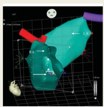
The Quick Star deflectable catheter was introduced again into the LA and advanced into the LV cavity via the right femoral vein. Left atrial and LV anatomical and activation maps were recorded (Figure 2). The latest activation of the left ventricle was localized at the lateral wall in three patients and at the posterolateral wall in one patient.
An 11 F-long sheath (SCOUT Pro 8 Fr, Biotronik GmbH&Co, Berlin, Germany) was introduced via the left subclavian vein. The Quick Star catheter was advanced into the sheath and directed to the location of the previously marked transseptal puncture site applying CARTO map guidance.
The long sheath was forced through the interatrial septum into the LA and further into the left ventricle over the deflectable Quick Star catheter. When the sheath did not go through the site of the puncture, angioplasty balloon (6 mm×20 mm Maverick, Boston Scientific, Natick, MA, USA) was positioned to the puncture site through the previously applied guide-wire from the femoral access and it was inflated and deflated again, as needed. The Quick Star catheter was used to relocate the LV segment with the latest activation. When the latest activation site was found, the Quick Star catheter was with- drawn into the sheath and the sheath was pushed against the LV wall to ensure stable position. Active fixation LV leads were fixed at the basal or mid-basal portion of the left ventricle in all patients where the longest delays were detected on the activation map. Using the long
sheath pushed against the LV wall to have a stable support facilitated the position of the LV lead close to the mitral valve. Standard bipolar screw-in leads were used in all patients (Medtronic 5076 – 65 cm, Medtronic, Minneapolis, MN, USA, n¼4). The LV lead was connected to the CRT device placed in the left pectoral area. The atrial lead was implanted in the right atrial appendage in all patients.
The right ventricular lead was positioned in the right ventricular septum (Figure3AandB).
Data are presented as mean+standard deviation. Changes in LV pacing threshold, LV pacing impedance at implantation, and at last patient visit were analysed using pairedt-test. New York Heart Asso- ciation (NYHA) functional class and mitral regurgitation were analysed using the Wilcoxon’s signed rank test, as appropriate. Statistical signifi- cance was considered atP,0.05. Statistical analyses were performed using GraphPad Prism software (GraphPad Software Inc., La Jolla, CA, USA).
Results
LV endocardial leads were successfully implanted in all patients.
Cardiac resynchronization therapy pacemaker was implanted in one patient (Stratos LV-T, Biotronik GmbH&Co, Berlin, Germany), while three patients received CRT-D devices (Cognis, Boston Scientific, Miami, FL, USA,n¼1; Concerto, Medtronic,n
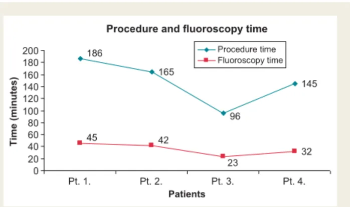
¼1; Atlas HF, St Jude, Sylmar, CA, USA,n¼1). Electrical para- meters during device implantation were as follows: LV signal amp- litude 8.5+3.0 mV, LV pacing threshold 0.78+0.18 V, impulse width of 0.5 ms, and LV lead impedance 520+177V. No phrenic nerve stimulation occurred at 10 V, 0.5 ms with rapid LV pacing (100 b.p.m.). The procedure times were 186, 165, 96, and 145 min, and fluoroscopy times were 45, 42, 23, and 32 min, re- spectively (Figure4).
Figure 1 Patient 1. CARTO image, antero-posterior projec- tion. The location of the transseptal puncture is indicated with a single white arrow on the CARTO map.
Figure 2 Patient 1. CARTO image, left lateral projection. Right and the LV activation map: the earliest activation site is the right ventricular anteroseptal region; the latest site is the mid-basal part of the posterolateral wall.
In all patients, the international normalized ratio was maintained between 3.5 and 4.5 as for those with mechanical valve prostheses and high thrombotic risk. Neither pericardial fluid nor intracardiac thrombi were observed during echocardiography in the early post- operative period or during follow-up. Neither major haematoma nor significant post-procedural bleeding occurred. During the mean follow-up of 18.3 months, stable sensing and pacing para- meters were found. The mean LV pacing threshold was 0.6+ 0.1 V at impulse width of 0.5 ms (P¼0.44), LV lead impedance was 439+119V(P¼0.12).
We did not observe lead dysfunction, insulation failure, or dis- location of the LV lead. There were no signs of lead infection during the follow-up period. Heart failure symptoms improved at least one NYHA class in all patients, LV systolic function improved
significantly from a mean LV ejection fraction of 28+5.2 to 41+ 6.6% (P¼0.015). The grade of mitral regurgitation did not change significantly during the follow-up period (P¼0.28) (Table1). Small left – right shunt was detected in all patients immediately after the procedure. We did not observe residual left – right shunts by trans- thoracal echocardiography; no embolism, cyanosis, right-heart failure was observed during the follow-up. No thromboembolic or haemorrhagic events occurred.
Discussion
Difficulties may be encountered when attempting LV lead place- ment during CRT. If transvenous LV lead placement via the CS is not possible and epicardial approach is contraindicated, LV endo- cardial lead implantation is an alternative method in selected patients. Early case reports and small series showed the safety and feasibility of this procedure. No report has shown the feasibil- ity of electroanatomical mapping guidance during endocardial LV lead implantation, yet.
The first successful endocardial LV lead implantation was reported by Jaiset al.11in 1998 was performed via the right jugular vein after a transfemoral interatrial septal puncture. Leclerqet al.12 described three cases of LV lead placement applying a transseptal approach from the right jugular vein. Van Gelderet al.8and Nuta et al.13 developed a new technique, performing the transseptal puncture via the femoral vein and positioning the LV electrode from the subclavian vein into the LV cavity through the interatrial septum and the mitral valve. In our study, we used the latter ap- proach; however, electroanatomical mapping technique was used additionally to enhance proper LV lead placement and facilitating the LV lead implantation from a subclavian access. The implant- ation time and also the fluoroscopy time was less than expected.
There are potential advantages of endocardial LV lead place- ment compared with epicardial pacing. Endocardial LV lead pos- ition is not limited by the anatomy of the CS. Experimental14 and clinical15observations also suggested that endocardial pacing is more physiologic than epicardial pacing. Garrigue et al.16 and Jais et al.17 reported better haemodynamic results with higher aortic and mitral time velocity integral, improvement of LV frac- tional shortening and reduction of regional electromechanical delay in patients with endocardial LV pacing. In our patient Figure 3 Patient 4. Typical final lead positions in right-anterior
oblique projection and in left-anterior oblique projection. RA lead, right atrial lead is positioned in the right atrial appendage;
RV lead, right ventricular lead is positioned in the right ventricular apical septum; LV lead, LV lead is positioned in the mid-basal portion of the lateral wall.
Figure 4 Procedure and fluoroscopy time.
cohort, we also reported significant improvement of the LV systol- ic function and a marked improvement of the patient’s functional status during the long-term follow-up.
The drawbacks of this approach include the risk of transseptal catheterization, the current lack of appropriate implantation tools and the possible need for lifetime anticoagulation, as previ- ously reported.8However, anticoagulation is indicated in the ma- jority of these patients due to severe LV systolic dysfunction and/or the presence of atrial fibrillation. Long-term follow-up data regarding thromboembolic or haemorrhagic complications were so far not available. Our study showed that during 1.5-year follow-up no haemorrhagic or thromboembolic events occurred.
Using the transseptal CRT approach, the LV lead crosses the atrial septum, mitral valve and is actively fixed to the LV endocar- dial surface. It is controversial whether mitral regurgitation might be worsened with this technique: however, we did not observe any worsening of mitral regurgitation or echocardiographic evi- dence of the mitral valve being partially kept open. The risk of in- fective endocarditis might be increased,18 but no data of more frequent endocarditis are currently available in this patient popula- tion. We did not observe any lead infection during the follow-up period.
Transseptal punctures are associated with iatrogenic atrial septal defects and concomitant left – right shunts.19 It is important to evaluate this in patients with LV leads crossing the atrial septum.
In our study cohort, small left – right shunt was detected in all patients immediately after the procedure. During follow-up, we did not observe residual left – right shunts; however, it was diag- nosed using two-dimensional transthoracal echocardiography, and tranoesophageal echocardiography was not performed. We used an 8F sheath for the transseptal puncture during the proced- ure. Several earlier studies indicated that using 8F sheaths for trans- septal punctures create iatrogenic atrial septal defects.19 The incidence of atrial septal defects is as high as 87% immediately after the procedure, while it is decreasing over time. At 6-month follow-up, only 21% of iatrogenic atrial septal defects persisted.20 Residual septal defects are well tolerated and not associated with increased risk of embolism, cyanosis or right-heart failure.19 We do not have additional information about the impact of balloon dilatation on the incidence and persistence of atrial septal defects in this patient population undergoing transseptal CRT implantation. We assume that using balloon dilatation in this highly mobile region of the atrial septum facilitates more easy penetration in the short term while it is not associated with larger atrial septal defects in the long term, therefore not expected to be clinically relevant. In the future, serial transoesophageal echo- cardiography studies are needed to further evaluate this.
Clinical studies showed appropriate positioning of the LV lead to be of high importance to increase the number of CRT respon- ders.21,22 Recent studies also demonstrated that an ‘individually’
based LV pacing approach compared with conventional CS pacing, echo-guided or lateral area strategy might result in better short-term haemodynamic response in non-ischaemic cardiomy- opathy patients.23The authors reported a benefit of endocardial pacing. Our study also emphasizes the positive effects of endocar- dial pacing.
Spragg et al.24 recently found that electroanatomical mapping with colour-coded dP/dtmax response is a feasible approach to identify LV endocardial sites with the highest peak of LV pressure increase (dP/dtmax). Although only acute haemodynamic response was evaluated in this study, optimal LV pacing site was more often found in a basal location (in 8 of 11 patients). In our study, electroanatomical activation mapping was used to identify the latest activation area to find optimal LV lead position for CRT.
Functional assay (dP/dtmax) might be an alternative approach to guide LV lead implantation and the two approaches may give dis- cordant results.24However, there is no direct comparison or long- term data available yet.
Singhet al.25showed that clinical benefit from CRT was similar with LV leads along the anterior, lateral or posterior wall in mildly symptomatic HF patients. However, LV leads positioned in the apical region were associated with subsequent worse outcome.
Our study also supported the hypothesis that basal, mid-basal LV lead position might be associated with a favourable echocardio- graphic and clinical improvement during a mean follow-up of 1.5 years.
An alternative approach is reported to implant the LV lead transapically with a minimally invasive surgical technique. This tech- nique has the advantage of avoiding mitral valve crossing, but bears a higher surgical and post-operative risk compared with our new approach.26However, our small cohort does not allow a real com- parison with other techniques.
Because of the technical complexity of this technique, this ap- proach remains a rare exception in CRT candidates and indicated only if transvenous implantation is not successful. More patients and longer follow-up are needed to provide additional data of this approach.
Limitations of the report
This report includes four patients who had undergone electroana- tomical mapping-guided transseptal endocardial LV lead implant- ation. One of the major limitations of this report is the few patients included, which did not allow us to compare this tech- nique with the standard approach. The mean follow-up of 18.3 months is relatively short; however; earlier studies reported even shorter follow-up periods. With this technique, the position of the LV lead cannot be tracked within the LV cavity using the CARTO electroanatomical mapping system.
Conclusion
We report successful electroanatomical mapping-guided LV endo- cardial lead implantation in four patients after unsuccessful transve- nous or epicardial LV lead placement. Electroanatomical mapping to implant LV endocardial leads is proven to be useful in shortening the procedure and fluoroscopy time by identifying the location of the transseptal puncture. Activation map might help to identify the optimal LV lead position for CRT. No major complications such as bleeding, thromboembolism, or infections were observed. Pacing parameters remained stable over long-term follow-up. Clinical symptoms of HF and cardiac function improved significantly. We conclude that transseptal endocardial LV lead implantation
guided by electroanatomical mapping might be a feasible method for CRT if transvenous LV lead placement is not possible. More data are needed to assess the safety and long-term efficacy of this new approach.
Conflict of interest:V.K. has received consultant fees/honoraria from Biotronik and Servier and research support from Boston Sci- entific. B.M. has received consultant fees/honoraria from Biotronik, Boston Scientific, Medtronic, and St. Jude Medical, and serves in the speakers bureau of Boehringer Ingelheim. L.G. has received consultant fees/honoraria from Biotronik, Medtronic, St. Jude Medical, and Johnson & Johnson. E.Z. has received consultant fees and honoraria from Boston Scientific, Innomed, Biotronik, Medtronic, and St. Jude Medical for lectures, training, and participa- tion in clinical trials. I.O. has received consultant fees/honoraria from Biotronik, Medtronic, Boston Scientific, and St. Jude Medical. A.R., G.S., A.K., S.S. and L.M. have nothing to disclose.
Funding
The scientific work was granted by a grant from the National Development Agency of Hungary.
References
1. Abraham WT, Fisher WG, Smith AL, Delurgio DB, Leon AR, Loh Eet al.Cardiac resynchronization in chronic heart failure.N Engl J Med2002;346:1845 – 53.
2. Bristow MR, Saxon LA, Boehmer J, Krueger S, Kass DA, De Marco Tet al.
Cardiac-resynchronization therapy with or without an implantable defibrillator in advanced chronic heart failure.N Engl J Med2004;350:2140 – 50.
3. Cleland JG, Daubert JC, Erdmann E, Freemantle N, Gras D, Kappenberger Let al.
The effect of cardiac resynchronization on morbidity and mortality in heart failure.N Engl J Med2005;352:1539 – 49.
4. Gras D, Bocker D, Lunati M, Wellens HJ, Calvert M, Freemantle Net al.Implant- ation of cardiac resynchronization therapy systems in the CARE-HF trial: proced- ural success rate and safety.Europace2007;9:516 – 22.
5. Meisel E, Pfeiffer D, Engelmann L, Tebbenjohanns J, Schubert B, Hahn Set al.In- vestigation of coronary venous anatomy by retrograde venography in patients with malignant ventricular tachycardia.Circulation2001;104:442 – 7.
6. Higgins SL, Hummel JD, Niazi IK, Giudici MC, Worley SJ, Saxon LAet al.Cardiac resynchronization therapy for the treatment of heart failure in patients with intra- ventricular conduction delay and malignant ventricular tachyarrhythmias.J Am Coll Cardiol2003;42:1454 – 9.
7. Koos R, Sinha AM, Markus K, Breithardt OA, Mischke K, Zarse Met al.Compari- son of left ventricular lead placement via the coronary venous approach versus lateral thoracotomy in patients receiving cardiac resynchronization therapy.Am J Cardiol2004;94:59 – 63.
8. van Gelder BM, Scheffer MG, Meijer A, Bracke FA. Transseptal endocardial left ventricular pacing: an alternative technique for coronary sinus lead placement in cardiac resynchronization therapy.Heart Rhythm2007;4:454 – 60.
9. Hameedullah I, Chauhan VS. Clinical considerations for allied professionals:
understanding and optimizing three-dimensional electroanatomic mapping of complex arrhythmias – part 1.Heart Rhythm2009;6:1249 – 52.
10. Dickstein K, Vardas PE, Auricchio A, Daubert JC, Linde C, McMurray Jet al.2010 Focused Update of ESC Guidelines on device therapy in heart failure: an update of the 2008 ESC Guidelines for the diagnosis and treatment of acute and chronic heart failure and the 2007 ESC guidelines for cardiac and resynchronization therapy. Developed with the special contribution of the Heart Failure Association and the European Heart Rhythm Association.Europace2010;12:1526 – 36.
11. Jais P, Douard H, Shah DC, Barold S, Barat JL, Clementy J. Endocardial biventri- cular pacing.Pacing Clin Electrophysiol1998;21(Part 1):2128 – 31.
12. Leclercq F, Hager FX, Macia JC, Mariottini CJ, Pasquie JL, Grolleau R. Left ven- tricular lead insertion using a modified transseptal catheterization technique: a totally endocardial approach for permanent biventricular pacing in end-stage heart failure.Pacing Clin Electrophysiol1999;22:1570 – 75.
13. Nuta B, Lines I, MacIntyre I, Haywood GA. Biventricular ICD implant using endo- cardial LV lead placement from the left subclavian vein approach and transseptal puncture via the transfemoral route.Europace2007;9:1038 – 40.
14. Fish JM, Di Diego JM, Nesterenko V, Antzelevitch C. Epicardial activation of left ventricular wall prolongs QT interval and transmural dispersion of repolarization:
implications for biventricular pacing.Circulation2004;109:2136 – 42.
15. Fish JM, Brugada J, Antzelevitch C. Potential proarrhythmic effects of biventricular pacing.J Am Coll Cardiol2005;46:2340 – 47.
16. Garrigue S, Jais P, Espil G, Labeque JN, Hocini M, Shah DCet al.Comparison of chronic biventricular pacing between epicardial and endocardial left ventricular stimulation using Doppler tissue imaging in patients with heart failure.Am J Cardiol2001;88:858 – 62.
17. Jais PSF, Laborderie J, Reuter S, Bordachar P, Hsu LF, Sanders Pet al.Tailored endocardial left ventricular pacing is superior to coronary sinus in heart failure patients needing cardiac resynchronization.Heart Rhythm2006;3:S247.
18. Musci M, Siniawski H, Pasic M, Grauhan O, Weng Y, Meyer Ret al.Surgical treat- ment of right-sided active infective endocarditis with or without involvement of the left heart: 20-year single center experience.Eur J Cardiothorac Surg2007;32:
118 – 25.
19. McGinty PM, Smith TW, Rogers JH. Transseptal left heart catheterization and the incidence of persistent iatrogenic atrial septal defects.J Interv Cardiol2011;24:
254 – 63.
20. Rillig A, Meyerfeldt U, Kunze M, Birkemeyer R, Miljak T, Jackle Set al.Persistent iatrogenic atrial septal defect after a single-puncture, double-transseptal approach for pulmonary vein isolation using a remote robotic navigation system: results from a prospective study.Europace2010;12:331 – 6.
21. Butter C, Auricchio A, Stellbrink C, Fleck E, Ding J, Yu Yet al.Effect of resynchro- nization therapy stimulation site on the systolic function of heart failure patients.
Circulation2001;104:3026 – 9.
22. Dekker AL, Phelps B, Dijkman B, van der Nagel T, van der Veen FH, Geskes GG et al.Epicardial left ventricular lead placement for cardiac resynchronization therapy: optimal pace site selection with pressure-volume loops.J Thorac Cardio- vasc Surg2004;127:1641 – 7.
23. Derval N, Steendijk P, Gula LJ, Deplagne A, Laborderie J, Sacher Fet al.Optimiz- ing hemodynamics in heart failure patients by systematic screening of left ven- tricular pacing sites: the lateral left ventricular wall and the coronary sinus are rarely the best sites.J Am Coll Cardiol 2010;55:566 – 75.
24. Spragg DD, Dong J, Fetics BJ, Helm R, Marine JE, Cheng Aet al.Optimal left ven- tricular endocardial pacing sites for cardiac resynchronization therapy in patients with ischemic cardiomyopathy.J Am Coll Cardiol 2010;56:774 – 81.
25. Singh JP, Klein HU, Huang DT, Reek S, Kuniss M, Quesada Aet al.Left ventricular lead position and clinical outcome in the Multicenter Automatic Defibrillator Im- plantation Trial-Cardiac Resynchronization Therapy (MADIT-CRT) Trial.Circula- tion2011;123:1159 – 66.
26. Kassai I, Mihalcz A, Foldesi C, Kardos A, Szili-Torok T. A novel approach for endocardial resynchronization therapy: initial experience with transapical implant- ation of the left ventricular lead.Heart Surg Forum2009;12:E137 – 140.