Article
Response of Human Neutrophil Granulocytes to the Hyphae of the Emerging Fungal Pathogen
Curvularia lunata
Eszter Judit Tóth1,2, Mónika Varga2, Miklós Takó2 , Mónika Homa1, Olivér Jáger1,2, Edit Hermesz3 , Hajnalka Orvos4, Gábor Nagy1,2 , Csaba Vágvölgyi2 and
Tamás Papp1,2,*
1 MTA-SZTE Fungal Pathogenicity Mechanisms Research Group, Hungarian Academy of Sciences—University of Szeged, 6726 Szeged, Hungary; forimistio@gmail.com (E.J.T.);
homamoni@gmail.com (M.H.); jager.oliver.biology@gmail.com (O.J.); nagygab86@gmail.com (G.N.)
2 Department of Microbiology, Faculty of Science and Informatics, University of Szeged, 6726 Szeged, Hungary; varga.j.monika@gmail.com (M.V.); tako78@bio.u-szeged.hu (M.T.); csaba@bio.u-szeged.hu (C.V.)
3 Department of Biochemistry and Molecular Biology, Faculty of Science and Informatics, University of Szeged, 6726 Szeged, Hungary; hermeszedit@gmail.com
4 Department of Obstetrics and Gynecology, Faculty of Medicine, University of Szeged, 6725 Szeged, Hungary;
orvosh@obgyn.szote.u-szeged.hu
* Correspondence: pappt@bio.u-szeged.hu
Received: 6 March 2020; Accepted: 20 March 2020; Published: 21 March 2020
Abstract: Curvularia lunata is an ascomycete filamentous fungus causing local and invasive phaeohyphomycoses in both immunocompromised and immunocompetent patients. Neutrophils are crucial participants of the first line host defense against fungal infections. They migrate to the infected site and eliminate the infectious agents by various mechanisms including phagocytoses, oxidative damage, or formation of neutrophil extracellular trap (NET). Neutropenia may be a risk factor for phaeohyphomycoses, and restoration of the neutrophil function can improve the outcome of the infection. In the present study, interaction of primary human neutrophil granulocytes with the hyphaeC. lunatawas examined and compared to that with the well characterized filamentous fungal pathogenAspergillus fumigatus. Neutrophils could recognize the serum opsonized hyphae ofC. lunata and attach to them. Myeloperoxidase release was also activated by a soluble factor present in the culture supernatant of the fungus. Induction of the oxidative burst was found to depend on serum opsonization of the hyphae. Although extracellular hydrogen peroxide production was induced, the fungus efficiently blocked the oxidative burst by acidifying the reaction environment. This blockage also affected the NET forming ability of the neutrophils.
Keywords: Curvularia; phaeohyphomycosis; neutrophil extracellular trap; oxidative burst;
myeloperoxidase; hydrogen peroxide; acidification
1. Introduction
Curvulariais an ascomycete fungal genus, which mainly includes saprotrophic and plant pathogenic species. However, it is also considered as one of the most relevant filamentous fungal group involved in local and invasive phaeohyphomycoses, i.e., human infections caused by dematiaceous fungi [1–4].
Curvulariainfections can manifest as mycotic keratitis [5], cutaneous and subcutaneous mycosis [6], onychomycosis [7], peritonitis [8], and allergic sinusitis [8,9]. Deep infections frequently affect the central nervous system [8–10]. Development of disseminated mycoses has been reported in both immunocompromised and immunocompetent patients [11–14]. Within the genus,C. lunatais among
Pathogens2020,9, 235; doi:10.3390/pathogens9030235 www.mdpi.com/journal/pathogens
Pathogens2020,9, 235 2 of 12
the species most frequently identified from clinical samples [5,15]. Despite their abovementioned role as the causative agents of phaeohyphomycoses, there are only very limited information available about the immunological aspects and the pathomechanisms ofCurvulariainfections [16].
Immunosuppression and immunodeficiencies result in predisposition to opportunistic fungal infections [17], including those ofCurvulariaspecies [4]. Action of monocytes and macrophages phagocyting the conidia and neutrophils causing hyphal damage has a key role in the prevention of invasive mycotic infections [18,19]. Neutrophils are among the first immune cells, which migrate to the infected site and they eliminate the microbes by various mechanisms involving phagocytoses, formation of neutrophil extracellular trap (NET) or release of oxidative molecules (such as hydrogen peroxide or hydroxyl radicals), enzymes or antimicrobial peptides [20,21]. Action of neutrophil granulocytes againstAspergillusis a well-studied field of host–fungal pathogen interactions [17,22,23]
but little is known about their role in the defense toCurvulariaspecies and other dematiaceous molds.
Neutropenia was found to be associated with high risk of death in phaeohyphomycosis, while recovery from neutropenia improved the chance of the survival [24].
In a recent study, we examined the interaction ofC. lunatawith human monocytes and compared it to that ofA. fumigatusand otherCurvulariaspecies [16]. In that study, monocytes attached to the hyphae ofC. lunataproduced interleukin (IL)-8 in an elevated level. The primary role of this cytokine is to recruit neutrophils to the site of infection [25]. Here, we report the interaction ofC. lunatawith human neutrophil granulocytes, which represent another key component of the first line defense in the elimination of the fungal pathogens.
2. Results
2.1. Activation of the Neutrophils by the C. lunata Hyphae
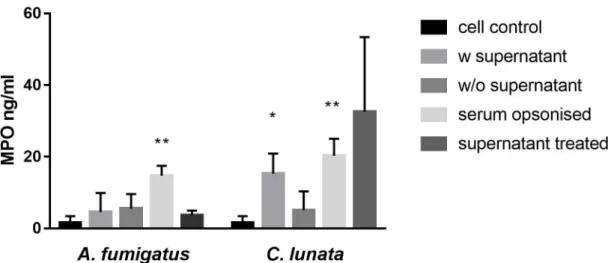
The culture supernatant ofC. lunatawith the hyphae and the serum opsonized hyphae induced the myeloperoxidase (MPO) release of the neutrophils (Figure1). If the culture supernatant was changed to modified Krebs-Ringer phosphate glucose (mKRPG) buffer over the hyphae, MPO release did not increase compared to the untreated neutrophils. Presence of theA. fumigatushyphae induced significant MPO release only after serum opsonization, while the culture supernatant of the germinated conidia could not activate the neutrophils.
2.2. Generation of Hydrogen Peroxide (H2O2)
BothC. lunataandA. fumigatushyphae induced the H2O2release only after opsonization (Figure2).
Interestingly, the released H2O2almost completely disappeared at the 60th min of the interaction with C. lunata, while its level proved to be more or less constant in the case ofA. fumigatus. Without the fungi, H2O2release of the neutrophil cell could not be detected.
2.3. Detection of Extracellular Melanin
AsC. lunatahyphae can produce dihydroxynaphthalene (DHN)-type melanin, which may affect the oxidative damage by the neutrophils, melanin production during the interaction was tested.
Acidification causes precipitation of the melanin pigment. However, by acidifying the supernatant of the interaction, no precipitation was formed, and no visible residue left on the polytetrafluoroethylene (PTFE) membrane. Acidification and filtering of the culture media from 7 days long cultivations in mKRPG showed the same result.
Figure 1. Neutrophil granulocytes are activated by serum opsonizedC. lunata and by the culture supernatant of the hyphae. Myeloperoxidase (MPO) release of neutrophil granulocytes was measured after 60 min of interaction withA. fumigatusandC. lunata. Cell control: untreated neutrophils; w supernatant: neutrophils were added directly to the germinated conidia; w/o supernatant: germinated conidia were washed with fresh mKRPG before addition of neutrophils; serum opsonized: young hyphae were serum opsonized before addition of neutrophils; supernatant treated: cell free culture supernatant of the germinated conidia was added to the neutrophils. Error bars represent standard deviations calculated from three biological parallels. Significance was calculated with multiple t-test compared to the cell control using the GraphPad Prism 7 software, FDR (Q=10%), *p<0.05 **p<0.0001.
Figure 2.The amount of H2O2decreases after 60 min of interaction withC. lunata. H2O2concentration was measured after 30 and 60 min of interaction with the serum opsonized hyphae ofA. fumigatusand C. lunataand it was normalized to 106neutrophils. Error bars represent standard deviations calculated from three biological parallels.
2.4. NET Formation in the Presence of the Hyphae
Serum opsonized A. fumigatus hyphae induced the NET formation, as it was published previously [17] (Figure 3a). In contrasts, NET formation was not observed in the presence of theC. lunatahyphae (Figure3b).
Pathogens2020,9, 235 4 of 12
Pathogens2020,9, x FOR PEER REVIEW 3 of 12
bars represent standard deviations calculated from three biological parallels. Significance was calculated with multiple t-test compared to the cell control using the GraphPad Prism 7 software, FDR (Q = 10%), *p< 0.05 **p< 0.0001.
2.2. Generation of Hydrogen Peroxide (H2O2)
Both C. lunata and A. fumigatus hyphae induced the H2O2 release only after opsonization (Figure 2). Interestingly, the released H2O2 almost completely disappeared at the 60th min of the interaction with C. lunata, while its level proved to be more or less constant in the case of A.
fumigatus. Without the fungi, H2O2release of the neutrophil cell could not be detected.
Figure 2. The amount of H2O2 decreases after 60 min of interaction with C. lunata. H2O2
concentration was measured after 30 and 60 min of interaction with the serum opsonized hyphae of A. fumigatusandC. lunataand it was normalized to 106neutrophils. Error bars represent standard deviations calculated from three biological parallels.
2.3. Detection of Extracellular Melanin
As C. lunata hyphae can produce dihydroxynaphthalene (DHN)-type melanin, which may affect the oxidative damage by the neutrophils, melanin production during the interaction was tested. Acidification causes precipitation of the melanin pigment. However, by acidifying the supernatant of the interaction, no precipitation was formed, and no visible residue left on the polytetrafluoroethylene (PTFE) membrane. Acidification and filtering of the culture media from 7 days long cultivations in mKRPG showed the same result.
2.4. NET Formation in the Presence of the Hyphae
Serum opsonized A. fumigatus hyphae induced the NET formation, as it was published previously [17] (Figure 3a). In contrasts, NET formation was not observed in the presence of theC.
lunatahyphae (Figure 3b).
Figure 3.Neutrophil extracellular trap (NET) formation is missing when neutrophils are confronted with the hyphae ofC. lunata. Fluorescent microscopy was performed about after 3 h of interaction of serum opsonizedA. fumigatus(A) andC. lunata(B) with neutrophil granulocytes. Hyphae and conidia were labeled with Calcofluor white (blue); neutrophil’s cell membrane was stained with CellMask Deep Red Plasma Membrane Stain (red) and the DNA was stained with SYTOX Green (green). NET formation was detected in the interaction withA. fumigatusbut only gathering of the cells to the hyphae could be observed in case ofC. lunata.
It was even not induced if H2O2was added to the interactions. Seemingly, neutrophils recognized the serum opsonized hyphae and attached to it, but the lobular structure of the nuclei remained intact and no chromosome decondensation was observed. After 3 h of co-incubation with the neutrophils, the 17% and 33% of the nuclear areas ofA. fumigatusindicated decondensation and NET formation, respectively (Figure4). At the same time, 100% of the nuclei remained condensed in case ofC. lunata.
Without serum opsonization, neutrophils did not gather around the hyphae of both fungi.
Figure 4.Ratio of the nuclear areas measured after 3 h of interaction of serum opsonizedA. fumigatus (a) andC. lunata(b) with neutrophil granulocytes. Nuclear areas of≤50µm2were considered as condensed nuclei, those which were between 50 and 90µm2, were regarded as decondensed nuclei, while those measured to be≥90µm2indicated NET formations.
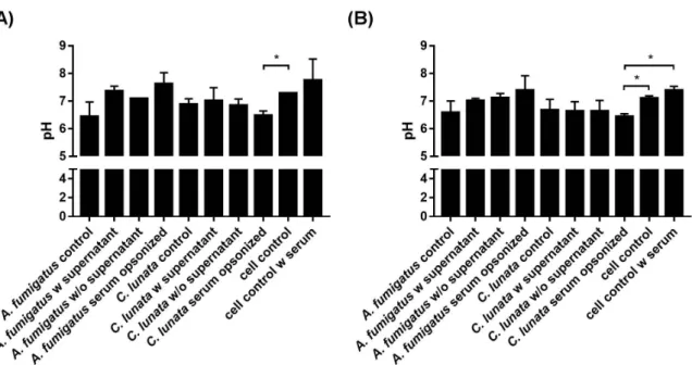
2.5. Extracellular pH of the Neutrophil–Fungus Interaction
Infection of the neutrophils withA. fumigatusdid not change significantly the extracellular pH compared to that of the uninfected cells at the tested timepoints (Figure5a). However, serum opsonized hyphae ofC. lunatadecreased the pH of the interaction environment even after 60 min and this decrease proved to be significant after 3 h of interaction (Figure5b).
Figure 5.Extracellular pH of the environment after 60 min (A) and 3 h (B) of interaction withA. fumigatus andC. lunata. Addition of serum alkalifies the media; significance of serum opsonization conditions were calculated compared to the untreated neutrophils with and without serum as well. Control: untreated hyphae or neutrophils; w supernatant: neutrophils were added directly to the germinated conidia; w/o supernatant: germinated conidia were washed with fresh modified Krebs-Ringer phosphate glucose (mKRPG) buffer before addition of neutrophils; serum opsonized: young hyphae were serum opsonized before addition of neutrophils. Error bars represent standard deviations calculated form three biological parallels. Multiple t-test was used in GraphPad Prism 7 software, FDR (Q=10%), *p<0.05.
2.6. Killing Efficiency of the Neutrophil Granulocytes
While the neutrophils could effectively killA. fumigatusin case of serum opsonization, viability of C. lunatadid not decreased after 3 h of interaction under the examined conditions (Figure6).
Figure 6. Viability ofC. lunatais not affected by 3 h-long interaction with neutrophils. Neutrophils were added to the serum opsonized hyphae. As a control, fungal strains were cultivated under the same conditions without immune cells. Viability is expressed as the percentage normalized to the control conditions. Error bars represent standard deviations calculated from three biological parallels.
Significance was calculated with multiple t-test with False Discovery Rate (FDR) (Q=10%) in the GraphPad Prism 7 software, *p<0.05.
Pathogens2020,9, 235 6 of 12
2.7. Viability of the Neutrophils in the Presence of the Fungal Hyphae
At 60 min post-inoculation, significant death of the neutrophils was detected only in the presence of serum opsonized hyphae.C. lunataandA. fumigatuscaused a cell death of 68% and 26% compared to the control, respectively.
3. Discussion
Neutrophils are key components of the host defense against fungal pathogens and neutropenia or inadequate neutrophilic response generally are considered as risk factors of opportunistic fungal infections. Therefore, the interaction of neutrophil granulocytes with opportunistic human pathogenic fungi is an intensively researched field [26]. In this respect, the most studied filamentous fungus isA.
fumigatus[27]. Patients having chronic granulomatous disease (CGD), which is caused by a deficiency in the NADPH-oxidase system of phagocytes, making them unable to produce reactive oxygen species, are at high risk having invasive aspergillosis (IA) or other fungal infection. CGD also affects the NET forming capacity of neutrophils [17,28]. Disseminated phaeohyphomycoses including those caused by Curvulariaspp. also occur with a higher prevalence in immunosuppressed patients, in which case, the survival rate was found to be higher when the patient recovered from neutropenia (24).
It is known that neutrophils recognize theA. fumigatushyphae only after serum opsonization, and efficient killing mechanisms are then activated [17]. Consistently to this, activation of the neutrophils in the presence of theA. fumigatushyphae could be observed only after opsonization. Opsonization of theC. lunatahyphae also led to the MPO release of the neutrophils indicating that the recognition may be similar to that ofA. fumigatus.The fact that we could detect release of MPO when the supernatant of the hyphae was not removed before the infection or even only the supernatant was added to the neutrophil cells suggests that neutrophils could also recognize a soluble factor produced byC. lunata.
MPO forms a complex with the H2O2produced during respiratory burst, which is responsible for the oxidation of halides forming hypohalous acids like hypochlorous acid (HOCl) [29,30]. NADPH oxidase of neutrophils produce high amount of superoxide (O2-), which is converted into H2O2by superoxide dismutase or by a spontaneous reaction [20]. By measuring the amount of H2O2produced by the neutrophils in response to fungi, we observed that oxidative burst was activated only in the presence of the opsonized hyphae of both fungi. However, while the H2O2level was quite constant in case ofA. fumigatus, it reduced radically in the presence ofC. lunata, indicating a potent antioxidant activity for the latter fungus.
C. lunataproduces DHN-type melanin. Antioxidant capacity of melanin is well documented [31].
Moreover, the protective effect of melanin was proven against H2O2 and HOCl in the case ofA.
nidulans [32]. However, as the presence of released melanin was not detected in the interaction environment, it can be suggested that the drop of the H2O2level was not the consequence of the antioxidant activity of this pigment in our study.
Large microbes and fungal hyphae trigger extracellular reactive oxygen species (ROS) release, which leads to recruiting further neutrophils and NET formation [33]. Interestingly, we detected H2O2
production in not all those cases when activation of neutrophils was seen. MPO release induced by the supernatant of the hyphae did not result in H2O2production. MPO has important roles in the proinflammatory regulation. In a non-enzymatic way, it activates further neutrophils via an autocrine signaling pathway [34], mediates the recruitment of other leukocytes binding to their CD11b/CD18 receptors [35], and induces the IL-6 and IL-8 production of endothelial cells [36]. It is possible that the MPO functions as a regulating molecule when cells are activated by the soluble factor ofC. lunata.
NET formation was observed in the presence of opsonizedA. fumigatus but not in case ofC.
lunata, although neutrophils attached to the hyphae. NET formation is mainly regulated by the presence of ROS [37]. It is not clear if there is a specific ROS regulating the process. According to Nishinaka [38], singlet oxygen is the main inducer of NET production, but there are also data contradicting this hypothesis [39,40]. In parallel with the lack of NET formation, an acidification of the reaction environment was observed in the case ofC. lunata. It was recently reported that acidification
of the environment can inhibit the NET formation of neutrophils via the impeding of ROS production, but addition of H2O2was enough to induce NET appearance [41]. Considering that H2O2almost disappeared and the extracellular pH was found around 6.5 during the interaction withC. lunata, this can explain the lack of NET formation in response to this strain.
Viability of fungal strains was determined after three hours of interaction. While neutrophils were able to killA. fumigatusefficiently, viability ofC. lunatadid not decrease after the interaction.
This data is in line with our other results, suggesting thatC. lunatacan efficiently block the neutrophil functions. Cell death of neutrophils was also measured after the interaction. In the presence ofC.
lunata, neutrophils were more viable than those in the interaction withA. fumigatus. This result is in agreement with the observation that acidification of the reaction environment prolongs the lifespan of neutrophils [41] and supports the possible role of acidification in the protection of the fungus against neutrophil effector functions.
In a previous study, we found that THP-1 monocytes responded only to the hyphae ofC. lunataby attaching to them and producing elevated levels of the anti-inflammatory cytokine IL-10 [16]. Induction of IL-10 together with the observation that monocytes could not reduce the viability of the fungus correlates well to the fact thatC. lunatafrequently cause chronic infections even in immunocompetent persons [8]. Neutrophils also could not decrease the fungal viability and their NET formation was blocked by the hyphae. This efficient evasion of the monocytic and the neutrophil attacks may contribute in the ability of this fungus to cause disseminated infections even in immunocompetent patients [8,11,12,14].
4. Materials and Methods
4.1. Fungal Strains, Culture Conditions, and Inoculum Preparation
The fungal strains,Curvularia lunataSZMC 23759 andAspergillus fumigatusSZMC 23245 isolated from human eye infections were used (SZMC, Szeged Microbiology Collection, Szeged, Hungary;
http://www.wfcc.info/ccinfo/collection/by_id/987). To harvest the conidia, fungi were grown on rice flour agar (1% rice flour, 0.2% yeast extract, 1.5% agar) at room temperature. Conidia were washed from 7 days old colonies with modified Krebs-Ringer phosphate glucose (mKRPG) buffer (145 mM NaCl, 5.7 mM Na2HPO4, 4.86 mM KCl, 5.5 mM glucose, pH 7.35) and the spore suspensions were filtered through a Millipore filter with a pore size of 45µm (Merck, Burlington, USA) to eliminate the hyphal debris.
4.2. Isolation of Human Neutrophil Granulocytes and Serum
Human neutrophils were isolated from heparinized venous blood of healthy donors (n= 6) with dextran (Sigma-Aldrich, St. Louis, MI, USA) sedimentation followed by centrifugation over Ficoll-Paque PLUS (GE Healthcare, Chicago, IL, USA) and hypotonic lysis of erythrocytes [42]. Isolation was performed at room temperature. After isolation, the cells were resuspended in mKRPG. Viability and purity of the cells were checked with trypan blue (Sigma-Aldrich, St. Louis, MI, USA) [43] and Wright staining (Sigma-Aldrich, St. Louis, MI, USA) [44,45], respectively. Serum was isolated from venous blood of the same donors taken into serum separation blood collection tubes (BD Vacutainer, Becton Dickinson, Franklin Lakes, NJ, USA). Then, coagulation tubes were centrifuged at 1200 rpm for 15 min at room temperature and the serum was collected.
4.3. Interaction of the Neutrophils and the Hyphae
To produce young hyphae, 2×10⁴conidia were inoculated in 100µL mKRPG in a 96-well plate (Sarstedt, Nümbrecht, Germany) and incubated at 37◦C. Considering that the time of the germ tube formation from the conidia is different forAspergillusandCurvularia, the incubation time was set to 4 and 8 h in case ofC. lunataandA. fumigatus, respectively, to obtain the same length of hyphae for both strains. The isolated neutrophils were added to the germinated conidia in an effector:target (E:T)
Pathogens2020,9, 235 8 of 12
ratio of 5:1 in 100µL mKRPG in a 96-well plate. Interactions were examined after 30, 60, or 180 min under three conditions: (a) neutrophils were added directly to the germinated conidia; (b) neutrophils were added to germinated conidia previously washed with fresh mKRPG buffer; and (c) neutrophils were added to the germinated conidia, which was previously serum opsonized with 10% serum of the corresponding donor in the last hour of germination. For detection of neutrophil activation, a fourth condition was also examined, in which the cell free supernatant of the germinated conidia was added to the neutrophils. Final volume of the interactions was 200µL.
To detect possible NET formation, 7×10⁴conidia were germinated in CELLview dishes (Greiner Bio-One, Mosonmagyaróvár, Hungary) in 400µL mKRPG buffer and neutrophils were added to them in an E:T ratio of 5:1. Final volume of the reaction was 800µL.
4.4. Detection of Neutrophil Activation
To measure the activation of neutrophils, the MPO activity was determined using 3,30,5,50-tetramethylbenzidine (TMB; Sigma-Aldrich, St. Louis, MI, USA) at the 30th and 60th min of the co-incubation according to the method of Suzuki et al. [46]. To exclude other peroxidase activities from the measurement, the MPO inhibitor 4-aminobenzoic hydrazide (ABH) was added to a parallel measurement in a concentration of 1 mM. MPO activity of the untreated neutrophils was considered as a control. The standard curve was generated based on the reaction of Recombinant Human Myeloperoxidase Protein (R&D Systems, Minneapolis, MN, USA) using 4-parametric logistic regression.
4.5. Measurement of the H2O2Release of Neutrophils
Release of H2O2 was measured with the Amplex Red Hydrogen Peroxide/Peroxidase Assay Kit (Invitrogen, Carlsbad, CA, USA) 30 and 60 min after the infection according to the manufacturer’s recommendation.
4.6. Detection of Extracellular Melanin Release
Extracellular melanin release ofC. lunatawas analyzed during the fungi–neutrophil granulocyte interaction, as well as after the cultivation of the fungi in mKRPG for 7 days. Conditions of the interaction were the same as described in the Section4.3. The supernatant from the interaction was centrifuged at 6000 rpm for 10 min, while the mKRPG from the cultivation was filtered through gauze.
The flowthrough was acidified to pH 2.0 with addition of HCl and the resulting precipitate was filtered on a 0.2µm PTFE membrane [47].
4.7. Visualization of NET Formation
NET formation was analyzed after 3 h of co-incubation (the E:T ratio was 5:1). Samples were fixed with 1% formaldehyde solution for 15 min.
To measure the chromosome decondensation and visualize the netosis, staining solutions were prepared in 10 mM sodium citrate buffer (pH 7.2). To stain the NET, 167 nM SYTOX Green nucleic acid stain (Life Technologies, Carlsbad, CA, USA) was applied for 30 min, while the fungal and the neutrophil cells were stained with 5µg/mL Calcofluor white (Cyanamid, NJ, USA) for 15 min and a 0.5-fold solution of the CellMask Deep Red Plasma Membrane Stain (Thermo Scientific, Waltham, MA, USA) for 15 min, respectively. Between the staining steps and after the whole process, samples were washed two times with 10 mM sodium citrate buffer.
To induce NET formation by hydrogen peroxide (H2O2), the latter was added to the interactions after 0, 1, and 2 h of co-incubation in a final concentration of 30µM [41].
Microscopic examinations were carried out with an Axio Observer 7 (Zeiss, Oberkochen, Germany) fluorescent inverted microscope.
4.8. Measurement of Chromosome Decondensation
Chromosome decondensation was analyzed on the pictures of samples stained with SYTOX green, Calcofluor white, and CellMask Deep Red and taken by fluorescent microscopy. For each sample, at least 35 cells were analyzed. SYTOX Green positive area was measured with ImageJ for each cell as described before [48]. Nuclear areas found to be≤50µm2were considered as condensed nuclei, those, which were between 50 and 90µm2, were regarded as decondensed nuclei, while those measured to be
≥90µm2indicated NET formations.
4.9. Measurement of Extracellular pH
Extracellular pH was determined from the cell-free supernatant of the interaction at 60 min and 3 h post-inoculation with litmus solution (CPAChem, Stara Zagora, Bulgaria). Calibration curve was generated from a pH scale of mKRPG with the linear regression model. To 100µL of supernatant or mKRPG 5µL of 2% litmus solution was added in a 96-well plate. Absorption was measured at 580 nm with a SPECTROstar Nano Microplate Reader (BMG Labtech, Ortenberg, Germany).
4.10. Viability Assay for the Hyphae
To measure the hyphal damage after incubation with the neutrophils, a colorimetric assay using 3-(4,5-dimethylthiazol-2-yl)-2,5-diphenyltetrazolium bromide (MTT; Sigma-Aldrich, St. Louis, MI, USA) was performed in 96-well plates [49]. After 3 h of coincubation, neutrophils were lysed with 0.5% sodium deoxycholate (Sigma-Aldrich) and the wells were washed three times with PBS.
Two hundredµL of mKRPG buffer supplemented with 0.5% MTT was added to the samples and the plates were incubated at 37 ◦C for 3 h. After removing the supernatant, wells were washed two times with PBS and stored overnight at −20◦C. Before detection, 200µL acidic isopropanol (95% isopropanol and 5% 1 N HCl) was added into the wells and the plates were incubated until the blue color dissolved from the hyphae. Absorbance of the supernatant was measured at the wavelength of 550 nm using a spectrophotometer. Viability of the hyphae was calculated using the following formula, whereOD550samplewas the absorbance measured for the hyphae incubated with neutrophils andOD550controlwas the absorbance measured for the hyphae incubated without neutrophils: Viability =
OD550sample OD550control
×100.
4.11. Neutrophil Viability Assay
Cell death of neutrophil granulocytes following 60-min co-incubation was measured by the LDH Cytotoxicity Detection Kit (Takara Bio, Shiga, Japan) according to the manufacturer’s instructions. For the measurement of the lactate dehydrogenase (LDH) released into the medium due to the cell death, the cell free supernatant of the cultures was used in a 10-fold dilution. The cell culture supernatant incubated without the hyphae were used as the low control. For high control, neutrophils incubated without the hyphae were lysed with Triton X-100 in a final concentration of 1%. From the data of the low and high controls, a calibration curve was presented, and viability was calculated from the equation of the line.
4.12. Statistical Analysis
All measurements were performed in three biological and two technical parallels. Significance was calculated with multiple t-test with False Discovery Rate (FDR) (Q=10%) in the GraphPad Prism 7 software (GraphPad Software, San Diego, CA, USA).
4.13. Ethics Statement
Conforming to the principles outlined in the Declaration of Helsinki, and the healthy volunteers informed consent, blood was taken in the Department of Obstetrics and Gynecology at the University
Pathogens2020,9, 235 10 of 12
of Szeged, Hungary. The Ethics Committee of the Department of Obstetrics and Gynecology approved the study protocol (20/2016-SZTE).
5. Conclusions
Our results show that neutrophils are able to recognize the serum opsonized hyphae ofC. lunata and can attach to them. They are also activated by a soluble factor produced by the fungus, although induction of oxidative burst is dependent on the presence of the opsonized hyphae. These results suggest the regulatory role of MPO in the immune response againstC. lunata. Nevertheless, H2O2is generated after the activation of the cells. However, oxidative burst seems to be blocked by the fungus after 60 min of interaction. This blockage also influences the NET formation of neutrophils, with which C. lunatacan efficiently survive the neutrophil attack.
Author Contributions: Conceptualization, E.J.T. and T.P.; methodology, E.J.T., T.P. and T.M.; validation, E.J.T. and T.P.; formal analysis, E.J.T., T.P. and G.N.; investigation, E.J.T., M.V., M.H., O.J., C.V. and G.N.; resources, E.J.T., M.T., E.H. and H.O.; writing—original draft preparation, E.J.T. and T.P.; writing—review and editing, T.P. and C.V.; supervision, T.P.; project administration, T.P., G.N. and M.H.; funding acquisition, T.P. and G.N. All authors have read and agreed to the published version of the manuscript.
Funding:This study was funded by the “Lendület” Grant of the Hungarian Academy of Sciences (LP2016-8/2016), the project NKFI K131796 and the FIKP program (TUDFO/4738-1/2019 ITM) of the Ministry of Human Capacities.
MT was supported by the grants J. Bolyai Research Scholarship of the Hungarian Academy of Sciences and the UNKP-18-4 New National Excellence Program of the Ministry of Human Capacities. EJT was supported by the UNKP-18-3 New National Excellence Program of the Ministry of Human Capacities.
Acknowledgments:The authors would like to thank Katalin Szabóand Alexandra Hoffmann for their help in the preparation of the blood samples and the interaction studies.
Conflicts of Interest:The authors declare no conflict of interest.
References
1. Perfect, J.R.; Alexander, B.D.; Schell, W.A. Phaeohyphomycoses (Brown-Black Moulds). InEssentials of Clinical Mycology, 2nd ed.; Kauffman, C.A., Pappas, P.G., Sobel, J.D., Dismukes, W.E., Eds.; Springer: New York, NY, USA, 2011; pp. 305–317.
2. Paredes, K.; Capilla, J.; Sutton, D.A.; Mayayo, E.; Fothergill, A.W.; Guarro, J. Experimental treatment of Curvulariainfection.Diagn. Microbiol. Infect. Dis.2014,79, 428–431. [CrossRef]
3. Krizsan, K.; Toth, E.; Nagy, L.G.; Galgoczy, L.; Manikandan, P.; Chandrasekaran, M.; Kadaikunnan, S.;
Alharbi, N.S.; Vágvölgyi, C.; Papp, T. Molecular identification and antifungal susceptibility ofCurvularia australiensis,C. hawaiiensisandC. spiciferaisolated from human eye infections.Mycoses2015,58, 603–609.
[CrossRef] [PubMed]
4. Revankar, S.G.; Baddley, J.W.; Chen, S.C.; Kauffman, C.A.; Slavin, M.; Vazquez, J.A.; Seas, C.;
Morris, M.I.; Nguyen, M.H.; Shoham, S.; et al. A mycoses study group international prospective study of phaeohyphomycosis: An analysis of 99 proven/probable cases. Open Forum. Infect. Dis. 2017,4, ofx200.
[CrossRef] [PubMed]
5. Khan, I.D.; Makkar, A.; Malik, A.; Khan, S.; Mehdi, I.; Arif, S.; Aden, D.; Somayaji, P.; Roomi, K.Curvularia keratomycosis after cataract surgery.J. Arch. Milit. Med.2017,5, e57331.
6. Rasheeduddin, M.; Visalakshi, P. Cutaneous phaeohyphomycosis of foot web byCurvularia lunata.Glob. J.
Med. Clin. Case Rep.2017,4, 074–075. [CrossRef]
7. Vineetha, M.; Palakkal, S.; Sobhanakumari, K.; Celine, M.I.; Letha, V. Verrucous onychomycosis caused by Curvulariain a patient with congenital pterygium.Indian J. Dermatol.2016,61, 701. [CrossRef] [PubMed]
8. Krizsán, K.; Papp, T.; Manikandan, P.; Shobana, S.; Chandrasekaran, M.; Vágvölgyi, C.; Kredics, L.
Clinical importance of the genusCurvularia. InMedical Mycology: Current Trends and Future Prospects;
Razzaghi-Abyaneh, M., Shams-Ghahfarokhi, M., Rai, M., Eds.; CRC Press: Boca Raton, FL, USA, 2015;
pp. 147–204.
9. Cavanna, C.; Seminari, E.; Pusateri, A.; Mangione, F.; Lallitto, F.; Esposto, M.C.; Pagella, F. Allergic fungal rhinosinusitis due toCurvularia lunata.New Microbiol.2014,37, 241–245.
10. Skovrlj, B.; Haghighi, M.; Smethurst, M.E.; Caridi, J.; Bederson, J.B.Curvulariaabscess of the brainstem.
World Neurosurg. 2014,82, e9–e241. [CrossRef]
11. Revankar, S.G.; Patterson, J.E.; Sutton, D.A.; Pullen, R.; Rinaldi, M.G. Disseminated phaeohyphomycosis:
Review of an emerging mycosis.Clin. Infect. Dis.2002,34, 467–476. [CrossRef]
12. Kobayashi, H.; Sano, A.; Aragane, N.; Fukuoka, M.; Tanaka, M.; Kawaura, F.; Fukuno, Y.; Matsuishi, E.;
Hayashi, S. Disseminated infection byBipolaris spiciferain an immunocompetent subject.Med. Mycol.2008, 46, 361–365. [CrossRef]
13. Balla, A.; Pierson, J.; Hugh, J.; Wojewoda, C.; Gibson, P.; Greene, L. Disseminated cutaneousCurvularia infection in an immunocompromised host; diagnostic challenges and experience with voriconazole.J. Cutan.
Pathol.2016,43, 383–387. [CrossRef] [PubMed]
14. Beckett, A.R.; Kahn, S.A.; Seay, R.; Lintner, A.C. InvasiveCurvulariainfections in burn patients: A case series.
Surg. Infect. Case Rep.2017,2, 76–79. [CrossRef]
15. Shrivastava, A.; Tadepalli, K.; Goel, G.; Gupta, K.; Gupta, P.K. Melanized fungus as an epidural abscess: A diagnostic and therapeutic challenge.Med. Mycol. Case Rep.2017,16, 20–24. [CrossRef] [PubMed]
16. Tóth, E.J.; Boros, E.; Hoffmann, A.; Szebenyi, C.; Homa, M.; Nagy, G.; Vágvölgyi, C.; Nagy, I.; Papp, T.
Interaction of THP-1 monocytes with conidia and hyphae of differentCurvulariastrains.Front. Immunol.
2017,8, 1369. [CrossRef] [PubMed]
17. Gazendam, R.P.; van Hamme, J.L.; Tool, A.T.; Hoogenboezem, M.; van den Berg, J.M.; Prins, J.M.; Vitkov, L.;
van de Veerdonk, F.L.; van den Berg, T.K.; Roos, D.; et al. Human neutrophils use different mechanisms to killAspergillus fumigatusconidia and hyphae: Evidence from Phagocyte Defects. J. Immunol.2016,196, 1272–1283. [CrossRef] [PubMed]
18. Brakhage, A.A.; Bruns, S.; Thywissen, A.; Zipfel, P.F.; Behnsen, J. Interaction of phagocytes with filamentous fungi.Curr. Opin. Microbiol.2010,13, 409–415. [CrossRef]
19. LeibundGut-Landmann, S.; Wuthrich, M.; Hohl, T.M. Immunity to fungi. Curr. Opin. Immunol.2012,24, 449–458. [CrossRef]
20. Babior, B.M.; Lambeth, J.D.; Nauseef, W. The neutrophil NADPH oxidase.Arch. Biochem. Biophys.2002,397, 342–344. [CrossRef]
21. Kolaczkowska, E.; Kubes, P. Neutrophil recruitment and function in health and inflammation. Nat. Rev.
Immunol.2013,13, 159–175. [CrossRef]
22. Braem, S.G.; Rooijakkers, S.H.; van Kessel, K.P.; de Cock, H.; Wosten, H.A.; van Strijp, J.A.; Haas, P.J.A.
Effective neutrophil phagocytosis ofAspergillus fumigatusis mediated by classical pathway complement activation.J. Innate Immun.2015,7, 364–374. [CrossRef]
23. Feldmesser, M. Role of neutrophils in invasive aspergillosis.Infect. Immun.2006,74, 6514–6516. [CrossRef]
[PubMed]
24. Ben-Ami, R.; Lewis, R.E.; Raad, I.I.; Kontoyiannis, D.P. Phaeohyphomycosis in a tertiary care cancer center.
Clin. Infect. Dis.2009,48, 1033–1041. [CrossRef] [PubMed]
25. Morton, C.O.; Bouzani, M.; Loeffler, J.; Rogers, T.R. Direct interaction studies between Aspergillus fumigatus and human immune cells; what have we learned about pathogenicity and host immunity?Front. Microbiol.
2012,3, 413. [CrossRef] [PubMed]
26. Kobayashi, S.D.; Malachowa, N.; DeLeo, F.R. Influence of microbes on neutrophil life and death.Front. Cell Infect. Microbiol.2017,7, 159. [CrossRef]
27. Dagenais, T.R.; Keller, N.P. Pathogenesis ofAspergillus fumigatusin invasive aspergillosis.Clin. Microbiol.
Rev.2009,22, 447–465. [CrossRef]
28. Bianchi, M.; Hakkim, A.; Brinkmann, V.; Siler, U.; Seger, R.A.; Zychlinsky, A.; Reichenbach, J. Restoration of NET formation by gene therapy in CGD controls aspergillosis.Blood2009,114, 2619–2622. [CrossRef]
29. Aratani, Y. Role of myeloperoxidase in the host defense against fungal infection.Nihon Ishinkin Gakkai Zasshi 2006,47, 195–199. [CrossRef]
30. Klebanoff, S.J.; Kettle, A.J.; Rosen, H.; Winterbourn, C.C.; Nauseef, W.M. Myeloperoxidase: A front-line defender against phagocytosed microorganisms.J. Leukoc. Biol.2013,93, 185–198. [CrossRef]
31. Revankar, S.G.; Sutton, D.A. Melanized fungi in human disease. Clin. Microbiol. Rev. 2010,23, 884–928.
[CrossRef]
32. de Cassia, R.G.R.; Pombeiro-Sponchiado, S.R. Antioxidant activity of the melanin pigment extracted from Aspergillus nidulans.Biol. Pharm. Bull.2005,28, 1129–1131.
Pathogens2020,9, 235 12 of 12
33. Warnatsch, A.; Tsourouktsoglou, T.D.; Branzk, N.; Wang, Q.; Reincke, S.; Herbst, S.; Gutierrez, M.;
Papayannopoulos, V. Reactive oxygen species localization programs inflammation to clear microbes of different size.Immunity2017,46, 421–432. [CrossRef] [PubMed]
34. Lau, D.; Mollnau, H.; Eiserich, J.P.; Freeman, B.A.; Daiber, A.; Gehling, U.M.; Brümmer, J.; Rudolph, V.;
Münzel, T.; Heitzer, T.; et al. Myeloperoxidase mediates neutrophil activation by association with CD11b/CD18 integrins.Proc. Natl. Acad. Sci. USA2005,102, 431–436. [CrossRef] [PubMed]
35. Johansson, M.W.; Patarroyo, M.; Oberg, F.; Siegbahn, A.; Nilsson, K. Myeloperoxidase mediates cell adhesion via the alpha M beta 2 integrin (Mac-1; CD11b/CD18).J. Cell Sci.1997,110, 1133–1139. [PubMed]
36. Lefkowitz, D.L.; Roberts, E.; Grattendick, K.; Schwab, C.; Stuart, R.; Lincoln, J.; Allen, R.C.; Moguilevsky, N.;
Bollen, A.; Lefkowitz, S.S. The endothelium and cytokine secretion: The role of peroxidases as immunoregulators.Cell Immunol.2000,202, 23–30. [CrossRef] [PubMed]
37. Fuchs, T.A.; Abed, U.; Goosmann, C.; Hurwitz, R.; Schulze, I.; Wahn, V.; Weinrauch, Y.; Brinkmann, V.;
Zychlinsky, A. Novel cell death program leads to neutrophil extracellular traps.J. Cell Biol.2007,176, 231–241.
[CrossRef] [PubMed]
38. Nishinaka, Y.; Arai, T.; Adachi, S.; Takaori-Kondo, A.; Yamashita, K. Singlet oxygen is essential for neutrophil extracellular trap formation.Biochem. Biophys. Res. Commun.2011,413, 75–79. [CrossRef]
39. Palmer, L.J.; Cooper, P.R.; Ling, M.R.; Wright, H.J.; Huissoon, A.; Chapple, I.L. Hypochlorous acid regulates neutrophil extracellular trap release in humans.Clin. Exp. Immunol.2012,167, 261–268. [CrossRef]
40. Tripathi, J.K.; Sharma, A.; Sukumaran, P.; Sun, Y.; Mishra, B.B.; Singh, B.B.; Sharma, J. Oxidant sensor cation channel TRPM2 regulates neutrophil extracellular trap formation and protects against pneumoseptic bacterial infection.FASEB J.2018,32, 6848–6859. [CrossRef]
41. Behnen, M.; Moller, S.; Brozek, A.; Klinger, M.; Laskay, T. Extracellular acidification inhibits the ROS-dependent formation of neutrophil extracellular traps.Front. Immunol.2017,8, 184. [CrossRef]
42. Clark, R.A.; Nauseef, W.M. Isolation and functional analysis of neutrophils.Curr. Protoc. Immunol.2001,7.
[CrossRef]
43. Strober, W. Trypan blue exclusion test of cell viability. Curr. Protoc. Immunol. 2015,111, A3.B.1–A3.B.3.
[CrossRef]
44. Conn, H.J.Biological Stains, 8th ed.; Williams and Wilkins: Baltimore, MD, USA, 1969; p. 498.
45. Clark, G.Staining Procedures, 3rd ed.; Williams and Wilkins: Baltimore, MD, USA, 1973; p. 418.
46. Suzuki, K.; Ota, H.; Sasagawa, S.; Sakatani, T.; Fujikura, T. Assay method for myeloperoxidase in human polymorphonuclear leukocytes.Anal. Biochem.1983,132, 345–352. [CrossRef]
47. Goncalves, R.C.; Lisboa, H.C.; Pombeiro-Sponchiado, S.R. Characterization of melanin pigment produced by Aspergillus nidulans.World J. Microbiol. Biotechnol.2012,28, 1467–1474. [CrossRef] [PubMed]
48. Gonzalez, A.S.; Bardoel, B.W.; Harbort, C.J.; Zychlinsky, A. Induction and quantification of neutrophil extracellular traps.Methods Mol. Biol.2014,1124, 307–318. [PubMed]
49. Levitz, S.M.; Diamond, R.D. A rapid colorimetric assay of fungal viability with the tetrazolium salt MTT.J.
Infect. Dis.1985,152, 938–945. [CrossRef] [PubMed]
©2020 by the authors. Licensee MDPI, Basel, Switzerland. This article is an open access article distributed under the terms and conditions of the Creative Commons Attribution (CC BY) license (http://creativecommons.org/licenses/by/4.0/).