Immunobiology 226 (2021) 152032
Available online 23 November 2020
0171-2985/© 2020 The Author(s). Published by Elsevier GmbH. This is an open access article under the CC BY-NC-ND license
(http://creativecommons.org/licenses/by-nc-nd/4.0/).
Cytotoxic activity of human dendritic cells induces RIPK1-dependent cell death
Zs ´ ofia Varga
a,b, Evelin R ´ acz
a, Anett M ´ azl o ´
a,b, M onika Korodi ´
c, Anik ´ o Szab ´ o
a, Tam as Moln ´ ´ ar
a,b, Arp ´ ´ ad Sz ¨ o or ˝
d, Zolt ´ an Ver ´ eb
e,f, Attila B ´ acsi
a, G ´ abor Koncz
a,*aDepartment of Immunology, Faculty of Medicine, University of Debrecen, Debrecen, Hungary
bUniversity of Debrecen, Doctoral School of Molecular Cellular and Immune Biology, Debrecen, Hungary
cUniversity of P´ecs, Doctoral School of Chemistry, Department of Chemistry, Faculty of Sciences, P´ecs, Hungary
dDepartment of Biophysics and Cell Biology, Faculty of Medicine, University of Debrecen, Debrecen, Hungary
eRegenerative Medicine and Cellular Pharmacology Laboratory (HECRIN), Department of Dermatology and Allergology, University of Szeged, Szeged, Hungary
fInstitute for Translational Medicine, University of Szeged, Szeged, Hungary
A R T I C L E I N F O Keywords:
Dendritic cells Cytotoxicity
Immunogenic cell death RIPK1
Immune tolerance
A B S T R A C T
Dendritic cells (DCs), as potent phagocytes engulf dead cells and present peptide fragments of tumor antigens or pathogens derived from infected cells to naïve CD8+T-lymphocytes. Dendritic cells can also induce apoptosis in target cells, thus getting an opportunity to sample their microenvironment. Here, we present that the superna- tants of LPS- or CL075-activated DCs induced cell death in different cell lines, but during the differentiation to mature DCs, they lost their cytotoxic potential. Dexamethasone-pre-treated tolerogenic DCs induced less inten- sive death indicating that the tissue microenvironment can downregulate DC-mediated killing. Exploring the signaling of DC-induced cell death, we observed that the supernatant of activated DCs induced TNF-dependent cell death, since TNF antagonist blocked the cytotoxic activity of DCs, contrary to inhibitors of Fas and TRAIL receptors. We identified that the DC-induced killing is at least partially a RIPK1-dependent process, as RIPK1 positive target cells were more susceptible to DC-induced cell death than their RIPK1 deficient counterparts.
Moreover, both the elevated phosphorylation of RIPK1 and the increase in RIPK1-caspase-8 interaction in target cells suggest that RIPK1-mediated signals contribute to DC supernatant-induced cell death. We also proved that the cytotoxic activity of DC-derived supernatant induced apoptosis in the target cells and not necroptosis, as it was completely abrogated with the pan caspase inhibitor (Z-VAD), while the necroptosis inhibitor (Nec-1) had no effect.
Our work revealed that the supernatant of activated DCs induces the apoptosis of target cells in a RIPK1- dependent manner. This phenomenon could be relevant for the initiation of cross-presentation and may broaden the plethora of cytotoxic mechanisms acting against tumor cells.
1. Introduction
Dendritic cells (DCs) work as sentinels of the immune system and provide connection between innate and adaptive immunity. Immature DCs (iDCs) detect and phagocyte incoming pathogens in peripheral tis- sues. Upon activation, iDCs leave the site of infection and migrate into lymph nodes, during which time they fully differentiate into mature DCs (mDC), and where they present antigens derived from the phagocyted pathogen to naïve T cells (Clatworthy et al., 2014). DCs are also able to engulf dead cells and present antigenic peptides both on MHCII and MHCI molecules. The latter, so called cross-priming process initiates
cytotoxic T cell responses against viruses that do not infect DCs and tumors that originate from non-DCs (Bousso, 2008). In recent years, various regulated cell death pathways have been described and char- acterized with different immunological outcomes including receptor- interacting protein kinase 1 (RIPK1)-mediated apoptosis and nec- roptosis (Wang et al., 2008). These special, RIPK1-dependent routes of cell death have been published to increase the cross-presentation and the immunogenicity of DCs and in addition to playing a physiological role in enhancing innate immunity, regulate T lymphocyte activation (Yatim et al., 2015).
Thus, cell death is inevitably the very first step in the induction of a
* Corresponding author at: Department of Immunology, Faculty of Medicine, University of Debrecen, H-4032 Debrecen, 1 Egyetem Square, Hungary.
E-mail address: konczgb@gmail.com (G. Koncz).
Contents lists available at ScienceDirect
Immunobiology
journal homepage: www.elsevier.com/locate/imbio
https://doi.org/10.1016/j.imbio.2020.152032
Received 4 June 2020; Received in revised form 5 October 2020; Accepted 18 November 2020
2 cytotoxic T cell response. Cell death providing cellular debris for cross- priming can occur as a result of a passive, intrinsic process via a pro- grammed apoptotic pathway or due to active mechanisms via the cytotoxic effect of the pathogens or induced by the immune system itself.
In addition to their efficient antigen-presenting function, the contribution of DCs to the generation of various cell death signals has also been widely demonstrated (Vidalain et al., 2001; Chan and Hous- seau, 2008; Chauvin and Josien, 2008). However, the physiological role of DC-induced killing is still not clearly understood. The direct cytotoxic capacity of DCs is mainly related to their immature phenotype localized in tissues (Janjic et al., 2002), but an effective pathogen clearance by iDCs is unlikely, as activated dendritic cells leave the site of infection (Chan and Housseau, 2008). The exact molecular background underly- ing the cytotoxicity of DCs is still yet to be defined, as it can be related to either a direct cell–cell contact-mediated action or soluble cytotoxic mediators released by DCs (Lakomy et al., 2011). The contribution of the known death ligands, such as TNF (Vidalain et al., 2001; Joo et al., 2002;
Lu et al., 2002), TRAIL (Vidalain et al., 2001, 2000; Lu et al., 2002), Fas (Burdelya et al., 2013; Salamone et al., 2010); lymphotoxin α and β (Lu et al., 2002) in DC-induced killing mechanisms has already been demonstrated.
DC-mediated cross-presentation can be either up- or down-regulated by the immunological microenvironment (Anderson et al., 2008);
however, it is not known how the immunological conditions modify the cytotoxic potential of DCs. Infected cells produced type I interferons as alarm signals for DCs. Indeed, DCs express high levels of MHC and co- stimulatory molecules in the presence of Interferon-α (IFN- α), there- fore they stimulate naïve CD4+T cells (Carbonneil et al., (Jul 2004).);
and naïve CD8+T cells via cross-presentation (Schiavoni et al., 2013).
Glucocorticoids (GCs) induce immune suppression by modulating DC differentiation, maturation, and function in the very first steps of the immune responses (Hunzeker et al., 2011). GCs may also suppress CD8+ T cell-mediated immunity, at least in part because they reduce the cross- presenting ability of DCs (Im et al., 2014).
We suppose that DC-induced killing is rather a mechanism to sample the surrounding microenvironment for intracellular pathogen- or tumor- derived antigens, by allowing the iDC to phagocytose and cross-present dead cell-derived antigens. Here, we provide evidence that supernatants of short-term activated moDCs induce TNF-dependent apoptosis of Jurkat cells in a RIPK1-dependent manner. We also demonstrate that GC-induced tolerogenic conditions decrease the cytotoxic capacity of DCs.
2. Materials and methods 2.1. Antibodies and reagents
Antibodies and reagents used were: Z-VAD (ApexBio), TNFR1:Fc (Adipogen), Trail:Fc (ProSpec Protein Specialist), Fas:Fc (R&D System), TNF-α, IL-10 and TGFß (PeproTech), propidium iodide /PI/ (Sigma- Aldrich), a SMAC mimetic /Birinapant/ (LC Laboratories), necrostatin-1 (Abcam), Anti-RIPK1 (BD Biosciences), anti-pRIPK1 (Cell Signaling), anti-Caspase-8 (Cell Signaling) and anti-β-actin (Sigma-Aldrich), anti- rabbit and anti-mouse antibodies conjugated to horseradish peroxi- dase (GE Healthcare).
2.2. Generation of monocyte-derived DCs
Monocyte-derived DCs (moDC) were separated as it was published (Bene et al., 2017). Briefly, monocytes were purified by anti-CD14 microbeads from PBMCs and were cultured in the presence of 100 ng/
ml IL-4 (PeproTech) and 80 ng/ml GM-CSF (Gentaur Molecular Prod- ucts) for 5 days.
2.3. Generation of monocyte-derived IFN DCs
IFN-moDCs were generated by culturing isolated monocytes in the presence of 1000 U/ml rhIFN-a (Roferon-A) and 40 ng/ ml GM-CSF and for 5 days.
2.4. Generation of monocyte-derived dexamethasone DCs
Dexamethasone DCs (dexa DCs) were generated by culturing isolated monocytes with 20 ng/ml IL-4, 0.25 µM dexamethasone (Sigma-Aldrich) and 100 ng/ml GM-CSF for 5 days.
2.5. Collection of DCs (moDC, IFN DC, dexamethasone DC) supernatants DCs (moDC, IFN DC, dexa DC) were stimulated on day 5 of the dif- ferentiation with Toll-like receptor ligands CL075 (1 µg/ml, Sigma) and LPS (0.5 µg/ml, InvivoGen) for 30 min. After 3 washing steps 4.0 ×10⁶ cells/ml DCs were incubated in fresh serum-free RPMI 1640 medium (Sigma-Aldrich) for 2, 8 or 24 h and the supernatants of DCs were harvested.
2.6. Generation of monocyte-derived mDCs
To generate LPS preconditioned DCs, moDCs were treated on the fifth day of differentiation for 24 h with 100 ng/ml LPS. To collect the supernatant, 24 h LPS pre-conditioned DC was reactivated with TLR- ligands for 30 min and after washing steps the supernatants were har- vested for 2 h, in the same way as in the case of short term activated DCs.
2.7. Cell lines
SVT35 Jurkat and HT-29 cell lines were cultured in RPMI 1640 medium (Biosera). The media was supplemented with 10% FCS (Gibco), 2 mM L-glutamine (Biosera) and 40 mg/l Gentamicin (Sigma-Aldrich).
HUVECs (Sigma-Aldrich) were cultured in M199 (Gibco) media sup- plemented with 20% FCS (Gibco), 2 mM L-glutamine (Biosera), 1%
penicillin/streptomycin (Gibco) and with EGMTM-2 Endothelial Single- Quots™ Kit (Lonza) following the manufacturer’s instructions. These cell lines were maintained at 37 ◦C in a humidified atmosphere con- taining 5% CO2.
2.8. Measurement of cytokine concentration
Supernatants of moDCs were harvested 2 h after activation, and the concentrations of TNF-α, IL-6 and IL-10 cytokines were measured using OptEIA kits (BD Biosciences) following the manufacturer’s instructions.
2.9. Membrane integrity
Total cell death was examined based on propidium iodide (PI) uptake by BD FACS Calibur flow cytometer (BD Biosciences). Data were analyzed using FlowJo software (Tree Star).
2.10. SubG1 measurement
DNA fragmentation was detected based on subG1 peak. Following fixation in ice-cold 70% ethanol, cells were washed in a 38 mM citrate buffer (pH 7.4) and incubated for 15 min with 50 µg/ml PI. The subG1 population was determined using flow cytometer. Representative fig- ures of this method are shown in Supplementary Fig. 1.
2.11. Caspase 3/7 assay
SVT35 Jurkat cells were incubated in the presence of the supernatant of activated DCs or 20 ng/ml TNF-α for 24 h at 37 ◦C. In 24 h, the caspase-3/7 activity was measured using an Apo-ONE Homogeneous
Caspase-3/7 assay kit (Promega) following the manufacturer’s instructions.
2.12. Immunoprecipitation
SVT35 Jurkat cells were incubated in the presence of the supernatant derived from 2 h activated DCs or the combination of TNF-α and SMAC mimetic for 1 h at 37 ◦C, then resuspended in a lysis buffer with 1%
Triton-X 100 (Sigma-Aldrich) in the presence of protease inhibitor cocktail (Sigma-Aldrich) and incubated for 30 min at 4 ◦C. The lysate was recovered by centrifugation for 10 min at 13.000 rpm at 4 ◦C and mixed with Protein G beads (GE Healthcare) pre-coated with the anti- caspase-8 (Cell Signaling) antibody. The content was gently rotated at 4 ◦C overnight. Next day, the beads were washed, 2X Laemmli sample buffer was added to them, were subjected to boiling for 5 min at 95 ◦C, and then samples were recovered by centrifugation for 10 min at 13.000 rpm. Protein extractions were fractionated by SDS-PAGE. The RIPK1- Caspase-8 interaction was visualized by western blot using anti-RIPK1 antibody (BD Biosciences). Immunoprecipitation with anti-RIPK1 was performed the same way and the intensity of Caspase-8 binding was examined by western blot.
2.13. Western blotting
Western blot analysis of proteins was performed according to
standard protocols (Varga et al., 2020). Briefly, cells were lysed in 2X Laemmli sample buffer and were fractionated by SDS-PAGE and trans- ferred onto nitrocellulose membranes (Bio-Rad Laboratories). Mem- branes were developed with the indicated antibodies using the SuperSignal ECL system (Thermo Fischer Scientific).
2.14. Statistical analysis
Two-way ANOVA or one-way ANOVA was used for multiple com- parisons followed by Sidak’s multiple test. Mean of ±SD are depicted.
GraphPad Prism software was used for data analyses. Significance was indicated as *P <0.05; **P <0.01; ***P <0.005; and ****P <0.0001.
3. Results
3.1. The supernatant of dendritic cells induces cell death
To demonstrate the ability of DCs to induce cell death, moDCs were activated with ligands of intracellular and cell surface PRRs, namely with CL075 or LPS for 30 min. Following activation, ligands were washed out completely and the cells were incubated for an additional 2 h in fresh serum-free medium. SVT35 Jurkat cells were then activated with the collected supernatants (DCsup) of these short term activated DCs for 24 h. While the supernatants of untreated DCs exhibited minimal cytotoxic activity, supernatants collected after activation with either Fig. 1. The supernatant of immature dendritic cells induces cell death. (A-B) SVT35 Jurkat, (C) HT-29 cells or (D) HUVECs were treated with the superna- tants of unstimulated, CL075-(1 μg/ml) or LPS-(0.5 μg/ml) activated dendritic cells. After 24 h, the extent of cell death was determined by measuring the subG1 peak or (B) the caspase 3/7 activation was determined by measuring the luminescence intensity. (E) Jurkat cells were triggered with the supernatants of unsti- mulated, CL075-(1 μg/ml) or LPS- (0.5 μg/ml) acti- vated DC and the supernatant of LPS preconditioned DCs. After 24 h, cell death was determined by measuring the subG1 peak. The figures show the average of five independent experiments.
4 CL075 or LPS induced intense cell death of SVT35 Jurkat cells as we detected by subG1 peak or caspase 3/7 activity (Fig. 1A-B). DCsup- mediated cytotoxicity was not limited to the Jurkat cells, as DCsup also induced significant cell death in the HT-29 cell line and HUVECs (Fig. 1C-D). However, the DCs themselves were resistant to the cytotoxic effect induced by DCsup (Supplementary Fig. 2). Next, we compared the effects of supernatants of short term activated DC and LPS precondi- tioned DCs. The supernatant of LPS preconditioned induced less efficient killing of Jurkat cells than that of short term activated DCs. These results indicate that DCs acquire cytotoxic ability after PRR-induced short term activation (Fig. 1E).
3.2. Supernatant of short term activated DCs induces cell death in a TNF- dependent manner
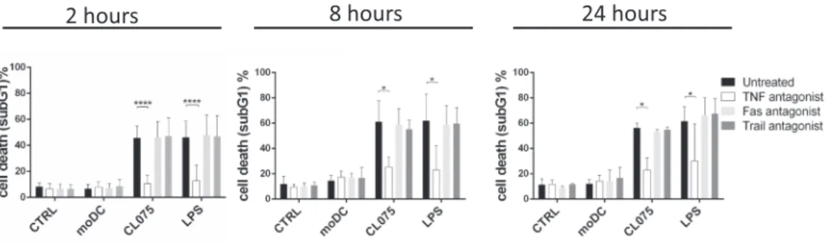
It has been published that DC-induced cell death is induced by death receptor (DR) activation, but the results as to how DR achieves this effect are contradictory (Chan and Housseau, 2008). Because Jurkat cells are sensitive to both TNF-α, FasL and TRAIL (Supplementary Fig. 3A), we monitored the role of each DR in the cytotoxic effect of DCsup using soluble death ligand antagonists. MoDCs were activated with CL075 or LPS for 30 min and supernatants of DCs were collected after 2 and 8 or 24 h, respectively. The actDC supernatant was pre-treated with TNF:Fc, Fas:Fc or TRAIL:Fc to neutralize the possible ligands in the supernatant.
All these supernatants were cytotoxic to Jurkat cells and TNF:Fc alone inhibited this effect. Fas:Fc or TRAIL:Fc were unable to block cell death regardless of how long the supernatant was produced (Fig. 2).
Supporting the observed TNF-dependent cytotoxicity, we detected significant TNF-α production by DCs after CL075 or LPS pre-activation (Supplementary Fig. 3B). These observations suggest that PRR activation-induced TNF-α production is responsible for short-term acti- vated DC-mediated cytotoxicity.
3.3. RIPK1 is required for DCsup-induced cell death
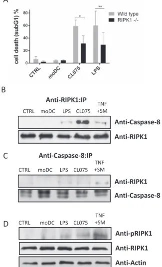
It has been published that TNF receptor ligation induces both RIPK1- dependent and independent cell death (Wang et al., 2008). We previ- ously reported that the supernatant of activated T cells induced RIPK1 dependent Fas-mediated apoptosis (Koncz et al., 2012) and we were curious to know whether RIPK1 was also required for DCsup-induced cell death. Therefore, we compared the susceptibility of Jurkat cells and their RIPK1 deficient subclones to DCsup-induced cytotoxicity.
Treatment with the supernatants of both CL075- and LPS-activated DCs resulted in more intensive cell death in wild-type cells than in their RIPK1-deficient counterparts (Fig. 3A).
During RIPK1-dependent apoptosis, RIPK1 recruits procaspase-8 and FADD, thus forming a cell death-inducing platform, referred to as a
“ripoptosome” (Tummers and Green, 2017). To analyse the interaction
between RIPK1 and pro-caspase-8, RIPK1 was immunoprecipitated from Jurkat cells after DCsup-induced activation. We found that caspase-8 is associated with RIPK1 after treatment of cells with the supernatants of CL075- or LPS-activated DCs, but not in the presence of the supernatant of unstimulated moDC (Fig. 3B). The intensity of RIPK1/caspase-8 as- sociation was similar in DCsup-treated and TNF plus SMAC mimetic- treated samples, the latter is a frequently used stimulus to activate RIPK1-dependent cell death (Wang et al., 2008). We confirmed these results by immunoprecipitating caspase-8 from Jurkat cells after DCsup- induced activation (Fig. 3C). RIPK1 phosphorylation is a widely accepted marker to characterize RIPK1-mediated cell death pathways (Geng et al., 2017). We detected increased phosphorylation of RIPK1 in Jurkat cells treated with either CL075 or LPS-activated DCsup but not after treatment with unstimulated DC supernatant (Fig. 3D). Altogether our results indicate that RIPK1-driven signaling contribute to DCsup- induced cell death.
3.4. The supernatant of dendritic cells activates RIPK1-dependent apoptosis
In addition to apoptosis, RIPK1 also mediates necroptosis when caspase-8 activity is blocked, and consequently it is unable to inhibit the necroptotic proteins (Geng et al., 2017). To determine whether DCsup stimulates RIPK1-mediated apoptosis or necroptosis, we treated the target cells with the DCsup in the presence of the pan caspase inhibitor (Z-VAD) and the necroptosis inhibitor (necrostatin-1). Pre-treatment of Jurkat cells with 10 μM Z-VAD inhibited DCsup-induced apoptosis (Fig. 4A). In addition, Z-VAD pre-treatment almost completely reduced cell death detected by PI-uptake (Fig. 4B). Necrostatin-1, however, did not have any effect on cell death (Fig. 4 A-B) nor was it additive to the Z- VAD-mediated inhibition. These results indicate that DCsup induces caspase-dependent apoptotic but not necroptotic cell death.
3.5. Tolerogenic microenvironment reduces the cytotoxic capacity of DCs We hypothesized that DCs induce cell to allow the sampling of dead cell-derived antigens from the surrounding microenvironment. To check whether controlling the availability of intracellular antigens is a constitutive function of DCs or it is also regulated by the surrounding tissues, we modelled the effects of tolerogenic and also immunogenic conditions. For this, according to previously published protocols (Sauter et al., 2019; Carbonneil et al., 2004), DCs were pre-treated with dexa- methasone to induce the differentiation of tolerogen DCs, or with type I IFNs to mimic intracellular infection. To confirm the differentiation of these cells, we examined the production of inflammatory and tolero- genic cytokines in each differentiated DC population following LPS or CL075 activation. IFN-treated DCs produced significantly higher level of IL-6 than the tolerogen DCs, but dexamethasone-pre-treated DCs
Fig. 2.moDC supernatant induces cell death in a TNF-dependent manner. DCs were stimulated on day 5 of the differentiation with Toll-like receptor ligands CL075 (1 μg/ml, Sigma) and LPS (0.5 μg/ml, InvivoGen) for 30 min. After washing steps DCs were incubated in fresh serum-free RPMI 1640 medium (Sigma) for 2, 8 or 24 h and the supernatants of DCs were harvested. The supernatants of DCs were pre-treated with TNF:Fc, Fas:Fc or TRAIL:Fc for 1 h. SVT35 Jurkat cells were activated with TNF:Fc, Fas:Fc or TRAIL:Fc pre-treated supernatants for 24 h. Cell death was determined by measuring the subG1 peak. The figures show the average of at least three independent experiments.
secreted more IL-10 (Supplementary Fig. 4A-B). We checked the cyto- toxic capacity of polarized DCs, by treating all these subpopulations with LPS or CL075 and examined the effect of their supernatants on Jurkat cells. The supernatant of dexamethasone-pre-treated DCs induced significantly less intense cell death than that of normal DCs, while cell death increased slightly, but not significantly after exposure of Jurkat cells to the supernatant of IFN-pre-treated DCs (Fig. 5A).
To determine whether the immunomodulatory environment also affects the susceptibility of target cells to DC-induced cytotoxicity, we treated the Jurkat cells were with type I IFN or with tolerogenic cyto- kines such as IL-10 and TGFβ. Regardless of pre-treatment of the target cells, the DC supernatant induced comparable cell death. However, IFN pre-treatment slightly, though not significantly, elevated cell death in- tensity (Fig. 5B). We can conclude that the tolerogenic microenviron- ment reduces the DC-induced cytotoxicity, and this effect is due to the reduced killing capacity of DCs rather than an acquired resistance of Fig. 3. RIPK1 is required for DCsup-induced cell death. (A) SVT35 Jurkat cells and their RIPK1-negative sub-clones were treated with the supernatants of unstimulated, CL075-(1 μg/ml) or LPS- (0.5 μg/ml) activated DCs for 24 h and cell death was determined by measuring the subG1 peak. The figure shows the average of five independent experiments. (B-C) SVT35 Jurkat cells were acti- vated with the supernatants of unstimulated, CL075-(1 μg/ml), or LPS- (0.5 μg/
ml) activated DCs or with the combination of TNF-α (20 ng/ml) and SMAC mimetic (0.5 μM) (TNF +SM) for 1 h. Following immunoprecipitation with anti- RIPK1 (B) or anti-Caspase-8 (C), the recruited molecules were visualized by western blotting. (D) The phosphorylation of RIPK1 was detected from total cell lysates by western blotting following the activation with the supernatants of unstimulated, CL075-(1 μg/ml), or LPS-(0.5 μg/ml) activated DCs or with the combination of TNF-α (20 ng/ml) and SMAC mimetic (0.5 μM) (TNF +SM) for 1 h. Representative images of three independent experiments are shown.
Fig. 4.The supernatant of dendritic cells activates apoptosis. (A) SVT35 Jurkat cells were pre-treated with either 10 μM Z-VAD or 40 μM necrostatin-1 alone or in combination for 1 h and were treated with the supernatants of unstimulated, CL075-(1 μg/ml), or LPS-(0.5 μg/ml) activated DCs for 24 h. Cell death was determined by measuring the subG1 peaks or (B) by the uptake of PI. The figures show the average of five independent experiments.
Fig. 5.Tolerogenic microenvironment reduces the cytotoxic capacity of DCs.
(A) SVT35 Jurkat cells were treated with the supernatants of untreated, CL075- (1 μg/ml) or LPS-(0.5 μg/ml) activated, control DCs (moDC), IFN-pre-treated DCs (IFN DC), or dexamethasonepre- treated (dexa) DCs for 24 h. Cell death was determined by measuring the subG1 peak. (B) SVT35 Jurkat cells were pre- treated with 100 ng/ml IL-10 and/or 100 ng/ml TGFß or 0.6 million U/ml IFN- α for 1 h. Following pre-treatment, these ligands were washed out completely and SVT35 Jurkat cells were triggered with the supernatant of unstimulated, CL075-(1 μg/ml) or LPS- (0.5 μg/ml) activated DCs for 24 h. Cell death was determined by measuring the subG1 peaks. The figures show the average of three independent experiments.
6 target cells.
4. Discussion
Dying cells may also activate innate and adaptive immune response in addition to tolerogenic apoptosis. Accidental necrosis or regulated necrotic pathways result in the release of damaged molecules (DAMP) from dying cells and consequently induce inflammation. RIPK1- dependent processes (Galluzzi et al., 2018; Yatim et al., 2015; Hancz et al., 2018), which facilitate effective uptake and presentation of dead cell-derived antigens, induce immunogenic cell death and initiate the response of naive CD8+T cells (Galluzzi, 2020). Consequently, various cell death inductions can be determinative in the regulation of immune response.
DCs are known to have cytotoxic potential, but the immunological role of this process is highly questionable (Chan and Housseau, 2008;
Chauvin and Josien, 2008). The limited number of DCs already pre- cludes the role of DCs in the effective killing of tumors or virus-infected cells. In addition, DCs migrate from the site of infection shortly after activation. Therefore, DC-induced cytotoxicity may be more important for efficient cross-representation by facilitating the acquisition of anti- gens, but is unlikely to function in the elimination of pathogens.
Accordingly, 24 hours LPS preconditioned DCs lose their killer function while losing their phagocytic ability. To confirm this hypothesis, we examined RIPK1-mediated signaling in dying cells, which has been shown to be a specific mode of cell death that promotes efficient cross- representation (Yatim et al., 2015). Comparing RIPK1 positive and RIPK1 deficient Jurkat cell lines, we found that DC-induced killing is at least partially a RIPK1-dependent process. These results confirm that DCs induce immunogenic cell death to facilitate their access to intra- cellular antigen fragments. It is important to note that PRR activation is required for inducing DCs’ cytotoxicity, so killing for sampling is not a continuous process. However, the external LPS signal, which is recog- nized primarily by cell surface PRR receptors, was effective in activating the cytotoxic function of DCs, indicating that the DCs themselves do not need to be infected to initiate their killer function.
If DC-induced cytotoxicity is not a constitutive surveillance mecha- nism, then we can assume that the tissue environment is able to regulate this process. Pre-treatment of DCs with dexamethasone is a widely used method for modelling tolerogenic DCs (Im et al., 2014). We compared the cytotoxic capacity of dexamethasone- and type I IFN-treated DCs. We have shown that tolerogenic stimuli reduces the DCs’ killing capacity.
Previous reports have shown that DCs can directly eliminate tumor cells and induce anti-tumor responses by taking up apoptotic tumor cells. Our results indicate that the immunosuppressive environment that charac- terizes most of tumors (Galluzzi, 2020) can significantly block DCs’ cytotoxic activity.
Immunomodulatory cytokines, type I IFNs are originally described as inducers of the antiviral response (Isaacs and Lindenmann, 1957). Sur- prisingly, the presence of type I IFN did not significantly change DC- induced cytotoxicity. PRR-induced activation of DCs triggers the killing function of DCs even without the paracrine effect of interferon produced by infected cells. It is well known that type I IFN mediates potent immunostimulatory effects, but this depends more on increased MHC expression, efficient cross-presentation, or directed chemotaxis of dendritic cells (Schiavoni et al., 2013) than on the elevated cytotoxic function of DCs. However, IFN also causes the maturation of DCs, a process that can down-regulate and compensate for the increased killing potential (Chauvin and Josien, 2008).
Mechanistically, DC-driven cytotoxicity was mediated by TNF secretion, but we could not detect the role of FasL or TRAIL in DCsup- induced killing. Depending on the gathered molecular complexes, TNF receptors can induce either apoptosis or necroptosis (Wang et al., 2008).
Using specific inhibitors for apoptotic and necroptotic processes, we demonstrated that DCsup induces almost exclusively apoptosis.
In summary, we have shown that DC-induced cytotoxicity is at least
in part a RIPK1-dependent process, which confirms the hypothesis that the ambition of DC-mediated killing is to facilitate the cross- presentation of intracellular antigens to CD8+T cells. The tolerogenic microenvironment downregulates the cytotoxic ability of DCs and thereby prevents the presentation of intracellular antigens. This toler- ogen mechanism can play a role in reducing unwanted reactions to auto- antigens, but it can also inhibit the effective cross-presentation of tumor antigens.
CRediT authorship contribution statement
Zsofia Varga: Methodology, Data curation, Writing - original draft, ´ Visualization, Investigation, Validation, Writing - review & editing. : . Evelin Racz: ´ Investigation, Validation, Writing - review & editing.
Anett M´azlo: ´ Investigation, Writing - review & editing. M´onika Kor- odi: Investigation, Validation, Writing - review & editing. Aniko Szab´ ´o:
Investigation, Validation, Writing - review & editing. Tamas Moln´ ´ar:
Investigation, Validation, Writing - review & editing. Arp´ ´ad Sz¨o˝or:
Investigation, Writing - review & editing. Zolt´an Vereb: Investigation, ´ Validation, Writing - review & editing. Attila B´acsi: Funding acquisi- tion, Writing - review & editing. G´abor Koncz: Conceptualization, Funding acquisition, Writing - review & editing.
Declaration of Competing Interest
The authors declare that they have no known competing financial interests or personal relationships that could have appeared to influence the work reported in this paper.
Acknowledgements
We thank Zsuzsanna Debreceni for the excellent technical assistance and Attila Sz¨oll˝osi for the language editing. The European Regional Development Fund GINOP-2.3.2-15-2016-00005, the National Research, Development and Innovation Office – NKFIH, 125224 are acknowledged for financial support of this work.
Appendix A. Supplementary data
Supplementary data to this article can be found online at https://doi.
org/10.1016/j.imbio.2020.152032.
References
Clatworthy, M.R., et al., 2014. Immune complexes stimulate CCR7-dependent dendritic cell migration to lymph nodes. Nat. Med. 20, 1458.
Bousso, P., 2008. T-cell activation by dendritic cells in the lymph node: lessons from the movies. Nat. Rev. Immunol. 8, 675.
Wang, L., Du, F., Wang, X., 2008. TNF-alpha induces two distinct caspase-8 activation pathways. Cell 133, 693.
Yatim, N., et al., 2015. RIPK1 and NF-kappaB signaling in dying cells determines cross- priming of CD8(+) T cells. Science 350, 328.
Vidalain, P.O., Azocar, O., Yagita, H., Rabourdin-Combe, C., Servet-Delprat, C., 2001.
Cytotoxic activity of human dendritic cells is differentially regulated by double- stranded RNA and CD40 ligand. J. Immunol. 167, 3765.
Chan, C.W., Housseau, F., 2008. The ’kiss of death’ by dendritic cells to cancer cells. Cell Death Differ. 15, 58 (Jan.
Chauvin, C., Josien, R., 2008. Dendritic cells as killers: mechanistic aspects and potential roles. J. Immunol. 181, 11.
Janjic, B.M., et al., 2002. Innate direct anticancer effector function of human immature dendritic cells. I. Involvement of an apoptosis-inducing pathway. J. Immunol. 168, 1823.
Lakomy, D., et al., 2011. Cytotoxic dendritic cells generated from cancer patients.
J. Immunol. 187, 2775.
Joo, H.G., et al., 2002. Human dendritic cells induce tumor-specific apoptosis by soluble factors. Int. J. Cancer 102, 20.
Lu, G., et al., 2002. Innate direct anticancer effector function of human immature dendritic cells. II. Role of TNF, lymphotoxin-alpha(1)beta(2), Fas ligand, and TNF- related apoptosis-inducing ligand. J. Immunol. 168, 1831.
Vidalain, P.O., et al., 2000. Measles virus induces functional TRAIL production by human dendritic cells. J. Virol. 74, 556.
Burdelya, L.G., et al., 2013. Central role of liver in anticancer and radioprotective activities of Toll-like receptor 5 agonist. Proc. Natl. Acad. Sci. U.S.A. 110, E1857.
Salamone, G.V., et al., 2010. Flagellin delays spontaneous human neutrophil apoptosis.
Laboratory Investigation 90, 1049.
Anderson, A.E., et al., 2008. Differential regulation of naive and memory CD4+T cells by alternatively activated dendritic cells. J. Leukoc. Biol. 84, 124.
Carbonneil, C., Saidi, H., Donkova-Petrini, V., Weiss, L., 2004. Dendritic cells generated in the presence of interferon-alpha stimulate allogeneic CD4+T-cell proliferation:
modulation by autocrine IL-10, enhanced T-cell apoptosis and T regulatory type 1 cells. Int. Immunol. 16, 1037.
Schiavoni, G., Mattei, F., Gabriele, L., 2013. Type I interferons as stimulators of DC- mediated cross-priming: impact on anti-tumor response. Front. Immunol. 4, 483.
Hunzeker, J.T., et al., 2011. A marked reduction in priming of cytotoxic CD8+T cells mediated by stress-induced glucocorticoids involves multiple deficiencies in cross- presentation by dendritic cells. J. Immunol. 186, 183.
Im, S.A., Gerelchuluun, T., Lee, C.K., 2014. Evidence for direct inhibition of MHC- restricted antigen processing by dexamethasone. Immune Netw 14, 328.
Bene, K., Varga, Z., Petrov, V.O., Boyko, N., Rajnavolgyi, E., 2017. Gut microbiota species can provoke both inflammatory and tolerogenic immune responses in human dendritic cells mediated by retinoic acid receptor alpha ligation. Front. Immunol. 8, 427.
Varga, Z., et al., 2020. Differences in the sensitivity of classically and alternatively activated macrophages to TAK1 inhibitor-induced necroptosis. Cancer Immunol.
Immunother.
Koncz, G., et al., 2012. Vesicles released by activated T cells induce both Fas-mediated RIP-dependent apoptotic and Fas-independent nonapoptotic cell deaths. J. Immunol.
189, 2815.
Tummers, B., Green, D.R., 2017. Caspase-8: regulating life and death. Immunol. Rev.
277, 76.
Geng, J., et al., 2017. Regulation of RIPK1 activation by TAK1-mediated phosphorylation dictates apoptosis and necroptosis. Nat. Commun. 8, 359.
Sauter, A., Yi, D.H., Li, Y., Roersma, S., Appel, S., 2019. The culture dish surface influences the phenotype and cytokine production of human monocyte-derived dendritic cells. Front Immunol 10, 2352.
Galluzzi, L., et al., 2018. Molecular mechanisms of cell death: recommendations of the Nomenclature Committee on Cell Death 2018. Cell Death Differ. 25, 486.
Hancz, D., et al., 2018. Flagellin increases death receptor-mediated cell death in a RIP1- dependent manner. Immunol. Lett. 193, 42.
Galluzzi, L., et al., 2020. Consensus guidelines for the definition, detection and interpretation of immunogenic cell death. J. Immunother. Cancer 8.
Isaacs, A., Lindenmann, J., 1957. Virus interference. I. The interferon. Proc. R. Soc. Lond.
B Biol. Sci. 147, 258.