Tamas Molnar1,2, Petra Pallagi3,4, Balint Tel3,4, Robert Kiraly5, Eszter Csoma6, Viktoria Jenei1, Zsofia Varga1,2, Peter Gogolak1, Anne Odile Hueber7, Zoltan Mate8, Ferenc Erdelyi8, Gabor Szabo8, Aladar Pettko-Szandtner9, Attila Bacsi1, Laszlo Virag10, Jozsef Mal eth3,4and Gabor Koncz1
1 Department of Immunology, Faculty of Medicine, University of Debrecen, Hungary 2 Doctoral School of Molecular Cellular and Immune Biology, University of Debrecen, Hungary 3 First Department of Medicine, University of Szeged, Szeged, Hungary
4 HAS-USZ Momentum Epithelial Cell Signalling and Secretion Research Group, University of Szeged, Szeged, Hungary 5 Department of Biochemistry and Molecular Biology, Faculty of Medicine, University of Debrecen, Hungary
6 Department of Medical Microbiology, Faculty of Medicine, University of Debrecen, Hungary 7 CNRS, Inserm, iBV, Universite C^ote d’Azur, Nice, France
8 Medical Gene Technology Unit, Institute of Experimental Medicine, Budapest, Hungary 9 Laboratory of Proteomics Research, Biological Research Centre, Szeged, Hungary 10 Department of Medical Chemistry, Faculty of Medicine, University of Debrecen, Hungary
Keywords
Caspase-9; cell death; inflammation;
necroptosis; pancreatitis Correspondence
G. Koncz, Department of Immunology, Faculty of Medicine, University of Debrecen, 1 Egyetem Square, Debrecen H- 4032, Hungary
Tel:+36 52 417 159, E-mail: konczgb@gmail.com
(Received 22 September 2020, revised 4 March 2021,
doi:10.1111/febs.15898
Necroptosis is a regulated necrotic-like cell death modality which has come into the focus of attention since it is known to contribute to the pathogene- sis of many inflammatory and degenerative diseases as well as to tumor regulation. Based on current data, necroptosis serves as a backup mecha- nism when death receptor-induced apoptosis is inhibited or absent. How- ever, the necroptotic role of the proteins involved in mitochondrial apoptosis has not been investigated. Here, we demonstrated that the stimu- lation of several death and pattern recognition receptors induced necropto- sis under caspase-compromised conditions in wild-type, but not in caspase- 9-negative human Jurkat and murine MEF cells. Cerulein-induced pancre- atitis was significantly reduced in mice with acinar cell-restricted caspase-9 gene knockout. The absence of caspase-9 led to impaired association of receptor-interacting serine/threonine-protein kinase 1 (RIPK1) and RIPK3 and resulted in decreased phosphorylation of RIP kinases, but the overex- pression of RIPK1 or RIPK3 rescued the effect of caspase-9 deficiency.
Inhibition of either Aurora kinase A (AURKA) or its known substrate, glycogen synthase kinase 3b (GSK3ß) restored necroptosis sensitivity of caspase-9-deficient cells, indicating an interplay between caspase-9 and AURKA-mediated pathways to regulate necroptosis. Our findings suggest that caspase-9 acts as a newly identified regulator of necroptosis, and thus, caspase-9 provides a promising therapeutic target to manipulate the immunological outcome of cell death.
Abbreviations
AP, acute pancreatitis; AURKA, Aurora kinase A; CHX, cycloheximide; CYLD, cylindromatosis protein; DR, death receptor; GSK3ß, glycogen synthase kinase 3b; HHV-1 or HSV-1, human herpesvirus 1; IBD, inflammatory bowel disease; LPS, lipopolysaccharide; MEF, mouse embryonic fibroblasts; MLKL, mixed lineage kinase domain-like protein; Nec-1, necrostatin-1; NSA, necrosulfonamide; PI, propidium iodide;
PRR, pattern recognition receptor; RIPK1, receptor-interacting serine/threonine-protein kinase 1; TNF-a, tumor necrosis factor-a; TRAIL, tumor necrosis factor-related apoptosis-inducing ligand.
6476 The FEBS Journal288(2021) 6476–6491ª2021 The Authors.The FEBS Journalpublished by John Wiley & Sons Ltd on behalf of
Introduction
In the past few years, novel cell death pathways have been discovered and classified and these cell death modalities have been shown to differently affect the activation of immune responses [1,2]. In addition to intensity, the immunological outcome of cell death also has fundamental impact on the evolution of various inflammatory diseases, autoimmune processes, degen- erative disorders, and immune surveillance of tumors.
The molecular programs that govern switches between the different modes of cell death remain obscure despite the enormous potential of putative therapeutic applications.
Apoptosis is mediated by both intrinsic and extrin- sic pathways. The extrinsic pathway is induced by cell death receptors (DR) interacting with their speci- fic ligands such as tumor necrosis factor-a (TNFa), Fas ligand and tumor necrosis factor-related apopto- sis-inducing ligand (TRAIL) [3,4]. These interactions lead to the recruitment of the adaptor protein FAS- associated death domain, which activates apoptosis initiator enzymes caspase-8 and caspase-10, resulting in the activation of effector caspases (type I cells), or initiation of the mitochondrial apoptotic pathway (type II cells) [5,6]. The intrinsic pathway can be activated by various stimuli which induce mitochon- drial membrane permeabilization [7]. Cytochrome c released from the mitochondria interacts with the apoptotic protease-activating factor 1, which recruits caspase-9 [8]. The resulting molecular complex forms the apoptosome, which maintains caspase-9 in an active conformation resulting in the activation of effector caspases [9,10].
Cell death is controlled by multiple interconnected pathways, among them necroptosis, a highly regulated necrosis-like cell death mode [11–13]. Necroptosis uti- lizes a unique signaling pathway requiring the involve- ment of receptor-interacting serine/threonine-protein kinases 1 and 3 (RIPK1 and RIPK3)[2,14]and mixed lineage kinase domain-like protein (MLKL)[15]. Upon stimulation of DRs, necroptosis requires inhibited cas- pase activity because active caspase-8 blocks necrop- totic signaling[2,16]primarily through the cleavage of RIPK1 [17], RIPK3 [18,19], and cylindromatosis pro- tein (CYLD) [20] which acts as the deubiquitinase enzyme of RIPK1. Even though the crucial role of DR-mediated apoptosis in the negative regulation of necroptosis is investigated extensively, the function of caspase-9 in necroptosis has not been documented yet.
Perturbations in necroptotic signaling contribute to the pathogenesis of many diseases [21]. Upregulated necroptosis occurs in retinal disorders,
neurodegenerative diseases, and ischemia reperfusion [22]. Downregulation of necroptosis contributes to infectious diseases, carcinogenesis, or the resistance to anti-cancer treatments [23]. Dysregulation of the bal- ance between apoptosis and necroptosis plays a crucial role in inflammatory diseases such as inflammatory bowel disease (IBD), pancreatitis, acute kidney injury, hepatitis, and various skin disorders[21,24].
More than 70 human molecules have been docu- mented as regulators of necroptotic events along the RIPK1-RIPK3-MLKL axis[21]. Among them Aurora kinase A (AURKA) has recently been identified as a binding partner of RIPK3 [25–28] and an inhibitor against unwanted necroptosis. AURKA, with its downstream partner, GSK3b regulates the formation of RIPK1-RIPK3 complex [25]. Silencing or blocking AURKA or GSK3b leads to spontaneous necroptosis [25].
In the present study, we report that caspase-9 is required for death and pattern recognition receptor (PRR)-induced necroptosis in vitro and increases the severity of cerulein-induced pancreatitis in vivo, even though caspase activity is inhibited under necroptotic conditions. We provide evidence that caspase-9 regu- lates the formation of the necrosome. Our results may offer novel strategies for regulating necroptosis through targeting caspase-9 and provide relevant infor- mation on the cross-talk of stress-induced apoptosis and programmed necrosis.
Results
Caspase-9 plays a role in death receptor- mediated necroptosis
It is well known that DR-mediated apoptosis domi- nates necroptosis[2,13], but the link between necropto- sis and intrinsic apoptosis has not been studied in detail [21–24]. Several substrates of caspase-8 have been documented as critical regulators of necroptosis, such as RIPK1, RIPK3, and CYLD [20]; however, its most well-known substrates, the downstream caspases, have been only marginally studied during necroptosis.
Silencing of caspase-3 and caspase-7 did not influence TNF-induced necroptosis[29], while the effect of cas- pase-9 on necroptosis has not yet been investigated.
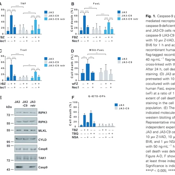
To assess this, we induced cell death in JA3 Jurkat cells and in their caspase-9-negative subclone (JA3- C9). Cells were treated with the classical necroptosis inducing cocktail of TNF, SMAC mimetic (BV6), and pan-caspase inhibitor Z-VAD (TBZ) [30,31] or cas- pase-8 inhibitor Q-IETD-Oph [32]. In the culture of caspase-9-deficient cells, the rate of cell death was
significantly lower compared with that of caspase-9- positive ones (Fig.1A). We used a necroptosis inhibi- tor, necrostatin-1 (Nec-1), and validated that it suc- cessfully blocked TBZ-induced cell death. To confirm that the observed cell death is strictly caspase-9 depen- dent, we retransfected the JA3-C9 subclone with wild- type caspase-9. Reconstitution of caspase-9 expression restored the sensitivity of transfected cells (JA3-C9 retr) to TBZ-induced necroptosis (Fig.1A).
To verify the essential role of caspase-9 in the necroptotic signaling of other DRs, we triggered necroptosis with recombinant FasL or TRAIL in the presence of BV6 and Z-VAD (FBZ or TrBZ, respec- tively). Treatment with either FasL or TRAIL acti- vated necroptosis only in the wild-type cells, whereas in the absence of caspase-9 significantly alleviated necroptosis was detected in both cases. Retransfection of caspase-9 into JA3-C9 cells also restored the inten- sity of cell death after these stimuli (Fig.1B,C). Jurkat cells were also cocultured with FasL-overexpressing
human B-cell line (WSU-FasL) in the presence of Z- VAD, because this stimulus was able to induce necrop- tosis without the synergic effect of BV6. Significantly decreased necroptosis was detected in caspase-9-defi- cient cells compared with wild-type cells upon WSU- FasL induction (Fig.1D). In these Jurkat cells, the same expression level of the most widely studied necroptosis regulatory molecules (RIPK1, RIPK3, MLKL, caspase-8, CYLD, TAK1) was detected regardless of the presence of caspase-9 (Fig. 1E). We also checked the intensity of necroptosis under cas- pase-8 compromised conditions. For this, wild-type and caspase-9-deficient Jurkat cells were treated with a specific caspase-8 inhibitor (Q-IETD-Oph) in combina- tion with BV6 and TNF-a. Following this treatment, the intensity of necroptosis and its caspase-9 depen- dence was comparable to TBZ-induced cell death (Fig. 1F).
To study whether caspase-9 acts on necroptosis only or controls necrosis in general, we treated the cells
A B
C D
TBZ – + + – + + – + + Nec1 – – + – – + – – +
TrBZ – + + – + + – + + Nec1 – – + – – + – – +
FBZ – + + – + + – + + Nec1– – + – – + – – +
E F
****
****
****
****
0 2 0 4 0 6 0 8 0
F a s L
Cell death (%)
J A 3 J A 3 - C 9 J A 3 - C 9 r e tr
****
****
***
****
****
****
**** ****
0 2 0 4 0 6 0 8 0
T N F
Cell death (%)
J A 3 J A 3 - C 9 J A 3 - C 9 r e tr
**
***
****
****
**** ****
0 2 0 4 0 6 0 8 0
T r a il
Cell death (%)
J A 3 J A 3 - C 9 J A 3 - C 9 r e tr
wFZ – + + – + + Nec1 – – + – – +
**
*** ***
0 1 0 2 0 3 0 4 0
W S U - F a s L
Cell death (%)
J A 3 J A 3 - C 9
RIPK1 JA3 JA3 JA3
-C9 retr
RIPK3 MLKL CYLD Casp8 TAK1 Casp9 kDa
72 55 55
95 55
72 43
Q - IE T D -O P h
Cell death (%)
0 2 0 4 0 6 0 8 0 1 0 0
J A 3 J A 3 - C 9
TBZ – + + – – – + + – – TBQ – – – + + – – – + + NSA – – + – + – – + – +
Fig. 1.Caspase-9 plays a role in DR - mediated necroptosis. (A) JA3 cells, its caspase-9-deficient counterpart (JA3-C9), and JA3-C9 cells retransfected with caspase-9 (JA3-C9 retr) were pretreated with 10lMZ-VAD, 40lMNec-1, and 1lM BV6 for 1 h and activated with 50 ngmL 1 recombinant human TNF-a, (B) 30 ngmL 1 flag-tagged recombinant FasL or (C) 40 ngmL 1flag-tagged recombinant Trail cross-linked with the anti-flag (M2) antibody.
After 24 h, cell death was determined by PI staining. (D) JA3 and JA3-C9 cells were pretreated with 10lMZ-VAD and cocultured with cell tracker green-stained human FasL expressing WSU B-cell line (wF) at a ratio of 1 : 5. After 24 h, the extent of cell death was determined by PI staining in the cell tracker-negative population. (E) The expression of the indicated molecules was determined by western blotting of total cell lysates.
Representative images of three independent experiments are shown. (F) JA3 and JA3-C9 cells were pretreated with 10lMZ-VAD, 10lMQ-IETD-OPh, 1lM BV6, and 1lMNSA for 1 h and activated with 50 ngmL 1human TNFa. After 24 h, cell death was determined by PI staining.
Figure A-D, F shows the mean plus SD of at least three independent experiments.
Significance is indicated by**P<0.01;
***P<0.005;****P<0.001.
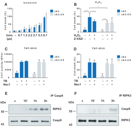
with ionomycin and observed that the intensity of ion- omycin-induced necrosis was comparable in both JA3 and JA3-C9 cells (Fig.2A). Treatment with H2O2 has been shown to induce both apoptosis and classical necrosis in Jurkat cells [33]. The role of caspase-9 is well-known in stress-induced apoptosis, and conse- quently, H2O2induces apoptosis only in caspase-9-pos- itive cells, but in the presence of Z-VAD, H2O2 treatment resulted in equally intense necrosis in both JA3 and JA3-C9 cells (Fig.2B).
In addition to RIPK1-mediated necroptosis, RIPK1- dependent apoptosis has also been documented, pri- marily in the presence of SMAC mimetics under con- ditions that allow caspase activation[34]. JA3 and its caspase-9-negative subclone were stimulated with TNF and BV6, and the activity of effector caspases was investigated. In accordance with published results [35],
the absence of caspase-9 did not modify the intensity of RIPK1-mediated apoptosis (Fig.2C,D). Our results indicate that in addition to its well-known role in intrinsic apoptosis, caspase-9 is specifically required for necroptosis, whereas it has no role in necrosis or in RIPK1-mediated apoptosis.
Caspase-9 is associated with RIPK3 upon necroptotic stimulus
Considering that DR-induced necroptosis takes place at caspase-compromised conditions, the protease activ- ity of caspase-9 is not required for its role in the sig- naling. Therefore, we assumed that caspase-9 contributes to necroptosis as an adaptor protein. We checked the molecules recruited to caspase-9, in partic- ular, its association with RIPK1, RIPK3, and MLKL.
Iono. – 0.7 1.3 2.2 2.7 5.3 6.7 (μM)
T N F + B V 6
C3/7 activity (RFU)
0 5 0 0 0 1 0 0 0 0 1 5 0 0 0
J A 3 - C 9 J A 3 Io n o m ic in
Cell death (%)
0 5 0 1 0 0 1 5 0
J A 3 J A 3 - C 9
H2O2 – + + – + + Z-VAD – – + – – +
***
**
*
****
0 2 0 4 0 6 0
H2O2
Cell death (%) J A 3
J A 3 - C 9
TB – + + – + + Nec1 – – + – – +
T N F + B V 6
Cell death (%)
0 2 0 4 0 6 0
J A 3 J A 3 - C 9
TB – + + – + + Nec1 – – + – – +
A B
C D
E F
c 10’ 1h 3h
Casp9 IP RIPK3
RIPK3 kDa
55
43 RIPK3
Casp9
c 10’ 1h 3h
IP Casp9 kDa
55
43
Fig. 2.Caspase-9 interacts with RIPK3 during necroptosis. (A) JA3 and JA3-C9 cells were treated with the indicated doses of ionomycin or (B) pretreated with 10lMZ-VAD for 1 h and activated with 400lMH2O2. The degree of total cell death was quantified based on the uptake of PI. (C-D) JA3 and JA3-C9 cells were pretreated with 40lMNec-1 and 1lMBV6 for 1 h followed by stimulation with 50 ngmL 1TNF.
(C) Caspase-3/7 activity, expressed as relative fluorescence units (RFU), was measured 30 min after the addition of the profluorescent substrate Z-DEVD-R11. (D) The degree of total cell death was determined by PI staining. Panels A-D show the mean plus SD of at least three independent experiments. Significance is indicated by*P<0.05;**P<0.01;***P<0.005;****P<0.001. (E) JA3 and JA3-C9 cells were pretreated with 10lMZ-VAD and 1lMBV6 for 1 h and activated for the indicated times with 50 ngmL 1human TNF-a. Following immunoprecipitation from total cell lysates with anti-caspase-9 or (F) anti-RIPK3, the recruited molecules were detected by western blotting.
Figures E and F shows representative images of three independent experiments.
We were unable to detect any interaction between cas- pase-9 and RIPK1 or MLKL, but we observed a weak and transient association of RIPK3 with caspase-9, which peaked at 3 h after initiation of necroptosis (Fig.2E). Immunoprecipitation with anti-RIPK3 Ab has confirmed RIPK3-caspase-9 association (Fig.2F).
According to our data, caspase-9 can be considered as a regulator of necroptosis that interacts faintly with RIPK3.
Caspase-9 regulates death- and PRR-mediated necroptosis in MEF cells
Necroptosis has been published to highly depend on the cell type and on the sorts of stimuli. To determine whether the role of caspase-9 in necroptosis is a general require- ment or specific to the Jurkat cells, we studied the necrop- totic sensitivity of caspase-9-deficient MEF cells (MEF- C9) and their wild-type counterparts. Similarly to our observations in Jurkat cells, TBZ activation induced necroptosis in wild-type, but not in the caspase-9-deficient MEFs (Fig.3A). We also stimulated necroptosis with FasL and found that MEF-C9 cells were less susceptible to FBZ-induced necroptosis than wild-type MEFs (Fig.3B).
These cells are also sensitive to TNF, Z-VAD, and cyclo- heximide (CHX)-triggered (TCZ) necroptosis; thus, this treatment induced intense necroptosis in MEFs, while cas- pase-9-deficient cells were less sensitive to these stimuli (Fig.3C). All these results confirm that caspase-9 con- tributes to necroptosis, and provide evidence that its role is essential for necroptosis in both human and mouse cells.
Necroptosis has been observed not only after DR-me- diated signals, but also as a result of various other stim- uli, among them PRR activation has been investigated the most intensely[36]. Wild-type and caspase-9-nega- tive MEF cells were treated with lipopolysaccharide (LPS) in the presence of Z-VAD. Similar to the results detected after DR activation, in the culture of caspase-9- deficient cells significantly fewer dead cells were detected after LPS exposure compared with that of wild-type cells (Fig.3D). Mouse cells have been published to be sensitive to human herpesvirus 1 (HHV-1 or HSV-1)-in- duced necroptosis [37]. MEF and MEF-C9 cells were infected with HSV-1 in the presence of Z-VAD, and it was observed that the lack of caspase-9 protected MEF cells from HSV-1-induced necroptosis (Fig.3E). We also checked the expression of the main necroptosis reg- ulatory molecules (RIPK1, RIPK3, MLKL, caspase-8, CYLD, TAK1), and it was comparable in both wild- type and caspase-9-deficient MEF cells (Fig.3F). Our results indicate that caspase-9 plays a general role in necroptosis, as caspase-9 deficiency prevents necroptosis in both human and murine cells under various stimuli.
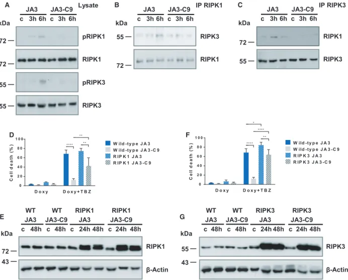
Caspase-9 regulates upstream events of necroptotic signaling
To dissect the role of caspase-9 in the molecular back- ground of necroptosis, we examined the phosphoryla- tion of the core components of the necroptotic pathway. RIPK1 phosphorylation (S166) was strongly elevated in JA3 cells following TBZ activation, but only marginal phosphorylation of RIPK1 was found in JA3-C9 cells (Fig. 4A). Similarly, more intense RIPK3 phosphorylation (S227) was detected in wild- type, than in JA3-C9 cells 6 h postactivation (Fig. 4A). We compared the association of RIPK1 with RIPK3 in the presence and absence of caspase-9 to analyze the regulated events in the course of the necroptotic process. We detected the RIPK1/RIPK3 interaction by immunoprecipitation 3–6 h following TBZ activation in wild-type Jurkat cells, but we could not observe this association in the absence of caspase- 9 (Fig.4B,C). The failure of RIPK1/RIPK3 interac- tion in JA3-C9 cells indicates that caspase-9 regulates the upstream events of necroptotic signaling.
We overexpressed RIPK1 and RIPK3 assisted by a Tet-On inducible system in both JA3 and JA3-C9 cells to verify the regulation of the necrosome assembly by caspase-9. Overexpression of either RIPK1 or RIPK3 reconstituted the sensitivity of JA3-C9 cells to TBZ-in- duced necroptosis (Fig. 4D-G). These results demon- strate that the downstream parts of the necroptotic pathway still function normally in the absence of cas- pase-9 in both RIPK1- and RIPK3-overexpressing cells.
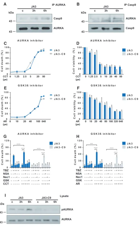
AURKA inhibitor restores the sensitivity of caspase-9-deficient cells to TNF-induced necroptosis
We have shown that caspase-9 regulates the initial phase of necroptotic signaling. Caspase-9 interacts weakly with the basic components of necroptotic sig- naling pathway; therefore, we hypothesized that it acts primarily on molecules that regulate the assembly of the necrosome. AURKA has been identified recently as an interaction partner of RIPK3 and RIPK1 [25,26]. We tested the association of caspase-9 with AURKA under necroptotic conditions in Jurkat cells and found that AURKA was associated with caspase- 9 in a stimulus-dependent manner (Fig.5A,B).
AURKA and its downstream target, GSK3b, have been published to downregulate necroptosis. We tested the TBZ-induced necroptosis in the presence of AURKA inhibitor (CCT137690) or GSK3b inhibitor (AR-A014418) in JA3 and JA3-C9 cells. Low doses of
CCT137690 (<2.5lM) or AR-A014418 (<20 lM) had only minimal effects on the cell survival; however, higher doses of CCT137690 (≥2.5lM) and AR- A014418 (≥20lM) elevated the ratio of dead cells (Fig.5C-F). Cotreatment with low doses of either CCT137690 or AR-A014418 with TBZ increased necroptosis, and this cell death was effectively blocked by all of the applied necroptosis inhibitors/necrostatin-1, GSK’872, and necrosulfonamide (NSA)/ (Fig.5G,H).
Importantly, cotreatment of either CCT137690 or AR- A014418 with TBZ restored the necroptosis sensitivity of the caspase-9-deficient cells.
Caspase-9-mediated regulation of AURKA was con- firmed by the investigation of AURKA phosphoryla- tion. AURKA phosphorylation on T288 was observed
only in JA3 but not in JA3-C9 cells, and this phospho- rylation was increased upon TBZ stimuli indicating that the activity of this kinase is regulated by caspase- 9 (Fig.5I). In summary, caspase-9 and the AURKA- GSK3bpathways interact in the regulation of necrop- tosis in a still unidentified way.
Knockout of caspase-9 in pancreatic acinar cells decreases the severity of cerulein-induced acute pancreatitis
To confirm the role of caspase-9 in necroptosisin vivo, we compared the severity of cerulein-induced experi- mental acute pancreatitis (AP) in wild-type and pan- creas acinus-specific caspase-9 knockout mice.
A B
C D
E F
HSV – + – +
Z-VAD – + – +
**** ***
0 1 0 2 0 3 0
H S V
Cell death (%) M E F
M E F - C 9
TBZ – + + – + + Nec1 – – + – – +
*
** **
0 2 0 4 0 6 0 8 0
T N F
Cell death (%) M E F
M E F - C 9
***
*
***
0 5 0 1 0 0 1 5 0
F a s L
Cell death (%) M E F
M E F - C 9
****
**** ****
0 2 0 4 0 6 0
T C Z
Cell death (%) M E F
M E F - C 9
FBZ – + + – + + Nec1 – – + – – +
TCZ – + + – + +
Nec1 – – + – – + LPS – + – +
Z-VAD – + – +
*** ***
0 1 0 2 0 3 0
L P S
Cell death (%) M E F
M E F - C 9
RIPK1
RIPK3 MLKL CYLD Casp8 TAK1 Casp9 MEF MEF kDa -C9
72 55 55
95 43 72
43 Fig. 3.Caspase-9 regulates death- and
PRR-mediated necroptosis in MEF cells. (A) MEF and their caspase-9-deficient counterpart (MEF-C9) were pretreated with 10lMZ-VAD, 40lMNec-1, and 1lMBV6 for 1 h and activated with 20 ngmL 1 human TNF-aor (B) 40 ngmL 1flag-tagged recombinant FasL. After 24 h, cell death was determined by PI staining. (C) MEF and MEF-C9 cells were pretreated 10lMZ- VAD, 40lMNec-1, and 0.4lgmL 1CHX for 1 h and activated with 20 ngmL 1 human TNFa. After 24 h, cell death was determined by PI staining. (D) MEF and MEF-C9 cells were pretreated 50lMZ-VAD for 1 h and were activated with
100 ngmL 1LPS or (E) were infected with HHV-1 at MOI 5. After 24 h, cell death was determined by PI staining. Panels A-E show the mean plus SD of at least three independent experiments. Significance is indicated by*P<0.05;**P<0.01;***P<
0.005;****P<0.001. (F) The expression of the indicated molecules was visualized by western blotting of total cell lysates.
Representative images of three independent experiments are shown.
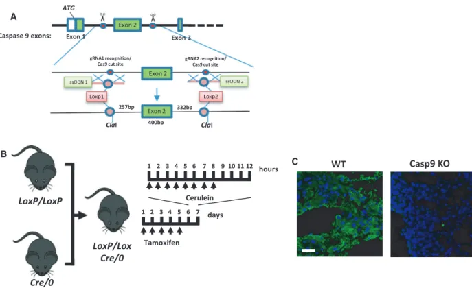
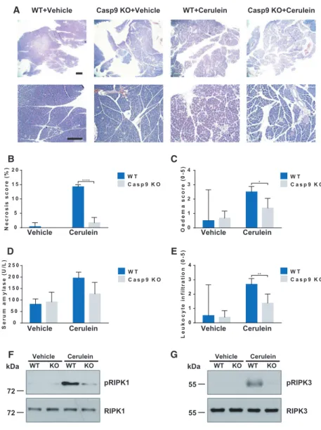
Pancreas acinus-specific knockout was achieved by crossing of caspase-9 LoxP animals with pancreatic acinar cell-specific-cre mice expressing tamoxifen-in- ducible Cre recombinase under the direction of the Cela1 promoter (Fig.6A,B). Caspase-9 knockout was confirmed by immunostaining (Fig.6C). In this experi- ment, mice were i.p. injected hourly (eight times) with either physiological saline (control) or 50µg/bwkg cer- ulein to induce AP. Overall, in this experimental model we observed a remarkable improvement of pancreatitis severity in the caspase-9 knockout animals. The
control mice had normal pancreatic histology in both groups, whereas cerulein hyperstimulation caused pan- creatic damage as demonstrated in Fig. 7A. Impor- tantly, knockout of caspase-9 in pancreatic acinar cells significantly decreased the histological scores and improved serum amylase activity. The extent of inter- stitial edema (2.5 0.14 for WT vs 1.380.28 for KO) and leukocyte infiltration (2.68 0.15 for WT vs 1.38 0.26 for KO) was significantly improved, and the extent of necrosis was completely restored to the control level (14.25 0.27 for WT vs 1.710.75 for
**** **
**
D o x y D o x y + T B Z 0
2 0 4 0 6 0 8 0 1 0 0
Cell death (%) W ild -ty p e J A 3
W ild -ty p e J A 3 -C 9 R IP K 1 J A 3 R IP K 1 J A 3 - C 9
****
**
****
*
D o x y D o x y + T B Z 0
2 0 4 0 6 0 8 0 1 0 0
Cell death (%) W ild -ty p e J A 3
W ild -ty p e J A 3 -C 9 R IP K 3 J A 3 R IP K 3 J A 3 - C 9
D F
C
RIPK1
RIPK3 IP RIPK3 kDa
72 55
JA3 JA3-C9 c 3h 6h c 3h 6h B
RIPK3
RIPK1 IP RIPK1 kDa
55
72
JA3 JA3-C9 c 3h 6h c 3h 6h pRIPK1
Lysate
RIPK1
pRIPK3
RIPK3 kDa
72
72 55 55
JA3 JA3-C9 c 3h 6h c 3h 6h A
E WT WT RIPK1 RIPK1 G
JA3 JA3-C9 JA3 JA3-C9 c 48h c 48h c 24h 48h c 24h 48h
RIPK1
β-Actin kDa
72 43
RIPK3
β-Actin
WT WT RIPK3 RIPK3
JA3 JA3-C9 JA3 JA3-C9 c 48h c 48h c 24h 48h c 24h 48h kDa
55 43
Fig. 4.Caspase-9 regulates the assembly of necrosome during necroptosis. (A) JA3 and JA3-C9 cells were pretreated with 10lMZ-VAD and 1lM BV6 for 1 h and activated for the indicated times with 50 ngmL 1human TNF-a. Phosphorylation of RIPK1 (S166) and RIPK3 (S227) was detected from total cell lysates. (B) Following immunoprecipitation from total cell lysates with anti-RIPK1 or (C) anti-RIPK3 antibodies, the recruited molecules were detected by western blotting. (D) RIPK1 or (F) RIPK3 overexpression was activated with 2lgmL 1doxycycline for 24 h, and cells were pretreated with 10lMZ-VAD and 1lMBV6 and activated with 50 ngmL 1human TNF-a. Cell death was determined by PI uptake, 24 h after TNF activation. Data are presented as the mean plus SD of three independent experiments. Significance is indicated by*P<0.05;**P<0.01;****P<0.001. (E) Cells were pretreated with 2lgmL 1doxycyclin for the indicated times, and the expression of RIPK1 or (G) RIPK3 was detected from total cell lysates by western blotting. Figure A-C, E, G is representative images of three independent experiments.
KO) (Fig.7B-E). To demonstrate that cerulein-in- duced necrosis occurred due to necroptosis, we exam- ined the phosphorylation of necrosome components.
We detected increased RIPK1 and RIPK3 phosphory- lation in pancreatic tissue samples following cerulein treatments in wild-type mice, but not in caspase-9-defi- cient animals (Fig.7F,G).
These results highlight the major role of caspase-9- mediated necroptosis in the development of experimen- tal AP.
Discussion
Switching between cell death modalities may have paramount importance to avoid unwanted inflamma- tory outcome of cell death in various diseases such as retinal disorders, neurodegenerative diseases, ischemia reperfusion, and inflammatory disorders such as IBD, pancreatitis, and hepatitis [22]. The mechanisms exe- cuting various cell death processes may be intercon- nected more strictly than previously thought[38].
G S K 3 ß in h ib it o r
Cell death (%)
0 2 0 4 0 6 0 8 0 1 0 0
J A 3 J A 3 - C 9
G S K 3 ß in h ib it o r
Cell viability (%)
0 2 0 4 0 6 0 8 0 1 0 0
J A 3 J A 3 - C 9 A U R K A in h ib ito r
Cell viability (%)
0 2 0 4 0 6 0 8 0 1 0 0
J A 3 J A 3 - C 9 A U R K A in h ib ito r
Cell death (%)
0 2 0 4 0 6 0 8 0 1 0 0
J A 3 J A 3 - C 9
**** ****
0 3 0 6 0 9 0 1 2 0
G S K 3 ß in h ib it o r
Cell death (%) J A 3
J A 3 - C 9
***
****
****
****
****
****
0 3 0 6 0 9 0 1 2 0
A U R K A in h ib ito r
Cell death (%) J A 3
J A 3 - C 9
A IP AURKA
JA3
c 3h 6h
Casp9
AURKA kDa
43
43
kDa 43
43
IP Casp9 JA3
c 3h 6h
AURKA
Casp9
CCT 0 1.25 2.5 5 20 80
(μM)
F
CCT 0 1.25 2.5 5 10 20 40 80 (μM)
AR 0 10 20 40 160 640
(μM)
AR 0 10 20 40 80 160 320 640 (μM)
H G
pAURKA Lysate
JA3 JA3–C9
c 3h 6h c 3h 6h
I
AURKA kDa
43
43
TBZ –+ + + + – + + + + –+ + + + – + + + + NSA – – + – – – – + –– – –+ – – – – + – – Nec1 – – –+ – – – – + – – – –+ – – – – + – GSK – – – – + – – – –+ – – – –+ – – – – + AR – – – – – + + + + + – – – – – + + + + + TBZ – + + + + – + + + + –+ + + + –+ + + +
NSA – – + – – – – + – – – –+ – – – –+ ––
Nec1– – – + – – – –+ – – – –+ – – – –+ – GSK– – – –+ – – – –+ – – – –+ – – – – + CCT – – – – – + + + + + – – – – – + + + + +
E
C D
B
Fig. 5.Aurora kinase A inhibitor restores TNF/BV6/Z-VAD-induced cell death in caspase-9-deficient Jurkat cells. (A) JA3 and JA3-C9 cells were pretreated with 10lMZ- VAD and 1lMBV6 for 1 h and activated for the indicated times with 50 ngmL 1human TNF-a. Following immunoprecipitation with anti-AURKA or (B) anti-caspase-9, the recruited molecules were detected by western blotting. Figure A and B shows a representative image of three independent experiments. (C, D) JA3 and JA3-C9 cells were treated with the indicated doses of CCT137690 (AURKA inhibitor) or (E, F) AR- A014418 (GSK3b) inhibitor 24 h. (C, E) The extent of cell death was determined by PI staining. (D, F) Cell cytotoxicity was measured with Cell Counting Kit-8, and cell viability (%) was expressed as a percentage relative to the untreated control cells. (G) JA3 and JA3-C9 cells were pretreated with 2.5lMCCT137690 (AURKA inhibitor) or (H) 20lMAR-A014418 (GSK3binhibitor) in combination with 10lMZ-VAD, 1lMBV6, 1lMNSA, 40lMNec-1, and 7.5lM GSKʹ872 for 1 h followed by activation with 50 ngmL 1human TNF-a. After 24 h, the extent of cell death was determined by PI staining. Panels C-H show the mean plus SD of at least three independent experiments.Significance is indicated by
***P<0.005;****P<0.001. (I) Phosphorylation of AURKA (T288) was detected from total cell lysates by western blotting. Representative images of three independent experiments are shown.
Caspase-9 has been discovered as the initiator caspase of the mitochondrial apoptotic pathway, but its role in both nonapoptotic cell death processes [39,40] and in autophagy has also been documented[41,42]. FasL plus caspase inhibitor[43]or cytotoxic T cell-induced necro- tic cell death [44] were also reduced in caspase-9-defi- cient cell lines. Here, we have shown that necroptosis is blocked in the absence of caspase-9. These results have been confirmed in different cell lines of human and mur- ine origin upon stimulation of all DRs, PRRs, and HSV1 infection. In all these experiments, necroptosis was induced under caspase-compromised conditions which indicate that caspase-9 acts as an adaptor protein in necroptotic signaling. In accordance with this scenar- io, previous studies have demonstrated that pharmaco- logical inhibition of caspase-9 did not modify necroptosis intensity[45,46].
It is reasonable to assume that the common molecules of different cell death signals regulate the transition for one cell death form to another, and thus, caspase-9 poses an attractive target for pharmacological interven- tions. We checked the role of caspase-9 in one of the most intensely studied inflammatory disorders, AP.
Necroptosis inhibitors, as well as RIPK3[14]or MLKL [47] deficiency, have been published to block AP, but
caspase inhibitors remain ineffective in in vivo pancre- atitis models[48]. Consistent with the demonstrated role of caspase-9 in in vitro experiments, we presented here that acinar cell-specific depletion of caspase-9 protected mice from cerulein-induced AP. Necroptosis is consid- ered as a backup mechanism of apoptosis, because the absence of caspase-8 or caspase inhibition activates necroptosis. We can assume that preventing one type of regulated cell death induces the activation of an alterna- tive cell death pathway to compensate the failure. In accordance with this hypothesis, decreased apoptosis markedly stimulated necrosis in pancreatitis model[49].
Surprisingly, our data indicate that the intensity of necroptosis was reduced or even ceased in the absence of caspase-9 both in in vitro and in vivo experimental settings. Our results suggest that caspase-9 is also involved in the signaling of two different cell death path- ways. It is reasonable to assume that it may play a role in the cross-regulation of apoptosis and necroptosis, and further studies should be performed to study its contribution to inflammatory disorders.
The in vivo appearance of necroptosis indicates that in addition to caspase-mediated processes, various reg- ulatory mechanisms control necroptosis that are inde- pendent of caspase activity [21]. The expression, the LoxP/LoxP
Cre/0
LoxP/Lox Cre/0 B
days
Tamoxifen
hours 1 2 3 4 5 6 7 8 9 10 11 12
Cerulein 1 2 3 4 5 6 7
A
WT Casp9 KO
C
Exon 2
Loxp1 Loxp2
ssODN 1 ssODN 2
gRNA1 recognion/
Cas9 cut site
gRNA2 recognion/
Cas9 cut site
400bp
257bp 332bp
ATG
Exon 2 Caspase 9 exons: Exon 1
Exon 2
Exon 3
ClaI ClaI
Fig. 6.Pancreatic acinar cell-specific deletion of caspase-9. (A) The gene modification strategy for generating the caspase-9 floxed animals.
(B) Schematic representation of the cerulein-induced AP experimental protocol. (C) Immunostaining of caspase-9 in pancreatic acinar cells.
Scale bar: 100µm.
interaction partners, the posttranslational modifica- tions, and the localization of necrosome components are tightly regulated to control necroptosis [21]. We demonstrated that the expression levels of RIPK1 and RIPK3 did not depend on caspase-9, but it was required for optimal RIPK1/RIPK3 association. We also identified caspase-9 as a new binding partner of RIPK3. The phosphorylation of RIPK1 and RIPK3 decreased in the absence of caspase-9. It is important to note that although RIPK1 initiates RIPK3 activa- tion during DR -driven necroptosis, it inhibits the acti- vation of RIPK3 in PRR-induced necroptosis[50]. We assume that caspase-9 may act more on RIPK3. This process can consequently controls RIPK1 phosphory- lation in DR-induced necroptosis because the stability of the necrosome has been reported to regulate the phosphorylation of both RIPK1 and RIPK3 [51]. The phosphorylation of at least one interaction partner of
RIPK3, namely of AURKA, was also dependent on caspase-9 during necroptosis. We revealed that AURKA was associated with caspase-9 upon TBZ stimulus, but further investigations are needed to determine the exact molecular background of caspase- 9 and AURKA interaction.
RIPK3 dimerization is the most critical points of necroptosis induction, and increased expression of RIPK3 can induce its oligomerization and can initiate necroptosis [52,53]. We have shown that overexpres- sion of RIPK1 and RIPK3 restores the necroptosis sensitivity of caspase-9-deficient cells and can bypass caspase-9-mediated regulation of necroptosis. It has been published that AURKA-GSK3ß axis downregu- lates necrosome formation [25]. Inhibition of either AURKA or its downstream substrate, GSK3ß, restored necroptosis sensitivity of caspase-9-deficient cells as did the overexpression of necrosome
Leukocyte infiltration (0-5)
0 1 2 3 4
** W T
C a s p 9 K O
WT+Vehicle
Vehicle Cerulein WT KO WT KO
A
B
pRIPK1
Vehicle Cerulein WT KO WT KO
pRIPK3
RIPK1 RIPK3
F
C
D
Serum amylase (U/L)
0 5 0 1 0 0 1 5 0 2 0 0 2 5 0
W T C a s p 9 K O
E
Vehicle Cerulein Vehicle Cerulein
Vehicle Cerulein Vehicle Cerulein
kDa
72 72
kDa 55
55
G
Necrosis score (%)
0 5 1 0 1 5 2 0
**** W T
C a s p 9 K O
Oedema score (0-5)
0 1 2 3 4
W T C a s p 9 K O
*
Casp9 KO+Vehicle WT+Cerulein Casp9 KO+Cerulein
Fig. 7.Knockout of caspase-9 in pancreatic acinar cells decreases the severity of cerulein-induced AP. (A) Representative images of pancreatic histology in cerulein- induced pancreatitis. WT and mice with pancreatic acinar cell-restricted caspase-9 gene inactivation were given eight hourly i.p. injections of either physiological saline (vehicle groups) or 50µg/bwkg cerulein.
Scale bar: 100µm. (B-E) Histological scores for edema, inflammatory cell infiltration, % of the total area for necrosis, and serum amylase activity were evaluated (n=6–8 mice per group). Panels B-E show the mean plus SD of three independent experiments.
(F-G) Phosphorylation of RIPK1 (S166) and RIPK3 (Thr231/Ser232) was detected from total cell lysates isolated from whole pancreatic tissue. WT refers toLoxP/LoxP animals, the vehicle group received sunflower oil only. Significance is indicated by*P<0.05;**P<0.01;****P<0.001.
components. Altogether, our results indicate that cas- pase-9 acts as a newly identified regulator in the fine- tuning of necroptotic signaling, and can control the cross-regulation of intrinsic apoptosis and necroptosis.
Materials and methods
Reagents
The following commercial reagents were used in this study:
Z-VAD (ApexBio, Houston, TX, USA; #A1902), Q-IETD- Oph (BioVision, Milpitas, CA, USA; #1176), cycloheximide (Sigma-Aldrich, St. Louis, MO, USA; #C6255), necrostatin- 1 (Abcam, Cambridge, UK; #ab141053), necrosulfonamide (Tocris Bioscience, Bristol, UK; #5025), CCT137690 (Apex- Bio; #A4132), AR-A014418 (Selleck Chemicals, Houston, TX, USA; #S7435), TNF-a (BioLegend, San Diego, CA, USA; #570104), FasL (Enzo Life Sciences, Farmingdale, NY, USA; #ALX-522-001-C010), trail (Enzo Life Sciences;
#ALX-522-003-C010), ionomycin (Sigma-Aldrich; #I0634), H2O2 (Sigma-Aldrich; #H1009), LPS (Sigma-Aldrich;
#L8643), polybrene (Sigma-Aldrich; #H9268), doxycycline (Duchefa Biochemie, Haarlem, The Netherlands; #D0121), puromycin (Santa Cruz Biotechnology, Dallas, TX, USA;
#sc-108071), blasticidin (Thermo Fisher Scientific, Waltham MA, USA; #R21001), CellTracker Green (Thermo Fisher Scientific; #C7025). BV6 was a kind gift from Genentech.
Cell lines
The human Jurkat T lymphocyte cell line (JA3) and its cas- pase-9-negative subclone (JA3-C9) were kindly provided by J.
Blenis (Harvard Medical School, Boston, MA, USA). Mouse embryonic fibroblasts (MEF) and their caspase-9-deficient counterparts (MEF-C9) were kindly provided by Andreas Strasser (The Walter and Eliza Hall Institute of Medical Research, Australia)[54]. Jurkat and WSU cells were cultured in RPMI-1640 (Sigma-Aldrich; #R5886), MEFs were cul- tured in Dulbecco’s Modified Eagle Medium (DMEM) low glucose (Sigma-Aldrich; #D5546), and HEK293FT cells were cultured in DMEM high glucose (Sigma-Aldrich; #D5796) in a humidified atmosphere of 5% CO2at 37°C, respectively.
All media were supplemented with 10 % FBS, 2 mM L-glu- tamine, and 40 mgL 1gentamicin.
Measurement of cell viability
A total of 59105cells were pretreated with the indicated reagents: 10–50lMZ-VAD, 1lMBV6, 0.4lgmL 1CHX, 2.5lM CCT137690, 20lM AR-A014418, 40lM Nec-1, or 1lMNSA for 1 h and activated with the indicated stimuli for 24 h: 30 ngmL 1 flag-tagged recombinant FasL cross- linked with the anti-FLAG M2 antibody (Sigma-Aldrich;
#F1804), 50 ngmL 1 human TNF-a, 40 ngmL 1 flag-
tagged recombinant Trail cross-linked with the anti-FLAG M2 antibody, 400lM H2O2, 0.74–6.7lM ionomycin, 100 ngmL 1 LPS or cocultured with cell tracker green- stained human FasL expressing WSU B cells or infected with HHV-1 (ATCC-VR-1493) at MOI 5.
To distinguish between Jurkat and WSU-FasL cells, WSU-FasL was stained using CellTrackerTM Green CMFDA Dye. Cells were loaded with 10 ngmL 1 Cell- Tracker at 37°C for 30 min. After washing, 2.59106cells in 0.5 mL volume were cocultured with 5 9105unlabeled Jurkat cells in 24-well tissue culture plates. Cell death was determined after 24 h by propidium iodide (PI, Sigma- Aldrich) staining (10lgmL 1) only in the cell tracker-neg- ative population. Cell death was measured by flow cytome- try using BD FACSCaliburTM flow cytometer (BD Biosciences, San Jose, CA, USA), and data were analyzed byFLOWJOsoftware (Tree Star, Ashland, OR, USA).
CCK8 assay
A total of 29104 Jurkat cells were treated with either 1.25–80lM CCT137690 or 10–640lM AR-A014418 for 24 h in a 96-well plate. Cell cytotoxicity was measured with Cell Counting Kit-8 (ApexBio; #K1018) according to the manufacturer’s protocol. Briefly, 10 lL CCK-8 solution was added to each well and then incubated for 4 h. Absor- bance was measured with EnVision2105 multimode plate reader (PerkinElmer, Waltham, MA, USA) at 450 nm. All experiments were performed in quadruplicate, and cell via- bility (%) was expressed as a percentage relative to the untreated control cells.
Caspase activity assay
We used Apo-ONE Homogeneous Caspase-3/7 Assay (Pro- mega, Madison, WI, USA; #G7792) to evaluate the activity of caspase-3 and caspase-7, respectively. Jurkat cells (2x10⁴) were pretreated with 10lMZ-VAD, 1lMBV6, and 40lM Nec-1 for 1 h and activated for 24 h with 50 ngmL 1 human TNF-a in 96-well black tissue culture plates (SPL Life Sciences, Pocheon-si, Korea; #31296). The reactions to detect caspase-3/7 activity were performed following the manufacturer’s protocol. Fluorescence was measured using Synergy HT Multimode Microplate Reader configured to detect caspase-3/7 activity at an excitation wavelength range of 48520 nm and an emission wavelength range of 52820 nm.
Western blotting
Protein extraction was performed by lysing the cells in 29 Laemmli sample buffer (4% SDS, 20% glycerol, 10% 2- mercaptoethanol, 0.004% bromophenol blue, 0.125MTris/
HCl, pH 6.8). Proteins were separated by SDS gel elec- trophoresis using 10% polyacrylamide gels and transferred