Sebocytes contribute to skin inflammation by promoting the differentiation of T helper 17 cells*
M. Mattii,1M. Lovaszi,2N. Garzorz,3A. Atenhan,1M. Quaranta,1F. Lauffer,3A. Konstantinow,3M. K€upper,1 C.C. Zouboulis,4L. Kemeny,5K. Eyerich,3C.B. Schmidt-Weber,1D. T€or}ocsik2and S. EyerichiD1
1ZAUM–Center for Allergy and Environment, Technische Universit€at and Helmholtz Center Munich, Member of the German Center for Lung Research (DZL), Biedersteinerstraße 29, 80802 Munich, Germany
2Department of Dermatology, Faculty of Medicine, University of Debrecen, Debrecen, Hungary
3Department of Dermatology and Allergy, Technische Universit€at Munich, Munich, Germany
4Departments of Dermatology, Venereology, Allergology and Immunology, Dessau Medical Center, Dessau, Germany
5Department of Dermatology and Allergology, University of Szeged, Szeged, Hungary
Correspondence Stefanie Eyerich.
E-mail: Stefanie.eyerich@tum.de
Accepted for publication 30 July 2017
Funding sources
This work was supported by OTKA NN117020 and the New National Excellence Program of the Ministry of Human Capacities, the German Research Foundation (EY97/3-1), the ‘Impuls und Netzwerk Fond’ of the Helmholtz Association and the Fondation Acteria.
Conflicts of interest None declared.
M.M. and M.L. contributed equally as first authors.
D.T. and S.E. contributed equally as senior authors.
*Plain language summary available online DOI 10.1111/bjd.15879
Summary
Background The main function of sebocytes is considered to be the production of lipids to moisturize the skin. However, it recently became apparent that sebocytes release chemokines and cytokines and respond to proinflammatory stimuli as well as the presence of bacteria.
Objectives To analyse the functional communication between human sebocytes and T cells.
Methods Immunofluorescence stainings for CD4 and interleukin (IL)-17 were per- formed on acne sections and healthy skin. Migration assays and T-cell-stimulation cultures were performed with supernatants derived from unstimulated or pres- timulated SZ95 sebocytes. Dendritic cells were generated in the presence of SZ95 supernatant and subsequently used in mixed leucocyte reactions.
Results We showed that CD4+ IL-17+ T cells accumulate around the piloseba- ceous unit and are in close contact with sebocytes in acne lesions. By using SZ95 sebocyte supernatant, we demonstrate a chemotactic effect of sebocytes on neutrophils, monocytes and T cells in a CXCL8-dependent manner. Further- more, sebocyte supernatant induces the differentiation of CD4+ CD45RA+ naive T cells into T helper (Th)17 cells via the secretion of IL-6, transforming growth factor-b and, most importantly, IL-1b. No direct effects of sebocytes on the function of CD4+ CD45RO+ memory T cells were detected. Moreover, sebocytes functionally interact with Propionibacterium acnes in the maturation of dendritic cells, leading to antigen-presenting cells that preferentially prime Th17 cells.
Conclusions Our study provides evidence that human sebocytes actively participate in inflammatory processes in the skin by recruiting and communicating with immune cells. This interaction leads to the generation of Th17 cells, which might contribute to the pathogenesis not only of acne vulgaris, but also of several inflammatory skin diseases.
What’s already known about this topic?
•
Sebocytes are part of the pilosebaceous unit and produce lipids for moisturizing the skin.•
They were long regarded as bystander cells during skin inflammation with no impact on the immune response.What does this study add?
•
We show that sebocytes actively contribute to inflammatory processes by recruit- ment of immune cells into the skin and by skewing T-cell differentiation towards T helper 17 cells.What is the translational message?
•
This interaction of sebocytes might be important in the pathogenesis of other inflammatory diseases.Sebocytes are specialized epithelial cells that construct the sebaceous gland acini and, together with hair follicles, form the pilosebaceous unit. The main function of sebocytes is con- sidered to be the synthesis and accumulation of lipid droplets, which are released along the hair shaft by cellular disintegra- tion to moisturize the skin surface.1 More recently, it has become apparent that sebocytes may also act as immunocom- petent cells regulating immunological and inflammatory pro- cesses in the skin by production of cytokines and inflammatory mediators.2,3 It was shown that sebocytes con- stitutively produce CXCL8,4 and after stimulation with proin- flammatory agents, also secrete interleukin (IL)-6.3 Furthermore, sebaceous triglycerides serve as nutrients forPro- pionibacterium acnes,5 and their presence stimulates the produc- tion of the inflammasome protein IL-1b.6 In addition, the presence ofP. acnes may amplify immune responses by stimu- lating the development of subclasses of T cells.7,8 It has recently been shown that P. acnes induces a T helper (Th)17 response in human peripheral blood mononuclear cells, the expression of key Th17 related genes and IL-17 secretion from CD4+T cells.9
Both sebocytes andP. acnesare key players in the pathogene- sis of acne vulgaris, which represents a chronic inflammatory disorder of the pilosebaceous unit.7,10 Various immune fac- tors, including both adaptive and innate immune responses, have been implicated in acne pathogenesis, and the piloseba- ceous gland itself also plays an active role in skin inflamma- tion as it releases certain immunological factors such as lipid mediators and proinflammatory cytokines.11,12 Several studies mapping the progression of inflammatory lesions at different time points revealed that, apart from neutrophils and macro- phages, CD4+cells infiltrate sites of early acne lesions.13,14
Although the role of sebocytes in acne inflammation and innate immunity has been widely studied, data regarding the implication of these cells in T-cell recruitment and activation are still missing. Herein, we evaluated whether sebocytes are able to recruit immune cells to sites of skin inflammation and to modulate their function. Moreover, we evaluated a possible synergism between human sebocytes andP. acnesin driving an inflammatory response, which may be active in skin
homeostasis (symbiosis) and/or in immune reactions such as acne vulgaris.
Materials and methods
SZ95 sebaceous gland cell culture
Immortalized human SZ95 sebocytes15were cultured at 37°C in a humidified atmosphere containing 5% (v/v) CO2, in Sebomedâ medium (Biochrom, Cambridge, U.K.) supple- mented with 10% fetal bovine serum (Biowest, Nuaille, France), 1 mmol L 1 CaCl2 solution, 1% penicillin/strepto- mycin and 5lg mL 1 epidermal growth factor (EGF) (both Sigma-Aldrich, St Louis, MO, U.S.A.). At approximately 80%
confluence, SZ95 sebocytes were stimulated with recombinant cytokines (50 ng mL 1 each), lipopolysaccharide (LPS)–
lipoteichoic acid (LTA) (1lg mL 1) or P. acnes strain 889 (50 : 1 ratio) for 6 h, extensively washed and cultured for additional 24 h in Sebomed medium. SZ95 sebocyte super- natants were collected, filtered using 02-lm syringe filters (Sarstedt, N€umbrecht, Germany) and frozen until use in sub- sequent experiments. Sebomed medium lacking EGF served as a control in stimulation experiments.
Migration assay
Neutrophils, monocytes and T cells were isolated from hep- arinized whole blood of healthy donors by density centrifuga- tion, CD14 microbeads and CD3 microbeads (Miltenyi Biotech, Bisley, U.K.), respectively, resuspended in complete RPMI 1640 buffer (Invitrogen, Carlsbad, CA, U.S.A.) plus 05% bovine serum albumin (Sigma-Aldrich) with a final con- centration of 29106 cells mL 1 and added to the top of a 5-lm pore polycarbonate membrane (ChemoTx Disposable Chemotaxis System; NeuroProbe, Gaithersburg, MD, U.S.A.).
After 2 h at 37 °C with 5% CO2, migrated cells were further analysed with an LSRFortessa flow cytometer (BD Biosciences, San Jose, CA, U.S.A.). Migrated T cells were additionally stained for CD4, CD8, CD56, CD45RO and CD45RA. Migra- tion was performed in duplicate.
Purification and stimulation of naive and memory CD4 cells
CD4+cells were magnetically sorted using the CD4 T Cell Iso- lation Kit II followed by a positive selection with CD45RO or CD45RA beads (Miltenyi Biotech). A sample of 29105 cells was seeded in a 96-well plate and stimulated with plate-bound human aCD3 and soluble aCD28 (each 075lg mL 1) (BD Biosciences) in the presence of 100lL SZ95 sebocyte super- natant and 100lL RPMI 1640 (Invitrogen) supplemented with 25% human serum (Lonza, Basel, Switzerland) and 05% penicillin–streptomycin solution (Invitrogen) at 37 °C with 5% CO2. The supernatant of CD4+CD45RO+memory T cells was collected at day 3. CD4+CD45RA+ naive T cells were kept in culture for 6 days, and restimulated for 72 h with plate-bound human aCD3 and soluble aCD28 (each 075lg mL 1) (BD Biosciences) before supernatant collec- tion. Samples were assayed in duplicate.
Dendritic cell generation and mixed leucocyte reaction
CD14+monocytes (1 9106) were seeded in a 24-well plate containing 500lL RPMI 1640 (Invitrogen) supplemented with 15% fetal calf serum (Biochrom), 05% penicillin–strep- tomycin solution (Invitrogen), 500lL of SZ95 sebocyte supernatant and IL-4 and granulocyte–macrophage colony-sti- mulating factor (100 U mL 1 of each) (PromoKine, Heidel- berg, Germany) and incubated for 5 days at 37 °C with 5%
CO2. At day 5, dendritic cells (DCs) were stimulated with LPS (1lg mL 1, Invitrogen) for 24 h, washed twice with phos- phate-buffered saline and plated in a 96-well plate in a 1 : 10 ratio with CD4+CD45RA+or CD4+CD45RO+T cells for the mixed leucocyte reaction. The supernatant of CD4+CD45RO+ memory T cells was collected at day 3. CD4+CD45RA+naive T cells were kept in culture for 6 days and restimulated for 72 h with plate-bound human aCD3 and soluble aCD28 (each 075lg mL 1) (BD Biosciences) before supernatant collection. Samples were assayed in duplicate.
Statistical analysis
Each experiment was performed in technical duplicate. The given n-number represents independent experiments per- formed with different donors. Statistical analysis was per- formed using GraphPad Prism software (GraphPad, La Jolla, CA, U.S.A.). Statistical significance between two stimulation conditions was determined using the Wilcoxon matched-pairs signed-rank test. Statistical analysis with more than two stimu- lation conditions was performed with the Kruskal–Wallis test and Dunn’s multiple comparison test to correct for multiple testing. Asterisks represent statistical significance and are defined as*P<005;**P<001;***P<0001. If no aster- isk is given, no statistical differences could be detected. Graph- ically, box plots with Tukey whisker plots are shown.
Further information on the culture of immunofluorescence staining,P. acnes, cytokine neutralization, protein digestion and
enzyme-linked immunosorbent assay/Bio-Plex can be found in Appendix S1 (see Supporting Information).
Results
T helper-17 cells surround the pilosebaceous unit in acne lesions
Staining of paraffin-embedded skin sections of acne lesions revealed a high number of CD4+IL-17+ double-positive T cells accumulating in close proximity to the pilosebaceous unit (Fig. 1). Isotype control stainings are given in Figure S1 (see Supporting Information). Despite the fact that healthy control skin did not show signs of inflammation, CD4+IL-17+ dou- ble-positive T cells were detected next to the sebaceous gland, indicating not only a potential cross-talk of Th17 cells and sebocytes during inflammation, but also homeostasis.
Sebocytes attract immune cells through CXCL8 release
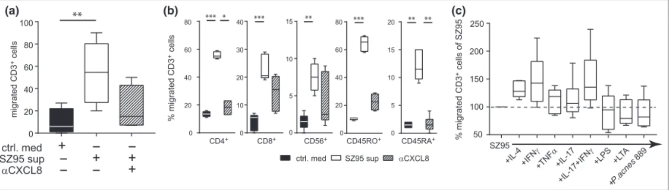
We next analysed whether sebocytes actively contribute to the recruitment of immune cells and especially T cells into the skin. Here we used cell-free supernatants from the human sebaceous gland cell line SZ95, which represents an accepted and widely used model in sebocyte research.15 In a first step, these supernatants were analysed for production of 27 cytoki- nes, chemokines and growth factors by Luminex technology (Luminex, Austin, TX, U.S.A.), revealing a robust secretion of chemokines such as CXCL8, CCL2, CCL5 and CXCL10 (Table S1; see Supporting Information). As these chemokines are important for granulocyte and leucocyte migration into tis- sues, we next analysed the migratory capacity of neutrophils, monocytes and T cells towards the SZ95 sebocyte supernatant.
Neutrophils, monocytes and T cells migrated towards the SZ95 sebocyte supernatant in a CXCL8-dependent manner (Fig. 2a; Fig. S2a; see Supporting Information). Among T cells, CD4+ and CD45RO+ effector T cells represented the main migratory subsets (Fig. 2b).
Next, we wanted to understand whether cultivation of SZ95 sebocytes in different proinflammatory environments alters the secretion of proteins and also the subsequent migration of cells. Therefore, SZ95 sebocytes were prestimu- lated with IL-4, interferon (IFN)-c, tumour necrosis factor (TNF)-a, IL-17, LPS, LTA andP. acnes. Whereas all conditions lead to increased secretion of CXCL8, CCL5 and CXCL10, IFN-c, IL-17, LPS and LTA were the predominant activators of sebocytes (Table S1). Whereas IFN-cstimulation of sebo- cytes seems to foster (not significantly) the migration of CD3+ T cells (Fig. 2c), with a significant migration of the CD45RO+ T-cell subset (Fig. S2c), LPS or LTA stimulation significantly induced migration of neutrophils and monocytes (Fig. S2b; see Supporting Information).
These data provide evidence that resting sebocytes can attract immune cells in vitro in a CXCL8-dependent manner, and that this chemoattractant effect is further raised in a proinflammatory environment.
Sebocytes do not influence CD4+CD45RO+effector T-cell cytokine secretion
With CD4+CD45RO+ being the largest T-cell subset that is attracted by sebocytes, we questioned whether their function is actively influenced by sebocytes. Therefore, human, blood- derived CD4+CD45RO+ cells were stimulated with plate- bound aCD3 and soluble aCD28 in the presence of SZ95 sebocyte supernatant or control medium for 72 h. Here, no significant alteration in the secretion of IL-17, IFN-c, TNF-a or IL-4 production compared with control medium could be detected. Only for IL-22 secretion was a significant induction detectable (Fig. S3a; see Supporting Information).
As, in this setting, T-cell-receptor stimulation alone might not be sufficient to induce alterations in cytokine secretion, we next investigated whether sebocytes trigger functional changes in T-cell activation via DCs. Therefore, CD14+mono- cytes were differentiated into DCs in the presence of SZ95 sebocyte supernatant or control medium and stimulated with LPS prior to coculture with allogeneic CD4+CD45RO+ cells (mixed leucocyte reaction). SZ95 supernatant did not impact
on DC maturation (Fig. S4; see Supporting Information) and also did not impact on T-cell activation, as no significant release of any of the cytokines analysed was detected (Fig. S3b).
Our data suggest that human sebocytes affect memory T-cell cytokine secretion neither directly nor mediated by DCs.
Sebocytes trigger a T helper-17 cell immune response
As naive T cells were also attracted in small numbers by sebo- cytes, we investigated the influence of sebocytes on T-cell dif- ferentiation. For this purpose, CD4+CD45RA+ T cells were stimulated withaCD3 andaCD28 in the presence of the SZ95 sebocyte supernatant or control medium. As also in our hands intracellular cytokine stainings of differentiated T cells did not work,16 secretion of effector cytokines was assessed by enzyme-linked immunosorbent assay. Whereas SZ95 sebocyte supernatant did not induce a Th1 or Th2 immune response, as no significant IFN-c, TNF-a and IL-4 release was detected in the supernatant of differentiated T cells, a significantly higher production of IL-22 and IL-17 was detected after Fig 1.CD4+interleukin (IL)-17+T cells
surround the pilosebaceous unit. Paraffin- embedded sections of patients with acne vulgaris were immunofluorescently stained for CD4 (green) and IL-17 (red). The nucleus was counterstained with DAPI (4ʹ,6- diamidino-2-phenylindole; blue).
Fluorescence images were obtained using an Olympus IX73 inverted fluorescence microscope equipped with cellSens software (Olympus, Center Valley, PA, U.S.A.) and processed with ImageJ software (https://imagej.
nih.gov/ij/). Shown is one representative staining for healthy (upper part) and lesional skin (lower part).
6 days in culture (Fig. 3a). In line with that, DCs that were generated in the presence of SZ95 sebocyte supernatant were also able to drive naive T-cell polarization towards the Th17 phenotype with significantly increased expression of IL-17 and IL-22 (Fig. 3b). Interestingly, IL-17 and IL-22 cytokine levels were even higher compared with the sole stimulation with aCD3 and aCD28, whereas levels of IFN-c and TNF-a were significantly reduced in this set-up.
Thus, our in vitrodata imply that human sebocytes have the capacity to skew immune responses towards a Th17 profile.
Sebocytes induce T helper-17 cell differentiation via secretion of interleukin-1b
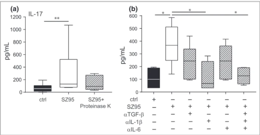
To address the contribution of lipids or proteins to the observed effect on Th17 differentiation, we incubated the SZ95 sebocyte supernatant with proteinase K. CD4+CD45RA+ naive T cells were stimulated withaCD3 andaCD28 and cul- tured in the presence of the protein-digested SZ95 sebocyte supernatant, resulting in a reduction of IL-17 secretion in the absence of the whole-protein fraction (Fig. 4a). This suggests that the sebocyte effect on naive T-cell polarization is primar- ily protein mediated. However, the effects of sebocytes pro- duced lipids, and interactions of lipids and proteins cannot be ruled out without further experiments.
As, in the steady state, SZ95 sebocytes secrete cytokines that are known to contribute to Th17 polarization, such as IL-1band IL-617(Table S1; see Supporting Information), we next neutral- ized these cytokines in SZ95 supernatant and performed a subse- quent naive T-cell differentiation. Neutralization of IL-1bled to a decrease of IL-17 secretion in differentiated T cells of 35%, whereas neutralization of TGF-band IL-6 alone had only mar- ginally effects. Conversely, the depletion of all three cytokines simultaneously abrogated IL-17 production by 44% (Fig. 4b).
Therefore, it is likely that sebocytes drive a Th17 immune response via the production of IL-6, TGF-band largely IL-1b.
Propionibacterium acnesdoes not influence immune cell recruitment, but affects the priming capacity of dendritic cells
To explore whetherP. acnessynergistically acts with sebocytes to reinforce the local symbiosis and/or immune response, SZ95 sebocytes were preincubated for 24 h withP. acnesson- icate 889, extensively washed to remove bacteria and further incubated for 24 h prior to supernatant collection. In migra- tion assays,P. acnesdid not alter the chemoattractant potential of sebocytes compared with SZ95 supernatant, as migration of neutrophils, monocytes or lymphocytes was not altered when sebocytes were preincubated with P. acnes (Fig. 2c;
Fig. S2b,c; see Supporting Information).
However, when DCs were generated in the presence of P.
acnes-prestimulated SZ95 supernatant and subsequently used for differentiation of allogeneic CD4+CD45RA+naive T cells, these T cells showed a slight, but not significant, increase in IL-17 and IL-22 production (Fig. 5). Interestingly, DCs matured with the SZ95 sebocyte supernatant (either unstimu- lated or prestimulated with P. acnes) induced a significant reduction of the Th1 cytokine IFN-c (P=003), whereas TNF-aand IL-4 levels were not significantly altered (Fig. 5).
These data indicate that sebocytes induce Th17 polarization, and P. acnes indirectly contributes to this phenomenon by inhibiting Th1 differentiation.
Discussion
Current research on human sebocytes has indicated that these cells are not only bystanders during skin inflammation, but also actively modulate immune responses via secretion of chemokines and cytokines.2,3,18In this study, we provide evi- dence of a functional communication between sebocytes and T cells resulting in the induction of a Th17-dominated immune response. On the other hand, we demonstrate an
(a) (b) (c)
Fig 2. SZ95 sebocytes induce the migration of T cells via secretion of CXCL8. (a) CD3+T cells migrated towards the SZ95 sebocyte supernatant after 2 h of incubation. Graphs show absolute numbers of migrated cells towards control medium, SZ95 sebocyte supernatant and CXCL8-depleted SZ95 sebocyte supernatant (n=3). (b) Flow cytometric analysis of CD3+cells. Migrated CD4+, CD8+, CD56+, CD45RO+and CD45RA+cells are expressed as the percentage of total migrated CD3+cells (n=3). (c) Flow cytometric analysis of migrated CD3+cells towards prestimulated SZ95 sebocyte supernatant expressed as the percentage of SZ95 migration. Statistical significance was determined using the Kruskal–Wallis test and Dunn’s multiple comparison test to correct for multiple testing, and expressed as*P<005; **P<001;***P<0001. IFN, interferon; LPS, lipopolysaccharide; LPA, lipoteichoic acid; TNF, tumour necrosis factor.
(a)
(b)
Fig 3.Sebocyte supernatant polarizes naive T cells into T helper-17 cells. (a) CD4+ CD45RA+naive T cells were cultured with SZ95 sebocyte supernatant or control medium and stimulated withaCD3 and aCD28. After 6 days, cells were restimulated for 72 h and supernatants analysed for levels of interleukin (IL)-17, IL-22, interferon (IFN)-c, IL-4 and tumour necrosis factor (TNF)-aby enzyme-linked immunosorbent assay (ELISA) (n=5). TCR, T-cell receptor. (b) Dendritic cells generated from monocytes in the presence of the SZ95 sebocyte supernatant and control medium were stimulated with lipopolysaccharide and cocultured with allogeneic CD4+CD45RA+ naive T cells. After 6 days, T cells were restimulated with aCD3 and aCD28 for 72 h and supernatants analysed by ELISA (n=3). Statistical significance was determined using the Wilcoxon matched-pairs signed-rank test and expressed as*P<005;**P<001.
(a) (b)
Fig 4.Sebocytes induce T helper-17 cell differentiation by release of key polarizing cytokines. (a) Proteins in SZ95 sebocyte supernatant were removed by digestion with proteinase K. CD4+CD45RA+naive T cells were cultured with the protein-depleted supernatant, the entire SZ95 sebocyte supernatant or control medium in the presence ofaCD3 andaCD28 antibodies for 6 days. Secretion of interleukin (IL)-17 was measured by enzyme-linked immunosorbent assay (ELISA) after restimulation withaCD3 andaCD28 antibodies (n=3). (b) SZ95 sebocyte supernatant was incubated for 1 h with transforming growth factor (TGF)-b, IL-1band IL-6 neutralizing antibodies either alone or in combination and used to differentiate CD4+CD45RA+naive T cells for 6 days. After restimulation, levels of IL-17 were measured in the supernatants by ELISA (n=2).
Statistical significance was determined using the Kruskal–Wallis test and Dunn’s multiple comparison test to correct for multiple testing and expressed as*P<005;**P<001.
indirect contribution ofP. acnesvia sebocytes and DCs towards acne-associated inflammation.
In the steady state, SZ95 sebocytes release several chemokines and cytokines. This is in line with previously published reports highlighting thein situproduction of, for example, CXCL8, IL-6 and IL-1bby sebocytes in the sebaceous gland,3,6and underlin- ing the importance of SZ95 sebocytes as anin vitro model for sebocyte research. Among the steady-state chemokines, CXCL8 has a key role in recruitment of immune cells such as neu- trophils and monocytes to sites of skin inflammation.
Although previous reports suggested that neutrophils are the first immune cells in acne lesions,19,20 some studies revealed that, along with macrophages, T lymphocytes also infiltrate sites of evolving inflammatory lesions.13,14 Indeed, we found that, in a CXCL8-mediated fashion, sebocytes recruit different subsets of T cells such as CD4+CD45RO+ effector, but also CD4+CD45RA+ naive, T cells to the skin. However, during inflammatory responses, sebocytes become further acti- vated by proinflammatory cytokines and/or bacterial products, leading to enhanced secretion of chemokines and cytokines. In line with that, prestimulated SZ95 sebocytes showed an increased chemoattractant potential on immune cells in vitro that mainly followed the concentration of CXCL8 and is reflectedin vivoby high numbers of immune cells surrounding the sebaceous gland in acne lesions.
Owing to the important role of T cells in the inflammatory tissue response, we investigated whether T cells are not only attracted to the sebaceous gland, but also influence their func- tion. We could demonstrate that factors released by sebocytes do not alter cytokine secretion of CD4+CD45RO+effector T cells, indicating that sebocytes do not impact on the previ- ously determined T-cell phenotype. One exception is a slight increase in IL-22 production, implying that sebocytes ensure barrier homeostasis by fostering the IL-22/TNF-aaxis.21
Unlike effector cells, we found that sebocytes impact on the differentiation of CD4+CD45RA+ naive T cells. Sebocytes secrete various cytokines, most importantly IL-6 and IL-1b, which represent the key cytokines forde novo differentiation of
Th17 cells.22,23 Because of this we could show that sebocyte supernatants alone are capable of fully inducing the Th17 phe- notype in naive T cells and that this interaction is dependent mainly on IL-1b production. As T-cell priming does not take place in peripheral tissues, we assume that sebocytes con- tribute to the generation of a local micromilieu that skews dif- ferentiation of naive T cells towards the Th17 phenotype in skin-draining lymph nodes.
The Th17 population bridges innate and adaptive immunity and has a key role in mediating host defence. Alone or in syn- ergy, the Th17 effector cytokines IL-17 and IL-22 induce an array of antimicrobial peptides to produce a robust antimicro- bial response.24–26 However, Th17 cells can also induce pathological inflammation and are associated with several inflammatory skin conditions such as psoriasis, atopic eczema and allergic contact dermatitis.27–29Moreover, a role for Th17 cells in acne pathogenesis has recently been described. Kelhala et al. showed enhanced expression of Th17-associated cytoki- nes and differentiation factors in lesional skin.30 In line with our data, Agaket al.showed that Th17 cells are present in the perifollicular infiltrate of comedones. However, and in con- trast to our findings, the authors hypothesized that the Th17 immune response is regulated mainly byP. acnes.9Similarly, a recent study showed thatP. acnesinduces a Th1/Th17 response even though acne pathogenesis has been associated with Th1- type immunity.31,32 Our data indicate that sebocytes induce differentiation of neither Th1 nor Th2 cells, but skew the immune response towards a Th17 profile that is further enhanced by the presence ofP. acnes.
Furthermore, it has been reported that P. acnes efficiently induces IL-1b secretion in sebocytes by activating the NLRP3 inflammasome.6 We could also detect an increase in IL-1b levels when sebocytes were pretreated withP. acnes; however, we did not find a further increase in Th17 differentiation, arguing for a quite high intrinsic production of IL-1b that is totally sufficient for Th17 priming, even in a resting state.
Our data argue for a steady-state induction of Th17 cells by sebocytes to maintain skin homeostasis. However, when the Fig 5. Dendritic cells (DCs) were generated in the presence of interleukin (IL)-4 and granulocyte–macrophage colony-stimulating factor and supernatant derived from SZ95 sebocytes that had been prestimulated withPropionibacterium acnesstrain 889. At day 5, DCs were stimulated with lipopolysaccharide and subsequently cocultured with CD4+CD45RA+naive T cells for 6 days. Differentiated T cells were restimulated withaCD3 andaCD28 antibodies prior to supernatant collection and analysis by enzyme-linked immunosorbent assay for IL-17, IL-22, interferon (IFN)-c, IL-4 and tumour necrosis factor (TNF)-alevels (n=3). Statistical significance was determined using the Kruskal–Wallis test and Dunn’s multiple comparison test to correct for multiple testing and expressed as*P<005.
pilosebaceous unit is colonized withP. acnesunder pathological conditions, the Th17 response is further enhanced in vivo. In line with this, we could detect CD4+IL-17+ cells only spar- sely around sebaceous glands in healthy individuals, whereas these cells were frequently colocalized with sebocytes in acne lesions.
Several in vitro studies show thatP. acnes whole cells or cell fractions stimulate cytokine release from immune cells, ker- atinocytes and sebocytes through binding to Toll-like receptor 2.2,33–35 However, the mechanism by whichP. acnesexerts its activityin vivo is still unknown. Propionibacterium acnescan reside in the deeper portions of sebaceous follicles,33 but rarely in the sebaceous gland.36 When this commensal bacterium pro- liferates, it can come into contact with DCs and activate their maturation, with the consequent immune response crucially depending on the presence of local commensals or pathogens, biofilm production and additional signals from tissue cells.37
It has been reported that DCs stimulated withP. acnesshow an increased expression of adhesive molecules and cytokines, which is similar to DCs activated by LPS.38,39In the presence of naive T cells,P. acnes-matured DCs induced a strong secre- tion of IFN-c that is comparable with LPS-matured DCs, confirming the capacity ofP. acnesin eliciting a powerful Th1- type immune response.40 However, the presence of sebocyte supernatant reduced the capability to induce Th1 responses and instead drove the symbiotic and/or immune response to P. acnes specifically towards a Th17 commitment. Taken together, we assume that sebocytes maintain the barrier integ- rity by: (i) homeostatic priming of Th17 cells; (ii) initiation of effective inflammatory responses; and (iii) reduction of pathogenic IFN-c production to reach homeostasis after inflammation.
The limitations of our study lie clearly in the in vitronature of the performed experiments and the use of a cell line. How- ever, the SZ95 sebaceous gland cell line is a widely accepted and used human sebocyte model that shows comparable beha- viour with other sebocyte lines and mostly with primary sebo- cytes that deliver only limited amounts of material.41 Furthermore, additionalin vivo mouse experiments are needed to characterize fully the contribution of sebocyte-induced Th17 cells to acne pathogenesis and, probably, other inflam- matory skin diseases.
Despite these limitations, our study provides evidence that sebocytes actively participate in inflammatory processes in the skin via recruitment of immune cells and a functional cross- talk with T cells leading to a pronounced Th17 differentiation.
This interaction might be of importance for the pathogenesis of acne vulgaris; however, further studies have to clarify whether the sebocyte–Th17 axis contributes to a beneficial host defence or the perpetuation of a vicious circle of inflam- mation.
Acknowledgments
The authors thank Jana S€anger, ZAUM – Center for Allergy and Environment, for excellent technical support. Technical
support in culturing P. acnes was kindly provided by Judit Szabo, Department of Medical Microbiology, University of Debrecen, Hungary.
References
1 Hong I, Lee MH, Na TY et al. LXRaenhances lipid synthesis in SZ95 sebocytes.J Invest Dermatol2008;128:1266–72.
2 Nagy I, Pivarcsi A, Kis Ket al. Propionibacterium acnesand lipopolysac- charide induce the expression of antimicrobial peptides and proin- flammatory cytokines/chemokines in human sebocytes. Microbes Infect2006;8:2195–205.
3 Alestas T, Ganceviciene R, Fimmel Set al.Enzymes involved in the biosynthesis of leukotriene B4 and prostaglandin E2 are active in sebaceous glands.J Mol Med (Berl)2006;84:75–87.
4 Bohm M, Schiller M, Stander S et al. Evidence for expression of melanocortin-1 receptor in human sebocytes in vitro andin situ.J Invest Dermatol2002;118:533–9.
5 Webster GF. Acne vulgaris.BMJ2002;325:475–9.
6 Li ZJ, Choi DK, Sohn KC et al. Propionibacterium acnes activates the NLRP3 inflammasome in human sebocytes. J Invest Dermatol 2014;
134:2747–56.
7 Zouboulis CC. Is acne vulgaris a genuine inflammatory disease?
Dermatology2001;203:277–9.
8 Jappe U, Ingham E, Henwood J et al. Propionibacterium acnes and inflammation in acne; P. acneshas T-cell mitogenic activity. Br J Dermatol2002;146:202–9.
9 Agak GW, Qin M, Nobe Jet al. Propionibacterium acnesinduces an IL- 17 response in acne vulgaris that is regulated by vitamin A and vitamin D.J Invest Dermatol2014;134:366–73.
10 Zouboulis CC, Eady A, Philpott Met al. What is the pathogenesis of acne?Exp Dermatol2005;14:143–52.
11 Ottaviani M, Camera E, Picardo M. Lipid mediators in acne.Media- tors Inflamm2010;2010:858176.
12 Zouboulis CC. Acne and sebaceous gland function. Clin Dermatol 2004;22:360–6.
13 Jeremy AH, Holland DB, Roberts SG et al. Inflammatory events are involved in acne lesion initiation. J Invest Dermatol 2003;
121:20–7.
14 Layton AM, Morris C, Cunliffe WJ et al. Immunohistochemical investigation of evolving inflammation in lesions of acne vulgaris.
Exp Dermatol1998;7:191–7.
15 Zouboulis CC, Seltmann H, Neitzel Het al.Establishment and char- acterization of an immortalized human sebaceous gland cell line (SZ95).J Invest Dermatol1999;113:1011–20.
16 Flores-Borja F, Bosma A, Ng Det al.CD19+CD24hiCD38hiB cells maintain regulatory T cells while limiting TH1 and TH17 differen- tiation.Sci Transl Med2013;5:173ra23.
17 Manel N, Unutmaz D, Littman DR. The differentiation of human TH-17 cells requires transforming growth factor-band induction of the nuclear receptor RORct.Nat Immunol2008;9:641–9.
18 Koreck A, Pivarcsi A, Dobozy Aet al.The role of innate immunity in the pathogenesis of acne.Dermatology2003;206:96–105.
19 Webster GF. Inflammation in acne vulgaris. J Am Acad Dermatol 1995;33:247–53.
20 Adisen E, Yuksek J, Erdem Oet al.Expression of human neutrophil proteins in acne vulgaris.J Eur Acad Dermatol Venereol2010;24:32–7.
21 Eyerich S, Wagener J, Wenzel Vet al.IL-22 and TNF-arepresent a key cytokine combination for epidermal integrity during infection withCandida albicans.Eur J Immunol2011;41:1894–901.
22 Veldhoen M, Hocking RJ, Atkins CJ et al.TGFbin the context of an inflammatory cytokine milieu supportsde novodifferentiation of IL-17-producing T cells.Immunity2006;24:179–89.
23 Wilson NJ, Boniface K, Chan JRet al.Development, cytokine pro- file and function of human interleukin 17-producing helper T cells.Nat Immunol2007;8:950–7.
24 Aujla SJ, Chan YR, Zheng M et al. IL-22 mediates mucosal host defense against Gram-negative bacterial pneumonia.Nat Med2008;
14:275–81.
25 Liang SC, Tan XY, Luxenberg DPet al.Interleukin (IL)-22 and IL- 17 are coexpressed by Th17 cells and cooperatively enhance expression of antimicrobial peptides.J Exp Med2006;203:2271–9.
26 Kao CY, Chen Y, Thai P et al. IL-17 markedly up-regulates b- defensin-2 expression in human airway epithelium via JAK and NF-jB signaling pathways.J Immunol2004;173:3482–91.
27 Guttman-Yassky E, Lowes MA, Fuentes-Duculan Jet al.Low expres- sion of the IL-23/Th17 pathway in atopic dermatitis compared to psoriasis.J Immunol2008;181:7420–7.
28 Pennino D, Eyerich K, Scarponi Cet al.IL-17 amplifies human con- tact hypersensitivity by licensing hapten nonspecific Th1 cells to kill autologous keratinocytes.J Immunol2010;184:4880–8.
29 Fitch E, Harper E, Skorcheva I et al.Pathophysiology of psoriasis:
recent advances on IL-23 and Th17 cytokines. Curr Rheumatol Rep 2007;9:461–7.
30 Kelhala HL, Palatsi R, Fyhrquist N et al. IL-17/Th17 pathway is activated in acne lesions.PLoS ONE2014;9:e105238.
31 Kistowska M, Meier B, Proust Tet al. Propionibacterium acnespromotes Th17 and Th17/Th1 responses in acne patients. J Invest Dermatol 2015;135:110–18.
32 Mouser PE, Baker BS, Seaton EDet al. Propionibacterium acnes-reactive T helper-1 cells in the skin of patients with acne vulgaris.J Invest Dermatol2003;121:1226–8.
33 Kim J, Ochoa MT, Krutzik SRet al.Activation of toll-like receptor 2 in acne triggers inflammatory cytokine responses. J Immunol 2002;169:1535–41.
34 Liu PT, Krutzik SR, Kim Jet al.Cutting edge: all-transretinoic acid down-regulates TLR2 expression and function. J Immunol 2005;
174:2467–70.
35 Lee SE, Kim JM, Jeong SKet al.Protease-activated receptor-2 medi- ates the expression of inflammatory cytokines, antimicrobial pep- tides, and matrix metalloproteinases in keratinocytes in response toPropionibacterium acnes.Arch Dermatol Res2010;302:745–56.
36 Alexeyev OA, Lundskog B, Ganceviciene Ret al. Pattern of tissue invasion by Propionibacterium acnes in acne vulgaris. J Dermatol Sci 2012;67:63–6.
37 Smits HH, van Beelen AJ, Hessle Cet al.Commensal Gram-negative bacteria prime human dendritic cells for enhanced IL-23 and IL-27 expression and enhanced Th1 development. Eur J Immunol 2004;
34:1371–80.
38 Michalak-Stoma A, Tabarkiewicz J, Olender Aet al. The effect of Propionibacterium acneson maturation of dendritic cells derived from acne patients’ peripheral blood mononuclear cells. Folia Histochem Cyto2008;46:535–9.
39 Verhasselt V, Buelens C, Willems Fet al. Bacterial lipopolysaccha- ride stimulates the production of cytokines and the expression of costimulatory molecules by human peripheral blood dendritic cells – evidence for a soluble CD14-dependent pathway. J Immunol 1997;158:2919–25.
40 Kitagawa H, Yamanaka K, Kakeda Met al. Propionibacterium acnesvac- cination induces regulatory T cells and Th1 immune responses and improves mouse atopic dermatitis.Exp Dermatol2011;20:157–8.
41 Xia L, Zouboulis CC, Ju Q. Culture of human sebocytesin vitro.Der- matoendocrinol2009;1:92–5.
Supporting Information
Additional Supporting Information may be found in the online version of this article at the publisher’s website:
Fig S1. Immunofluorescence isotype controls.
Fig S2. Neutrophils and monocytes migrate towards SZ95 supernatant.
Fig S3. CD4+CD45RO+ effector T-cell function is not altered by SZ95 supernatant.
Fig S4.SZ95 supernatant does not affect dendritic cell mat- uration.
Table S1Bio-Plex results of chemokines and cytokines ana- lyzed in SZ95 supernatant after 24 h of culture.
Appendix S1Supplementary methods.
Powerpoint S1Journal Club Slide Set.