The complex role of N-cadherin and endocannabinoid signaling during cortical development
Ph.D. thesis
Zsófia László
Semmelweis University
János Szentágothai Doctoral School of Neurosciences
Supervisor: Zsolt Lele, Ph.D.
Official Reviewers of the Ph.D.
Dissertation: Tamás Bíró, M.D., D.Sc.
István Adorján, M.D., Ph.D.
Head of the Final Examination
Committee: András Csillag, M.D., D.Sc.
Members of the Final Examination
Committee: Krisztina Herberth-Minkó, Ph.D.
Krisztián Tárnok, Ph.D.
Budapest
2019
2
Table of contents
List of abbreviations ... 6
1. Introduction ... 10
1.1. Generation of excitatory neurons in the pallium ... 11
1.1.1. Neuroepithelial stage (E8-10) ... 11
1.1.2. Main neurogenic phase (E11-E16) ... 11
1.1.2.1. Apical radial glia progenitor cells ... 11
1.1.2.2. Basal progenitor cells ... 13
1.1.3. Gliogenic proliferative stage (E17-P3) ... 13
1.2. Transcriptional regulation of proliferation and differentiation in the pallium ... 14
1.3. Glutamatergic cell migration ... 15
1.3.1. Delamination and leaving the VZ ... 16
1.3.2. Morphological transitions during radial migration ... 16
1.3.3. Glial-guided locomotion ... 17
1.3.4. Terminal translocation ... 18
1.4. Progenitor pool in the subpallium, generation of GABAergic cells ... 18
1.5. Interneuron migration to the cerebral cortex ... 22
1.5.1. Tangential migration ... 22
1.5.2. Cortical invasion ... 24
1.5.3. Laminar allocation... 25
1.6. Developmental cell death during cortical development... 26
1.7. Cadherin superfamily ... 27
1.7.1. Cadherin subfamilies ... 28
1.7.2. Cadherin-binding proteins ... 29
1.8. N-cadherin function during embryonic cortical development ... 31
1.8.1. N-cadherin in the neuroepithelium and in progenitor pools ... 31
1.8.2. N-cadherin as a regulator of cell fate commitment ... 32
1.8.3. N-cadherin role during cell migration ... 32
1.9. The endocannabinoid system ... 34
3
1.9.1. Endocannabinoid mediated retrograde neuronal transmission... 34
1.9.2. The function of the endocannabinoid system during cortical development... 36
1.9.2.1 Endocannabinoid function during proliferation and differentiation ... 36
1.9.2.2 Endocannabinoid signaling in axonal guidance and behavior... 37
1.9.3. The medical relevance of cannabinoids ... 38
2. Aims ... 39
3. Material and Methods ... 40
3.1. Animals ... 40
3.2. Genomic DNA extraction and genotyping ... 40
3.3. Sample preparation and sectioning ... 40
3.4. DNA constructs and cloning protocols ... 41
3.5. In vitro transcription and in situ hybridization ... 42
3.6. In utero electroporation ... 44
3.7. Fluorescent single-cell mRNA detection (RNAscope) ... 44
3.8. Immunohistochemistry ... 45
3.9. Cell death detection - TUNEL assay ... 45
3.10. BrdU (5-bromo-2'-deoxyuridine) treatment and staining ... 46
3.11. In vitro studies ... 46
3.12. Western blot ... 48
3.13. Maternal alcohol consumption model ... 49
3.14. Phylogenetic tree ... 49
3.15. Image acquisition and editing ... 50
3.16. Image analysis ... 50
3.17. Statistical analysis ... 52
3.18. Personal contributions for the results ... 52
4. Results ... 54
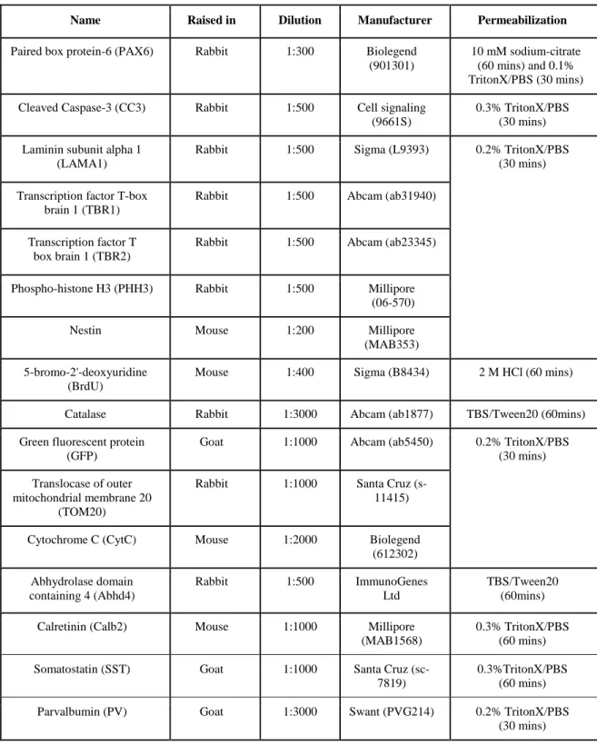
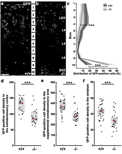
4.1. The role of N-cadherin (Cdh2) during tangential migration and interneuron differentiation in the somatosensory cortex ... 54
4.1.1. Lack of Cdh2 in postmitotic interneurons effects their tangential migration but not proliferation in the ganglionic eminences ... 54
4.1.2. Gad65-GFP-positive cell number is changed in the triple transgenic adult brain ... 56
4
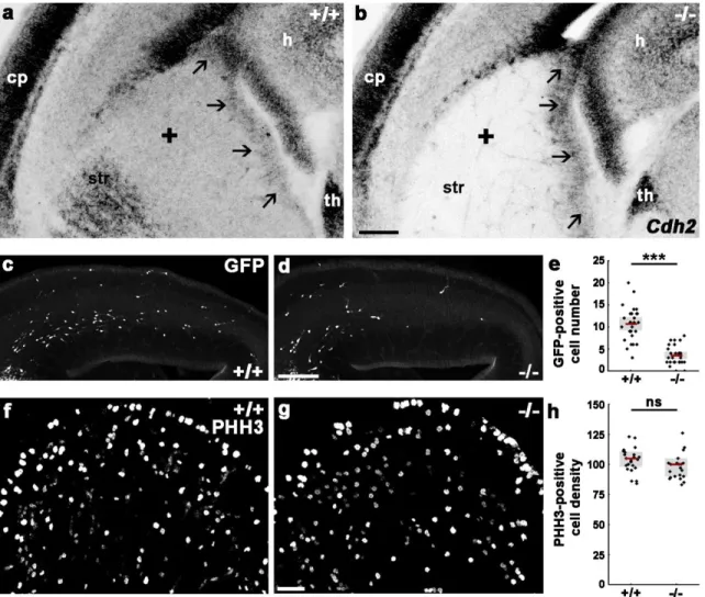
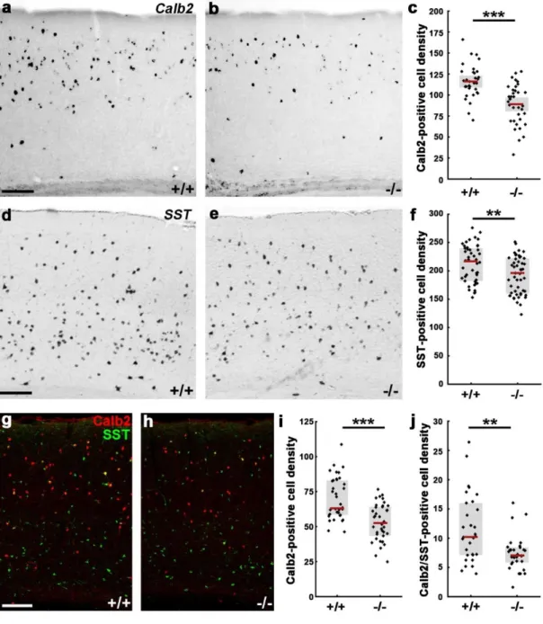
4.1.3. N-cadherin regulates interneuron composition of the adult primer somatosensory
cortex ... 56
4.1.4. Disruption of N-cadherin signaling in postmitotic cells causes a migration delay in the somatosensory cortex ... 61
4.1.5. The fate commitment of the arrested cells in the postnatal SVZ ... 63
4.2. The consequences of abnormal delamination in the developing mouse cortex ... 65
4.2.1. In vivo cadherin-based adherens junction disruption model ... 65
4.2.2. Pathophysiological delamination causes apoptosis and migration defect in the embryonic dorsal telencephalon ... 68
4.3. Investigation of the pathological delamination-evoked cell death mechanism ... 70
4.3.1. The identification of potential molecular players in cadherin-loss induced apoptosis ... 70
4.3.2. Characterization of Abhd4 knockout animals ... 72
4.3.3. Abhd4 is sufficient to trigger caspase-dependent cell death ... 77
4.3.4. The mechanism of Abhd4-induced cell death ... 81
4.3.5. Abhd4 is necessary for loss-of adherens junction-induced cell death ... 83
4.3.6. Fetal alcohol exposure induces Abhd4-dependent cell death in the embryonic cortex ... 88
5. Discussion ... 90
5.1. The role of N-cadherin during cortical development ... 90
5.1.1. Cdh2 as a regulator of proliferation and differentiation ... 90
5.1.2. The role of N-cadherin in the postmitotic phase of neuronal development ... 90
5.1.3. The long-term effects of N-cadherin elimination ... 91
5.2. The role of N-cadherin in cellular survival ... 94
5.2.1. Mechanism of the anti-apoptotic effect of N-cadherin-based connections ... 94
5.2.2. The concept of developmental anoikis ... 94
5.2.3. Outlook: The functional impact of N-cadherin research in brain disease ... 95
5. 3. Abhd4 function in normal and pathophysiological development of the cortex ... 97
5.3.1. Abhd4 as a pro-apoptotic protein ... 97
5.3.2. The potential mechanism and medical aspects of delamination-induced cell death ... 99
6. Conclusions ... 103
7. Summary ... 104
5
8. Összefoglalás ... 105
9. References ... 106
10. List of publications ... 135
11. Acknowledgements ... 136
6
List of abbreviations
2-AG 2-arachydonoyl-glicerol
Abhd4 Abhydrolase domain containing 4
AEA Anandamide
AJ Adherens juncton
aRGPC Apical radial glia progenitor cell
bRGPC Basal radial glia progenitor cell
ATGL Adipose triglyceride lipase
bIP Basal intermediate progenitor cell
BP Basal progenitor cell
BrdU 5-bromo-2'-deoxyuridine
Calb2 Calretinin
CBR1 Cannabinoid receptor 1/Cnr1
CC3 Cleaved Caspase-3
CCK Cholecistokinin
Cdh2 Cadherin-2
CGE Caudal ganglionic eminence
ChR2 Channelrhodopsin-2
CP Cortical plate
CR Cajal-Retzius cell
CytC Cytochrome C
DAPI 4′,6-diamidino-2-phenylindole
DGL Diacylglycerol-lipase
Dlx5/6 Distal-less homeobox 5/6
ΔnCdh2 Dominant-negative cadherin-2
ECM Extracellular matrix
ER Endoplasmic reticulum
7
ERK Extracellular-signal regulated kinase
EtOH Ethanol
FAAH Fatty acid amide hydrolase
FWHM Full width at half maximum
GABA γ-aminobutyric acid
GAD65 Glutamic acid decarboxylase 65
GE Ganglionic eminence
GFP Green fluorescent protein
Glast1 Glutamate Aspartate Transporter 1
HEK-293 Human embryonic kidney 293 cells
IN Interneuron
IZ Intermediate zone
KO Knockout (-/-)
LAMA1 Laminin subunit alpha 1
LGE Lateral ganglionic eminence
MGE Medial ganglionic eminence
MGL Monoacylglycerol lipase
MP Multipolar cell
mTOR Mammalian target of rapamycin
MZ Marginal zone
NAE N-acylethanolamine
NAPE N-acyl phosphatidylethanolamine
NAPE-PLD N-acyl phosphatidylethanolamine phospholipase D
NGN2 Neurogenin 2
NLP Number of localization point
NSC Neuronal stem cell
NPY Neuropeptide Y
PAX6 Paired box protein-6
8
PBS Phosphate-buffered saline/ buffer solution
PCR Polymerase chain reaction
PHH3 Phospho-histone H3
PI3K Phosphoinositide 3-kinase
PLA2 Phospholipase A2
PLIN1 Perilipin 1
PSB Pallial-subpallial boundary
PtdSer Phosphatidylserine
PV Parvalbumin
Reln Reelin
RGPC Radial glia progenitor cell
ROI Region of interest
SSC Saline-sodium citrate buffer
SST Somatostatin
STORM Stochastic Optical Reconstruction Microscopy
SVZ Subventricular zone
TAG Triacylglycerol
TBR1 T-box brain protein 1
TBR2 T-box brain protein 2/Eomes
TBS Tris-buffered saline/ buffer solution
THC ∆9-tetrahydrocannabinol
TOM20 Translocase Of Outer Mitochondrial Membrane 20
TRPV1 Transient receptor potential vanilloid receptor 1
TUNEL Terminal deoxynucleotidyl transferase dUTP nick end
labeling
vGlut1 Vesicular glutamate transporter 1
VIP Vasoactive intestinal peptide
VZ Ventricular zone
WT Wild type (+/+)
9
ZIKV Zika virus
Z-VAD-FMK Benzyloxycarbonyl-Val-Ala-Asp(OMe)
fluoromethylketoneclose
10
1. Introduction
The cerebral cortex is the most complex structure in the mammalian brain. During evolution, cortical expansion allowed humans to become a conscious and one of the most versatile species in the world (Van Essen et al., 2018). Naturally, these attributes require the balanced and coordinated activity of excitatory and inhibitory neurons which in turn depend on the proper development of these cells. At the beginning of brain development, the telencephalon is dorso-ventrally divided into two parts, named the pallium and subpallium, respectively (Figure 1). More than 30 years ago scientists were convinced that both excitatory pyramidal neurons and inhibitory interneurons (INs) are originated from the pallium (Rakic, 1988). With the advancement of technology it has been shown, that these two major cell types of the cortex are actually born in distinct germinative niches of the telencephalon (Anderson et al., 1997). Excitatory projection neurons or pyramidal cells of the cortex use glutamate as neurotransmitter and are born in the progenitor pools of the pallium. In contrast, inhibitory interneurons that synthesize GABA (γ-aminobutyric acid) as their main neurotransmitter invade the cortex from the subpallial ganglionic eminences (GE; Marín and Rubenstein, 2003).
The developing embryonic cortex has a multi-layered structure each featuring their own distinct cell types (Figure 1; Buchsbaum and Cappello, 2019). The most apical layer of the pallium is called ventricular zone (VZ) consists of dividing progenitors and newborn daughter cells (Götz and Barde, 2005). Above the VZ, there is another germinative layer called the subventricular zone (SVZ) which is responsible for the generation of both deep (neurons in the layer 5 and 6) and upper layer neurons (layer 2-4; Hevner, 2019). After their birth, newborn neuroblasts start their radial migration by using the elongated fibers of radial glial cells (Hatanaka et al., 2016). They travel through the intermediate zone (IZ), which at later embryonic stages contains a dense neuropil of thalamocortical axons and acts as a barrier between proliferative zones and the forming cortex (Iwashita et al., 2014). The IZ is followed by a transient layer, called subplate (SP), which contains the earliest generated neurons (Hoerder-Suabedissen and Molnár, 2015). Next, pyramidal cells enter the cortical plate (CP), translocate to distinct places and begin their differentiation (Ayala et al., 2007).
11
Finally, the uppermost layer of the embryonic cortex is the marginal zone (MZ) where Cajal- Retzius cells (CRs) are settled and regulate the final phase of pyramidal cell migration (Gupta et al., 2002). Pyramidal cells are stopped just beneath the MZ and the next generation of excitatory neurons will continuously bypass them in an inside-out manner, hence deep-layer neuron generation is followed by the production of upper-layer neurons (Huang, 2009). In contrast, subpallial interneuron precursors migrate tangentially from the subpallium (Figure 1) and invade the cortex via distinct migratory routes, either through the subventricular or the marginal zone (Marín, 2013). Then, using the radial glia scaffold interneurons migrate radially to their destined places and undergo final differentiation which is strongly influenced by their synaptic interactions with pyramidal cells (Wong et al., 2018).
1.1. Generation of excitatory neurons in the pallium 1.1.1. Neuroepithelial stage (E8-10)
After neural tube closure, the developing neocortex consists of neuronal stem cells (NSCs) forming a single-layer thick neuroepithelium (Johansson et al., 2010). They have apico-basal polarity and are tethered to the pial and the ventricular surface (Arai and Taverna, 2017). At the apical side of the cortex, NSCs are bound to each other via classic cadherin- based adherens junctons (AJs) which together form the adherens junction belt (Chen et al., 2013). NSCs are multipotent and divide symmetrically in order to self-renew and enlarge the stem cell pool of the ventricular zone (Yamaguchi and Miura, 2013). During symmetrical division cells inherit a complete cellular architecture including their apical cadherin-based adherens junctions (Huttner and Kosodo, 2005).
1.1.2. Main neurogenic phase (E11-E16)
1.1.2.1. Apical radial glia progenitor cells
Around embryonic day 10-11 in mice, NSCs start to acquire some astroglial properties (Malatesta et al., 2003) such as the expression of Glast1 (Glutamate Aspartate Transporter 1) and GFAP (Glial fibrillary acidic protein) and become apical radial glia progenitor cells (aRGPCs).
12
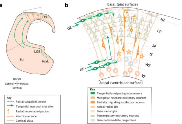
Figure 1. Distinct cell types and migration routes during cortical development
(a) Schematic figure of anterior telencephalon visualizes the distinct migration routes of inhibitory (green arrows) and excitatory neurons (orange arrows). Grey trapezoid is magnified in panel b. (b) Schematic representation of the cortical cellular organization on embryonic day 14. Projection neurons are migrating radially (dark orange cells) by using the radial glia scaffold (elongated cell with light orange color). Interneurons (green cells) are coming from the subpallial ganglionic eminencies via tangential migration and invade the cortical layers radially through the SVZ and CP migration routes. MGE medial ganglionic eminence; LGE: lateral ganglionic eminence, Str: striatum; Ctx: cortex; GE: ganglionic eminence; VZ: ventricular zone, SVZ: subventricular zone, IZ: intermediate zone; SP:
subplate, CP: cortical plate; MZ: marginal zone. Figure was adapted from Buchsbaum and Cappello, 2019.
Despite this, they still preserve NSC attributions such as apico-basal polarity (Götz and Huttner, 2005) albeit with a unique bipolar morphology and they attach to the pial surface with their elongated radial fiber (Rakic, 1972). In the neurogenic phase of cortical development between embryonic day 11 to 17, most aRGPCs divide asymmetrically and produce one radial glia progenitor and a basal progenitor cell (BP) or a neuron (Götz and
13
Huttner, 2005; Noctor et al., 2004; Tamamaki et al., 2001). During asymmetric division the apical ciliary membrane together with the mother centriole and some important regulatory molecules (eg. Par3) are also inherited asymmetrically, which actually determine the fate of daughter cells (Kosodo et al., 2004; Wang et al., 2009).
1.1.2.2. Basal progenitor cells
Two types of basal progenitor cells exist: the basal intermediate progenitor cells (bIPs), which are the most abundant BPs in the rodent embryonic cortex and the basal radial glia progenitor cells (bRGPCs), which are more abundant in primates than in rodents. bIPS are fate-restricted, symmetrically dividing, transient amplifying cells and produce most of the pyramidal cells (Haubensak et al., 2004; Kowalczyk et al., 2009; Vasistha et al., 2015). They do not have a polarized cell body or attachments to the cortical surfaces (Arai and Taverna, 2017), but they have neurites and filopodia which are important to guide the radially or tangentially migrating cells in the SVZ (Hevner, 2019; Kowalczyk et al., 2009). In contrast, bRGPCs are located at the VZ-SVZ border and have unipolar morphology with a basal fiber connecting to the pial surface (Miyata et al., 2001). They have similar proliferative potential as bIPS and responsible for the majority of neuronal production in primates (Ostrem et al., 2017). In contrast to rodents, human SVZ is thick and split into the inner and outer SVZ (iSVZ and oSVZ, respectively, Miller et al., 2019). The oSVZ contains mostly bRGPCs, also called outer radial glia-like cells in humans (Miller et al., 2019). They divide asymmetrically to increase the progenitor pool in the oSVZ (Hansen et al., 2010).
1.1.3. Gliogenic proliferative stage (E17-P3)
At the end of neurogenesis around embryonic day 17.5, the ventricular zone disappears and the SVZ takes over its proliferative function. Progenitor cells in the SVZ mainly produce astrocytes and oligodendrocytes until early postnatal stages (Murao et al., 2016). Recently, it was reported that these progenitor cells are actually present at the beginning of cortical development (around 9.5) but become dormant during the neurogenic phase and get reactivated postnatally (Furutachi et al., 2015).
14
1.2. Transcriptional regulation of proliferation and differentiation in the pallium
Cell fate is mostly genetically determined and gets directed by sequential waves of transcription factors (Telley et al., 2016). The fluctuation of Notch signaling is one of the key pathways to maintain the NSC self-renewal (Kageyama et al., 2008; Shimojo et al., 2008).
Activation of Notch receptor by a single-pass transmembrane protein such as Deltalike-1 (Dll1) instructs progenitor cells to remain in neuronal stem cell state (Andersson et al., 2011;
Yoon et al., 2008). Consequently, the expression of Hes1, a Notch-target member of the basic helix-loop-helix (bHLH) transcription factor family, is oscillating in parallel with Notch signaling (Shimojo et al., 2008; Telley et al., 2016). Hes1 is a stem cell marker and inhibits neurogenic commitment via repressing the proneural gene neurogenin 2 (Ngn2) and Dll1 (Shimojo et al., 2008). The fate of the neighbor cell is changed towards differentiation due to repression of Dll-Notch signaling generated lateral inhibition, creating a salt-and-pepper pattern-like distribution of progenitors and differentiating cells (Ohtaka-Maruyama and Okado, 2015).
After the asymmetric division, other transcriptional waves control the further fate of the daughter cells. One example is the sequential expression of PAX6, TBR2 and TBR1 which determine the aRGPCs to BP to differentiated pyramidal cell process (Englund et al., 2005;
Hevner, 2019). Paired box protein 6 (PAX6) expression is restricted to RGPCs and this factor is an important regulator of cell division (Asami et al., 2011; Götz et al., 1998) but it is also able to activate the expression of the basal progenitor cell marker, Eomesodermin or T-box brain protein 2 (TBR2) in a dose-dependent manner (Quinn et al., 2007; Sansom et al., 2009).
TBR2 is a transcriptional repressor responsible for BP production, specification and necessary for pyramidal cell differentiation (Bulfone et al., 1999; Englund et al., 2005;
Mihalas et al., 2016). However, TBR2 also functions as a negative feedback signal as it directly downregulates the expression of PAX6 (Elsen et al., 2018). Finally, T-box brain protein 1 (TBR1) is expressed by postmitotic neurons in the cortical plate (Bulfone et al., 1995) and its activation is regulated by TBR2 (Englund et al., 2005). TBR1 function is related
15
to the differentiation of pyramidal cells and it regulates several genes partaking in this process (Bedogni et al., 2010).
1.3. Glutamatergic cell migration
At the beginning of cortical development, the first wave of excitatory cells forms the so- called preplate above the VZ. These early born neurons utilize their bipolar morphology and migrate to the MZ by somal translocation (Hirota and Nakajima, 2017). Additionally, another cell type appears in the embryonic MZ, the Cajal-Retzius cells (CRs). In rodents between E10.5 and E12.5, glutamatergic CRs tangentially invade the marginal zone from multiple extra-neocortical sources in a complementary manner (Barber and Pierani, 2016) including the pallial septum, the developing hippocampus (cortical hem; Griveau et al., 2010; Yoshida et al., 2006) and the pallium-subpallium border (Bielle et al., 2005; Griveau et al., 2010). The main function of CR cells is to coordinate radial and tangential migration events. They are transient in nature and get eliminated by programmed cell death during postnatal development (Chowdhury et al., 2010).
Excitatory projection neurons find their final position in the cortical network due to the tightly regulated radial migration process from the VZ to the CP (Rakic, 1972, 1988;
Shoukimas and Hinds, 1978). Four subsequent phases of pyramidal cell migration have been described:
1. After asymmetric division daughter cells are delaminated and migrate to the VZ-SVZ border (Shoukimas and Hinds, 1978).
2. Here, postmitotic cells become multipolar and remain for approximately 24 hours, then undergo multipolar-bipolar transition before leaving towards the cortical plate (Nadarajah et al., 2001; Tabata and Nakajima, 2003).
3. Multipolar cells establish their leading and trailing processes (De Anda et al., 2010;
Hatanaka et al., 2004; Namba et al., 2014) and migrate to the CP guided by the radial glia scaffold (O’Rourke et al., 1992).
4. Once in the cortical plate the final phase of migration is carried out by terminal translocation (Franco et al., 2011; Noctor et al., 2004).
16
1.3.1. Delamination and leaving the VZ
To start the migration process, daughter cells need to lose their inherited apical and basal connections and delaminate from the mother cell. They achieve this by downregulating the molecular complex of the adherens junction belt containing Cdh1 and Cdh2 (E- and N- cadherin, respectively; Itoh et al., 2013; Rogers et al., 2018; Rousso et al., 2012). In parallel with neural differentiation, Ngn2 upregulates the levels of Scratch 1 and Scratch 2 transcription factors of the Snail family which induce cell movement via the repression of E- cadherin (Itoh et al., 2013). In addition, it was shown, that repression of Numb proteins, regulators of Notch signaling (Berdnik et al., 2002), also decreases the level of both E- and N-cadherins and causes delamination in parallel with differentiation (Rasin et al., 2007).
Loss of the cell adhesion complexes leads to a change in polarity along with the reorganization of the whole cytoarchitecture (Das and Storey, 2014). Shh (Sonic Hedgehog) signaling via primary cilium and the activation of Ngn2 together regulate the apical fiber abscission and cilium disassembly in daughter cells (Das and Storey, 2014). Meanwhile, intracellular actin dynamics are regulated by the Rho family of GTPases (Ridley, 2015). This family contains three members: Cdc42, Rac1 and RhoA, which are coordinating the connection between cytoskeleton and cell adhesion via cycling between their active (guanosine triphosphate – GTP) and passive (guanosine diphosphate) phases (Hodge and Ridley, 2016).
1.3.2. Morphological transitions during radial migration
During radial migration cell shape and the movement direction can rapidly change. After a short migration from the VZ cells arrive to the SVZ and become multipolar with several neurites extending in every direction (Tabata and Nakajima, 2003). Multipolar cells (MP) can migrate laterally which is independent from the radial glia scaffold (Tabata and Nakajima, 2003). Horizontal movement of MPs is controlled by cell-surface binding ephrin signaling (Dimidschstein et al., 2013; Torii et al., 2009) in particular EfnA (Ephrin-A) ligands and EphA (Ephrin Type-A Receptor) receptors which are both expressed in the SVZ/IZ in a spatial gradient.
17
It has been reported that two distinct postmitotic populations of daughter cells travel through the SVZ in a different manner. After division, postmitotic neurons accumulate at the apical part of the SVZ while basal progenitor cells immediately migrate to the basal side of the SVZ, where they transform to multipolar cells and undergo further proliferation (Tabata et al., 2009). MP to BP transition is established by transcriptional activation of small GTPases. Activation of Rac1 (Ras-related C3 botulinum toxin substrate 1) promotes the exit from the multipolar stage (Kawauchi et al., 2003) and its interaction with several scaffold proteins properly localizes the activated Rac1 at the basal part of the leading process (Yang et al., 2012). Cdc42 on the other hand, is mainly localized next to the centrosome and is connected to the microtubule organizing center (MTOC) via microtubules. Cdc42 moderates actin dynamics via MTOC, thereby identifying the direction of migration (De Anda et al., 2010; Konno et al., 2005). Rnd2 (Rho-related GTP-binding protein RhoN) is a unique member of the RhoA GTPase family as it lacks intrinsic GTPase activity and regulates neurite extension by the repression of RhoA signaling (Xu et al., 2019). Ngn2 directly activates Rnd2, therefore promotes the MP-BP transition (Heng et al., 2008). Accordingly, modulation of microtubule dynamics by small GTPases is also essential in the elongation of migrating cells, they determine the basal (future dendrite) and apical (future axon) processes (De Anda et al., 2010; Konno et al., 2005; Namba et al., 2014).
1.3.3. Glial-guided locomotion
After MP-BP transition, bipolar cells follow their route to the CP in a glia fiber-guided manner (Noctor et al., 2001; Tabata and Nakajima, 2003). This phenomenon operates coordinated adhesions between the scaffold and the bipolar cell, alternatively utilizing contractile and pulling activity (Dogterom and Koenderink, 2019). Saltatory movement of bipolar cells is achieved via a cyclic two-step process: the centrosome moves forward in the direction of the leading process then the nucleus follows (Tsai et al., 2007). Microtubules are pointing their plus ends towards the leading process and encompass the soma as a cage-like network (Tsai et al., 2007; Xie et al., 2003). Microtubule minus-end associated proteins such as dynein and lissencephaly-1 (Lis1) are located at the dilation zone of the leading process and behave like motors (Dantas et al., 2016; Tsai et al., 2007). Upon contraction from the
18
lateral regions caused by the activation of the actin-myosin system, microtubule motor proteins pull towards the centrosome followed by the movement of the nucleus and the trailing process (Jiang et al., 2015; Tsai et al., 2007).
Connection between the postmitotic neuron and the radial glia scaffold is crucial for the proper migration process. Gap junction protein, Connexin 43 (Cx43) has an important role in intercellular communications through chemical or electrical coupling of the cells (Valiente and Marín, 2010). However, Cx43 also functions as an adhesion molecule to stabilize the leading process on the radial glial cell fiber and helps nuclear translocation (Elias et al., 2007). Moreover, further experiments showed that Cx43 regulates microtubule dynamics and promotes cell motility via of p27 signaling (Liu et al., 2012).
1.3.4. Terminal translocation
In the final phase, migrating neurons enter the cortical plate and switch to terminal translocation in a radial glia-independent manner (Nadarajah et al., 2001; Sekine et al., 2011).
They anchor their leading process to the MZ and move their soma rapidly to their final destination (Hirota and Nakajima, 2017). Cajal-Retzius cells are coordinating the final translocation of migrating neurons by secreting an extracellular glycoprotein, reelin.
Mutations in the Reelin gene can cause several pathological symptoms, such as ataxia, locomotion deficits and developmental disorders (Caviness and Sidman, 1973; D’Arcangelo et al., 1995; Magdaleno et al., 2002). In mouse, the spontaneous mutant reeler has inverted cortical layering suggesting that the function of reelin is to coordinate the proper radial migration which maintains the inside-out pattern (D’Arcangelo et al., 1995; Hirotsune et al., 1995; Kubo et al., 2010).
1.4. Progenitor pool in the subpallium, generation of GABAergic cells
Although INs represent only 20-30% of the total neurons in the adult cortex, they are the most diverse population in the brain. Recently, high-throughput RNA-sequencing data identified more than 20 distinct molecular classes of GABAergic cells (Tasic et al., 2016;
Zeisel et al., 2015). INs are mostly locally projecting inhibitory cells with different morphological and electrophysiological characteristics. Their main function is to regulate pyramidal cell activity and to control neuronal networks (Tremblay et al., 2016).
19
Around embryonic day 10 in rodents, the neuroepithelium in the ventral part of the telencephalon starts to protrude into the third ventricle and forms three distinct anatomical parts, called the ganglionic eminences (GEs). The dorsal-most area of the GE is the lateral ganglionic eminence (LGE) which also determines the boundary between the pallial and subpallial regions (Guillemot, 2005). The ventral part of the subpallium is the medial ganglionic eminence (MGE) which produces most of the future cortical interneurons (Miyoshi and Fishell, 2011). The LGE and MGE fuse together at the dorso-caudal part of the GE and establish a common third region, called caudal ganglionic eminence (CGE). This area gives the second highest proportion of interneurons located mostly in the upper cortical layers of the adult cortex (Miyoshi, 2019). The ventral-most part of the GEs is the preoptic region, which is the birthplace of a small amounts (approximately 5-10%) of highly diverse cortical interneurons (Gelman et al., 2009).
The GE contains two germinal zones (VZ, SVZ) but also a molecularly mapped progenitor subdomain structure which produce the different interneuronal subtypes (Figure 2; Martynoga et al., 2012). As in the dorsal telencephalon, subpallial neuroepithelium is composed of neuronal stem cells, which transform into radial glia progenitors (Anthony et al., 2004). However, due the expansion of the ventral telencephalon, radial glia fibers can only reach the pial surface at the beginning of the neurogenic phase (Tamamaki et al., 2001).
In order to maintain the radial glia scaffold and enable cell migration at later stages they attach to blood vessels with their basal endfeet (Tan et al., 2016). Subpallial RGPCs have dynamically changing basal endfeet structure, with several protrusions, which are continuously searching for vascular anchoring possibilities (Tan et al., 2016). Four different progenitor cell types were described with distinct proliferative potentials: there are VZ located short neuronal progenitors which undergo symmetric division and generate aRGPCs with the same function as in the pallium (Gal et al., 2006; Turrero García and Harwell, 2017).
There are subapical progenitors at the basal part of the VZ which produce more intermediate or basal progenitor cells in the SVZ (Petros et al., 2015; Pilz et al., 2013). Postmitotic inhibitory neurons are mainly produced by the SVZ-located basal progenitor cells and each progenitor subdomain determines the future cell-type specific progeny in the future (Brown
20
et al., 2011; Harwell et al., 2015). The proliferative capacity and activity differ between the parts of the GE. In the LGE and MGE cell cycles are shorter and they provide more progenitor cells than those in the CGE (Ross, 2011).
The dorsal part of the LGE (dLGE) is an important regulatory region during cortical development because dLGE defines the pallial-subpallial boundary (PSB). This area is characterized by the gradient expression of PAX6 and GSH2 (Genetic-screened homeobox 2) genes and regulates the pallial or subpallial cell fate (Toresson et al., 2000; Yun et al., 2001). Transgenic mouse models showed that PAX6 and GSH2 can cross-repress each other, which is an important molecular mechanism in the positioning of the PSB and the establishment of correct patterning during early telencephalic development (Carney et al., 2009; Corbin et al., 2000).
Similarly to the pallium, cell fate determination in the subpallium is also highly dependent on transcriptional cascades. However, the transcription cascades between the two parts of the telencephalon are suppressing each other, thus stabilizing regional progenitor pool fate (Chouchane and Costa, 2019; Kovach et al., 2013). Ngn2 outlines the glutamatergic regions in the dorsal telencephalon, meanwhile another proneural factor from the bHLH family, Ascl1 (Achaete-Scute-like family bHLH transcription factor 1) promotes GABAergic engagement. At the next level, members of another transcription factor family are expressed sequentially during cell transition in all parts of the GE, called Dlx genes (distal-less homeobox). Dlx2 is expressed by NSCs and RGPCs in the VZ, Dlx1/2/5 appear in SVZ cells and Dlx5/6 visualize postmitotic differentiating cells (Eisenstat et al., 1999; Long et al., 2009;
Wang et al., 2010). Dlx genes regulate one another (Lindtner et al., 2019; Zhou et al., 2004) and maintain cell differentiations by repressing proneural genes (Yun et al., 2002). Sequential expression of Dlx genes promotes GABAergic cell fate commitment by direct transcriptional activation of glutamic acid decarboxylase genes (Gad, Le et al., 2017), Gad1 which encodes Gad67 protein and Gad2 which produces Gad65 (Erlander et al., 1991). Their expression in different interneuron populations vary but both proteins synthesize GABA from glutamate (López-Bendito et al., 2004; Rowley et al., 2012; Tamamaki et al., 2003). Although, distinct parts of the GE share common features at the early phase of differentiations, later they acquire
21
individual transcription factor sets involved in determining interneuron subtype specificity (Mayer et al., 2018).
The MGE produces the majority of cortical neurons, approximately 60-70% of the total cell population that are classified into two main groups (Figure 2): somatostatin-positive (SST) interneurons originating from the dorsal part of the MGE, and parvalbumin-expressing (PV) inhibitory cells rising from the ventral part (Gelman and Marín, 2010). Using single- cell RNA-sequencing, 7 classes of MGE interneurons were determined in total at embryonic stage; 4 subclasses of PV cells and 3 subclasses of SST-positive interneurons (Mi et al., 2018). PV-positive interneurons possess fast-spike firing characteristic and consist of morphologically three different cells types in the adult brain. The chandelier cells or axo- axonic cells have extended axonal arbors and they form synapses on the axon initial segment of the pyramidal cells (Taniguchi et al., 2013). The second group consist of basket cells, which are the most abundant interneuron type. As their name suggests, they form synapses on the soma and proximal dendrites of the excitatory cells, completely surrounding them like baskets (Freund and Katona, 2007). Members belonging to the third group of PV interneurons, the translaminar interneurons have a single elongated axon and establish synapses with several pyramidal cells from the upper layers (Lim et al., 2018). SST-positive interneurons are sorted into three groups: Martinotti cells, which located in layer 2/3 and 5 and arborize with cells in the layer 1, meanwhile non-Martinotti cells are abundant in layer 2 to 6 and they project locally. Finally, GABAergic projection neurons are situated in the deep layers and form inter-regional connections in the neocortex (Lim et al., 2018).
The CGE is the source of a more heterogenous interneuron population, giving 30-40%
of all cortical interneurons (Figure 2). CGE-driven interneurons are all Htr3a-positive and divided into six subclasses based on specific protein markers. VIP (vasoactive intestinal peptide) interneurons can be cholecystokinin (CCK) – positive basket cells or CCK- negative/calretinin (CR)-positive bipolar cells. (Tremblay et al., 2016). Neurogliaform and single bouquet cells are located mainly in layer 1 and both express reelin (Lim et al., 2018).
NPY (neuropeptide Y) – positive cells are multipolar and restricted to layer 1 and 2 (Tremblay et al., 2016). There is a recently identified new group of 5Ht3aR interneurons
22
settled under the subplate cells on the edge of the white matter. They have been named as Meis2 – positive (Myeloid Ecotropic viral Integration Site 1 homolog 2) cells (Frazer et al., 2017).
The LGE does not participate in the production of cortical interneurons rather it provides GABAergic cells for the olfactory bulb and medium spiny neurons for the striatum (Bandler et al., 2017; Marín et al., 2000).
The preoptic region consists of two distinct parts: the medially situated preoptic area (PoA) and the most caudally located preoptic-hypothalamic border (PoH, Figure 2). PoA produces a small proportion (approx. 10%) of cortical interneurons (Gelman et al., 2011, 2009) and shares transcriptional codes with MGE, like Nkx2.1(Gelman and Marín, 2010).
These transcription factors might regulate the cell fate of this heterogenous population and differentiate these cells from MGE-driven progeny. To corroborate this notion, PoA- produced PV- and SST-positive interneurons are located solely in deep cortical layers, suggesting that these cells may have different electrophysiological characteristics than their MGE-driven relatives (Gelman et al., 2011). In contrast, PoH has molecular and cellular similarities with CGE, furthermore some neurogliaform and multipolar NPY-positive interneurons are originated possibly from this preoptic region (Flames et al., 2007).
1.5. Interneuron migration to the cerebral cortex
The exit of newborn GABAergic cells from the GE is followed by the process of tangential migration which can be divided into three different stages. First, postmitotic cells are detached from the radial scaffold, migrate through the subpallial areas and arrive to the PSB. Second, they invade the cortex tangentially via different cortical migratory streams (Figure 1) and third, they integrate into the cortical layers and differentiate (Marín, 2013).
1.5.1. Tangential migration
Postmitotic interneurons rearrange their morphology (become bipolar and form a leading process) and leave the radial glia scaffold in the GEs, which process is regulated by both intrinsic and environmental signals (Nadarajah et al., 2002). The leading process contains several branches each possessing a growth-cone like structure.
23
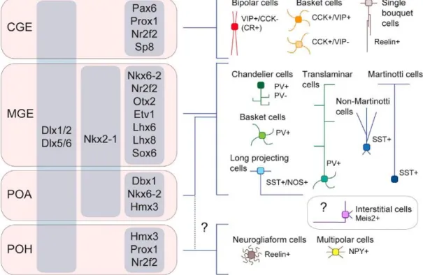
Figure 2. Cortical interneuron subtypes and their developmental origin
This schematic figure displays the distinct parts of the embryonic subpallium and their interneuron derivates. On the left side, regional transcription factors are indicating the different molecular regulation of each subpart of the ganglionic eminences (grey columns).
The right side displays the distinct interneuron subtypes with their specific protein profile and morphology. Developmental lineages are represented with blue connecting lines. The precise lineage of Meis2-positive interstitial cells (purple), reelin-positive neurogliaform cells and multipolar cells (brown) is unclear (dashed lines), they might originate from the CGE or/and the preoptic region. CGE: caudal ganglionic eminences; MGE: medial ganglionic eminence; POA: preoptic area; POH: preoptic-hypothalamic border domain; VIP:
vasoactive intestinal peptide; CCK: cholecystokinin; PV: parvalbumin; SST: somatostatin;
NOS:, NPY: neuropeptide Y; Meis2: myeloid ecotropic viral integration site 1 homolog 2.
Figure is adapted from Lim et al., 2018.
Upon chemo-attractive signaling, one of the branches stabilizes and the cell body moves towards that direction. Physiologically this movement is very similar to radial migration mentioned earlier. The regulation of tangential migration is region-specific (Marín, 2013) and is driven by chemoattractant and chemorepulsive signaling including the Ephrin, Semaphorin-neuroligin and Slit-Robo signaling pathways (Rudolph et al., 2014; Steinecke et al., 2014; Zimmer et al., 2008). MGE-driven interneurons leave the germinative niche upon
24
sensing a chemorepulsive signal provided by the Eph/ephrin family (Steinecke et al., 2014).
Semaphorin 3A and semaphorin 3F are membrane-targeted proteins, expressed by striatal cells. Migrating interneurons detect the repulsive signal via semaphorin receptors such as neuropilins and they avoid the striatum during tangential migration (Marín et al., 2001).
Another guiding molecular complex, the protein Slit (Slit guidance ligand) and its receptor Robo (Roundabout homolog) which is also responsible for striatal avoidance (Hernández- Miranda et al., 2011). Activation of both semaphorin and Slit/Robo signaling results in the actin-dependent collapse of the leading process, through small GTPAses (Peyre et al., 2015).
In contrast to all the repulsive signals, the dorsal cortex secretes chemoattractive signals, such as brain-derived neurotrophic factor (BDNF) and neuregulins (Flames et al., 2004; Polleux et al., 2002). Different isoforms of neuregulins are expressed along the migration paths, which activate the ErbB4 (Erb-B2 receptor tyrosine kinase 4) receptor on the leading processes of interneurons (Flames et al., 2004).
1.5.2. Cortical invasion
Once migrating neurons reach the PSB, they enter the cortex through two distinct migration streams carefully avoiding the cortical plate (Marín, 2013). Instead, they use the MZ and SVZ tracks (Lavdas et al., 1999; López-Bendito et al., 2004). The choice of migratory stream is based on the subtype and origin of the interneuron. Although fluorescently labelled GABAergic cells of all kinds of origins migrate through the SVZ stream, researchers found that half of the SST-positive interneurons, especially future Martinotti cells and PV-positive translaminar cells, preferentially use the MZ route (Lim et al., 2018).
Cortical tangential migration is regulated by chemokine signaling. SVZ and MZ migratory tracks contain high concentration of the chemoattractant Cxcl12 (C-X-C Motif Chemokine Ligand 12) which is secreted by TBR2-positive intermediate progenitor cells and the leptomeninges (López-Bendito et al., 2008; Sessa et al., 2010). The activation of chemokine receptors Cxcr4 and Cxcr7 on the surface of leading protrusion by Cxcl12 promotes the locomotion of interneurons (Wang et al., 2011). Moreover, ablation of both
25
receptors results in an altered laminar and regional distribution of interneurons (López- Bendito et al., 2008; Wang et al., 2011).
1.5.3. Laminar allocation
The different types of interneurons share the same cortical layers albeit with different connectivity (Tremblay et al., 2016). Previously, it was thought that interneurons, like pyramidal cells, populate the cortical layers in an inside-out manner (Kriegstein and Noctor, 2004), however it was shown that CGE-driven interneurons preferentially populate the superficial layers, therefore the origin or birthdate do not determine the layering distribution of interneurons (Miyoshi and Fishell, 2011). Nevertheless, GABAergic cells from different parts of the GE arrive to the cortex at different timepoints. First, the MGE-driven SST- positive cells enter the cortex, followed by the PV-positive population (Wonders and Anderson, 2006). Later, the VIP- and the NPY-positive cells invade the cortex from the CGE and lastly the reelin-positive CGE-driven cells arrive and localize the cortical layer 1 (Yozu et al., 2004). The laminar allocation of interneurons is regulated by several factors.
Chemokine signaling is important during tangential dispersion, but when cells change to radial migration they lose their responsiveness to Cxcl12 (Li et al., 2008). Parallel to this, developing excitatory cells control the position of interneurons in a subtype specific manner (Lodato et al., 2011). This idea is proven by the fact that aberrant cortical layering modifies the locations of inhibitory cells (Lodato et al., 2011; Pla et al., 2006). Moreover, pyramidal cells secrete neuregulin 3 during tangential migration, which guides allocating interneurons via the ErbB4 signaling (Bartolini et al., 2017). Once interneurons change to radial migration, they start to express Cx43, which establishes tight connections between the radial glia fibers and the cells (Elias et al., 2010).
The two main neurotransmitters, GABA and glutamate, are both promoting neuronal migration as excitatory signals, through depolarizing the plasma membrane and evoking Ca2+
mediated transients in the cells (Wang and Kriegstein, 2009). Interneurons begin to express KCC2, a potassium-calcium exchanger during their final phase of migration. Upregulation of KCC2 reverses the chloride potential in the migrating interneurons and makes GABA as hyperpolarizing signal (Ben-Ari, 2002; Bortone and Polleux, 2009). This phenomenon is
26
termed as “GABA switch” is indispensable for the proper maturation and differentiation of interneurons (Bortone and Polleux, 2009; Miyoshi and Fishell, 2011).
1.6. Developmental cell death during cortical development
In the process of brain development, a lot more cells are born than get utilized in the final neuronal circuits. In addition, transient cell populations like Cajal-Retzius cells and subplate cells also have to be eliminated for normal postnatal development. Accordingly, a complex cell death mechanism is developed to maintain the balance between the cell production and the functionally active cell number (Wong and Marín, 2019). Three classical cell death types are responsible for maintaining this balance: apoptosis, necrosis and autophagy (Galluzzi et al., 2018). Programmed cell death or apoptosis is the most common mechanism in the developing brain (Wong and Marín, 2019).
Programmed cell death is achieved in two different manners, the intrinsic and the extrinsic signaling pathways. The intrinsic apoptotic pathway is initiated by endogenous processes, which can change the permeability of the mitochondrial membrane, therefore pro – and anti-apoptotic proteins can be released from the outer membrane space and interact with each other (Hollville et al., 2019). After pro – apoptotic proteins, such as cytochrome c (CytC) is released to the cytoplasm, they interact with apoptosome forming proteins and trigger initiator and effector caspases. The cleavage of caspase-3 (CC3) is the last step in this pathway, functioning as a point of no return, causing irreversible DNA and mitochondria fragmentation which leads to the ultimate disruption of cell morphology (Kondratskyi et al., 2015). In contrast, the extrinsic pathway is mediated by death receptors. Upon ligand binding, these receptors undergo conformational changes and establish a complex with initiator caspases which in turn are activating the effector caspases (Galluzzi et al., 2018).
Three typical cell death waves are present during corticogenesis. The first occurs in the VZ and SVZ during the proliferation and controls the number of progenitors. The second is responsible for the elimination of dispensable pyramidal cells, and the third wave is the disposal of inactive interneurons (Wong and Marín, 2019). A recent study using stereological methods found that approximately 13% of pyramidal cells undergo programmed cell death between postnatal day 2 and 5 (Wong et al., 2018). It is important to note that pyramidal cell
27
death is different between cortical layers and it is highly dependent on neuronal activity (Blanquie et al., 2017). Approximately 30% of interneurons die the first two weeks of postnatal development (Southwell et al., 2012), however the elimination pathway and its time window differs between interneuron subtypes. The survival of interneurons from the MGE is mostly determined by local pyramidal cell activity. In the absence of pyramidal synaptic transmission, inactive interneurons upregulate the AKT pathway inhibitor PTEN (Phosphatase And Tensin Homolog) which triggers cell death between postnatal day 5 and 10 (Wong et al., 2018). Meanwhile, CGE-driven interneurons undergo programmed cell death later than MGE interneurons, around P12-22 (Priya et al., 2018). The survival of these cells is also activity-dependent, and regulated by a calcium-dependent phosphatase, called calcineurin (Priya et al., 2018). Conclusively, subtype-specific interneuron cell death seems to proceed based on their neurogenic sequence (Wong and Marín, 2019).
1.7. Cadherin superfamily
Cadherins are Ca2+-dependent adhesion proteins that are responsible for establishing cell to cell connections. The first cadherin-like ancestor appeared approximately 600 million years ago in early metazoans. Since then, the cadherin superfamily evolved and expanded greatly, owing the emergence of multi-layered tissues and functionally highly specialized organs (Gul et al., 2017). The human genome encodes 114 different cadherin proteins, which are classified into three functionally and structurally different groups. The major cadherin class includes 32 members, the protocadherin branch has 65 proteins and the cadherin-related subfamily contains 17 molecules (Gul et al., 2017). Despite the high diversity of cadherin molecules, they also share structural similarities. With the exception of Cdh13, they all have a transmembrane domain which is connected to cadherin motifs or extracellular (EC) domains at the N-terminal. The number of EC domains shows high variation between cadherin subgroups (Hirano and Takeichi, 2012). EC domains maintain the connection with the partner cell through a conserved tryptophan residue settled in the so-called adhesion arm.
Cadherin molecules usually are connected in a homophilic manner however evidence shows that different types of cadherins can also interact in a heterophilic way (Katsamba et al.,
28
2009). At the cytoplasmic side of the transmembrane domain cadherins interact with several different proteins to regulate adhesion-based intracellular signaling (Gul et al., 2017).
1.7.1. Cadherin subfamilies
The major cadherin branch consists of the classic cadherins, desmosomal cadherins, T- cadherin, Flamingo or Celsr cadherins and the 7D family (Hirano and Takeichi, 2012). The classical cadherins, such as E-cadherin and N-cadherin have five EC domains which provide the platform for Ca2+ binding. Once Ca2+ attaches, grooves between the EC domains, it stabilizes the cadherin EC structure and allows it to interact with the connection partner (Loh et al., 2019). The Flamingo/Celsr has seven transmembrane domains and a long complex extracellular part which is important in the formation of cell polarity. T-cadherin is a unique member of the cadherin family as it lacks the cytoplasmic domain, instead it is connected to the extracellular surface via a GPI (Glycosylphosphatidylinositol) anchor (Hirano and Takeichi, 2012).
The protocadherin family is divided into two subclasses, the clustered and non-clustered groups. Clustered protocadherin encoding genes are located in chromosome 5 in three clusters, each including more than ten genes. Meanwhile non-clustered protocadherins are translated from different chromosomes (Mountoufaris et al., 2018). Although protocadherins and classic cadherins share some common features, protocadherin-based adhesion is weaker, therefore the general view is that protocadherins act as specific “bar codes” on the plasma membrane and mediate cellular signaling in a cell type specific manner (Rubinstein et al., 2017).
The cadherin-related family is the most diverse group. Proteins from the calsyntenin subfamily have two EC domains and they have a major role in kinesin dependent vesicular transport. Calsyntenin was reported as an important molecular player of axonal branching, synaptogenesis and synaptic plasticity (de Ramon Francàs et al., 2017). Dachsous (DCHS1,2) and FAT (Protein Fat Homolog 1-4) are the longest of the cadherins, with more than 20 EC domains. They interact with each other in a heterophilic manner. Mutations in DCHS1 and FAT4 cause the Van Maldergem syndrome with serious developmental deficits such as cortical migration defects and increased proliferation which lead to periventricular
29
heterotopia, the displacement of neurons below the white matter (Cappello et al., 2013).
Cadherin-related 15 (Cdhr15) and cadherin-related 23 (Cdhr23) bind to each other and have an important regulatory role in inner-ear mechanotransduction (Araya-Secchi et al., 2016).
1.7.2. Cadherin-binding proteins
The cytoplasmic domain of classic cadherins is connected to cadherin-associated proteins with diverse cellular functions. p120catenin (or CTNND1, Catenin Delta 1) and β- catenin directly bind to the juxtamembrane region and the C-terminal of cadherins, respectively. Both proteins contain armadillo repeats, a repetitive amino acid sequence which includes the cadherin-catenin binding site (Gul et al., 2017). Secondary catenin-binding proteins are the α-catenin and the vinculin, both of which link the adhesion complex to the actin cytoskeleton (Figure 3; Shapiro and Weis, 2009). This cadherin-catenin molecular complex allows the control of cell fate determination by direct transcriptional regulation via β-catenin/Lef binding and cell motility through p120 which accesses to the microtubular system (Hirano and Takeichi, 2012; Stocker and Chenn, 2015). In vertebrates, β-catenin is duplicated and its paralogue gamma-catenin or plakoglobin creates an adhesion complex called desmosome or macula adherens with the two desmosomal cadherins, desmocollin and desmoglein (Schmidt and Jäger, 2005).
Cadherin based adhesion positively regulates the canonical Wnt (Wingless-Type MMTV Integration Site Family) signaling pathway (Figure 3; Gao et al., 2014). Wnt ligands are approximately 400 amino acid long glycoproteins. In the classical view, Wnt ligands bind to the seven transmembrane receptor Frizzled (Fzd) and its co-receptor the lipoprotein receptor related protein 6 (LRP6) which together interact with the intracellular scaffold protein dishevelled (DVL; Acebron and Niehrs, 2016). This interaction leads to the phosphorylation of LRP6 by glycogen synthase kinase 3β (GSK3β) and casein kinase 1 (CK1). The phosphorylated LRP6 accretes the scaffold proteins, axin and APC (Adenomatous Polyposis Coli Protein) to the plasma membrane and allows the cytoplasmic accumulation and nuclear transport of β-catenin where it can activate gene transcription through the LEF1/TCF transcription factor complex. In the absence of a Wnt ligand, β-
30
catenin is associated with a destruction complex composed of axin, APC and phosphorylated by GSK3β and CK1 for final degradation by the proteasome (Acebron and Niehrs, 2016).
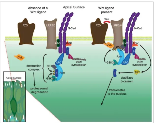
Figure 3. Canonical Wnt/ β-catenin signaling
The adherens junction is built up by cadherin dimers (here N-cadherin) and its associated molecules localized at the apical surface of neural progenitor cells. In the absence of Wnt ligand, β-catenin (β) undergoes phosphorylation by the destruction complex which leads to its ubiquitination and proteasomal degradation. In contrast, in the presence of Wnt ligand, FZD (Frizzled) and LPR5/6 (lipoprotein receptor related protein 5/6) are connected by DVL (Dishevelled) and the phosphorylated by glycogen synthase kinase 3β (GSK3β) and casein kinase 1 (CK1). This leads to the attachment of scaffold protein axin and APC (Adenomatous Polyposis Coli Protein) to the plasma membrane. In this case the destructive complex is unable to eliminate β-catenin, therefore it can regulate gene expression after nuclear translocation. In addition, N-cadherin also triggers β-catenin transcriptional activation through the phosphorylation of AKT by PI3K (Phosphoinositide 3-kinases). β-catenin mediated signaling regulates the actin cytoskeleton via α-catenin (α). Figure was adopted and modified from Stocker and Chenn, 2015.
31
1.8. N-cadherin function during embryonic cortical development
During the neurogenic phase of the developing embryonic cortex, N-cadherin is highly expressed in the adherens junction belt at the ventricular surface of both the pallium and subpallium, as well as in the intermediate zone and in the cortical plate (Kadowaki et al., 2007; László et al., 2019). Interestingly, these layers include mainly polarized cells, which suggest the functional importance of cadherin in regulating morphological changes. N- cadherin is vital in progenitor maintenance and identifies the apical side of progenitor cells (Zhang et al., 2010). After asymmetric cell division the position of N-cadherin also directs the formation of the leading process to face the basal surface (Gärtner et al., 2015). Moreover, N-cadherin at the basal side regulates the recruitment of the centrosome and the Golgi apparatus, which are indispensable for neuronal migration. This dual cellular distribution of the protein highlights its diverse function in every step of cortical development (Hansen et al., 2017).
1.8.1. N-cadherin in the neuroepithelium and in progenitor pools
After neuronal tube closure, classic epithelial cells downregulate the tight junction protein, so-called occludin and upregulate N-cadherin resulting in a rosette-like shape at the ventricular surface of the developing cortex (Aaku-Saraste et al., 1996; Gänzler-Odenthal and Redies, 1998). The role of N-cadherin-based adhesions and signaling during progenitor pool expansion is highly investigated in different animal models. Blocking N-cadherin function in chicken embryos resulted in the disruption of proper neuroepithelial organization (Gänzler-Odenthal and Redies, 1998). In addition, zebrafish and murine models revealed, that mutations in the Cdh2 gene cause neuronal displacement, increased mitosis and embryonic lethality at approximately 72 hours and 10 days post-fertilization respectively (Lele et al., 2002; Radice et al., 1997). Later, a conditional knockout study using a neocortical selective promoter showed gross morphological changes in the developing cortex, clusters of cells protruded into the lateral ventricles and the radial glia organization was also disrupted. Furthermore, mispositioned progenitor cells, increased proliferation and an overall thicker cortex was observed in these animals (Gil-Sanz et al., 2014; Kadowaki et al., 2007).
32
1.8.2. N-cadherin as a regulator of cell fate commitment
N-cadherin-connected catenins not only support cell-cell connections but are also able to recruit several molecules to form a multi-functional signaling hub (Figure 3). This network is crucial in progenitor pool maintenance in the developing telencephalon (Stocker and Chenn, 2015). N-cadherin homophilic binding activates protein kinase B (PKB, also known as AKT) thereby inducing pro-survival signaling through the phosphorylation of β-catenin (Zhang et al., 2010). In addition, activation of Wnt signaling also promotes progenitor state maintenance and stabilizes cell-cell connections via PAX-6 mediated positive feedback regulation (Gan et al., 2014; Gao et al., 2014) therefore progenitor cells are able to inhibit their own differentiation (Zhang et al., 2010, 2013). Accordingly, loss of β-catenin causes progenitor pool disassembly and brain malformations, in contrast gain-of-function experiments reveal increased progenitor pool and gyrification-like phenotypes in the embryonic mouse brain (Chenn and Walsh, 2003; Junghans et al., 2005). It has been proposed that Notch signaling-associated Numb proteins are important regulators of N-cadherin localization. Numb is localized primarily in the proximity of N-cadherin and interacts with p120catenin to maintain the N-cadherin-based intercellular connections thereby preserving the progenitor state (Rasin et al., 2007). N-cadherin connections also contribute to neurogenesis via Notch signaling (Hatakeyama et al., 2014). In contrast, the proneural gene, Ngn2 negatively regulates the levels of N-cadherin via the expression of Foxp2 and 4 (forkhead domain protein 2 and 4) transcription factors, in this way promoting differentiation and delamination (Rousso et al., 2012). During physiological delamination, the downregulation of N-cadherin fosters the cilium disassembly and apical abscission (Das and Storey, 2014). Finally, the localization of the remaining N-cadherin determines the origin of the future leading trail for radial migration by repositioning and stabilizing centrosomes (Gärtner et al., 2012).
1.8.3. N-cadherin role during cell migration
Neuronal migration is a dynamic and well-regulated process, and without proper adhesion it cannot be completed (Franco et al., 2011; Luccardini et al., 2013). Both excitatory and inhibitory cell migration are regulated by cadherin-based adhesion (Kon et al., 2017;
33
Luccardini et al., 2015). However, it is important to note, that the role of N-cadherin- mediated signaling during glutamatergic cell development has been more thoroughly investigated than in migration of interneuron precursors.
During glial-guided locomotion of glutamatergic precursors, N-cadherin is expressed in the leading process and maintains a reversible adhesion between the radial glia scaffold and the postmitotic cell. The turnover of N-cadherin is mediated by endocytic vesicle-associated Rab-GTPases (Hor and Goh, 2018; Kawauchi et al., 2010; Linford et al., 2012). Internalized proteins can undergo lysosomal degradation or they can get recycled into the plasma membrane (Cadwell et al., 2016). In addition, metalloproteases ADAM9 and 10 (disintegrin and metalloprotease domain 9 and 10) directly control the shedding of N-cadherin in a Rab14-dependent manner (Linford et al., 2012). Cleavage of the extracellular domains of N- cadherin by ADAM proteins results in the redistribution of β-catenin from the cell membrane to the cytoplasmic pool and initiates β-catenin-mediated gene expression (Linford et al., 2012; Reiss et al., 2005).
MZ-derived reelin promotes neuronal migration through Rap1 GTPase and triggers the AKT signaling pathway to enhance N-cadherin-based connection forming (Jossin and Cooper, 2011; Matsunaga et al., 2017). Once bipolar cells arrive to the CP, they peel off from the radial fiber by lysosomal degradation of N-cadherin in a Rab7-dependent manner (Kawauchi et al., 2010). This process is also induced by reelin, which regulates cytoskeletal dynamics through the phosphorylation of Dab1 (disabled homolog 1). Activation of Dab1 promotes the direct phosphorylation of cofilin and inhibits the actin-depolymerization, causing the leading process attachment to the MZ (Chai et al., 2009). Activation of Dab1 recruits Rap1 GTPase which has a dual function. It stabilizes the growth cone of the leading process via integrin – fibronectin connections (Sekine et al., 2012) and helps to establish homophilic N-cadherin connections between the neurons and Cajal-Retzius cells (Franco et al., 2011; Gil-Sanz et al., 2013; Jossin and Cooper, 2011). This tight connection allows postmitotic neurons to translocate their soma and begin their integration into the cortical layers (Franco et al., 2011).
34
In case of interneuron migration, in vivo data showed that selective elimination of N- cadherin from MGE-driven cells causes tangential migration delay and the disruption of cortical invasion (Luccardini et al., 2013). Accordingly, in vitro results refer that the presence of N-cadherin promotes IN migration, as its downregulation leads to impaired cell motility and leading process formation. These changes are mainly caused by defects in the polarization and the centrosome localization in these cells (Luccardini et al., 2013, 2015).
1.9. The endocannabinoid system
1.9.1. Endocannabinoid mediated retrograde neuronal transmission Endocannabinoids are plasma membrane-derived lipid molecules, which mediate specific retrograde signaling in the mature synapses via cannabinoid receptors. These molecules have similar molecular architecture to the psychoactive compound of Cannabis sativa, called THC (∆9-tetrahydrocannabinol). The two main endocannabinoids present in the brain are 2-arachydonoyl-glicerol (2-AG) which has high affinity to the G protein- coupled cannabinoid receptor 1 and 2 (CBR1, CBR2) and anandamide (AEA) which serves as a partial agonist of CBRs (Di Marzo, 2018) but binds to TRPV1 (transient receptor potential vanilloid receptor 1) and PPAR (peroxisome proliferator-activated receptor) receptors (Di Marzo, 2018) with higher affinity. The endocannabinoid system is evolved to control neuronal network activity (Figure 4) as a negative feedback regulator of synaptic transmission (Katona and Freund, 2008). Briefly, Ca2+ influx into the boutons during neuronal activity triggers neurotransmitter-containing vesicle release at the synapse.
Receptors at the postsynaptic density bind these neurotransmitters and transmit the signal via secondary messenger molecules (Piomelli, 2003). These GABA or glutamate receptors are connected to the so-called postsynaptic machinery which includes a molecular cascade carrying out the synthesis of 2-AG. Then, the postsynaptically released endocannabinoid travels back to the presynapse through the synaptic cleft and activates the CBR1, which in turn inhibits the voltage-gated calcium channels via G-proteins thereby decreasing synaptic transmission (Katona and Freund, 2012). 2-AG is mainly synthesized by DGLα or β (diacylglycerol-lipase α or β) and is degraded in the presynapse by MAGL (monoacylglycerol lipase) to arachidonic acid and glycerol (Figure 4; Wang and Ueda, 2009).
35
In contrast, AEA is mainly produced by NAPE-PLD (N-acyl phosphatidylethanolamine phospholipase D) and is cleaved by FAAH (fatty acid amide hydrolase) to arachidonic acid and ethanolamine (Figure 4,Hussain et al., 2017). It is important to note, that meanwhile 2- AG synthesis is dependent on DGL availability, AEA can be produced via several pathways incorporating many other lipases (Tsuboi et al., 2018).
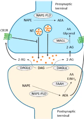
Figure 4. CB1 receptor-mediated endocannabinoid signaling
Schematic figure shows the main synaptic functions of endocannabinoids. 2-AG (2- arachydonoyl-glicerol) is synthesized by DGLα (diacil-glycerol lipase α) from diacil- glycerol (DAG) at the postsynaptic region, diffuses through the synaptic cleft and binds to presynaptic CB1 receptors (cannabinoid receptor 1). Activation of CB1 receptor leads to the termination of neurotransmitter release. Then, 2-AG is cleaved by MAGL (monoacylglycerol lipase) to arachidonic acid and glycerol. NAPE-PLD (N-acyl phosphatidylethanolamine phospholipase D) is responsible for the synthesis of AEA (anandamide) both pre- and postsynaptically. AEA might have both autocrine and paracrine effect on CB1 receptors.
Finally, fatty acid amide hydrolase (FAAH) degrades AEA to AA and ethanolamine. Figure was adapted from Zou and Kumar, 2018.
36
1.9.2. The function of the endocannabinoid system during cortical development
1.9.2.1 Endocannabinoid function during proliferation and differentiation
The level of the two main endocannabinoids is increased during embryonic development in rodents and peaks in the first two weeks of postnatal development. Accordingly, enzymes which are responsible for the metabolism of 2-AG display the same expressional tendency (Maccarrone et al., 2014). However, the AEA synthesizing enzyme NAPE-PLD becomes enzymatically active only in the first postnatal week (Morishita et al., 2005), hence the enzyme responsible for embryonic production of AEA is still unknown.
In the embryonic brain CB1 receptor shows gradual expression along the apico-basal axis of the embryonic cortex with the lowest levels being displayed in the ventricular zone (Berghuis et al., 2007; Galve-Roperh et al., 2013). Genetical ablation of CBR1 causes the disappearance of SVZ progenitors and the decrease of proliferating cells in the VZ. In contrast, more progenitor cells were found in the embryonic cortex of FAAH knockout animals, which reveals a function of AEA in proliferation (Mulder et al., 2008). Moreover, not just FAAH and CBR1 knockout animals display radial migration defects but using CBR1 and FAAH antagonists also produces the same effect (Mulder et al., 2008). In addition, prenatal exposure to the CBR1 agonist WIN 55,212-2 impairs the radial and tangential migration, furthermore increases the number of TBR2-positive intermediate progenitor cells (Saez et al., 2014). Another study used transient silencing of the CBR1 via in utero electroporation of small interfering RNA, which resulted in the overall impairment of radial migration (Díaz-Alonso et al., 2017). Overall, these in vivo experiments propose the existence of an endocannabinoid regulatory pathway which controls embryonic cortical proliferation and migration. Additionally, in vitro studies claimed that activation of CB1- and CB2- receptor mediated signaling promotes neuronal progenitor cell proliferation and differentiation (Díaz-Alonso et al., 2012) through the activation the AKT-pathway, which leads to the phosphorylation of Gsk3β and triggers the nuclear translocation of β-catenin (Trazzi et al., 2010).