The complex role of N-cadherin and endocannabinoid signaling during cortical development
Ph.D. thesis
Zsófia László
Semmelweis University
János Szentágothai Doctoral School of Neurosciences
Supervisor: Zsolt Lele, Ph.D.
Official Reviewers of the Ph.D.
Dissertation: Tamás Bíró, M.D., D.Sc.
István Adorján, M.D., Ph.D.
Head of the Final Examination
Committee: András Csillag, M.D., D.Sc.
Members of the Final Examination
Committee: Krisztina Herberth-Minkó, Ph.D.
Krisztián Tárnok, Ph.D.
Budapest
2019
2
1. Introduction
The cerebral cortex is the most complex structure in the mammalian brain. During evolution, cortical expansion allowed humans to become a conscious and one of the most versatile species in the world. Naturally, these attributes require the balanced and coordinated activity of excitatory and inhibitory neurons which in turn depend on the proper development of these cells. At the beginning of brain development, the telencephalon is dorso-ventrally divided into two parts, named the pallium and subpallium, respectively. More than 30 years ago scientists were convinced that both excitatory pyramidal neurons and inhibitory interneurons are originated from the pallium. With the advancement of technology it has been shown, that these two major cell types of the cortex are actually born in distinct germinative niches of the telencephalon. Excitatory projection neurons or pyramidal cells of the cortex use glutamate as neurotransmitter and are born in the progenitor pools of the pallium. In contrast, inhibitory interneurons that synthesize GABA (γ-aminobutyric acid) as their main neurotransmitter invade the cortex from the subpallial ganglionic eminences.
The embryonic cortex has a multi-layered structure each featuring their own distinct cell types. The most apical layer of the pallium is called ventricular zone (VZ) consists of dividing progenitors and newborn daughter cells. Above the VZ, there is another germinative layer called the subventricular zone (SVZ) which is responsible for the generation of both deep (neurons in the cortical layer 5 and 6) and upper layer neurons (layer 2-4). After their birth, newborn neuroblasts start their radial migration by using the elongated fibers of radial glial cells. They travel through the intermediate zone (IZ), which at later embryonic stages contains a dense neuropil of thalamocortical axons and acts as a barrier between proliferative zones and the forming cortex. The IZ is followed by a transient layer, called subplate (SP), which contains the earliest generated neurons. Next, pyramidal cells enter the cortical plate (CP), translocate to distinct places and begin their differentiation. Finally, the uppermost layer of the embryonic cortex is the marginal zone (MZ) where Cajal-Retzius cells are settled and regulate the final phase of pyramidal cell migration. Excitatory cells are stopped just beneath the MZ and the next generation of neurons will continuously bypass them in an inside-out manner, hence deep- layer neuron generation is followed by the production of upper-layer neurons. In contrast, subpallial interneuron precursors migrate tangentially from the subpallium and invade the cortex via distinct migratory routes, either through the subventricular or the marginal zone.
Then, using the radial glia scaffold, interneurons migrate radially to their destined places and undergo final differentiation which is strongly influenced by their synaptic interactions with pyramidal cells.
3
Cortical development cannot be fulfilled without distinct molecular pathways such as adhesion molecules and endocannabinoid-related signaling. Cadherins are Ca2+-dependent homophilic adhesion proteins that are responsible for establishing cell to cell connections in both the embryonic and adult brain. Neuronal-cadherin (N-cadherin, Cdh2) is highly expressed in the adherens junction belt at the ventricular surface of both the pallium and subpallium, as well as in the intermediate zone and in the cortical plate during brain development. N-cadherin is vital in progenitor maintenance, however after asymmetric cell division the position of N- cadherin also directs the leading process to face the basal surface in migrating postmitotic cells.
Both excitatory and inhibitory cell migration are regulated by cadherin-based adhesion.
However, it is important to note that the role of N-cadherin-mediated signaling during glutamatergic cell migration has been more thoroughly investigated then in the development of interneurons. During glial-guided locomotion of glutamatergic precursors, N-cadherin is expressed in the leading process and maintains a reversible adhesion between the radial glia scaffold and the postmitotic cell. Once migrating neurons arrive to the CP, N-cadherin establish homophilic connection between neurons and Cajal-Retzius cells which help the postmitotic neurons to translocate their soma and begin their integration into the cortical layers.
Endocannabinoids are plasma membrane-derived lipid molecules, which mediate specific retrograde signaling in the mature synapses via cannabinoid receptors (CBR1 and CBR2). The level of the two main endocannabinoids is increased during embryonic development in rodents and peaks in the first two weeks of postnatal development. Accordingly, enzymes which are responsible for the metabolism of 2-AG (2-arachydonoyl-glicerol) display the same expressional tendency. However, the AEA (anandamide) synthesizing enzyme NAPE-PLD becomes enzymatically active only on the first postnatal week, hence the enzyme responsible for embryonic production of AEA is still unknown. In the embryonic brain, CB1 receptor shows gradual expression along the apico-basal axis of the embryonic cortex with the lowest levels being displayed in the ventricular zone. Although the paracrine endocannabinoid signaling is crucial for fine-tuned neuronal communication in the mature brain, in the embryonic brain this is done in an autocrine manner. Members from the endocannabinoid system were described as regulators of proliferation and radial migration of excitatory cells. Furthermore, CBR1- mediated 2-AG signaling was reported to regulate axonal growth in the cortex, these incoming axonal tracks also maintain the migratory route of CB1 receptor-positive interneurons.
2. Aims
2.1. To investigate the role of N-cadherin in postmitotic interneuron migration and differentiation
4
Establish a mouse model in which N-cadherin-deficient migrating postmitotic interneurons can be monitored
Describe the interneuron migration phenotype in the absence of N-cadherin
Investigate the consequences of N-cadherin abolishment from postmitotic neurons in the early postnatal and adult mouse brain
2.2. To examine the outcome of adherens junction disruption and its mechanism during cortical development
Evaluate and characterize the effect of dominant-negative model of cadherin-based adherens junction disruption on glutamatergic cell development
Examine the role of endocannabinoid signaling in the molecular mechanism of pathophysiological delamination
Investigate the signaling of a potential teratogenic insult which is known to initiate delamination and migration defects in the embryonic cortex
3. Material and Methods
3.1. Animals
All animals were kept in standard laboratory conditions and were maintained according to protocols approved by the Hungarian Committee of the Scientific Ethics of Animal Research (license numbers: XIV-1-001/2332-4/2012 and PE/EA/354-5/2018). C57BL/6 mouse line was ordered from Charles River Laboratories. The Abhd4 transgenic line was made and validated by Benjamin Cravatt’s laboratory. The triple transgenic Dlx5/6i-Cre/Ncad-floxed mouse line was made by crosses of Dlx5/6i-Cre mouse line with Cdh2fl/fl then bred with Gad65-GFP mice.
In this study +/+ (wild type) and -/- (knock out) refers to Dlx5/6i-Cre+/+;Cdh2fl/fl and Dlx5/6i- CreCre/+;Cdh2fl/fl, respectively.
3.2. Sample preparation and sectioning
Adult and postnatal day 8 and 10 (P8-10) animals were anesthetized with a suitable dose of 1,25% Avertin (2,2,2, -tribromoethanol and 2-methyl-2-butanol in autoclaved water, Sigma) intraperitoneal injection, perfused transcardially with 4%PFA/ 0,1M PB and brains were postfixed overnight in the same solution. Next day brains were washed in PBS and were sectioned at 40µm for in situ hybridization and immunohistochemistry on a Leica1000 vibratome (Leica). Sections were collected in 24-well cell culture plates and stored at 4°C until the experimental procedures. For cryostat sectioning embryonic heads were cryoprotected in 15 and 30% sucrose/PBS solution overnight. Afterwards, tissue was embedded in Tissue-TEK OCT compound (Sakura) and 20 μm thick cryosections were collected on Superfrost Ultra Plus
5
glass slides (Thermo Fisher Scientific) with MICROM HM 550 cryostat (Thermo Fisher Scientific) and stored at -20°C until further experiments.
3.3. DNA constructs and cloning protocols
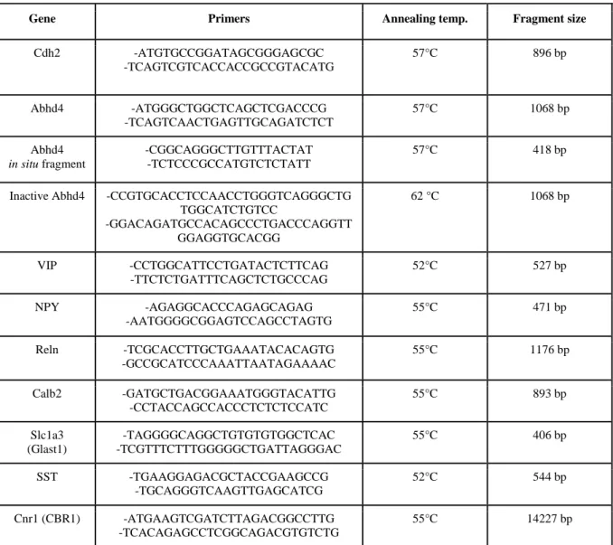
Mouse N-cadherin was cloned via RT-PCR using embryonic and adult cDNA as template with Long-template PCR mix according the manufacturer’s instructions (Roche) (Table 1.).
Mouse Abhd4 was also cloned via RT-PCR from E16 embryonic cDNA as template using the same way as described above. A shorter, 418bp fragment of Abhd4 was cloned separately to use as a template for in vitro transcription of the probe used in situ hybridization experiments.
The hydrolase dead version of Abhd4 (inactive Abhd4) was created by site-directed mutagenesis of serine 159 into glycine. All the fragments were cloned into pGEM-T Easy vector (Promega) and sequenced. ΔnCdh2, Abhd4 and Inactive-Abhd4 was then subcloned from pGEMT into the pCAGIG mammalian expression vector (Addgene # 1115) and purified with NucleoBond Xtra Plus EF kit (Macherey-Nagel) based on the manufacturer’s protocol.
For in situ hybridization on adult mouse brain, RNA was prepared by Ambion RNAqueous- 4PCR kit (Thermo Fisher Scientific). Reverse transcription was performed by Maxima RT Kit (Thermo Fisher Scientific) and fragments were amplified via RT-PCR, using appropriate primers (Table 1.), then cloned into a pGEMT-Easy vector (Promega).
3.4. In vitro transcription and in situ hybridization
To generate riboprobes, plasmid was linearized by the 5’ end of the insert with proper restriction enzyme and the product was separated with agarose gel electrophoresis followed by fragment isolation (GeneJET Gel Extraction Kit, Thermo Fisher Scientific). In vitro transcription was performed by DIG-UTP labeling mix (Roche) and the appropriate RNA polymerase (T3, T7, SP6, Promega). In situ hybridization assay was made as described earlier in Mayer et al. 2010. Sections were pulled onto glass slides (Thermo Fisher Scientific), mounted with Aqua-Poly/Mount (Polysciences, Inc.) and sealed with nail polish.
3.5. In utero electroporation and injections
Timed-pregnant mouse females were anesthetized (embryonic day 14.5) with 1,25%
avertin intraperitoneal injection or isoflurane vaporization. Abdominal cavity was opened next to the linea alba and uterine horns were exposed. Using glass capillary and mouth pipette, approximately 1 μl of expression vector (1-3 μg/μl for all of the constructs, dissolved in endotoxin-free water) with or without the general caspase inhibitor Z-VAD-FMK (5 μM final concentration; BD Biosciences) in endotoxin-free water containing Fast Green (Roth 1:10000) was injected into the lateral ventricle of the embryo. Electroporation was performed by SP-3c electroporator (Supertech) and tweezer electrodes with 5 pulses of 50 V for 50 millisecond
6
duration with 950 millisecond intervals. After the electrical pulses, uterine horns were returned into the abdominal cavity, muscle walls and skin were sutured, embryos were allowed to develop for the required time (1-3 days).
BrdU in 0.9% saline solution (200 mg/kg –Sigma) was prepared, passed through on a 0.2- μm pore size filter (Millipore) then intraperitoneally injected in the pregnant female mouse at day 14. Two hours later the embryos were decapitated, and heads were fixed with 4% PFA overnight.
Table 1. Primers used for cloning
3.6. Fluorescent single-cell mRNA detection (RNAscope)
In order to visualize the plasma membrane of the cells in the ventricular zone, ChR2-GFP (Addgene #26929) was electroporated in the embryos. One day later (E15.5) embryos were sacrificed by decapitation and heads were immediately frozen on isopentane/dry ice combination. Frozen embryonic heads were equilibrated in the cryostat at -20°C for 2-3 hours, then embedded in OCT and 20 μm thick cryosections was cut as described above. Cryosections
Gene Primers Annealing temp. Fragment size
Cdh2 -ATGTGCCGGATAGCGGGAGCGC
-TCAGTCGTCACCACCGCCGTACATG
57°C 896 bp
Abhd4 -ATGGGCTGGCTCAGCTCGACCCG
-TCAGTCAACTGAGTTGCAGATCTCT
57°C 1068 bp
Abhd4 in situ fragment
-CGGCAGGGCTTGTTTACTAT -TCTCCCGCCATGTCTCTATT
57°C 418 bp
Inactive Abhd4 -CCGTGCACCTCCAACCTGGGTCAGGGCTG TGGCATCTGTCC
-GGACAGATGCCACAGCCCTGACCCAGGTT GGAGGTGCACGG
62 °C 1068 bp
VIP -CCTGGCATTCCTGATACTCTTCAG
-TTCTCTGATTTCAGCTCTGCCCAG
52°C 527 bp
NPY -AGAGGCACCCAGAGCAGAG
-AATGGGGCGGAGTCCAGCCTAGTG
55°C 471 bp
Reln -TCGCACCTTGCTGAAATACACAGTG -GCCGCATCCCAAATTAATAGAAAAC
55°C 1176 bp
Calb2 -GATGCTGACGGAAATGGGTACATTG -CCTACCAGCCACCCTCTCTCCATC
55°C 893 bp
Slc1a3 (Glast1)
-TAGGGGCAGGCTGTGTGTGGCTCAC -TCGTTTCTTTGGGGGCTGATTAGGGAC
55°C 406 bp
SST -TGAAGGAGACGCTACCGAAGCCG
-TGCAGGGTCAAGTTGAGCATCG
52°C 544 bp
Cnr1 (CBR1) -ATGAAGTCGATCTTAGACGGCCTTG -TCACAGAGCCTCGGCAGACGTGTCTG
55°C 14227 bp
7
were held in the cryostat until the sectioning was finished. Sections were fixed immediately with cold 10% PFA solution for 30 minutes at 4°C. Next day RNAscope assay (Advanced Cell Diagnostic, Manual Fluorescent Assay AP-FastRed) was performed based on the manufacturer's instructions. Abhd4 RNAscope probe was custom designed for Abhd4 full sequence and used with Tbr2 or Glast1 separately. After the last washes slides were fixed with 10% PFA for 10 mins and immunohistochemistry was performed to improve the electroporated GFP signal.
3.7. Immunohistochemistry
Embryonic cryosections, adult free-floating samples or free-floating embryonic sections for confocal and STORM imaging were permeabilized by different concentration of Triton X- 100/PBS solution and/or with 10mM sodium-citrate at 65 °C depending on the antibody (Table 2., permeabilization column). Sections were then blocked with 5% Normal Donkey Serum (NDS; Sigma) in PBS for one hour. After several washes with PBS, sections were incubated in primary antibody (Table 2.) at 4°C, overnight. The following day samples were treated with secondary antibodies in PBS for 4 hours at room temperature. Finally, sections were washed with PBS and mounted with Vectashield Hard Set and sealed with nail polish. For STORM super-resolution microscopy, sections were mounted onto coverslips and stored uncovered at 4°C until the day of imaging. The primary and secondary antibodies which used in this study and their properties are listed in Table 2. and Table 3, respectively.
3.9. In vitro studies
HEK-293 cells were a kind gift from Balázs Gereben (IEM-HAS) and were maintained under normal conditions in plastic dishes with Dulbecco's Modified Eagle Medium (4.5 g/L glucose, L-glutamine & sodium pyruvate, Corning) containing 10% heat inactivated Fetal Bovine Serum (Biosera) in a 5% CO2 incubator at 37 °C. Cells were seeded a day before transfection, on poly-D-lysine coated coverslips in 24-well cell culture plate. The cells were held in Opti-MEM Media (Gibco) for an hour then transfection was made by using 2 μl Lipofectamine 2000 Reagent (Invitrogen) mixing with1 μg plasmid DNA in Opti-MEM Media.
After 20 hours, the transfected cells were washed with PBS and fixed with 4% PFA for 10 minutes. Fixed cells were treated with 0.2 % Triton X 100 for 15 mins in room temperature, and block with 1% Human Serum Albumin (HSA, Sigma) in PBS for 30 mins. Primary antibodies were diluted as listed below (Table 2.) for 1.5 hours at RT. After PBS washing, cells were incubated in secondary antibody solution for an hour (Table 3.). Then cells were washed with PBS and mounted on coverslips or in case of STORM super-resolution microscopy coverslips were stored in PBS at 4°C until the day of imaging.
8 Table 2. Used primary antibodies and their properties
3.10. Western blot
Embryonic telencephalon from Abhd4 +/+ and -/- animals and Abhd4 transfected HEK- 293 cells (as positive control) were homogenized in RIPA lysis buffer (50mM Tris-HCl, 150mM NaCl, 1% Tx-100, 0.1% SDS, 1mM DTT) containing 1x Protease inhibitor cocktail (Roche). Samples were denatured in Laemmli sample buffer (Bio-Rad) at 95°C for 5 mins and immediately loaded onto a 12% SDS-polyacrylamide gel. The amount of 15 μg protein was separated with PowerPac HC High-Current Power Supply (Bio-Rad) at 160 V, 400 mA then transferred to nitrocellulose membrane electrophoretically. For immunoblotting, we used 5 % Bovine Serum Albumin (BSA, Sigma) in TBST to block nonspecific binding sites, then incubated with primary antibodies in TBST overnight at 4°C. Washes with TBST were followed by horseradish peroxidase labeled secondary antibodies for 2 hours at RT, then membranes were developed by Supersignal West Dura Extended Duration Substrate Kit (Thermo Fisher Scientific).
Name Raised in Dilution Manufacturer Permeabilization paired box protein-6 (PAX6) Rabbit 1:300 Biolegend (901301) 10 mM sodium-
citrate and 0.1%
TritonX/PBS cleaved Caspase-3 (CC3) Rabbit 1:500 Cell signaling (9661S) 0.3% TritonX/PBS
calretinin (Calb2) Mouse 1:1000 Millipore (MAB1568) somatostatin (SST) Goat 1:1000 Santa Cruz (sc-7819)
laminin subunit alpha 1 (LAMA1) Rabbit 1:500 Sigma (L9393) 0.2% TritonX/PBS transcription factor T-box brain 1 (Tbr1) Rabbit 1:500 Abcam (ab31940)
transcription factor T box brain 1 (Tbr2)
Rabbit 1:500 Abcam (ab23345)
phospho-histone H3 (PHH3) Rabbit 1:500 Millipore (06-570)
nestin Mouse 1:200 Millipore (MAB353)
green fluorescent protein (GFP) Goat 1:1000 Abcam (ab5450) Translocase of Outer Mitochondrial
Membrane 20 (TOM20)
Rabbit 1:1000 Santa Cruz (s-11415)
cytochrome c (CytC) Mouse 1:2000 Biolegend (612302) parvalbumin (PV) Goat 1:3000 Swant (PVG214)
5-bromo-2'-deoxyuridine (BrdU) Mouse 1:400 Sigma (B8434) 2 M HCl
catalase Rabbit 1:3000 Abcam (ab1877) TBS/Tween20
abhydrolase domain containing 4 (Abhd4)
Rabbit 1:500 ImmunoGenes Ltd TBS/Tween20
9
Table 3. Secondary antibodies used in the experiments
3.11. Maternal alcohol consumption model
Abhd4 littermate embryos came from heterozygous breeding and were identically exposed to ethanol in utero. In the subchronic model, timed-pregnant mice received daily two vehicle or 2.5g/kg ethanol in saline intraperitoneal injections for two days (E13.5-E15.5) then embryos were collected at E16.5. The acute model was performed with a single 5g/kg vehicle or ethanol in saline injection at E14.5 and embryos were fixed 12 hours later. Maternal blood ethanol content was determined enzymatically, mother animals were decapitated, 0.1 ml blood was collected and immediately mixed with 900 ul of 6.25% trichloroacetic acid and centrifuged at 2000 rpm for 2 min to obtain the supernatant. The sample was then mixed with an alcohol reagent using the Synchron System Ethanol assay kit (Beckman) and assayed at 340 nm wavelength. Blood ethanol standards were created by mixing alcohol standards (0,1 ‰; 0,5 ‰;
1 ‰; 1,5 ‰; 2‰) with the reagent and immediately assayed. Maternal blood alcohol levels were between 0.5‰-1‰ in the subchronic model and it reached 1.5‰-2‰ in the acute model.
3.12. Phylogenetic tree
Protein sequences of species representing various phylogeny levels were collected from UniProt ). Sequence similarities were determined using protein alignment by Mega7 software, and the evolutionary history was inferred by using the Maximum Parsimony method. The analysis involved 12 amino acid sequences. All positions containing gaps and missing data were eliminated. There was a total of 310 positions in the final dataset.
Name Raised in Dilution Company Cat. number
Anti-Rabbit Alexa 488 Donkey 1:400 Jackson ImmunoReserach Ltd
711-545-152
Anti-Rabbit Alexa 594 711-585-152
Anti-Mouse Alexa 488 715-545-150
Anti-Mouse Alexa 594 711-585-150
Anti-Mouse Alexa 647 715-605-150
Anti-Goat Alexa 488 705-545-147
Anti-Goat Alexa 594 705-545-147
Anti-Goat Alexa 647 705-605-147
Anti-Rabbit CF568 1:1000 Biotium 20098-1
DAPI - 1:2000 Millipore 508741
Anti-Rabbit IgG, HRP-linked antibody
Goat 1:3000 Cell Signaling 7074
Alexa Fluor 568 Phalloidin - 1:500 Thermo Fisher Scientific A12380
10 3.13. Image acquisition and editing
In situ hybridization samples were imaged by Nikon Eclipse Ti80 upright microscope equipped with a Nikon DS-Fi1 CCD camera or a Zeiss Axioscope with an Axiocam Hrc digital camera. Fluorescent images were taken with Nikon A1R laser-scanning confocal system built on a Ti-E inverted microscope and operated by Nikon NIS-Elements AR software. STORM images with correlated confocal stacks were acquired via a CFI Apo TIRF 100x objective (1.49 NA) on a Nikon Ti-E inverted microscope with a Nikon C2 confocal scan head and an Andor iXon Ultra 897 EMCCD. The setup was controlled by Nikon N-STORM module in NIS- Elements AR software. Prior to imaging, sections were covered with imaging medium containing 0.1 M mercaptoethylamine and components of an oxygen scavenging system (5 m/v% glucose, 1 mg/ml glucose oxidase, 2.5 µl/ml catalase), and the coverslips were sealed with nail polish. Correlated confocal and STORM image acquisition and analysis were done as described earlier by Dudok et al., 2015. Pictures were always made and edited by equal settings between +/+ and -/- samples. Figure compositions were done by Photoshop CS5 (Adobe).
3.14. Statistical analysis
Experimental results were tested for statistical significance by Statistica 13.1 (TIBCO) and Prism 5.0 (Graphpad) programs. All experiments were repeated at least in 3 independent cases on different animals. Shapiro–Wilk normality test or in case of low item number, Kolmogorov–
Smirnov test was used to measure the normality of the samples. To determine the poolability of the raw datasets Kruskal-Wallis test was used. Unpaired comparisons were tested using two- tailed unpaired Student’s t-tests in case of normal distribution and Mann–Whitney tests based on nonparametric results. Multiple comparisons were examined based on their normality by one-way ANOVA with post hoc Tukey's Multiple Comparison Test or with nonparametric Kruskal-Wallis test followed by post hoc Dunn’s Multiple Comparison Test.
4. Results
4.1. The role of N-cadherin (Cdh2) during tangential migration and interneuron differentiation in the somatosensory cortex
4.1.1. Lack of Cdh2 in postmitotic interneurons effects their tangential migration
N-cadherin is highly expressed in several parts of the embryonic brain including the germinative zones of the pallium and subpallium. To investigate its role during tangential migration, we eliminated Cdh2 specifically in postmitotic interneuron precursors using a Cre- flox system where Cre expression was driven by the Dlx5/6i regulatory element. As a result, Cdh2 expression was completely absent in all postmitotic interneurons and striatal cells in the subpallium, meanwhile other regions, most importantly the subpallial ventricular zone as well
11
as the thalamus or pallial structures were not influenced. In order to visualize tangentially migrating interneurons in the absence of N-cadherin, Dlx5/6i-Cre/Cdh2-floxed animals were crossed to a GABAergic cell-type specific Gad65-GFP mouse line. Examining the pallium of these embryos showed that significantly less interneurons were able to migrate past the pallium – subpallium border (+/+: n = 3, -/-: n = 3; two-sided Mann-Whitney U test; ***P < 0.0001).
These data corroborate an earlier report indicating that N-cadherin is necessary for proper tangential migration. Previously, several studies showed that N-cadherin has a role in the regulation of proliferation and its absence could be an explanation behind the decreasing number of Gad65-GFP-positive interneurons in the pallium. To test this hypothesis, we performed phospho-histone H3 immunostaining (mitosis M phase marker) on the embryonic cortices at E14, however we could not detect any changes in proliferation between the genotypes (+/+: n = 3, -/-: n = 3; two-sided Mann-Whitney U test; P = 0.1389). These data show that lack of Cdh2 in postmitotic interneurons cause only a migration delay without affecting progenitor proliferation.
4.1.2. Gad65-GFP-positive cell number is changed in the triple transgenic adult brain
Our previous experiments demonstrated a migration delay of GFP-positive interneurons in the embryonic dorsal telencephalon of knock out animals, which might also have a long-term consequence. To confirm this, we counted the GFP-positive interneurons in the primer somatosensory cortex of adult transgenic animals and indeed found significantly less cells in the -/- mice compared with +/+ littermates (+/+: n = 6, -/-: n = 6; two-sided Student's t-test;
***P < 0.0001).
4.1.3. N-cadherin regulates interneuron composition of the adult primer somatosensory cortex
It was previously described that main interneuron subtypes are originated from distinct parts of the ganglionic eminences. To understand which cell population is affected by the loss of N-cadherin, we analyzed the interneuron densities in the adult cortex with widely accepted markers of different GABAergic interneuron subtypes. We did not detect any changes in the number of parvalbumin (+/+: n = 6, -/-: n = 6; two-sided Mann-Whitney U test; P = 0.8182) – or reelin – positive interneurons between genotypes (+/+: n = 7, -/-: n = 7; two-sided Mann- Whitney U test; P = 0.2086). Furthermore, these experiments did not reveal any changes in the cell densities of VIP- and NPY-expressing cell types either (both experiments: +/+: n = 7, -/-: n
= 7; two-sided Mann-Whitney U test; P = 1). In order to mark VIP-negative and cholecystokinin (CCK)-positive interneuron population, we chose cannabinoid receptor 1 as a marker, considering its perfectly overlapping expression with CCK, but excluding the CCK-positive
12
glutamatergic population. However, this comparison also did not show any alteration between +/+ and -/- (+/+: n = 8, -/-: n = 8; two-sided Mann-Whitney U test; P = 0.9591).
In contrast to the results described above, there was a sharp decrease in the number of calretinin (Calb2) – positive interneurons (+/+: n = 7, -/-: n = 7; two-sided Student's t-test; ***P
< 0.0001), not just in RNA, but in protein level as well (+/+: n = 6, -/-: n = 6; two-sided Mann- Whitney U test; ***P < 0.0001). Moreover, we also detected a significant decrease in the number of somatostatin (SST) – expressing interneurons in the adult somatosensory cortex of triple transgenic -/- animals (+/+: n = 11, -/-: n = 11; two-sided Mann-Whitney U test; **P = 0.0041). Furthermore, double immunostaining against Calb2 and SST revealed that the number of double positive cells in the -/- animals was altered as well (+/+: n = 6, -/-: n = 6; two-sided Mann-Whitney U test; **P = 0.0024). Taken together, these results indicate that loss of Cdh2 affects postmitotic interneuron development in a cell-type specific manner.
4.1.4. Disruption of N-cadherin signaling in postmitotic cells causes a migration delay in the postnatal somatosensory cortex
Beforehand, we observed a migration delay in the embryonic cortex and fewer SST/Calb2- positive interneurons in the adult somatosensory cortex. To investigate the fate of these cells between the embryonic migration delay and the adult phenotype, we measured the number of GFP-positive cells at an early postnatal age (P8) where we found significantly less GFP-positive interneurons in the cortices of the -/- littermates (+/+: n = 4, -/-: n = 4; two-sided Mann-Whitney U test; ***P < 0.0001). Moreover, we found that not only the cell number changed, but so did the laminar allocation of the GFP-positive cells in -/- mice. Distribution analysis also demonstrated a shift in the localization of these cells from the upper towards the deeper layers indicating that their migration has been arrested in the subventricular zone of the postnatal cortex (+/+: n = 4, -/-: n = 4; Bin4 and Bin10: two-sided Mann-Whitney U test; ***P < 0.0001).
Next, we considered if the absence of Cdh2 could cause a migration route change and the missing of their target cortical area. To test this hypothesis, we analyzed other brain areas which can serve as alternative destinations, such as the more lateral, supplementary somatosensory cortical area and the striatum. Interestingly, the number of GFP–positive interneurons was also decreased in both of these areas (+/+: n = 4, -/-: n = 4; two-sided Mann-Whitney U test;
supplemental somatosensory area (S2) ***P < 0.0001; striatum ***P = 0.0002), indicating that the precursors did not alter their migration route, rather probably got arrested before radial migration. All together these results show that loss of N-cadherin from subpallial postmitotic interneuron precursors affects various brain areas.
13
4.1.5. The fate commitment of the arrested cells in the postnatal SVZ
In the developing neocortex, pyramidal neuron activity can influence the fate and number of local interneurons. In an early critical period (P7-10) excitatory inputs to distinct interneuron cell-types can decide their survival or death. We assumed that the missing cells from the adult somatosensory cortex in triple transgenic -/- mice were eliminated at some point, because they did not change their fate or get misdirected to other possible target areas. In order to examine this, we performed TUNEL assays at embryonic and early postnatal stages to visualize cell death. At embryonic day 14, we analyzed two regions of the caudal ganglionic eminences, in a quadrate next to the ventricular surface and in the future striatal area. However, this analysis didn’t show any difference between genotypes at embryonic age (+/+: n = 3, -/-: n = 3; two- sided Mann-Whitney U test; CGE P = 0.7535; striatum P = 0.5429). In contrast, at P8 when we observed the migration arrest before, we also measured higher level of TUNEL-positive cell numbers in the somatosensory cortex (+/+: n = 5, -/-: n = 5; two-sided Student's t-test; ***P <
0.0001). Moreover, the laminar distribution of the TUNEL-positive cells reveals, that most of the dead cells were in the area where the migration was arrested (+/+: n = 5, -/-: n = 5; Bin1 and 10: two-sided Mann-Whitney U test; ***P < 0.0001). In summary, we showed evidence that loss of Cdh2 from postmitotic interneurons causes not just migration arrest, but also the elimination of the cells stuck in the postnatal SVZ.
4.2. The consequences of abnormal delamination in the developing mouse cortex 4.2.1. In vivo cadherin-based adherens junction disruption model
N-cadherin function during pallial development is highly investigated in physiological circumstances. However, much less is known about its potential contributions to cortical malformations. In order to examine the outcome of abnormal delamination we utilized an in vivo cadherin disruption model in the embryonic telencephalon. N-cadherin is one of the major molecular components of the adherens junction belt which anchors radial glia progenitor cells to each other forming the ventricular wall. In utero electroporation of a dominant-negative form of N-cadherin (ΔnCdh2-GFP) was able to disconnect classic cadherin-based connections, therefore we could avoid the potential functional redundancy between N-cadherin (Cdh2) and E-cadherin (Cdh1). Perturbation of N-cadherin connections resulted in a specific adherens junction destruction around the targeted RGPCs, as indicated by decreased expression of the fibrillar actin marker phalloidin. Next, we performed STORM super-resolution imaging on the radial glia scaffold. Reconstruction of the nanoscale architecture of nestin intermediate filaments however showed unaltered radial glia scaffold following ΔnCdh2-GFP electroporation. Previously it was shown that elimination of integrin-laminin connections at the
14
pial surface influences the morphology and survival of progenitor cells, nevertheless the basal endfeet-basal lamina connections of the RGPCs, visualized by the electroporated GFP and laminin subunit alpha 1 (LAMA1) respectively, were also intact. As a result of adherens junction belt elimination 48 hrs after electroporation, the PAX6-positive progenitor cells dispersed from the VZ and accumulated at the SVZ (n = 3; two-sided Mann-Whitney U test;
***P = 0.0004 in Bin4 and *** P = 0.0003 in Bin5). Taken together, these results demonstrate, that elimination of cadherin-based cell-to-cell connections at the ventricular surface leads to abnormal delamination of RGPCs.
4.2.2. Pathophysiological delamination causes apoptosis and migration defect in the embryonic dorsal telencephalon
Based on the fact that abnormally delaminated cells are generally eliminated via apoptosis to prevent possible malformations in an epithelial tissue environment, we asked whether a similar mechanism evolved to remove abnormally dispersed progenitor cells in the developing cortex. To test this hypothesis, we performed TUNEL assay to visualize cell death. Two days after ΔnCdh2-GFP electroporation, we found an approximately 2- fold cell death increase in the electroporated area compared with control conditions. This phenomenon was prevented by co-injection of the general caspase inhibitor, Z-VAD-FMK (GFP and GFP+Z-VAD-FMK n = 3-3; ΔnCdh2-GFP and ΔnCdh2-GFP+Z-VAD-FMK n = 4-4 animals; Kruskal-Wallis test with post hoc Dunn's Multiple Comparison Test; ΔnCdh2-GFP vs all the controls and treatments:
***P < 0.0001; between controls: P ≈ 1). Moreover, the observed migration defect, also described previously by others was rescued by the presence of the caspase inhibitor, indicating that disrupted cell migration is a consequence of the apoptotic process caused by the breakdown of adherens junctions (n = 3 in each group; Kruskal-Wallis test with post hoc Dunn's Multiple Comparison Test , Bin1 and 4: GFP vs ΔnCdh2-GFP, ΔnCdh2-GFP vs ΔnCdh2-GFP + Z-VAD- FMK ***P < 0.0001; Bin2: GFP vs ΔnCdh2-GFP: ***P = 0.0002; ΔnCdh2-GFP vs ΔnCdh2- GFP + Z-VAD-FMK **P = 0.0031; Bin5: GFP vs ΔnCdh2-GFP, ΔnCdh2-GFP vs ΔnCdh2- GFP + Z-VAD-FMK: ***P = 0.0001). These results confirm our hypothesis that there is a protective caspase-dependent cell death mechanism which eliminates inappropriately delaminating cells in the developing cortex.
4.3. Investigation of the pathological delamination-evoked cell death mechanism 4.3.1. The identification of potential molecular players in cadherin-loss induced apoptosis
To reveal the molecules involved in adherens junction breakdown-induced cell death, we carried out manual in silico analysis of available expression databases and single-cell RNA-
15
sequencing studies searching for genes from the endocannabinoid system with ventricular zone- restricted expression. Different expression analysis from mouse, human and cerebral organoids revealed a gene called Abhydrolase domain containing 4 (Abhd4), a serine-hydrolase previously implicated in N-arachidonoyl-ethanolamide (anandamide, AEA) synthesis.
Chromogenic in situ hybridization experiments provided evidence that Abhd4 mRNA was specifically restricted to the germinative zones of the embryonic telencephalon during cortical development and this expression was absent in Abhd4 knockout littermates. Furthermore, immunoblotting analysis of tissue samples revealed that Abhd4 was present in the E16.5 brain at the protein level as well.
Considering the fact that these germinative niches are the most active, highly proliferative hence very crowded regions of the developing brain, analyzing the expression of Abhd4 at the single-cell level is extremely challenging. To characterize the cell-type specific expression, we combined membrane targeting in utero electroporation with fluorescent single-mRNA detection, called RNAscope. Our experiments revealed that Abhd4 expression correlates positively with the Glast1 (Slc1a3 gene) radial glia marker. In contrast, inverse correlation was observed with the intermediate progenitor marker Tbr2 (n = 4 mice; Spearman’s rank correlation, Abhd4/Glast1: R = 0.48, P < 0.0001; Abhd4/Tbr2: R = -0.27, **P = 0.0086). Next, we wanted to know whether the previously observed specific expression of Abhd4 is changing during brain development. We found however, that Abhd4 expression pattern remained confined to the proliferative zones throughout cortical development and decreased in parallel with the number of proliferating progenitors in postnatal animals. Interestingly, the two main germinative niches, the SVZ of dorsal telencephalon and the subgranular zone (SGZ) of the hippocampus where neurogenesis still occurs in the adult mouse brain also expressed Abhd4.
4.3.2. Characterization of Abhd4 knockout animals
Radial glia cells serve two functions during cortical development. They are proliferating multipotent progenitor cells, but also provide a scaffold for postmitotic neuroblast migration.
In the previous section, we presented evidence that Abhd4 colocalizes with the radial glia marker Glast1, so next we examined if it plays a critical role in RGPC functions. Unexpectedly, neither single-pulse bromodeoxyuridine (BrdU, mitosis S phase marker) labeling of proliferating precursors nor immunohistochemistry using the M-phase marker, phospho- histone H3 (PHH3) revealed any quantitative differences between E14.5 +/+ and -/- cortices (BrdU, +/+: n = 6; -/-: n = 4; two-sided Student's t-test, P = 0.323; PHH3, +/+: n = 3; -/-: n = 3;
two-sided Student's t-test, P = 0.6882). To analyze the nanoscale architecture of the radial glia scaffold, we made STORM super-resolution microscopy and the composition of nestin
16
intermediate filament was reconstructed. Comparing the Abhd4 +/+ and -/- cortices however, we could not discover any difference neither in the number of nestin localization points nor in the size of the nestin bundle (Nestin NLP, +/+: n = 3; -/-: n = 3; two-sided Student's t-test, P = 0.297; FWHM, +/+: n = 3; -/-: n = 3; two-sided Mann-Whitney U test, P = 0.6882). Notably, we could also not detect any abnormalities in the structure of the adherens junction belt visualized by Alexa568-phalloidin staining in the Abhd4 -/- animals. Furthermore, to examine the possible changes in the organization of Abhd4 -/- cortex, we measured the cell number and distribution by distinct cell-fate markers. PAX6 the marker of proliferating progenitors showed the same distribution and quantity in both genotypes. (+/+: n = 4; -/-: n = 4; two-sided Student's t-test, P = 0.2598). In addition neither TBR1, which is the marker of deep-layer postmitotic neurons in the cortical plate, nor TBR2, an intermediate progenitor cell marker expressed in the SVZ, revealed significant changes in the embryonic Abhd4-/- cortex (TBR2, +/+: n = 3; -/-: n
= 3; two-sided Student's t-test, P = 0.1942; TBR1, +/+: n = 3; -/-: n = 3; two-sided Mann- Whitney U test, P = 0.0741). These results demonstrate that Abhd4 is not involved in classic radial glia cell functions.
4.3.3. Abhd4 is sufficient to trigger caspase-dependent cell death
As described before, we found no visible phenotype in the loss-of-function experiments but considering the specific expression of the enzyme, we hypothesized that Abhd4 function must be restricted to the ventricular zone before postmitotic daughter cells starts their radial migration. To test this idea, we expressed Abhd4 outside of the ventricular zone by using in utero electroporation with a plasmid containing a strong promoter which remains active outside the ventricular zone and followed its effect at different time points during development. Three days after the surgery, we observed a massive migration defect in the Abhd4-electroporated cortices compared with GFP control (n = 4 animals in each conditions; Kruskal-Wallis test with post hoc Dunn's Multiple Comparison Test; Bin1, 2, 4, 5: ***P < 0.0001). To prove that enzymatic function of Abhd4 is responsible for this migration phenotype, we mutated the catalytic serine in the conserved hydrolase domain to glycine to produce a non-functional form of the protein. Expression of the inactive Abhd4-GFP did not affect radial migration indicating that the enzymatic function of Abhd4 generated the migration arrest (n = 3; Kruskal-Wallis test with post hoc Dunn's Multiple Comparison Test; Bin1, 2, 5: ***P < 0.0001; Bin4: **P = 0.0063). Moreover, ectopic expression of Abhd4-GFP also induced characteristic morphological changes such as shrinkage, loss of extensions and rounded shape. After quantification of the occurrence of the two main morphological states of the cells (bipolar vs.
rounded), we found significantly decreased number of bipolar, migrating-cells after Abhd4
17
perturbation when compared with the controls (n = 3 in each treatment; Kruskal-Wallis test with post hoc Dunn's Multiple Comparison Test, Abhd4-GFP vs GFP or Inactive Abhd4-GFP:
*** P <0,0001; GFP vs Inactive Abhd4-GFP: P ≈ 1). The morphological changes described above can be a manifestation of a beginning cell death process so next, we performed TUNEL assay on the electroporated brain slices. Two days after electroporation we found higher level of TUNEL-positive cell density in the Abhd4-expressing samples, which we could prevent using the pan-caspase inhibitor, Z-VAD-FMK or the inactive form of the enzyme (GFP; GFP + Z-VAD-FMK and Inactive Abhd4-GFP n = 3;Abhd4-GFP and Abhd4-GFP + Z-VAD-FMK n = 4; Kruskal-Wallis test with post hoc Dunn's Multiple Comparison Test, Abhd4-GFP vs controls ***P < 0.0001; between controls P ≈ 1; except Abhd4-GFP + Z-VAD-FMK treatment vs Inactive Abhd4-GFP P = 0.607). Altogether, these results provide evidence that ectopic expression of Abhd4 evokes caspase-dependent apoptosis and radial migration defect in the mouse embryonic cortex.
4.3.4. The mechanism of Abhd4-induced cell death
Our previous experiment using a pan-caspase inhibitor suggested that Abhd4-mediated cell death might proceeded via the conventional apoptotic pathway. To test this hypothesis, we choose the two main apoptosis makers (Cytocrome C: CytC and Cleaved Caspase-3: CC3) to investigate the cell death signaling induced by Abhd4 in vitro. Using correlated confocal and super-resolution microscopy, we found that pixel intensity of the TOM20 mitochondrial outer membrane protein was significantly decreased after Abhd4 transfection when compared with the enzymatically inactive form (n = 4 experiments; two-sided Mann-Whitney U test, ***P
<0.0001). In parallel, the number of cytochrome c STORM localization points was lower after Abhd4 perturbation (n = 4 experiments; two-sided Mann-Whitney U test, ***P <0.0001).
Taking full advantage of super-resolution imaging, we could also separately quantify the distribution of intra- and extramitochondrial CytC localization points with a custom-made Python script. As we expected, the level of CytC NLP was higher in the cytoplasm after Abhd4 transfection then in the control condition, meanwhile in the control the majority of the CytC NLP remained in the mitochondria (n = 4 experiments; two-sided Mann-Whitney U test, ***P
= 0.0003). In addition, we examined the level of CC3 after Abhd4 perturbation and we found that the CC3 and GFP double positive cell ratio was higher in these samples than in the inactive Abhd4-GFP transfected ones (n = 3 experiment; two-sided Mann-Whitney U test, ***P
<0.0001). Important to note, that GFP-negative CC3-positive cell number was unchanged, conclusively, Abhd4-induced cell death is a cell autonomous phenomenon (n = 3 experiment;
two-sided Student's t-test, P = 0.5972). In summary, we established that Abhd4 triggers the
18
conventional intrinsic apoptotic pathway, which is mediated by CytC release from the mitochondria and the activation of the final effector, caspase-3.
4.3.5. Abhd4 is necessary for loss-of adherens junction-induced cell death Considering the lack of defects in Abhd4-/- embryonic cortex, but it has restricted ventricular zone expression and the protein’s ability to strongly induce cell death in gain-of- function experiments in vivo and in vitro, we hypothesized that Abhd4 might be involved in the cell death process evoked by loss of adherens junctions. So next, we examined if endogenous Abhd4 is required for AJ-loss induced apoptosis in vivo. As a preliminary control, we showed that ΔnCdh2-GFP electroporation initiated a similar dispersion of ectopic PAX6-positive RGPCs in both Abhd4 genotypes, indicating that Abhd4 is not required for delamination itself (n = 3 in both genotypes; two-sided Mann-Whitney U test, no significant difference between genotypes). In contrast, cell death levels induced by disruption of ventricular zone adherent junctions via ΔnCdh2-electroporation were much lower in Abhd4 -/- than in Abhd4 +/+
embryos (n=4 from each condition; Kruskal-Wallis test with post hoc Dunn's Multiple Comparison Test, ***P < 0.0001). Furthermore, exogenous replenishment of Abhd4 by co- electroporation of the two constructs into knockout animals restored the TUNEL-positive cell density to the Abhd4 +/+ level (n = 4; Kruskal-Wallis test with post hoc Dunn's Multiple Comparison Test, ***P < 0.0001; ns = not significant, P ≈ 1;).
Developmental cell death is an important regulator of the balance between the size of progenitor pools and final cortical cell number. Previously, we demonstrated that Abhd4 has a key role in delamination-induced cell death, so next we examined whether it has a similar function during developmental cell death. To this end, we examined the basal developmental cell death level in Abhd4 knockout animals. However, we could not detect any difference in basal, developmental cortical cell death levels of Abhd4+/+ vs Abhd4-/- embryos at E16.5 (n = 5 per genotype; two-sided Mann-Whitney U test, P = 0.834) or at P3 (+/+: n = 6; -/-: n = 5; two- sided Mann-Whitney U test, P = 0.792) when the peak of programmed cell death of excitatory neurons occur. Furthermore, the loss of Abhd4 does not influence the cellular composition of the adult cortex. By using cell population markers such as vGlut1 for excitatory neurons, Gad67 for inhibitory neurons and Glast1 for astrocytes we could not reveal any distribution differences between Abhd4 +/+ and -/- animals (n = 3 in both genotypes, two-sided Mann-Whitney U test , P > 0.05 in all bins of all experiments). These data demonstrate that Abhd4 is a necessary molecular player of cadherin-loss induced cell death but not required for normal, developmental cell death processes.
19
4.3.6. Fetal alcohol exposure induces Abhd4-dependent cell death in the embryonic cortex
Alcohol is the most commonly abused teratogen among pregnant women. Previously, it has been shown that alcohol exposure during pregnancy not only affects brain development, but also has negative long-term postnatal effects. In the embryonic cortex, alcohol exposure can cause abnormal delamination, cortical migration defect and increased cell death. Therefore, we next tested if Abhd4 is required for alcohol-induced changes in the embryonic cortex. Using a repeated drinking model over a span of three days resulted an elevation of cortical cell death levels in Abhd4+/+ embryonic cortices. However, lack of Abhd4 rescued the cell death (vehicle +/+: n = 5; vehicle -/-: n = 4; EtOH +/+: n = 4; EtOH -/-: n = 3; Kruskal-Wallis test with post hoc Dunn's Multiple Comparison Test, Abhd4 +/+ vs -/- and vehicles ***P < 0.0001; Abhd4 - /- vs vehicles P ≈ 1). Surprisingly, even a single dose of alcohol modeling binge drinking can elevate cell death levels in the ventricular zone of the developing cortex but again, lack of Abhd4 can prevent this (n = 3 in each genotype and condition; Kruskal-Wallis test with post hoc Dunn's Multiple Comparison Test, Abhd4 +/+ vs -/- and vehicles ***P < 0.0001; Abhd4 - /- vs vehicles P ≈ 1). These results demonstrate that Abhd4 is also essential for mediating maternal alcohol exposure-induced cell death in the embryonic neocortex.
5. Conclusions
This Ph.D. thesis provides insights into the importance of N-cadherin-based signaling during cortical development.
First, we generated a triple transgenic mouse line to investigate the function of N-cadherin during interneuron maturation. Our data indicate that selective elimination of Cdh2 from postmitotic interneuron precursors causes a delay in their migration in the mouse embryonic brain. During postnatal development, these migrating neuroblasts are arrested in the subventricular zone of the cortex and consequently get eliminated. As a result of this, the interneuron composition of the somatosensory cortex in adult triple transgenic animals is changed in a cell-type specific manner. Our results show that the number of somatostatin (SST) – and calretinin (Calb2) – positive interneurons were decreased while other interneuron populations are unaffected. Finally, we found that there is an overlap between the affected SST- and Calb2-populations as the number of double positive interneurons were also decreased.
To investigate the function of N-cadherin-related signaling during abnormal delamination we used in utero electroporation to disrupt the adherens junction belt around apical RGPCs.
Here, we provide evidence that selective elimination of cadherin-based connections causes a migration defect and caspase-dependent cell death in the ventricular and subventricular zones
20
of the embryonic mouse cortex. Furthermore, we identified a key molecular player in this process, called Abhydrolase domain containing 4 (Abhd4). This enzyme has a specific expression pattern in the germinative niches of the lateral and third ventricles, overlapping strongly with Glast1. We find that ectopic expression of Abhd4 triggers a radial migration defect and cell death. We also establish that Abhd4 evokes cytochrome c release from the mitochondria and enhances the level of cleaved caspase – 3, in vitro. Furthermore, we show that absence of Abhd4 can prevent the cell death caused by abnormal delamination in the embryonic cortex. Finally, we examine the effect of acute and subchronic maternal alcohol exposure and find that alcohol causes elevated cell death in the ventricular zone of the embryonic cortex in an Abhd4-dependent manner.
6. List of publications
Publications related to this thesis:
László ZI1, Bercsényi K1, Mayer M, Lefkovics K, Szabó G, Katona I, and Lele Z. (2019) N- cadherin (Cdh2) Maintains Migration and Postmitotic Survival of Cortical Interneuron Precursors in a Cell-Type-Specific Manner. Cerebral Cortex 30, 1318-1329.
Barna L1, Dudok B1, Miczán V, Horváth A, László ZI, and Katona I. (2016) Correlated confocal and super-resolution imaging by VividSTORM. Nature Protocols 11, 163–183.
1 equal contribution