ABHD4-dependent developmental anoikis safeguards the embryonic brain
Zsófia I. László 1,2,8, Zsolt Lele 1,8, Miklós Zöldi1,2, Vivien Miczán 1,3, Fruzsina Mógor1, Gabriel M. Simon4, Ken Mackie 5, Imre Kacskovics 6,7, Benjamin F. Cravatt 4& István Katona 1,5✉
A specialized neurogenic niche along the ventricles accumulates millions of progenitor cells in the developing brain. After mitosis, fate-committed daughter cells delaminate from this germinative zone. Considering the high number of cell divisions and delaminations taking place during embryonic development, brain malformations caused by ectopic proliferation of misplaced progenitor cells are relatively rare. Here, we report that a process we term developmental anoikis distinguishes the pathological detachment of progenitor cells from the normal delamination of daughter neuroblasts in the developing mouse neocortex. We identify the endocannabinoid-metabolizing enzyme abhydrolase domain containing 4 (ABHD4) as an essential mediator for the elimination of pathologically detached cells. Consequently, rapid ABHD4 downregulation is necessary for delaminated daughter neuroblasts to escape from anoikis. Moreover, ABHD4 is required for fetal alcohol-induced apoptosis, but not for the well-established form of developmentally controlled programmed cell death. These results suggest that ABHD4-mediated developmental anoikis specifically protects the embryonic brain from the consequences of sporadic delamination errors and teratogenic insults.
https://doi.org/10.1038/s41467-020-18175-4 OPEN
1Momentum Laboratory of Molecular Neurobiology, Institute of Experimental Medicine, 1450 Budapest Pf. 67., Budapest, Hungary.2School of Ph.D. Studies, Semmelweis University, Budapest, Hungary.3Faculty of Information Technology and Bionics, Pázmány Péter Catholic University, Budapest, Hungary.4The Skaggs Institute for Chemical Biology, Department of Chemical Physiology, The Scripps Research Institute, La Jolla, CA 92307, USA.5Department of Psychological and Brain Sciences, Indiana University, Bloomington, IN 47405, USA.6Department of Immunology, Eötvös Loránd University, Pázmány Péter stny 1/A., 1117 Budapest, Hungary.7ImmunoGenes Ltd, Makkosi út 86., 2092 Budakeszi, Hungary.8These authors contributed equally: Zsófia I. László, Zsolt Lele.✉email:katona@koki.hu
1234567890():,;
I
n the developing brain, radial glia progenitor cells (RGPCs) spawn various cell types including intermediate progenitor cells, neurons, astrocytes, oligodendrocytes, and ependymal cells1–4. Following asymmetric cell division at the ventricular surface, the self-renewed RGPCs remain anchored to their neighbors via cadherin-based adherens junctions5,6. This adhe- sion complex not only serves as a structural stabilizer, but it is also involved in important signaling mechanisms regulating cell cycle, proliferation, and differentiation, indicating that adherens junction assembly and disassembly are tightly coupled to cell fate decisions7–9. Accordingly, the non-RGPC-fated daughter cells need to break down their adherens junctions to delaminate from the ventricular wall and to migrate to their functional destinations along the radial scaffold of RGPCs6,9,10.Appropriately timed delamination is critically important for the mature daughter neuroblasts to follow their normal migratory route6. In contrast, pathological detachment and abnormal dis- persion of RGPCs, which retain their proliferative capacity at ectopic locations, represent a serious risk for brain malforma- tions, such as focal cortical dysplasia or periventricular hetero- topia, potentially predisposing to various forms of intellectual disabilities and neurological deficits11–14. Enormous number of cell division events and subsequent delamination steps are required for the generation of hundreds of billions of neurons and other cell types in the brain. Considering the remarkably low prevalence of congenital brain malformations, it is conceivable to hypothesize that a specific mechanism exists that protects against the consequences of sporadic delamination errors. Yet, how pathologically detached RGPCs are eliminated from the brain parenchyma and the related fundamental question of how fate- committed daughter cells become resistant to this defense mechanism both remain elusive.
Along a similar line of reasoning, it is plausible to predict that such a protective mechanism should also be activated when pervasive injuries or teratogenic insults damage the adherens junctions in the prenatal brain. For example, germinal matrix hemorrhage, the most common neurological disease of preterm infants as well as microcephaly-associated environmental terato- gens, such as the Zika virus and fetal alcohol exposure are all known to impair cell–cell adhesion in the germinative ventricular zone (VZ) leading to delamination and subsequent depletion of RGPC pools15–18. However, the molecular link between adherens junction damage and cell death induced by injury or by terato- genic insults remains unknown.
In the present study, we tested the hypothesis that a yet uni- dentified safeguarding mechanism determines distinct cell fate after normal delamination of daughter cells or pathological detachment of progenitor cells. We demonstrate that adherens junction impairment induces aberrant RGPC delamination, ectopic accumulation, and caspase-mediated apoptosis in the subventricular zone (SVZ) and VZs, whereas caspase inhibition not only prevents the evoked cell death, but also rescues radial migration into the cortical plate. Moreover, abhydrolase domain containing 4 (ABHD4), a serine hydrolase with a yet undefined neurobiological function is identified as a necessary and sufficient mediator for the elimination of pathologically detached cells.
Accordingly, healthy neuroblasts delaminating according to their normal developmental program switch off ABHD4 expression in parallel with their neurogenic commitment. ABHD4 is not required for the canonical form of developmentally controlled programmed cell death in the embryonic neocortex indicating that its function is specifically associated with pathological insults.
In agreement with this possibility, our findings elucidate that ABHD4 is also essential for prenatal alcohol exposure-induced apoptosis providing insights into the mechanisms underlying cell death associated with fetal alcohol syndrome.
Results
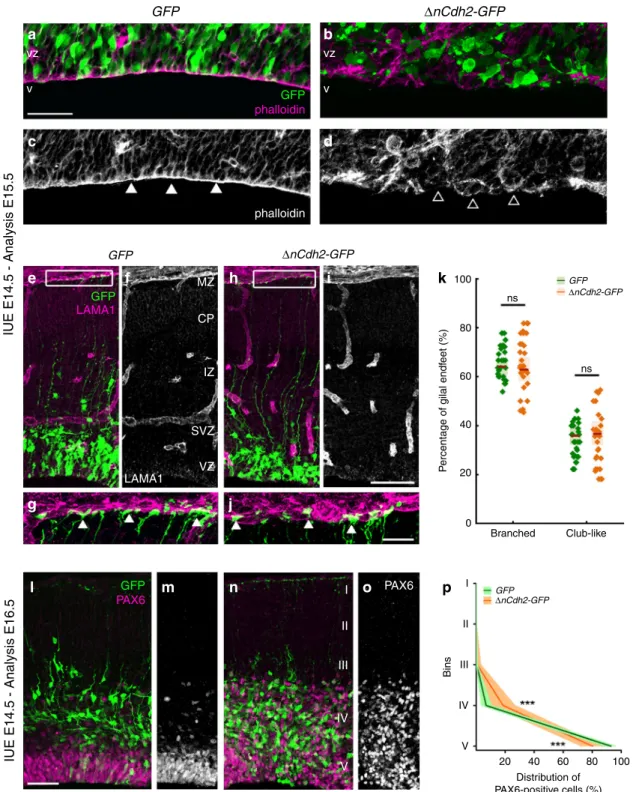
Pathological RGPC detachment triggers developmental anoi- kis. N-cadherin (encoded by the Cdh2gene) is the major mole- cular component of the adherens junction belt along the ventricular wall in the developing mammalian brain5. To interfere with cadherin-based cell-cell adhesions, we carried out in utero electroporation of a dominant-negative version of N-cadherin (ΔnCdh2-GFP) into the lateral ventricles at embryonic day 14.5 (E14.5). We exploited this conditional and sparse adherens junction injury protocol instead of the global loss-of-function approaches to restrict the effects in time and space to a selected population of RGPCs and to avoid potential compensatory mechanisms that are more likely to come into play upon systems- level perturbations. Accordingly, in utero electroporation (IUE or EP) of ΔnCdh2-GFP caused a destruction of adherens junctions limited to the electroporated area (Fig.1a–d; for comparison of electroporated and non-electroporated area see Supplementary Fig. S1a–f). Confocal and STORM super-resolution microscopy revealed a striking specificity of this experimental manipulation as basal processes of electroporated RGPCs still reached the basal surface in ΔnCdh2-electroporated cortices (Fig.1e–j). Moreover, the morphology of the basal endfeet at the pial surface (Fig.1g, j, k) and the nanoscale architecture of radial scaffold processes of transfected RGPCs both remained unaltered 24 h later (Supple- mentary Fig. S1g–n). As a result of the adherens junction dis- ruption, the detached RGPCs delaminated from the ventricular surface and accumulated in the SVZ. Most of these pathologically detached progenitor cells retained PAX6 transcription factor expression, a marker of RGPCs19even outside of the ventricular germinative niche (Fig. 1l–p). Immunostaining for the mitotic marker phospho-histone H3 (PHH3) revealed that the dispersed RGPCs are still undergoing proliferation (Supplementary Fig. S2a–d). In agreement with prior data that some RGPCs start differentiating as a result of adherens junction loss induced by ΔnCdh2-electroporation7, we also noticed an increase in the ratio of presumptive basal radial glia cells (bRG, based on the presence of the basal process in SVZ-located mitotic cells) and inter- mediate progenitor cells (IP, TBR2-positive cells with no apical or basal extensions in the SVZ) at the expense of RGPCs/apical radial glia precursors (mitotic cells in the VZ with both apical and basal processes; Supplementary Fig. S2e–i, j) together with a switch of fate based on the altered PAX6- and TBR2- immunostaining patterns (Supplementary Fig. S2k–q).
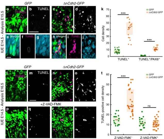
In order to investigate the fate of these pathologically detached RGPCs, we next tested whether cell death is also induced by adherens junction disruption. Notably,ΔnCdh2-GFP-electropora- tion caused a strong increase in the density of dead cells visualized by TUNEL-labeling when compared to GFP-electro- poration in control animals (Fig. 2a, b, c, d, k, l, m, n, o, t).
Although dying cells lose their protein content and consequently their immunogenicity, the majority of TUNEL-positive cells (73%; 232/317 cells) still had a relatively normal cell-like morphology and expressed PAX6. As a minimal estimation,
~30% of all TUNEL-positive signals overlapped with PAX6- immunostaining, indicating that many cells already arrived to a later stage of apoptosis (Fig.2e–k). In addition, administration of the pan-caspase inhibitor Z-VAD-FMK fully prevented the increase in cell death evoked by adherens junction disruption (Fig.2l–t). In contrast, basal cell death levels remained unaffected demonstrating that pathological detachment specifically triggers a caspase-dependent apoptotic process (Fig. 2l, m, p, q, t).
Moreover, the ectopic accumulation of RGPCs in the SVZ was also rescued by Z-VAD-FMK treatment and most of the surviving ΔnCdh2-GFP-electroporated cells could migrate to the cortical plate (Supplementary Fig. S3). These observations reinforce the idea that a specific cell death mechanism exists to
GFP
GFP
IUE E14.5 - Analysis E15.5IUE E14.5 - Analysis E16.5
GFP LAMA1
ns
ns GFP
80 100
60
40
Percentage of gilal endfeet (%)
20
0
Branched Club-like
a
c
e
g
l m n o
j
f MZ
CP
IZ
SVZ VZ
I PAX6 I
II
Bins III
IV
V
20 40 Distribution of PAX6-positive cells (%)
60 80 100
II III
IV
V LAMA1
GFP PAX6
h i k
p b
d vz
v GFP
phalloidin
phalloidin vz v
ΔnCdh2-GFP
ΔnCdh2-GFP
ΔnCdh2-GFP
GFPΔnCdh2-GFP
Fig. 1 Adherens junction disruption induces ectopic accumulation of PAX6-positive cells.High-powerfluorescent micrographs of the ventricular zone (VZ) show the adherens junction belt (white arrowheads) around the ventricle (V).ΔnCdh2-GFP-(b,d), but not control GFP- in utero electroporation (IUE) (a,c), demolishes adherens junctions (open arrowheads).e–jLaminin (LAMA1)-immunostaining of the developing cerebral cortex fromGFP- andΔnCdh2- GFP-electroporated embryos. High-power images (g,j) demonstrate that the attachment of the basal endfeet of RGPCs to the pial surface is not affected directly by the disruption of cadherin-based adherens junctions at the ventricular surface.kSelective damage of the apical adherens junction does not affect the two main morphological types of radial glia endfeet at the basal surface (two-sided Student’s unpairedttest,P=0.951;n=43 sections fromn= 3 animals per GFP treatment,n=37 sections fromn=3 animals perΔnCdh2-GFP-treatment). Graphs show box-and-whisker plots (including minima, maxima and median values, lower and upper quartiles) with single values.l–oBreakdown of adherens junctions prompts delamination (g–h) and ectopic accumulation of PAX6-positive cells in the subventricular zone.pDistribution of PAX6-positive cells infive equal bins (Roman numerals) (two-sided Mann–WhitneyUtest for all comparisons; 4th bin ***P=0.0004 and 5th bin ***P=0.0003;n=13 sections fromn=3 animals per GFP treatment;n= 11 sections fromn=3 animals perΔnCdh2-GFP-treatment). Data are shown as median (line) and interquartile range (transparent band). Scale bars:a–d, l–o:50μm,e–j: 25μm. Source data are provided as a Source Datafile.
eradicate pathologically detached progenitor cells in the devel- oping neocortex, and this mechanism must be switched off in normally delaminating fate-comitted daughter cells to permit their migration to their functional destinations. We termed this process developmental anoikis, because it conceptually resembles the specific form of apoptosis of epithelial cells induced by the loss of cell anchorage20,21. Anoikis has primarily been implicated as a protective mechanism in tumor biology, and pathologically detached tumor cells need to develop resistance to anoikis to become able to invade distant organs during metastasis formation22.
Selective ABHD4 expression in RGPCs in the prenatal brain.
The previous experiments led us to hypothesize that specific molecular players have evolved to mediate delamination error- induced cell death. These signaling components must be present in anchorage-dependent RGPCs to protect them from patholo- gical insults, but their expression should be downregulated in migrating healthy neuroblasts. Therefore, we searched public
single-cell RNA-Seq databases, and identified abhydrolase domain containing 4 (ABHD4), an endocannabinoid- metabolizing enzyme23with a yet unknown in vivo function, as a potential molecular candidate. Notably, Abhd4 mRNA levels were below detection threshold in more committed neuronal progenitor cell populations and in adult cortical neuronal types24,25, whereas Abhd4 was found to be highly expressed in putative RGPC pools in both mouse and human embryonic cortical samples and cerebral organoids26,27. The pattern of expression was very similar to the RGPC markerPax6, but was complementary to that ofTbr1, a marker of differentiated neu- rons (Supplementary Fig. S4). Moreover, a target-agnostic in vitro shRNA library screen in immortalized prostate epithelial cells suggested that downregulation of ABHD4 may potentially con- tribute to the resistance to anoikis28.
To experimentally determine whether the expression pattern of ABHD4 matches with its potential functional role in the developing neocortex, we first performed in situ hybridization on wild-type and littermate Abhd4-knockout embryonic brains.
GFP
IUE E14.5 - Analysis E15.5IUE E14.5 - Analysis E16.5
a
e
l
p q r s
m n o
f g h i j
GFP TUNEL
GFP TUNEL
TUNEL PAX6 TUNEL PAX6
TUNEL
TUNEL
Merged Merged
k
t
Cell density
70 GFP
ΔnCdh2-GFP
GFPΔnCdh2-GFP 60
50 40 30 20 10 0
60
50
40
30
TUNEL-positive cell density
20
10
0
TUNEL+
Z-VAD-FMK– Z-VAD-FMK+ TUNEL+/PAX6+
b c d
ΔnCdh2-GFP
GFP
+Z-VAD-FMK
ΔnCdh2-GFP
ns
Fig. 2 Adherens junction disruption induces apoptosis in the prenatal neocortex. a–dConfocal images demonstrate thatΔnCdh2-GFP- (c,d) triggers increased cell death compared to controlGFP-electroporation (a,b).e–gGFP-electroporated PAX6-expressing cells occasionally show TUNEL-labeling under control conditions.h–jIncreased TUNEL-labeling density is observed inΔnCdh2-electroporated samples. Scattered line encircles a still morphologically intactΔnCdh2-electroporated, TUNEL-positive cell that has retained PAX6-immunostaining.kDensity of TUNEL-positive and TUNEL/
PAX6 double-positive cells after electroporation at E15.5 (both cases: two-sided Mann-WhitneyUtest,P< 0.0001;n=22 samples fromn=3 animals per GFPelectroporation,n=21 samples fromn=3 animals perΔnCdh2-GFPelectroporation).l–oTwo days after the elimination of adherens junctions show elevated cell death in the electroporated area (n,o).p–sThe pan-caspase inhibitor Z-VAD-FMK prevents cell death induced byΔnCdh2-GFP-
electroporation.tDensity of TUNEL-positive dead cells in the electroporated area (Kruskal–Wallis test with post hoc Dunn’s Test;***P< 0.0001; ns=not significant,P≈1;n=18–18 sections fromn=3–3 animals perGFPandGFP+Z-VAD-FMK treatment;n=30–30 sections fromn=4–4 animals per ΔnCdh2-GFPandΔnCdh2-GFP+Z-VAD-FMK treatment). Graphs show box-and-whisker plots (including minima, maxima, and median values, lower and upper quartiles) with single values. Scale bars:a–d,l–s: 50μm,e–j: 10μm. Source data are provided as a Source Datafile.
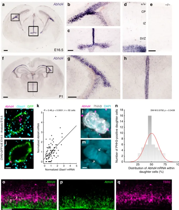
These experiments revealed that Abhd4 mRNA expression was remarkably abundant in the germinative niches of the tele- ncephalic and third brain ventricles, whereas it was absent in other regions and in control Abhd4-knockout embryonic brains (Fig. 3a–e). This spatially restricted expression pattern was
consistent throughout prenatal development (Supplementary Fig. S5a–f), butAbhd4expression markedly decreased postnatally in parallel with the reduced number of proliferating progenitors in the subventricular and subgranular zones (Fig. 3f–h; Supple- mentary Fig. S5g–i), reaching undetectable levels in adults.
a Abhd4
Abhd4 E16.5
P1
5 18 SW-W:0.9792 p = 0.5439
16 14 12 10 8
Number of PHH3-positive daughter cells
6 4 2
0 25
Distribution of Abhd4 mRNA within daughter cells (%)
50 75 100
4
3
Normalized Abhd4 mRNA
ChR2-GFP IUE E14.5 - Analysis E15.5
2
1
0 0
Abhd4 Abhd4
TBR2 TBR2
1
Normalized Glast1 mRNA
2 3 4 5
R = 0.48 p < 0.0001; n = 92 cells
Abhd4 Glast1 GFP Abhd4 PHH3PHH3 DAPI
+/+ –/–
CP
IZ
SVZ
f
k n
l
m i
j
o p q
b
c
g
d
h
e
Fig. 3Abhd4mRNA is expressed by radial glia progenitor cells. a–hAbhd4mRNA is present exclusively in the ventricular zone along with the lateral (b,g) and third ventricles (c,h) at both E16.5 (a–d) and P1 (f–h) wild-type (+/+) mice. The specificity of theAbhd4riboprobe is validated inAbhd4- knockout(−/−) animals (e). CP, cortical plate; IZ, intermediate zone; SVZ, subventricular zone; VZ, ventricular zone. High-power confocal imaging outlines the plasma membrane ofChR2-GFP-electroporated cells and delimits multi-color RNAscope analysis into single cells within the heterogeneous and densely packed cell layer of the ventricular zone.Abhd4mRNA typically colocalizes with the radial glia progenitor cell markerSlc1a3mRNA (encoding GLAST1 protein) (i), whereas other cells are often devoid of both markers (j).kCorrelation analysis ofAbhd4mRNA levels withGlast1mRNA levels in single cells (Spearman’s rank correlation,Abhd4/Glast1: R=0.48,P< 0.0001;n=92 cells fromn=4 mice). The scatter plot shows data from individual cells normalized to the median value of the respective mRNA levels.l,mAbhd4mRNA distribution in attached daughter cells marked by PHH3-immunostaining.
Arrows point to the mitotic cleavage furrow between the dividing cells.nQuantification ofAbhd4mRNA allocation within PHH3-positive daughter cells (Shapiro-Wilk normality test;W=0.9792;P=0.5439;n=24 sections fromn=3 animals).o–qRepresentative images forAbhd4in situ hybridization combined with TBR2-immunostaining.Abhd4mRNA shows complementary distribution to TBR2 protein-containing intermediate progenitor cells. Scale bars:a: 100μm,b–e,g–h,o–q: 50μm,f: 500μm,i,j,l,m: 2μm. Source data are provided as a Source Datafile.
Immunoblotting with a specific antibody raised against a conserved disordered motif of the ABHD4 protein further confirmed the presence of this serine hydrolase enzyme in the developing neocortex of wild-type, but not of Abhd4-knockout control embryos (Supplementary Fig. 5Sj, k).
Although RGPCs represent the majority of cells in the germinative niches, it is important to note that fate-committed daughter cells that are undergoing delamination still populate the VZ, where the high cellular abundance renders cell-specific quantitative mRNA analysis very difficult. In order to unequi- vocally identify the cell population expressing Abhd4, we developed an approach for single cell-restricted, high-resolution visualization of the plasma membrane via in utero electroporation combined with multi-color in situ hybridization (see “Online
“Methods”). We found thatAbhd4mRNA levels were positively correlated withGlast1expression (a marker of RGPCs29; Fig.3i, j). To test the possibility that Abhd4 mRNA is preferentially segregated either into self-renewing RGPCs or daughter cells during cell division, we also measured Abhd4 expression by quantifying RNAscope in situ hybridization signal within mitotic cells visualized by PHH3-immunostaining. The distribution analysis yielded a single-peak Gaussian curve with a peak value around 50% indicating the uniform spatial distribution ofAbhd4 mRNA within mitotic cell pairs (Fig. 3l–n). Subsequently, a combination of Abhd4 in situ hybridization with TBR2- immunostaining convincingly demonstrated the lack of overlap of Abhd4 mRNA expression with TBR2 protein, a marker of delaminated IP cells (Fig.3o–q). Thesefindings together indicate that the temporal expression of Abhd4 in RGPCs is tightly controlled and its downregulation in fate-committed daughter cells starts rapidly after their delamination.
ABHD4 is sufficient to trigger apoptosis. The previous experi- ments demonstrated strict spatial and temporal restriction of Abhd4 expression in RGPCs of the developing brain. RGPCs serve two major functions in the embryonic cerebral cortex: as progenitor cells1–3 and by providing a scaffold for postmitotic neuroblast migration10. By using Abhd4-knockout mice, we first tested the possibility that ABHD4 plays a role in RGPC proliferation. However, neither single-pulse bromodeoxyuridine- labeling of proliferating precursors (Fig. 4a–c), nor direct visua- lization of mitotic cells by PHH3-immunostaining (Supplemen- tary Fig. S6a–c) revealed any quantitative differences between wild-type andAbhd4-knockout mice. This corresponds well with the lack of any changes in the number of PAX6-positive RGPCs (Fig.4d–f) or in the structure of the adherens junction belt in the ventricular wall (Fig. 4g, h), together indicating an intact and functional neurogenic niche in Abhd4-knockout mice. Delami- nation and migration did not require ABHD4 either, because there was no change in the number and laminar distribution of TBR2-immunopositive intermediate progenitor cells30 in the SVZ, and the density of TBR1-immunopositive postmitotic neurons31 in the cortical plate of the Abhd4−/− embryonic cerebral cortex (Supplementary Fig. S6d–i). Moreover, the lami- nar distribution of pyramidal cells, GABAergic interneurons and astrocytes was also unaltered in the adult neocortex of knockout animals (Supplementary Fig. S7). Finally, quantitative STORM microscopy revealed that the nanoarchitecture of the radial glia processes also remained intact in the absence of ABHD4 (Fig. 4i–p). Together, these results demonstrate that despite its high expression in RGPCs, ABHD4 is not necessary for the two major developmental functions of these progenitor cells.
These unexpected findings, together with the spatially and temporally restricted expression of Abhd4 in RGPCs, raise the possibility that this serine hydrolase has evolved to fulfill a
hitherto undefined physiological function that is important in the cell biology of neurogenesis. Interestingly, phylogenetic analysis revealed that ABHD4 protein orthologues exhibit substantial homology that is especially high around the catalytic serine residue (S159 in mouse23) and the consensus nucleophile elbow sequence GXSXG, suggesting that the enzymatic function of ABHD4 is evolutionarily conserved (Supplementary Fig. S8).
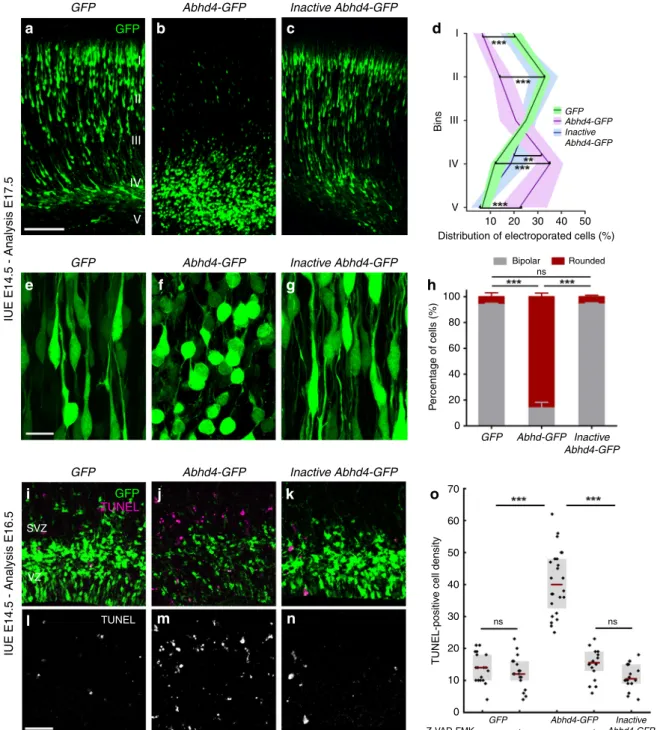
Because Abhd4 expression is rapidly downregulated in delaminated daughter cells right after their fate-commitment (Fig. 3o–q), we reasoned that counteracting this process by ectopic Abhd4 expression in these cells could shed light on the conserved function of ABHD4 in the developing brain. To test this idea, we in utero electroporated either GFP alone, or wild- type Abhd4-GFP, or a catalytically inactive form ofAbhd4-GFP (in which the catalytic serine residue that is required for hydrolase activity was mutated to glycine, S159G mutant) at E14.5. Interestingly, ABHD4, but not the inactive, hydrolase-dead form of ABHD4 caused a striking migration arrest within the SVZ similar to that of found after adherens junction disruption (Fig. 5a–d). Moreover, Abhd4-GFP-transfected cells lost the characteristic bipolar morphology of migrating neuroblasts and became shrunken, rounded, and had no leading processes (Fig. 5e–h). These morphological changes are hallmarks of imminent cell death. Indeed, we observed that ABHD4 alone, but not its inactive form could trigger a robust (295%) increase in caspase-dependent apoptosis (Fig.5i–o) when compared toGFP- electroporated controls, indicating that the enzymatic activity of ABHD4 is sufficient to elicit apoptosis in the delaminated cells.
A shift in the balance of proapoptotic and pro-survival molecular mechanisms is critically important in anoikis-related cell fate decisions during transition of cells to a metastatic phenotype32. We reasoned that some daughter cells whose transfection coincided with their delamination should already express the essential pro-survival molecular toolset to escape from anoikis and may be capable to balance the impact of exogenous Abhd4over-expression by its direct downregulation. To test this possibility, we next co-electroporated tdTomato either with a fusion protein construct (Abhd4-GFP-F) or with the bicistronic construct (Abhd4-GFP). Notably, we found that initially GFP expression in case of both of these constructs almost completely overlapped with the co-electroporated tdTomato marker 24 h after co-electroporation (Supplementary Fig. S9a–f). In striking contrast, only tdTomato-expressing, but GFP-negative cells were observed with the fusion construct in layer 2/3 in long-term survival experiments, whereas tdTomato extensively colocalized with GFP in case of the control bicistronic construct from which translation of ABHD4 and GFP occurs separately (Supplementary Fig. S9g–k). This observation suggests that only those postmitotic neurons could undergo delayed migration by postnatal day 3 that could degrade their ABHD4-GFP fusion protein and indicate that ABHD4 activity is non-permissive for radial migration into the cortical plate.
To determine how powerfully ABHD4 drives cell death even without a native tissue context (e.g., in the absence of additional proapoptotic signals), we next examined Human Embryonic Kidney-293 cells, which are relatively resistant to proapoptotic stimuli due to their deregulated pRB/p53 pathway and also lack endogenous Abhd4 expression33. Correlated confocal and STORM super-resolution imaging revealed that ABHD4, but not its inactive form caused a substantial loss of TOM20-positive mitochondria (Supplementary Fig. S10a–f, g) and a concomitant rise in extra-mitochondrial cytochrome c levels, signs of early stage apoptosis (Supplementary Fig. S10e, f, h–j). Moreover, ABHD4 also elicited a large increase in the number of cleaved caspase 3-immunopositive cells exhibiting nuclear fragmentation and condensation, features of late stage apoptosis (Supplementary
Fig. S10k–q). In contrast, the adjacent untransfected cells remained unaffected (Supplementary Fig. S10r). These results indicate that ABHD4 is capable of triggering an apoptotic program cell-autonomously even in cell types with a higher apoptotic threshold.
ABHD4 is necessary for developmental anoikis. It is well- established that cell type-specific consecutive waves of
developmentally controlled programmed cell death delete a sub- stantial proportion of neurons during cortical development34–36. Considering the proapoptotic activity of ABHD4, we therefore investigated the basal level of cell death in the developing neo- cortex. Notably, there was no difference in the density of dead cells in the subventricular and VZs of wild-type and Abhd4- knockout mice at embryonic day 16.5 (Fig. 6a–e). Devel- opmentally controlled programmed cell death of glutamatergic cells peaks early postnatally35but we could detect no differences a
d
g
m n
h e
+/+
+/+
+/+
+/+
Nestin STORM
–/–
–/–
phalloidin
+/+
–/– 150
120
BrdU-positive cell density
90 60 30 0
ns ns
25 20 15
Normalized PAX6 intensity
10 5 0
1.6 280
240 200 160 120 80 40 0
o p
1.4
ns ns
1.2 1 0.8
Normalized Nestin LP density Nestin bundle FWHM (nm)
0.6 0.4 0.2 0
+/+ –/– +/+ –/–
+/+ –/–
+/+ –/–
–/–
–/–
–/–
STORM Nestin BrdU
PAX6
Nestin
+/+
b c
f
i j
l k
Fig. 4 ABHD4 is not required for classical radial glia progenitor cell functions. a,bCoronal sections of the embryonic cortex at E.14.5 show replicating cells that incorporated bromodeoxyuridine (BrdU) during S phase of the cell cycle are shown.cQuantification of BrdU-positive cell density (two-sided Student’s unpairedttest,P=0.323;n=38 sections fromn=6 animals per wild-type (+/+) mice,n=29 sections fromn=4 animals perAbhd4- knockout (−/−) mice).d,eConfocal images andfquantification of PAX6-positive radial glia progenitor cells in the ventricular zone. (two-sided Student’s unpairedttest,P=0.2598;n=10 sections fromn=4 animals per wild-type (+/+) mice,n=10 sections fromn=4 animals perAbhd4-knockout (−/−) mice).g,hIdentical pattern of phalloidin-labeling of the adherens junction belt in the ventricular zone (VZ) of wild-type (+/+) andAbhd4-knockout (−/−) mice at E15.5.i,jLow-power confocal images show similar organization of nestin-positive radial processes in both genotypes at E15.5.k,lCorrelated confocal and STORM super-resolution microscopy images from both genotypes.m,nSTORM super-resolution images reveal that the nanoarchitecture of nestin intermediatefilament bundles at the core of the radial glia processes remains intact in the absence of ABHD4.oDensity of localization points (LPs) representing nestin (two-sided Student’s unpairedttest,P=0.297;n=39 segments fromn=3 animals per wild-type (+/+) mice,n=57 segments from n=3 animals perAbhd4-knockout (−/−) mice).pQuantification of full-width-at-half-maximum of nestinfilament bundles (two-sided Mann–WhitneyU test,P=0.684;n=28 segments fromn=3 animals per wild-type (+/+) mice,n=37 segments fromn=3 animals perAbhd4-knockout (−/−) mice).
Graphs show box-and-whisker plots (including minima, maxima and mean values with 2 × standard error, exceptpwhere median and lower, upper quartiles are presented) with single values. Scale bars:a–e,i–j: 50μm,g–h: 20μm,m,n: 200μm. Source data are provided as a Source Datafile.
GFP
Abhd4-GFP GFP
IUE E14.5 - Analysis E17.5IUE E14.5 - Analysis E16.5
I II III
IV V
a b c
e
i
SVZ
TUNEL GFP TUNEL
VZ
l
j
m n
k
f g
d
h
o Inactive Abhd4-GFP
I
II
GFP Abhd4-GFP Inactive Abhd4-GFP Bins III
IV
V
100 80
70 60 50 40
TUNEL-positive cell density
30 20 10
0 GFP
– + – +
Abhd4-GFP Inactive Abhd4-GFP Z-VAD-FMK
60
Percentage of cells (%)
40 20 0
GFP Abhd-GFP Inactive Abhd4-GFP 10
Distribution of electroporated cells (%) 20
Bipolar ns
Rounded 30 40 50
Abhd4-GFP
GFP Inactive Abhd4-GFP
Abhd4-GFP
GFP Inactive Abhd4-GFP
ns ns
Fig. 5 ABHD4 is sufficient to trigger migration arrest and apoptosis.Cells in utero electroporated withGFP-(a), or with anAbhd4-GFP-construct encoding an enzymatically inactive form of ABHD4 (c) migrate into the cortical plate, whereasAbhd4-GFP-electroporation causes radial migration defect (b).dLaminar distribution of electroporated cells infive equally-sized bins (Roman numerals) (Kruskal–Wallis test with post hoc Dunn’s test, 1st, 2nd, 4th, 5th bins for all comparisons: ***P< 0.0001, except 4th bin (Inactive Abhd4-GFPvsAbhd4-GFP): **P=0.006;n=19 sections fromn=4 animals perGFP- electroporation;n=18 sections fromn=4animals perAbhd4-GFP-electroporation;n=12 sections fromn=3 animals per InactiveAbhd4-GFP- electroporation). Data are shown as median (line) and interquartile range (transparent band in the same color). High-power images show bipolar migrating neurons (e,g), that become rounded uponAbhd4-GFP-electroporation (f).hQuantification of cell morphology (Kruskal–Wallis test with post hoc Dunn’s test, ***P< 0.0001; ns=not significant,P≈1;n=15–15 sections fromn=3–3 animals per treatment). Graphs show bar plots with median ± interquartile range.i–nRepresentative images show large density of TUNEL-positive cells uponAbhd4-GFPelectroporation in the subventricular (SVZ) and ventricular (VZ) zone (j,m), that is not replicated byGFP-(i,l), or by inactiveAbhd4-GFP-electroporation (k,n).oQuantification of TUNEL-positive cell density (Kruskal–Wallis test with post hoc Dunn’s test, ***P< 0.0001; ns=not significant,P≈1 except perAbhd4-GFP-electroporation with Z-VAD-FMK treatment vsInactive Abhd4-GFP P=0.607;n=18–18 sections fromn=3–3 animals perGFP-electroporation with and without Z-VAD-FMK treatment and per InactiveAbhd4-GFP-electroporation;n=24 sections fromn=4 animals perAbhd4-GFP-electroporation;n=18 sections fromn=4 animals perAbhd4- GFP-electroporation with Z-VAD-FMK treatment). Graphs show box-and-whisker plots (including minima, maxima, and median values, lower and upper quartiles) with single values. Scale bars:a–c: 100μm,e–g: 20μm,i n: 50μm. Source data are provided as a Source Datafile.
in cell death levels between the genotypes at postnatal day 3 either (Supplementary Fig. S11).
Importantly, physiologically controlled forms of programmed cell death or apoptosis induced by pathological insults do not necessarily share the same molecular mechanisms. Because our results have revealed that both adherens junction disruption- induced delamination and ectopic ABHD4 enzymatic activity in normally delaminating cells cause similar caspase-dependent cell death in the embryonic neocortex, we next tested the possibility that ABHD4 serves as a specific molecular link between pathological
detachment and subsequent cell death. To this end, ΔnCdh2-GFP was electroporated into the lateral ventricles of wild-type and littermate Abhd4-knockout embryos. Sparse adherens junction disruption initiated a marked increase in the density of dead cells that was restricted to the VZ and SVZs (see distribution analysis in Fig. 6m) of wild-type mice and was completely absent in Abhd4- knockout animals (Fig.6f, g, h, i, l). Importantly, this increase in cell death could be fully rescued by Abhd4 re-expression in Abhd4- knockout mice (Fig. 6j, k, l). These findings demonstrate that ABHD4 is not required for developmentally controlled Basal cell death
IUE E14.5 - Analysis E16.5
ΔnCdh2-GFP
+/+ –/–
CP
12
ns
ns 10
8 6 4 2 0
60 50 40 30 20 10 0
I
Abhd4 +/+
Abhd4 –/–
Rescued II
Bins III
IV
V 5
Distribution of TUNEL-positive cells
10 15 20 25
+/+ –/– Rescued
+/+ –/–
IZ
SVZ VZ
TUNEL-positive cell densityTUNEL-positive cell density
TUNEL
TUNEL GFP TUNEL
+/+
Rescued –/–
–/–
–/–
–/–
+/+
TUNEL
SVZ
VZ
a
c
f
h
j
i
k
b e
l
m d
g
Fig. 6 ABHD4 mediates apoptosis induced by adherens junction disruption. a–dBasal level of cell death is similar in coronal sections of the embryonic neocortex of wild-type (+/+) andAbhd4-knockout (-/-) mice at E16.5. CP, cortical plate; IZ, intermediate zone; SVZ, subventricular zone; VZ, ventricular zone.eQuantification of TUNEL-positive cell density in the VZ/SVZ (two-sided Student’s unpairedttest,P=0.6964;n=30 sample fromn=5 animals from both genotypes).f,g,i,j,ΔnCdh2-GFP in uteroelectroporation (IUE) triggers cell death in wild-type (f,i), but not inAbhd4-knockout mice (g–j) in the subventricular (SVZ) and ventricular (VZ) zones.h,kRe-expression ofAbhd4-GFPinAbhd4-knockout mice is sufficient to rescue impairedΔnCdh2-GFP- induced cell death.lQuantification of TUNEL-positive cell density (Kruskal–Wallis test with post hoc Dunn’s test, ***P< 0.0001; ns=not significant,P≈1;
n=32–32 sections fromn=4–4 animals per genotypes,n=28 sections fromn=4 animals perAbhd4-re-expression treatment). Graphs show box-and- whisker plots (including minima, maxima, and mean values with 2 × standard error, exceptlwhere median and lower, upper quartiles are presented) with single values.mDistribution of TUNEL-positive cells infive equal bins (Kruskal–Wallis test with post hoc Dunn’s test, 4th, 5th bins: ***P< 0.0001;Abhd +/+vs. RescuedP=1). Data are shown as median (line) and interquartile range (transparent band in the same color). Scale bars:a–d: 100μm,f–k: 50μm.
Source data are provided as a Source Datafile.
programmed cell death, but it is essential for developmental anoikis, a unique form of cell death triggered by the damage of cadherin- based adherens junctions in the prenatal brain.
Maternal alcohol exposure causes ABHD4-dependent cell death. The most common preventable teratogenic insult in humans is alcohol. Fetal alcohol spectrum disorder has a remarkably high prevalence and it is the foremost non-genetic cause of intellectual disabilities. Prenatal ethanol exposure can cause several brain malformations including cortical dysplasia/
heterotopias and microcephaly, all indicating impaired migration and neurogenesis16. Importantly, ethanol is known to induce disruption of the adherens junction signaling machinery, to reduce the RGPC pool and to increase cell death, including anoikis15,16,37,38. Therefore, we hypothesized that ABHD4 may play a role in cell death associated with prenatal alcohol exposure.
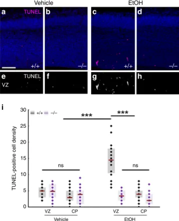
We tested this idea by applying two independent ethanol administration protocols to pregnant mice. Both a 3-day-long subchronic ethanol administration regime (Fig. 7) and a single
dose of acute ethanol administration (Supplementary Fig. S12a–i) resulted in a substantial (3–6 fold) increase in cell death and led to the scattered distribution of dead cells restricted to the VZs and SVZs in wild-type embryos. We did not detect elevated cell death in the cortical plate suggesting that mostly the proliferating progenitor pool was affected in the embryonic brain (Fig.7i). In striking contrast, maternal ethanol administration failed to induce a similar increase in cell death in Abhd4-knockout litter- mate embryos (Fig. 7d, h, i; Supplementary Fig. S12d, h, i).
Finally, we did not observe any increase in Abhd4 expression levels after ethanol-exposure (Supplementary Fig. S12j–l). These results strongly suggest that ABHD4 is also indispensable for ethanol-induced cell death in the embryonic brain.
Discussion
In the present study, we provide evidence for the existence of developmental anoikis, a distinctive mechanism for cell death in the prenatal brain. Ourfindings also identify ABHD4, an enzyme that has a vital function in this phenomenon by preventing the survival of misplaced cells that are produced by delamination errors or damaged by alcohol exposure (Fig. 8). Various envir- onmental stressors are known to affect the fetal brain and con- sidering the immense number of cell division events and subsequent delamination steps (>1011), the frequency of terato- genic and spontaneous delamination errors is likely to be con- siderably higher than the observed incidence of congenital brain anomalies (~0.25%,https://eu-rd-platform.jrc.ec.europa.eu/
eurocat/eurocat-data/prevalence). Thus, it is conceivable to pos- tulate the pivotal neurological importance of ABHD4-dependent developmental anoikis which eradicates those pathologically detached cells that may represent a risk for future brain mal- formations, such as dysplasias, heterotopias, or even brain tumors.
It is important to emphasize that RGPCs have a neuroepithelial origin. These proliferating cells maintain most of the epithelial characteristics including the apico-basal polarity via specific molecular anchors to the extracellular matrix and to each other6.
TUNEL
TUNEL VZ
i 30
+/+ –/–
ns ns
25 20
TUNEL-positive cell density
15 10 5 0
VZ
Vehicle EtOH
CP VZ CP
a
+/+ –/– +/+ –/–
b c d
e f g h
EtOH Vehicle
Fig. 7 ABHD4 is necessary for cell death caused by fetal alcohol exposure.Compared to vehicle-treated mice (a,b,e,f), increased number of TUNEL-positive dead cells is seen in ethanol-treated wild-type (+/+), (c,g), but not inAbhd4-knockout (−/−) mice (d,h) in the ventricular (VZ) zones of embryos derived from dams undergoing subchronic alcohol treatment, but not in the cortical plate (CP).iQuantification of the density of TUNEL-positive cells in the VZ and in the CP after subchronic ethanol- treatment (Kruskal–Wallis test with post hoc Dunn’s test, ***P< 0.0001; ns
=not significant,P≈1,n=31 sections fromn=5 animals per wild-type, vehicle-treated mice,n=27 sections fromn=4 animals perAbhd4- knockout, vehicle-treated mice,n=24 sections fromn=4 animals per wild-type, ethanol-treated mice,n=18 sections fromn=3 animals per Abhd4-knockout, ethanol-treated mice). Graphs show box-and-whisker plots (including minima, maxima, and median values, lower and upper quartiles) with single values. Scale bars:a–h: 100μm. Source data are provided as a Source Datafile.
Normal delamination and migration
Neurogenesis AJ VZ SVZ CP
Abhd4 expression
Abnormal detachment and cell death
b a
Fig. 8 Mechanism of ABHD4-dependent developmental anoikis. a,b Schematic drawing illustrates the distinct ABHD4-dependent fate of normally delaminated and abnormally detached cells in the developing cerebral cortex. After cell division, healthy daughter cells downregulate ABHD4, delaminate from the ventricular wall and migrate into the cortical plate along the radial processes of RGPCs. Random developmental errors and pathological insults such as fetal alcohol exposure often destroy the adherens junctions and trigger pathological detachment. As a central mediator of developmental anoikis, ABHD4 is necessary and sufficient to induce cell death that prevents the accumulation of misplaced progenitor cells at ectopic locations. CP cortical plate, SVZ subventricular zone, VZ ventricular zone.
Right after cell division, the delaminating non-RGPC-fated daughter cell dismantles its cell–cell connections, switches its transcriptional profile and undergoes a characteristic cytoskeletal reorganization to initiate migration. It is increasingly appreciated that several molecular components and the cellular processes that underlie daughter cell delamination are very similar in the so- called epithelial–mesenchymal transition phenomenon8,39. Our study further adds to this idea, because the ABHD4-dependent cell death mechanism described here is conceptually analogous to anoikis, a detachment-induced form of apoptosis that has long been considered as the central protective mechanism in epithelial–mesenchymal transition to prevent the proliferation and migration of pathologically detached epithelial cells20,21,40,41. Although the term has originally been coined for intrinsic apoptosis induced by the loss of the extracellular matrix- dependent anchorage of epithelial cells20,21, we propose to call this process developmental anoikis, because of its conceptual similarity and its functional importance during development.
Traditionally, anoikis has primarily been implicated as a protec- tive mechanism in tumor biology and obtaining resistance to anoikis is a critical step for cancer cells for tumor invasion and metastasis21,22,41. Thus, the results that ABHD4 is a proapoptotic molecule whose expression rapidly disappears in daughter neu- roblasts and coincidentally, that ABHD4 downregulation in immortalized prostate epithelial cells prevents cell death28toge- ther raise the intriguing possibility that a similar mechanism underlies anoikis resistance in healthy migrating neurons and in metastatic cancer cells. From a clinically relevant perspective, another interesting related finding is that ethanol-induced cell death also requires ABHD4 in the embryonic neocortex. Because alcohol turns on the epithelial–mesenchymal transition program that is thought to contribute to the metastatic aggressiveness of epithelial cancers associated with chronic alcohol consumption42, ABHD4-dependent cell death may play an important role in the attenuation of tumor progression. Notably, data from expression arrays indicate that theAbhd4gene is an important downstream target of the tumor suppressor protein p53 in cancer cells at the transcriptional level43,44. Thus, it remains to be investigated in future studies whether and how p53 and other related tran- scriptional regulators contribute to the tightly controlled spatial and temporal expression ofAbhd4mRNA in the developing brain and under pathological conditions.
At the biochemical level, ABHD4 is involved in N-acyl- ethanolamine and related N-acyl-phospholipid metabolism including the production of the endocannabinoid molecule anandamide23,45. Anandamide has various physiological func- tions including the regulation of synaptic plasticity and apopto- sis46. Interestingly, NAPE-PLD, another anandamide- synthesizing enzyme is primarily concentrated in presynaptic axon terminals47and its enzymatic activity only appears in the postnatal brain48. In contrast, our findings demonstrated that ABHD4 is present in radial glia progenitor cells in the prenatal brain, but it disappears postnatally. Together with the apoptosis- triggering function of ABHD4, it is plausible to propose that the multiple enzymatic routes of anandamide synthesis may have evolved to maintain a division of labor of anandamide signaling in time and space. It is likely that in the context of these two very distinct biological phenomena, synaptic plasticity and apoptosis, anandamide signaling requires different regulatory mechanisms, which can be achieved by independently controlling gene expression and enzymatic activity of the different molecular components of the endocannabinoid system.
Beside the potential proapoptotic role of the signaling molecule anandamide produced by ABHD4 activity46, one must also consider the function of N-acyl-phosphatidylethanolamines (NAPEs), which are the direct substrates of ABHD445. Early
biophysical studies reported a stabilizing effect of NAPEs on the integrity of biological membranes and increasing the NAPE content of liposomes inhibits their leakage49,50. Thus, ABHD4, as a NAPE lipase may also impair intracellular membrane com- partments by hydrolyzing NAPEs. This, combined with ana- ndamide triggering apoptosis via inducing endoplasmic reticulum stress51 makes it tempting to speculate that the synergistic upstream and downstream legs of ABHD4 activity in combina- tion would represent a very efficient and parsimonious mechan- ism that renders damaged cells more susceptible to additional proapoptotic processes. In light of the well-described proa- poptotic role of phosphatidylserine however, the potential sig- nificance of the N-acyl-phosphatidylserines, another substrate class of ABHD445, is also worthy of consideration in future studies.
Elucidation of the unique biological function of ABHD4 in the embryonic cerebral cortex will not only help to identify the specific lipid mediators and promote the discovery of additional molecular players cooperating with ABHD4, but will also pave the way to delineate the precise biochemical cascades and bio- physical processes of developmental anoikis. This is important, because the present findings indicate that ABHD4-mediated developmental anoikis is a safeguarding mechanism that protects the fetal brain from the effects of delamination errors. In this regard, developmental anoikis can be considered as a phenom- enon that contributes to biological robustness that helps to maintain the intactness of brain development against external and internal perturbations52,53. The astronomical number of cell divisions combined with the numerous detrimental environ- mental factors potentially affecting the developing brain puts an enormous stress on progenitor cells and inherently imply a relatively high probability of the occurrence of aberrant cell division and pathological detachment events. Because even occasional delamination errors could cause severe brain mal- formations due to misplaced proliferation of progenitor cells, protective mechanisms are likely to be very important to coun- terbalance the risk presented by the immense number of cell divisions and various environmental teratogens. Thus, ABHD4- mediated developmental anoikis may conceptually function similarly to other well-known biological phenomena such as the DNA repair mechanisms, the p53-pathway against genotoxic stress, or the unfolded protein response for protein quality con- trol. In summary, further research on the detailed mechanisms of developmental anoikis will therefore not only gain insights into neurodevelopmental processes underlying congenital brain anomalies and the related neurological and cognitive deficits, but could also extend our knowledge on biological robustness during cortical development.
Online methods
Animals. All experiments were approved by the Hungarian Committee of the Scientific Ethics of Animal Research (license numbers: XIV-1-001/2332-4/2012 and PE/EA/354-5/2018), and were carried out according to the Hungarian Act of Animal Care and Experimentation (1998, XXVIII, Section 243/1998), in accordance with the European Communities Council Directive of 24 November 1986 (86-609-EEC; Section 243/1998). Mice were kept under approved laboratory conditions and all efforts were made to minimize pain and to reduce the number of animals used. Both male and female mice were used throughout the study.
C57BL/6 and CD-1 mouse lines were obtained from Charles River Laboratories. Mice bearing a disruption in theAbhd4gene were generated from the 129S6/SvEvTac strain and were back- crossed into the C57BL/6 background for more than ten gen- erations prior to present experiments45.