The Role and Activities of Pseudopodia during Morphogenesis of the Sea Urchin Larva
TRYGGVE GUSTAFSON The Wenner-Gren Institute, University of Stockhohn, Steeden
We have quite sophisticated ideas about how deoxyribonucleic acid (DNA) codes ribonucleic acid (RNA), how RNA codes protein synthesis, how enzymes bring about metabolic reactions, and how metabolites regulate these events, but we are still ignorant about the final steps in the causal chain between the genes and the shape they control. In order to bridge the gap between molecular biology and morphology it seems logical to try to define the cellular forces that bring about changes in shape and how the forces become properly directed. If we could show that a morphogenetic process can be reduced, for example, to pseudopod activity, we could later on try to define the molecular basis for this ac- tivity, and how it becomes established at the right time and place. T h e sea urchin embryo is very useful for such studies: it has relatively few cells, there is little growth or cell division during the main morphogene- tic events, it is transparent, and its walls are only one cell layer thick.
It rapidly develops into a shape which is sufficiently complex to pose important and puzzling morphogenetic problems, but is not so complex as to overwhelm the observer. Our main experimental approach has been to study the development by means of time-lapse cinematography. A simple technique was worked out for holding the larvae in a constant position during filming. For bibliography of our papers see our review (Gustafson and Wolpert, 1963) where also the work of our colleagues from Japan, particularly Dan and Okazaki, is discussed.
The main morphological changes of the larva are summarized in Fig.
1 and comprise changes in thickness and curvature of the wall, strong, directed extension of some regions, release of cells from the wall which migrate and form a more-or-less regular mesenchyme pattern, the laying down of a complicated skeletal system, and the perforation of cell sheets and the surrounding hyaline membrane to give rise to a mouth. As to the forces that bring about these changes, moderate deformations of the wall are explicable in terms of forces arising within the cell sheet them- selves, "intrinsic forces." Changes in cell contact due to changes in ad-
333
hesion and/or tension in the cell membrane and changes in adhesion to the hyaline membrane appear to be responsible for variation in thickness and curvature of the wall and the release of cells from it. Strong deforma- tions of the wall, on the other hand, require "extrinsic forces," i.e., forces taking their origin outside the cell sheet. T h e main mechanism for these
"directed long-range translocations of a cell sheet," and also for the migration of 'free cells, is thus related to pseudopodial activities.
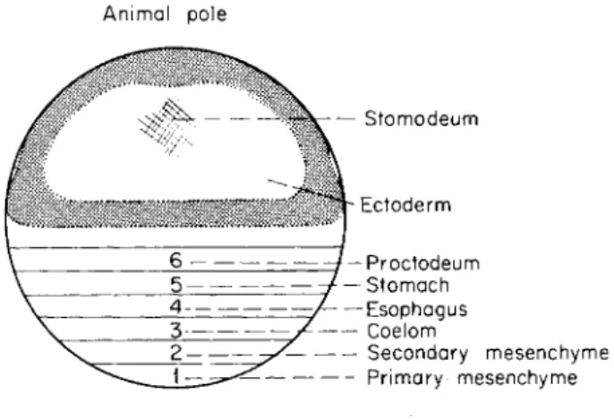
A map of the prospective significance of the different zones of the egg is shown in Fig. 2. Zone 1 gives rise to the primary mesenchyme cells
e f g h FIG. 1. Diagrammatic drawings of some stages of a developing sea urchin larva to show the main morphological changes, (a) An early mesenchyme blastula; the cells at the vegetal pole lose adhesion for each other and enter the blastocoele to form the primary mesenchyme, (b) The primary mesenchyme cells migrate by means of filo- pods. (c) An early gastrula in side-view, the primary mesenchyme cells have ar- ranged themselves into a characteristic pattern, and the archenteron rudiment has formed a rounded invagination due to a decrease in contact and hence a change in packing of its cells, (d) T h e rounded invaginated region extends by means of pseudo- podial activity at its tip; note the "cones of attachment" in the ectoderm formed where pseudopods attach, (e) T h e pseudopod-forming cells have detached from the archenteron tip which begins to attach to the mouth region by means of pseudopods.
(f) T h e archenteron tip is in direct contact with the mouth region where the ecto- derm is slightly invaginated. T h e uppermost region of the archenteron tip will later on become delineated by a constriction to form a sac. (g) (Ventral view) the sac has become extended and subdivided into two sacs by means of pseudopodial activity.
A skeleton has also become laid down by the primary mesenchyme, and the "arm buds" begin to protrude and will later on become extended due to the tension pro- duced by the growing skeleton, (h) (A pluteus larva) the mouth is completely formed, the archenteron is subdivided by constrictions, and the skeleton has extended the wall into characteristic processes, the four arms and the dorsal Scheitel. T h e coelom has become pulled out and subdivided into two sacs. Pseudopod-forming cells from the coelom pull out themselves and become free mesenchymal strands that line up around the esophagus and form a contractile network around it. T h e pseudopods have also extended the coelom (normally the left one) into a tube, the primary pore-canal.
Activities of Pseudopodia during Morphogenesis 335 which will lay down the skeleton. In the late blastula they begin to lose adhesion for each other and for the hyaline membrane and begin to round up and show a pulsatory activity. Thus they tend to force each other apart and enter the blastocoele (Fig. la). They pile up at the vegetal pole and then spread along the blastula wall to form a regular pattern comprising a ring with two clusters from each of which a branch of mesenchyme is formed (Fig. lc). The formation of this pattern of mesenchyme appears to require directed cell migration, and the sea ur-
Animal pole
— Stomodeum
τ Ectoderm Proctodeum Stomach Esophagus Coelom
Secondary mesenchyme Primary mesenchyme Vegetal pole
FIG. 2. A map of the presumptive regions of the egg. All the zones indicated have not yet been mapped by vital staining technique, cf. Hörstadius (1935), and the diagram should be considered as an extrapolation from available results. The rela- tive proportions of zones 1-6 is overscaled for convenience. T h e stippled zones cor- respond to thickened regions of the ectoderm, but must be considered entirely sche- matic.
chin larva, therefore, provides an almost unique system for studying such a process in vivo.
The piled up cells gradually begin to "shoot out" numerous filopods into the blastocoele (Fig. lb). T h e filopods are about 0.5 μ in diameter, but may be thinner. They may reach a length of 30-40 μ and can extend as quickly as 30 μ in 3 min. There is a considerable variability in their arrangement. The first ones project separately from the cell surface, but later on they may be confined to a small protrusion from the advancing end. Although quite thin, they may be stiff enough to be moved bodily across the blastocoele like long, thin bristles. They may also bend and show wavelike motions at their base, which contribute to their ability to explore the blastocoele and its wall. In the thickest filopods one may see a traffic of granules up and down. T h e filopods can attach to the wall,
but only with their tips. Upon attachment, the extension of the filopods appears to stop.
T h e filopods provide the basis of cell movement throughout the mi- gration of the primary mesenchyme. When a filopod has attached to the wall it contracts and brings about a movement of the cell body. Filopods that do not make contact or make very unstable contacts may continue to probe the wall but, more often, rapidly collapse (e.g. 25 μ shortening in 8 min) and are withdrawn into the cell body, or form an irregular mass, apparently incapable of reorganizing into a new filopod at once.
T h e filopodial exploration, attachment, and contraction does not only account for the cell movement, but also for its apparent directedness and thus for the development of the regular mesenchyme pattern. T h e filo- pods shoot across the blastocoele, and, since this is apparently liquid, they cannot be guided either by it or the wall. T h e random manner in which they shoot out is a further indication of this lack of guidance.
Their subsequent exploration also lacks any obvious directedness and appears to be quite random. T h e cells also show no tendency to continu- ous polarized movements along the wall, and after moving in one direc- tion they may move in any other. They may, therefore, transiently show a considerable dispersal and deviation from the final ringlike arrange- ment. Sometimes they migrate halfway to the animal pole, but sooner or later return to the ring level if they have surpassed it. T h e direction of their movements is, in fact, determined by the contraction of those filo- pods that make sufficiently stable contacts with the wall. If only one filopod is formed and brings about a movement, filopods formed later on may move the cell in another direction. If there are many filopods there is, in a sense, a competition between them, each trying to move the cell in its direction and the final movement is determined by the relative strength of the contact between the filopods and the wall. T h e cell bodies will, therefore, finally line up along the zones of the ectoderm where the adhesiveness is highest and so form the characteristic pattern. T h e in- crease in order goes hand in hand with a progressive fusion of the filopods into a syncytium that attains the shape of a cable from which innumera- ble thin short filopods extend, exploring the wall together, and, there- fore, with a high probability of finding the most adhesive zone.
The early dispersal of the mesenchyme cells past the ring level and their subsequent return is most difficult to account for in terms of mecha- nisms based on contact guidance, but is easily explained in terms of ran- dom filopodial exploration and the gradual development of the pattern of selective adhesiveness of the ectoderm in relation to the mesenchyme cells, or in the adhesive properties of the filopods themselves. T h e varia- bility in the manner in which the final pattern is reached probably reflects
Activities of Pseudopodia during Morphogenesis 337 the lack of precision in the timing of various events, e.g., the release of the mesenchyme cells from the blastula wall, their formation of filopods, their development of adhesiveness, their fusion into syncytia, and the changes in the adhesiveness of the ectoderm. In such a mechanism as that postulated here, the same final state will be reached regardless of varia- tion in the timing of the individual events.
It would be interesting to know the nature of the adhesiveness of the ectoderm. Studies on slightly compressed larvae, where the ectoderm is extended and its cells partially separated, show that the cells are mutually connected only at their inner and outer ends, but where the contact is low there are only outer connections (Gustafson, 1963). A strong packing
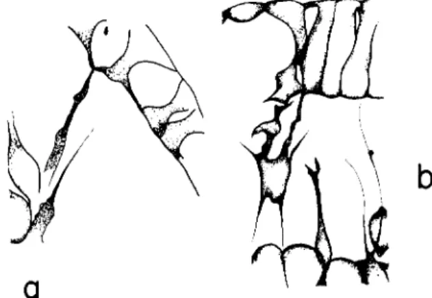
FIG. 3. Filopodial branches from a ventrolateral syncytial cable of the primary mesenchyme, and the filopodial attachments to the ectoderm at the inner points of contact between the ectoderm cells.
of the cells and hence a thickening of the ectoderm is evidently due to the presence of inner contact points. We have recently found that it is to these contact points that the filopods attach, particularly the inner ones (Fig. 3). T h e cell surface as a whole is not adhesive. This explains the important fact that the mesenchyme cells tend to line up along the thickened zones of the ectoderm. But why do the filopods attach so badly to the animal plate which is the thickest region? T o be accessible for the filopods, one may visualize that the adhesive surfaces must be partially uncovered by a tension in the sheet that tends to separate the cells or, that the tension is involved in some other way. In the animal plate the strong contact along the whole length of the cells may prevent changes.
Some justification for this hypothesis will be given later.
The ectoderm can evidently be regarded as a "template" for the mesenchyme pattern in the sense that it presents a pattern of variation in adhesiveness which is reflected in the distribution of the mesenchyme
cells, and the pseudopodial mechanism thus contributes to the coordina- tion of the embryo, quite as well as inductive stimuli do in other cases.
The mechanism for directed movement outlined here is not new in that it was suggested in general terms by Weiss (1947) as one of the three possible mechanisms by which cells could be guided, i.e., selective fixa- tion, selective conduction, and selective elimination. T h e observations we have described are perhaps the first clear-cut example of selective fixation, however, cf. Twitty and Niu (1958) and Twitty (1949). One of the novel features of a mechanism such as we have outlined is the use of long filopods to explore the substrate. The great length and number of the filopods and their slenderness make them a powerful and economic tool for this purpose. Another advantage is that they both localize the target and bring about the movement of the cells toward it. And, finally, they secure a constant end result in spite of an imperfect timing of the events in the embryo. It is, therefore, not surprising to find that pseudo- podial mechanisms also play a role in other events where directed long- range translocation is needed, cf. Nakai and Kawasaki (1959) and Nakai (1960).
Even when the mesenchyme pattern concerned is attained, pseudopods are continually making and breaking contacts with the ectoderm, there is a "turnover" of the contacts. T h e cells are now, however, joined by the cablelike structures and no longer move independently, but consider- able rearrangements are still possible, and as soon as the adhesiveness of the ectoderm changes, a corresponding change takes place in the distribu- tion of the cable-linked mesenchyme cells. These changes underly the development of the progressively complicating skeletal pattern. T h e skel- eton is formed as a triradiate crystal in the syncytial mass in the two mesenchyme clusters, and the three radii continue to grow within the syncytial cables and later on bend and branch in a characteristic way (Fig.
If—h). A crystallographic mechanism is responsible for the formation of its initial triradiate shape and for the tendency of its radii and branches to grow straight. T h e skeleton is, however, laid down with random orienta- tion, but within less than half an hour it attains a correct orientation so that two of its radii point along the ringlike cable and the third along a cable branch extending upward. This reorientation is brought about by the tension in the cables which meet at the point where the triradiate structure is laid down. T h e tension is produced by the filopods which are
"guided" by the adhesion of the ectoderm. Filopods attaching to adhesive regions are also responsible for all bends and branches of the skeleton including spine formation, since they deform the cable and bring about rearrangements of the mesenchyme.
It is interesting to find that experimental interference with the "vis-
Activities of Pseudopodia during Morphogenesis 339 cosity" of the filopods brings about a more complicated skeletal pattern
since the cells can form more complicated filopodial connections (cf. von Ubisch, 1957). Furthermore, mesenchyme cells from a species with a com- plicated skeleton transplanted to a species with a simpler skeletal pattern can, probably due to the characteristic "viscosity" of their cytoplasm, respond to adhesive regions which the host's own mesenchyme cells nor- mally do not detect, and a skeleton with some characteristics of the
"complicated" donor forms in the "simple" host (cf. von Ubisch, 1957).
Skeletal mesenchyme collects at some thickened regions of the ecto- derm, the "arm buds," and forms skeletal branches (Fig. lg), and the pressure of the growing branches with the mesenchyme at their tips extend the "arm buds" into long slender arms. Filopodial activity is thus, indirectly, responsible for the arm extension.
The archenteron rudiment corresponds to zones 2-6 in the diagram (Fig. 2). The first step in its invagination involves a decrease in contact between the cells (owing to changes in adhesion and possibly in tension) and their tendency for rounding up thus leads to a change in cell packing (Fig. lb and c). This change is particularly strong in zone 2 and is similar to that in zone 1, but the decrease in contact is not complete.
Pulsations also occur, probably due to the reduced contact, and these may help the invagination by breaking bonds between cells and by assist- ing in the transfer of cytoplasm from the outer to the inner edge. A further rounding up or change into pear-shape of the cells would only make the rudiment round up further and bring about no further elonga- tion, and invagination, therefore, tends to stop. The pulsatory activity of the cells, however, persists and may become extremely vigorous. Gradu- ally the pulsatory cells begin to shoot out pseudopods (filopods), often directly from the pulsatory "lobopodia." They therefore mainly shoot upward, though their direction is variable and they "explore" the blastocoele and the wall (Fig. Id). Many of the pseudopods attach to the wall. When they fail to do so, they tend to "collapse" and are withdrawn.
The pseudopods are of quite different dimensions and are often branched at their tips. T h e branches operate independently from each other in the sense that some of them may detach and collapse or continue to probe the wall, whereas other branches are still attached, holding the pseudopod extended. Sometimes the branches seem to fuse into a sheetlike perforated structure that seems to slide along the wall. The attached pseudopods shorten and exert a tension on the ectoderm and on the archenteron.
This tension is made evident by the formation of cones of attachment in the ectoderm, i.e., cells where the pseudopods attach tend to be pulled inward (Fig. Id), and sometimes the whole wall is deformed. The pseudo- pod-forming cells may also pull themselves out from the archenteron tip.
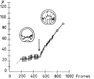
The correlation between the onset of pseudopodial activity and the onset of continued invagination is clear (Fig. 4) and, if the attachments of the pseudopods is prevented or the pseudopod-forming cells detach too early from the archenteron tip, the invagination stops. It is thus evident that it is the pseudopods that extend the archenteron. Calculations of the force required to bring about invagination have shown that they need only be very small, 1 0 ~2 dyne, and this is quite consistent with our knowledge of cellular forces. Work in progress with time-lapse cinematog- raphy in my laboratory by Dr. Shigeru Hamano, University of Hokkaido,
120
I
0 200 400 600 800 1000 F r a m e s
FIG. 4. Diagram of the invagination of the archenteron. T h e curve shows the length of the archenteron cavity plotted against time. T h e extent of the pulsatory activity is proportional to the length of the cross lines on the curve. T h e appearance of the first pseudopods at the archenteron tip is indicated by the arrow. The diagram shows the two steps in the process of invagination and the correlation between the onset of the second step and the appearance of pseudopods. A thousand frames* cor- respond to 5 hr, 33 min.
has shown that pseudopods also play a role in gastrulation of a fish em- bryo (Danio rerio) which is transparent enough to allow the observations of the cells in the mesendodermal region. This suggests that pseudopods play a more or less general role for long-range translocations during morphogenesis.
Gastrulation also involves the problem how movement becomes direc- ted, and one can, in principle, account for this problem in the same terms as the directed movement of the primary mesenchyme cells, i.e., in terms of random exploration of pseudopods for adhesive surfaces. In fact, both kinds of pseudopods attach to the inner contact points between the ectoderm cells (Fig. 5a) (Gustafson, 1963). That the archenteron ex-
Activities of Pseudopodia during Morphogenesis 3 4 1
tends along the dorsal and not along the ventral wall is probably partly a matter of relative distance (Fig. lc and d). The archenteron tip is brought closest to the dorsal wall owing to early changes in curvature of the ectoderm, and the probability for contacts is hence greatest there.
Furthermore, because of the extension of this wall, its cells tend to separate, and the contact points would, therefore, become partly un- covered and accessible for the pseudopods. This interpretation is sup- ported by the observation that a rupture in the animal plate makes it possible for pseudopods to attach there (Fig. 5b). When the invagination has proceeded almost to the animal pole, the pseudopod-forming cells
FIG. 5. (a) T h e attachment of an archenteron tip pseudopod at the contact points between adjacent ectoderm cells, (b) Relation between the animal plate (above) and pseudopods from the archenteron tip (below). T h e ectoderm cells in the animal plate are normally in close contact along the whole of their length, but owing to a rupture in the plate followed by a retraction of the cells to the left, an edge of a cell in the plate becomes available for pseudopod attachment.
detach and the archenteron tip thus becomes free (Fig. le). The remaining pseudopods attach to the mouth region which, for reasons still unknown, is very adhesive. T h e tension in these pseudopods is high and may pull the oral ectoderm inward, but also bends the archenteron to the mouth region where it becomes anchored (Fig. le and f). Gastrulation is a proc- ess that is quite variable, but the end result appears unaffected by this variability, and this is again explicable in terms of pseudopodial activity and adhesiveness of the wall.
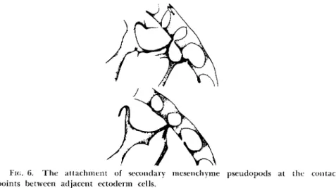
The secondary mesenchyme cells also migrate by means of pseudopods attaching to the points of junction of ectoderm cells (Fig. 6) (Gustafson, 1963), even to the same point as a primary mesenchyme filopod. That the two mesenchyme patterns are still not identical is probably because of the fact that the secondary mesenchyme cells are multipolar and their
pseudopods may attach at widely distant points of the wall making the pattern more irregular than that of the primary mesenchyme. In spite of this one may, with some imagination, see a similarity between the pattern of the two types of mesenchyme.
One class of the secondary mesenchyme cells are characterized by their content of pigment granules. These cells move by means of short pseudo- pods and long filopods. The granules can be used as markers for the cytoplasmic movements. We have seen how granules, thicker than the average diameter of a filopod, moved up through it to its tip, but the traffic was one way and the granulum remained at the tip when the filopod collapsed (Gustafson and Wolpert, unpublished). The pigment
FIG. 6. The attachment of secondary mesenchyme pseudopods at the contact points between adjacent ectoderm cells.
cells also illustrate how similar end results may be reached in different ways. In Psammechinus the pigment cells detach from the archenteron tip at the end of invagination, and later on some of them always collect at the arm tips. In Echinocardium the cells detach already before invag- ination has started and migrate in the spaces between the ectoderm cells, but their final distribution is similar to that in Psammechinus. This would again be the consequence of random filopodial exploration and attachment to adhesive regions within the ectoderm.
T o what extent do the principles discussed also account for apparently complicated processes later on in development? It seems that a dominant role is also played here by changes in cell contact, pulsatory and pseudo- podial activities. Much of the apparent complexity of these processes appears to arise from spatial relationships which make the study difficult.
The invagination of the archenteron was brought about mainly by activities in zone 2, the future secondary mesenchyme cells (Fig. 2). When
Activities of Pseudopodia during Morphogenesis 343 these have detached, the archenteron tip corresponds to zone 3, the coe-
lom region, which begins to show a pulsatory and pseudopodial activity as in the preceeding zones (Fig. lg). The pseudopods extend the sheet in a similar way as the archenteron is extended. Cones of attachment in the ectoderm indicate that they exert a considerable tension. The role of pseudopods in the coelom extension is illustrated by observations in vegetalized larvae where an everted coelomic rudiment often occurs at the vegetal pole. Pseudopods that may here shoot out into the sea water have no surface to attach to, and no extension occurs. On the other hand, if they attach to the slide, coelom sheet may become pulled out. The subdivision of the coelom into two sacs seems to be accounted for by the fact that the anterior sheet of the unpaired rudiment is attached to the oral ectoderm and by the inability of the pseudopods to attach to the animal plate (Fig. 8).
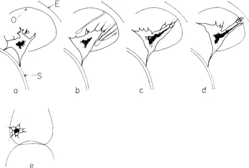
When the coelom sacs are pulled backward (Fig. lh), some of the pseudopod-forming cells pull themselves out of the coelom and form mesenchymal strands, a "coelom mesenchyme," attached to the esophagus wall (Fig. 7a and b). These strands and also the coelom pseudopods begin to show an intense, periodic contractility. The development of such a contractility in slowly contractile elements suggests a close relation be- tween the two types of contractility. Anyhow, the contractions apparently help the lining up of the strands around the esophagus. The situation may be likened to the tightening of elastic bands around a cylinder. Later on the strands form thin side-processes which fuse so that a muscular cage is formed around the esophagus, but the details of this process and how the contractility becomes peristaltic remains to be investigated. The great ability of the coelom mesenchyme strands to form side-links may reflect their origin from coelomic cells which normally attach to each other along the whole of their edges and form a sheet.
The backward extension of the coelom sacs along the esophagus by means of pseudopods (Fig. 7) is similar to gastrulation (Fig. Id). Since the left sac is larger than the right one it can be further extended and forms a tube, the direction of which would be determined in the same way as the direction of the free mesenchymal strands (Fig. 7). Finally, the pseudo- pods at the tip of the tube attach to the adjacent dorsal ectoderm and extend the tube right up to it. After that, these pseudopods have only the
"choice" to direct more or less radially (Fig. 7e), and their tension prob- ably contributes to break up the blind end of the tube as well as the ectoderm to which it attaches, leading to a free passage through the tube into the coelomic sac, and so the primary pore-canal has formed. T h e underlying processes appear to be simple and involve no novelty.
The double cell layer in the mouth region, the ectoderm, and the
coelom-esophagus sheet are thinned out and the cells are torn from each other by the rapid contractility in the coelomic pseudopods and the peristaltic activity of the esophagus muscles; and this, probably together with an enzymatic activity of the esophagus that softens the hyaline layer, finally causes a rupture of the double cell sheet, and thus the mouth perforation occurs (Fig. 8c-h). The invention of this important step does not appear to require basically new mechanisms, only a proper adjust-
FIG. 7. Semischematic drawings of a larva in profile, ventral side to the left, show- ing the rounded esophagus, O, and the left coelom sac which extends backward along the esophagus by means of pseudopods and finally forms a tube, the primary pore- canal that attaches to the dorsal ectoderm, E. In (e) the end of the tube is seen from the dorsal side; note the radiating pseudopods that tend to break up the end of the tube and the ectoderm to which the tube has attached. In (b) some of the mesen- chymal elements pulled out from the coelom are indicated.
ment of the time and intensity relations between the underlying mecha- nisms.
The following zone, 4, gives rise to the esophagus and zone 5 to the stomach (Fig. 2). The further morphogenesis of these zones also involves some pulsatory activity, but this is not followed by pseudopod formation.
However, their final shape is influenced by the contractile elements around the esophagus. For instance, the peristaltic activity pumps water into the thick-walled stomach rudiment which thereby becomes extended into a thin-walled vesicle (Fig. lh). T h e development of the sphincter
Activities of Pseudopodia during Morphogenesis 345 systems at the border between the compartments of the gut and the anal sphincter remains to be elucidated.
From our studies we got the general impression that the molding of the shape of the embryo is determined by the time-space distribution of a few cellular activities. Both the time when these activities become overt
a b c d
e
fg h
FIG. 8. Semischematic drawings to illustrate the development of the coelom and the mouth, (a-d) Larvae in optical median section [(c) somewhat to the right of the median plane]; (e) and (f) dorsoanimal view; (g) and (h) ventral view. T h e coelom rudiment becomes delineated and increases its contact with the ectoderm and be- comes pulled out by pseudopods into two sacs. As a result of the rapid periodic contractions in the pseudopods and the coelomic strands around the esophagus, the anterior coelom sheet and the ectoderm to which it attaches becomes torn and the cells separate leading to the formation of a mouth. T h e edges of the oral invagina- tion of the ectoderm is indicated by dotted contours in (h).
and their nature appear to be related to the position of the regions along the animal vegetal axis (Fig. 2). The changes are, in fact, time-graded in that decrease in contact, pulsatory activity, and pseudopod formation all start in zone 1 and progressively spread upward into the consecutive zones.
The "intensities" of the activities are also graded and fall off along the
axis although to a different extent. T h e decrease in contact is complete in zone 1, and the cells are released before pseudopods appear, whereas in zone 2 they have more or less to pull themselves out of the cell sheet, and in zone 3 only few cells can pull themselves out, most of them re- maining together forming a thin sheet, and in the following zones the cells keep together still more strongly. T h e pulsatory activity occurs in zones 1, 2, and 3, and also tends to occur in zone 4 and 5 at least. T h e pseudopod activity only occurs in zones 1, 2, and 3, occasionally in zone 4, and not in the following ones. It seems that the different activities are related, since a decrease in contact is always correlated with a pulsatory activity, and a strong pulsatory activity is followed by a pseudopod forma- tion. One may, therefore, suggest that these activities reflect quantitative variations of only one or two cellular parameters along the egg axis, perhaps in some properties of the cell membrane. It is possible that the cellular activities within the ectoderm can be added to this "spectrum"
of activities of the mesendoderm, since, also, the main morphogenetic differences between the ectodermal regions involve differences in cell con- tact. T h e primary mensenchyme and the animal plate cells would thus represent the opposite ends of this "spectrum," and the high adhesion between the ectoderm cells may also allow the mesenchyme pseudopods to adhere there, provided the contact points are accessible.
The justification for these extrapolations is that they urge us to think about pseudopodial activity in relation to other cellular phenomena.
And, we may also begin to visualize vaguely that there might be a close relation between the spectrum of morphogenetic activities of the cells and the biochemical and physiological gradients, so famous among embryologists. T o establish some kind of relation between the molecular and the morphological level was at least our point of departure and is still our goal. Some old results of ours, which seem to have stirred up some controversy through the years now appear to be closer to general acceptance. I refer to the role of mitochondria in differentiation and to the effect of the lower blastomeres upon the formation and distribution of mitochondria (Gustafson and Lenicque, 1952). Although electron mi- crographs only show some vague structural differences and no difference in the total number of mitochondria along the egg axis, the differences in Janus green staining (Czihak, 1962) indicate that the number of actively respiring mitochondria is greatest near the animal pole. (This is in line with our conclusions drawn in 1952 with regard to different stages in mitochondrial development.) T h e decrease in the proportion of ectoderm cell and the parallel decrease in "active" mitochondria brought about by subjecting the cleaving egg to lithium (Gustafson and Lenicque, 1952) perhaps follows from a block in the formation of functional RNA and
Activities of Pseudopodia during Morphogenesis 347 hence synthesis of new proteins, since chloramphenicol, which has this chemical effect, interferes with morphogenesis just as lithium does (cf.
Lallier, 1962; Hörstadius, 1963), and lithium retards the onset of formation of new proteins as chloramphenicol does in many systems.
If we return to adhesion, it seems that the high adhesiveness of the ectoderm cells corresponds to the formation of new proteins, perhaps related to the formation of actively respiring mitochondria, whereas cells with low adhesiveness—and also the ability to form pseudopods, perhaps a more primitive type of behavior—appear to have fewer respiring mitochondria. The relation among active respiration (or, more specifi- cally, oxidative phosphorylation), adhesion, and pseudopod formation will in any case be investigated. T h e ideas themselves only intend to symbolize a way of thinking about a link between molecular events and morphology.
One may ask how a system operating with such a restricted number of mechanisms may still give rise to such a sophisticated form as the pluteus larva. In the preceding discussion we have tried to outline the manner in which this is possible. A formal and perhaps amusing way to illustrate this problem more directly is to consider an irregular polygon for which one has only one rule: connect all junctions between lines. A progressively complicated pattern arises. This example also illustrates our basic approach which is essentially to view development as a historical process. One can only view, for example, the normal development of the coelomic sacs and the primary pore-canal as the results of a series of simple and partially interlinked steps, the results of the later ones being determined by what has occurred before and therefore more complicated and more difficult to predict. A correlate of this is that an early strong disturbance that is not corrected by the steady readjustments of the pseudopodial-ectoderm relations bring about the development of a shape that is hardly recognizable as a sea urchin larva, although all the instruc- tions are well considered by the cells. There is thus in one sense a certain
"all or none" character of normal development, the choice being deter- mined by the extent of the early disturbances. It is remarkable that the cells of dissociated early larvae can aggregate and later develop into a fairly normal larva (Giudice, 1962), but once a sorting out of cells and blastula formation have occurred there are no restrictions in the system that prevent the instructions from bringing about the whole series of consecutive processes of normal shape formation.
A special aspect of the self-complication is the possible inductive stimulus that the primary mesenchyme may emit to the ectoderm cells even after the blastula stage. T h e ectoderm acquires a pulsatory and pseudopodial activity in some regions of the ventral side, i.e., in the
curvature between the arm rudiments which are brought about by these activities. One may be permitted to suggest that the ectoderm cells have acquired certain mesenchymal properties as a result of their close earlier contact with the primary mesenchyme clusters.
But let us leave all these speculations and return to the central topic of this paper. I hope that this review has shown that pseudopods play an important role in morphogenesis, and that their advantage is con- nected with their ability to produce a force as well as to localize the target, even a distant one, and so to give the movement a proper direc- tion. And since they may be extremely thin and require little material they can be formed in large amounts. T h e pseudopods are evidently well- suited tools for integration of structures formed by regions far apart.
Their attachment does not require highly specific types of adhesiveness, and this makes the process of integration "cheap." Their continued ex- ploration, attachment, and detachment, furthermore, allow a rather ex- tensive variation and lack of precision of the underlying events in different regions and secure a rather constant end result. And, although my contribution to the understanding of central mechanisms of pseudo- pod activity has been more meager than it was intended to be, one may visualize that the pseudopod activity is closely related to other basic cellular activities.
REFERENCES
Czihak, G. (1962). Arch. Entwicklungsmech. Organ. 154, 29.
Giudice, G. (1962). Develop. Biol. 5, 402.
Gustafson, T . (1963). Zool. Bidrag Uppsala 35, 425.
Gustafson, T., and Lenicque, P. (1952). Exptl. Cell Res. 3, 251.
Gustafson, T., and Wolpert, L. (1963). Intern. Rev. Cytol. 15, 139.
Hörstadius, S. (1935). Pubbl. Staz. Zool. Napoli 14, 251.
Hörstadius, S. (1963). Develop. Biol. 7, 144.
Lallier, R. (1962). Experientia 18, 141.
Nakai, J . (1960). Z. Zellforsch. Mikroskop. Anat. 52, 427.
Nakai, J . , and Kawasaki, Y. (1959). / . Embryol. Exptl. Morphol. 7, 146.
Twitty, V. C. (1949). Growth 13, Suppl. 133.
Twitty, V. C , and Niu, M. C. (1958). / . Exptl. Zool. 108, 405.
von Ubisch, L . (1957). Pubbl. Staz. Zool. Napoli 30, 279.
Weiss, P. (1947). Yale J. Biol. Med. 19, 235.
DISCUSSION
DR. JAFFEE: I wonder how you fit exogastrulation into your view of how the archenteron extends?
DR. GUSTAFSON: T h e mechanism of exogastrulation is quite different from that of normal gastrulation. If we plot the length of the archenteron rudiment during normal gastrulation we get the curve in Fig. 4. If we plot exogastrulation we get a curve which starts off in the same way, but after the first phase of invagination the extension stops owing to a failure of the pseudopodial mechanism. Later on, when
Activities of Pseudopodia during Morphogenesis 349
there is an increase in the osmotic pressure within the blastocoele, the archenteron rudiment becomes reverted and extended outward. T h e elongation of the archen- teron rudiment during exogastrulation is thus brought about by an external force quite as the elongation during normal gastrulation, but it is an osmotic and not a pseudopodial force in normal larvae.
CHAIRMAN MARSLAND: Then it is an absence of pseudopodial activity at the critical time.
DR. GUSTAFSON: Yes; the archenteron rudiment does not become attached to the inside of the ectoderm, because of a premature release of the pseudopod-forming cells from its tip. T h e osmotic pressure that gradually increases in the blastocoele can, therefore, cause a reversion and outward extension of the archenteron rudiment. W e have many films that show this process.
DR. REBHUN: Has any electron microscopy been done on these pseudopods to see whether or not they do have internal structure of some sort?
DR. GUSTAFSON: I think Dr. Wolpert can answer this question. I have also some preliminary electron-microscopic observations of pseudopods. They look quite empty.
However, I know that Dr. Sylvan Nass has observed some structures inside the pseu- dopods. Another point of interest is that there are no signs of terminal bars or any other structure at the pseudopodial tips which may be involved in their attachments to the ectoderm.
DR. REBHUN: I wondered. Usually in electron micrographs, the microvilli appear empty. By several techniques, the freeze-thawing technique, and by use of acrolein fixation, it is possible to demonstrate rodlike structure within the center of almost all microvilli. Dr. Dougherty and I have looked at structures which I would tenta- tively suggest are similar to the microspikes of Dr. A. C . Taylor. They are not seen in formalin or ordinary osmium fixation.
DR. WOLPERT: We haven't looked at pseudopodia in great detail. Most pseudo- podia are seen in cross section. You sometimes see a central vesicle and begin to think it represents a central core. Dr. Mercer, with whom I have worked on this, prefers to believe it represents endoplasmic reticulum.
DR. REBHUN: In Dr. Dougherty's acrolein preparations, the cores look much more like Dr. Roth's "spindle tubules" than like endoplasmic reticulum.
DR. WOLPERT: I think Dr. Gustafson pointed out that you often find a granule moving rapidly foward in a pseudopod, and in certain cases moving back again. This argues against any fixed internal structure.
DR. GUSTAFSON: Yes, and the granules may be much thicker than the filopod causing a local extension. So I think there is no room for any thick solid rods within the filopods.
DR. NAKAI: I studied some electron micrographs of pseudopods from the nerve fiber in tissue culture, and the diameter was about from 0.25 to 0.3 μ. I couldn't find any structures in filopodia under the phase microscope, but electron micrographs show small vesicles which are probably E R .
Can you see the movement directly under the microscope? I mean is it moving fast?
DR. GUSTAFSON: Yes; the movements of the pseudopods may be particularly fast during bending. If the pseudopods bend at the base, their movements may be quite easy to follow in the microscope.
DR. NAKAI: What was the diameter of the pseudopodia?
DR. GUSTAFSON: On the average about 0.5 μ, but at the base they may exceed a micron; at the tips they may also approach the resolution limit of the microscope.

![FIG. 8. Semischematic drawings to illustrate the development of the coelom and the mouth, (a-d) Larvae in optical median section [(c) somewhat to the right of the median plane]; (e) and (f) dorsoanimal view; (g) and (h) ventral view](https://thumb-eu.123doks.com/thumbv2/9dokorg/1172735.85728/13.664.85.574.231.647/semischematic-drawings-illustrate-development-larvae-optical-somewhat-dorsoanimal.webp)
