Neuroanatomical Abnormalities in Violent Individuals with and without a Diagnosis of Schizophrenia
Victor A. Del Bene1,2, John J. Foxe1,3,4*, Lars A. Ross1, Menahem I. Krakowski3,5, Pal Czobor6, Pierfilippo De Sanctis1,3,7,8*
1 The Sheryl and Daniel R. Tishman Cognitive Neurophysiology Laboratory Children’s Evaluation and Rehabilitation Center (CERC) Departments of Pediatrics and Neuroscience Albert Einstein College of Medicine Van Etten, New York, United States of America, 2 Ferkauf Graduate School of Psychology Albert Einstein College of Medicine Bronx, New York, United States of America, 3 The Nathan S. Kline Institute for Psychiatric Research Orangeburg, NY, United States of America, 4 The Ernest J. Del Monte Institute for Neuromedicine Department of Neurobiology and Anatomy University of Rochester Medical Center Rochester, New York, United States of America, 5 New York University Langone Medical Center Department of Psychiatry New York, New York, United States of America, 6 Departments of Psychiatry and Psychotherapy Semmelweis University U¨ llőiWay 26, Budapest, Hungary, 7 Center for Psychiatric Neuroscience The Feinstein Institute for Medical Research Manhasset, NY, United States of America, 8 Department of Psychiatry Hofstra Northwell School of Medicine Zucker Hillside Hospital Glen Oaks, NY, United States of America
*John_Foxe@urmc.rochester.edu(JJF);pierfilippo.sanctis@einstein.yu.edu(PDF)
Abstract
Several structural brain abnormalities have been associated with aggression in patients with schizophrenia. However, little is known about shared and distinct abnormalities underlying aggression in these subjects and non-psychotic violent individuals. We applied a region-of- interest volumetric analysis of the amygdala, hippocampus, and thalamus bilaterally, as well as whole brain and ventricular volumes to investigate violent (n = 37) and non-violent chronic patients (n = 26) with schizophrenia, non-psychotic violent (n = 24) as well as healthy control subjects (n = 24). Shared and distinct volumetric abnormalities were probed by analysis of variance with the factors violence (non-violent versus violent) and diagnosis (non-psychotic versus psychotic), adjusted for substance abuse, age, academic achievement and negative psychotic symptoms. Patients showed elevated vCSF volume, smaller left hippocampus and smaller left thalamus volumes. This was particularly the case for non-violent individuals diagnosed with schizophrenia. Furthermore, patients had reduction in right thalamus size.
With regard to left amygdala, we found an interaction between violence and diagnosis. More specifically, we report a double dissociation with smaller amygdala size linked to violence in non-psychotic individuals, while for psychotic patients smaller size was linked to non-vio- lence. Importantly, the double dissociation appeared to be mostly driven by substance abuse. Overall, we found widespread morphometric abnormalities in subcortical regions in schizophrenia. No evidence for shared volumetric abnormalities in individuals with a history of violence was found. Finally, left amygdala abnormalities in non-psychotic violent individu- als were largely accounted for by substance abuse. This might be an indication that the a11111
OPEN ACCESS
Citation: Del Bene VA, Foxe JJ, Ross LA, Krakowski MI, Czobor P, De Sanctis P (2016) Neuroanatomical Abnormalities in Violent Individuals with and without a Diagnosis of Schizophrenia. PLoS ONE 11(12): e0168100.
doi:10.1371/journal.pone.0168100
Editor: Francisco J. Esteban, Universidad de Jaen, SPAIN
Received: February 19, 2016 Accepted: November 18, 2016 Published: December 28, 2016
Copyright:©2016 Del Bene et al. This is an open access article distributed under the terms of the Creative Commons Attribution License, which permits unrestricted use, distribution, and reproduction in any medium, provided the original author and source are credited.
Data Availability Statement: All relevant data are within the paper and its Supporting Information files.
Funding: Primary support for this work was provided by an RO1 grant from the U.S. National Institute of Mental Health (MH74767). Additional support was derived from MH85322.
Competing Interests: The authors have declared that no competing interests exist.
association between amygdala reduction and violence is mediated by substance abuse.
Our results indicate the importance of structural abnormalities in aggressive individuals.
Introduction
There is a growing interest to further our understanding of violence and aggression in patients with severe mental illness. Violent behavior is a public health concern, and there is evidence of increased rates of violence in patients with schizophrenia [1–3]. Although violence is deter- mined by multiple factors, emerging evidence suggests that emotional regulation, the ability to modulate one’s state or behavior in a given situation plays an important role in aggressive behavior [4–6]. Here, we investigate brain structures associated with emotional regulation comparing schizophrenia patients with (VS) and without a history of violence (NVS) as well as non-psychotic violent individuals (NPV) to delineate neuroanatomical abnormalities linked to psychosis and aggression.
Converging evidence from focal brain lesion studies [7], functional imaging [5,8] and neu- rotransmitter functioning [9–11] in aggressive individuals implicates the amygdala in the etiol- ogy of violence, with the amygdala considered a key structure in several neurobiological models of violence [12–14]. For example, the Siever model [14] proposes that violence and aggression occur when amygdala-mediated affective responses are not inhibited, and this lack of inhibition prevents the impulsive drive from being constrained. Deficits in inhibition, along with perceived urgency and difficulties in self-regulation associated with prefrontal cortex dys- function increase the risk of violence [15–18].
Studies of patients with schizophrenia have shown aberrant brain structures and functions in limbic and frontal regions. The hippocampus and whole brain volume are consistently found to be reduced in volume in patients with schizophrenia [19–23], particularly so in patients with a history of violence [24–27]. Applying resting-state functional magnetic reso- nance imaging (fMRI), Hoptman and colleagues [28] showed reduced functional connectivity (FC) between the amygdala and ventral prefrontal regions in patients with schizophrenia, with lower connectivity associated with higher levels of self-rated aggression. Recently, we used event-related brain potentials to show a delay of approximate 150 millisecond in sensory-per- ceptual responses to emotionally salient stimuli in schizophrenia patients with a history of vio- lence relative to their non-violent peers [29]. Given this deficit is related to the processing of negatively valenced stimuli, aberrant amygdala functioning must be considered. Hence, there is consistent evidence of aberrant brain functions linked to emotional regulation and its role in aggression.
Interestingly, structural imaging studies of the amygdala in violent patients with schizo- phrenia have yielded mixed results. One early study found temporal lobe volume reduction in violent patients [30] in contrast to more recent work [24–26,31]. Barkataki and colleagues [24] found that non-violent patients showed amygdala reductions relative to healthy controls and violent patients, but differences between patient groups were lost after controlling for pos- itive symptoms. Similar results were reported in two more investigations with amygdala reduc- tions seen in non-violent patients [25,26]. One of the studies also reported a weak trend towards a reduction in violent patients compared to healthy controls [25]. All the above studies unfortunately are characterized by small sample sizes (ranging from 10 to 13 for violent patients), which might contribute to these inconsistent findings. Considering the rather robust functional evidence implicating amygdala in aggression, further structural studies with larger sample sizes are clearly warranted.
The relationship between amygdala reduction and violence appears to be somewhat more consistent in non-clinical samples, which raises the important question about differential neu- ronal contributions to violence. Using the symptom check list to confirm the absence of psy- chiatric disorders (SCL-90-R)[32], Matthies and colleagues classified individuals as aggressive based on their Lifetime History of Aggression score (LHA>5)[33]. They reported smaller bilateral amygdala volumes in aggressive individuals. Lending support to the specificity of their findings, amygdala size correlated with LHA scores, but not with other clinical measures looking at general psychopathology, anxiety, and depression. Similar results were reported by Bobes Leon et al [34] applying the Reactive and Proactive Aggression Questionnaire [35] to measure aggressiveness in adult men. Amygdala size was found to be smaller in the violent group and negatively correlated with trait scores of reactive aggression. Notably, total score on the psychopathy checklist for the violent group at 10.6 was far below the commonly used cut- off score of 25 for psychopathy [36]. In a recent longitudinal study, amygdala volumes mea- sured at 26 years of age were associated with psychopathic features and levels of aggression measured in youth. Yet, group volumetric differences between adult individuals with a history of chronic, transient, and no serious violence were not significant [37].
Investigations on the issue of violence in schizophrenia have produced promising results, but studies with small sample size and inclusion of comorbid disorders [24–26,31] make inter- pretation of volumetric changes less clear. Including a non-psychotic, violent sample without a history of any major Axis I disorder, or any current substance abuse problems will help distin- guish neuroanatomical abnormalities associated with aggression and psychosis.
Methods Participants
Region of interest (ROI) analysis for non-violent patients with schizophrenia (NVS;n= 26), violent patients with schizophrenia (VS;n= 37), non-psychotic, violent participants (NPV;
n= 24), and healthy control participants (HC;n= 24) were carried out (for list of abbreviations seeTable 1). All participants, as reported in our previous studies using the same participants [29,38], had no significant co-morbid medical, psychiatric, or neurological illnesses. Partici- pants with any history of drug or alcohol dependence were excluded, along with any recent
Table 1. Abbreviations.
Healthy Controls HC
Nonviolent patients with schizophrenia NVS
Violent patients with schizophrenia VS
Non-psychotic violent participants NPV
Whole Brain Volume WBV
Ventricular Cerebrospinal Fluid vCSF
Positive and Negative Syndrome Scale PANSS
PANSS General Score panGS
PANSS Total Score panTS
PANSS Positive Score panPS
PANSS Negative Score panNS
Substance Abuse Disorder SUD
Wide Range Achievement Test WRAT-4
Life History of Aggression LHA
Barratt Impulsiveness Scale-11 BIS-11
doi:10.1371/journal.pone.0168100.t001
drug or alcohol abuse occurring 6 months or less to their participation in the study. The struc- tured clinical interview for DSM-IV-TR disorders (SCID-IV DSM-IV-TR)[39,40] was used for diagnostic purposes for all four groups, including the confirmation that our HC and NPV participants did not meet criteria for any Axis 1 psychiatric disorders. The patient population was recruited from the Rockland Psychiatric Center (Rockland, NY). In particular, patients were recruited from the Nathan S. Kline Institute (NKI) inpatient facilities, as well as outpa- tient centers in New York State. Non-patient participants were recruited from the volunteer recruitment pool at NKI. Informed written consent was obtained from all participants. NKI IRB approved consent forms were used for this process. The experimental protocol was approved by the institutional review board at NKI and is compliant with the tenets set forth in the Declaration of Helsinki.
Clinical Assessment
The Life History of Aggression (LHA) [41] form was completed for all participants to help determine grouping (e.g. violent or non-violent). Participants report during interview, avail- able official criminal records ("Record of Arrest and Prosecution"), staff interviews, and chart review was used to complete the LHA. The LHA scale has strong inter-rater and test-retest reli- ability, good internal consistency, and is considered a valid measure of overt aggression [41].
Scores20 on the LHA, plus a confirmed episode of physical assault in the past year, in con- junction with a score3 on the "Physical Aggression Against People" (PA-people) subscale, met criteria for inclusion in the two violent groups (e.g. VS and NPV). To be included in the non-violent group, there could be no evidence of a physical assault within the past year, or any lifetime episodes of severe physical aggression, accompanied with a score15 on the LHA, and2 on the PA-people. This study used the same inclusion and exclusion criteria found in our previous studies [29,38].
The Buss-Perry Aggression Questionnaire [42], is a 29-item measure, comprised of four dimensions (physical aggression, verbal aggression, hostility, and anger). There is moderate to high internal consistency, with stable test-retest scores over 7 months, and it has good conver- gent validity, although there is a moderate negative relationship with social desirability [43].
The Barratt Impulsiveness Scale (BIS-11) [44] is a widely used measure of impulsive person- ality traits that is multi-dimensional with strong internal consistency, test-retest reliability, and convergent validity [45].
The Wide Range Achievement Test–Fourth Edition, Reading Subtest (WRAT-4) was used to estimate academic achievement skills and IQ [46]. Age-adjusted T-scores were used for all analyses.
The Positive and Negative Syndrome Scale for Schizophrenics (PANSS) [47] was used to help characterize the schizophrenia patient population. The PANSS has been found to have good psychometric properties with no inter-correlation between the negative and positive scales suggesting separate dimensions of psychopathology [47,48]. Only patients with schizo- phrenia were administered the PANSS.
Neuroimaging Acquisition and Data Processing
T1-weighted magnetic resonance (MR) images were acquired with a 1.5T Siemens Vision scanner (Erlangen, Germany) at the NKI Center for Advanced Brain Imaging (Orangeburg, NY), which was equipped with a 30.5-cm i.d. three-axis local gradient coil and an end-capped quadrature birdcage radio-frequency coil. All participants were scanned with the following high-resolution T1-weighted magnetization prepared rapidly acquired gradient echo
(MPRAGE) parameters: TR = 11.6 ms, TE = 4.9 ms, flip angle 8˚, FOV = 300 mm, matrix = 256x256, slice thickness = 1.2 mm isotropic voxels, and 172 sagittal slices.
Individual volumes were concatenated using AFNI (to3d function; NIMH Bethesda, MD, USA,http://afni.nimh.nih.gov/afni/) to create a single 3D MRI image per participant, prior to using FSL (FMRIB—Oxford;http://fsl.fmrib.ox.ac.uk/fsl/fslwiki/) for segmentation. An auto- matic approach to the segmentation of subcortical structures, called FIRST was used [49,50]
(http://fsl.fmrib.ox.ac.uk/fsl/fslwiki/FIRST). A benefit of using an automated approach is that manual segmentation is time consuming, since it requires the accurate demarcation of struc- tures and associated anatomical landmarks. Automated packages, such as FIRST, can reliably segment volumes in a fraction of the time, and does not require expert neuroanatomists to manually draw masks. FIRST has the capabilities to automatically delineate and segment sub- cortical structures using a Bayesian based model that creates surface meshes on sub-cortical structures. A large training data-set of 336 T1-weighted MR images from the Center for Mor- phometric Analysis (CMA; Massachusetts General Hospital, Boston, USA) was used to manu- ally segment 15 structures [50]. Among those structures were the left and right amygdala, hippocampus, and thalamus. As part of the subcortical segmentation to MNI space, FIRST applies a 12 degrees of freedom affine registration of the T1 image to the FSL FIRST training library, followed by a second 12 degree of freedom registration using an MNI subcortical mask to ensure voxels outside of the regions of interest are not incorrectly included in the volumes.
In addition to utilizing FIRST, the cross-sectional version of SIENA ("Structural Image Evaluation using Normalization of Atrophy") called SIENAX was used to estimate whole brain tissue volume from a single T1-weighted image. The process segments brain tissue from non-brain tissue [51,52] before the brain is affine-registered to MNI152 space [53, 54]. Both, the normalized (e.g. MNI 152) and unnormalized (native) values are provided as outputs. This approach also extracts the grey and white matter, peripheral grey matter, and ventricular cerebrospinal fluid (vCSF) volumes using a partial volume estimation approach [55]. Since FIRST uses linear and non-linear transformations to normalize brain tissue prior to segmentation, we used the normalized values for WBV and vCSF for our analyses. SIENAX has been validated to differentiate clinical groups from a single time- point, with a 0.5–1% brain volume accuracy for SIENAX measures, compared with longi- tudinal SIENA brain volume change error of 0.15% [56]. It has been found that the estima- tion of the normalized brain volume accuracy of SIENAX is closely correlated with the SIENA measure [57]. vCSF volume was used as a proxy to measure ventricular size in our current study.
Statistical analyses were carried out using SPSS (IBM SPSS Statistics Version 20). Statis- tical outliers were excluded from analysis if their ROI volume was 2.5 or more standard deviations from the mean. Prior to excluding any outliers, T1-weighted MRI and the seg- mentation output quality were first verified. If the segmentation did not occur correctly, it was re-processed to ensure that an error did not occur during the segmentation. In this study, there were no segmentation errors. Due to motion artifact on T1-weighted MR images and significant statistical outliers we excluded six participants in total from the analysis (2 HCs, 1 VS, 1 NVS, and 2 NPV). To determine shared and distinct volumetric abnormalities, we applied analysis of variance (ANOVA) to test main and interactive effects between factors violence (non-violentversusviolent) and diagnosis (non-psychotic versuspsychotic). ANOVA was adjusted for substance abuse, age, academic achievement and negative psychotic symptoms. Bonferroni was used to correct for multiple post hoc pairwise comparisons.
Results
Demographic and Clinical Variables
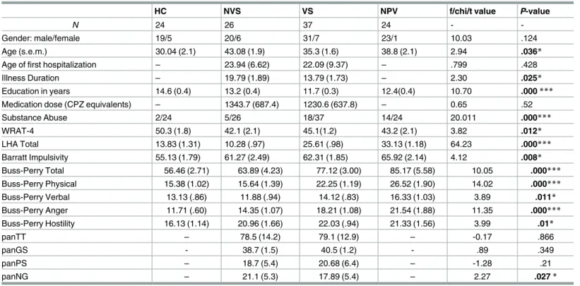
Demographic statistics are reported inTable 2. No difference in gender was observed between the groups, with the sample being largely comprised of men. No difference was also seen in medication dose, as measured using chlorpromazine equivalency values. Chlorpromazine equivalents were calculated following the guidelines published in [58]. The percentage of atypi- cal and typical antipsychotic medication was 70% and 30%, respectively. There was a signifi- cant difference in age among groups (F3,107= 2.94,p= .036), with NVS, older compared to HC, VS, and NPV. Groups also differed in years of education (F3, 109= 10.7,p<.001), with HC having spent more years at school than VS, NPV, and NVS. Age at first hospitalization did not differ between the VS and NVS (t57= 0.799,p= .428), although the duration of illness was lon- ger for the NVS group than the VS group (t56= 2.30,p= .025). Differences were also observed for WRAT-4 performances across groups (F3,106= 3.82,p= .012), with HC participants obtain- ing higher achievement scores than the other three groups. Age and WRAT-4 were used as covariates in all subsequent ROI analysis, along with substance abuse disorder.
Additionally, we saw group differences on the Buss-Perry Questionnaire Total Score (BPAQ;F3,107= 10.05,p<.001). BPAQ total scores were higher in VS and NPV relative to HC and NVS. For the physical aggression subscale of the BPAQ, NPV participants scored higher
Table 2. Demographic and clinical data.
HC NVS VS NPV f/chi/t value P-value
N 24 26 37 24 - -
Gender: male/female 19/5 20/6 31/7 23/1 10.03 .124
Age (s.e.m.) 30.04 (2.1) 43.08 (1.9) 35.3 (1.6) 38.8 (2.1) 2.94 .036*
Age of first hospitalization – 23.94 (6.62) 22.09 (9.37) – .799 .428
Illness Duration – 19.79 (1.89) 13.79 (1.73) – 2.30 .025*
Education in years 14.6 (0.4) 13.2 (0.4) 11.7 (0.3) 12.4(0.4) 10.70 .000***
Medication dose (CPZ equivalents) – 1343.7 (687.4) 1230.6 (637.8) – 0.65 .52
Substance Abuse 2/24 5/26 18/37 14/24 20.011 .000***
WRAT-4 50.3 (1.8) 42.1 (2.1) 45.1(1.2) 43.2 (2.1) 3.82 .012*
LHA Total 13.83 (1.31) 10.28 (.97) 25.61 (.98) 33.13 (1.18) 64.23 .000***
Barratt Impulsivity 55.13 (1.79) 61.27 (2.49) 62.31 (1.85) 65.92 (2.14) 4.12 .008*
Buss-Perry Total 56.46 (2.71) 63.89 (4.23) 77.12 (3.00) 85.17 (5.58) 10.05 .000***
Buss-Perry Physical 15.38 (1.02) 15.64 (1.39) 22.25 (1.19) 26.52 (1.90) 14.02 .000***
Buss-Perry Verbal 13.13 (.86) 11.88 (.94) 14.12 (.83) 16.33 (1.03) 3.89 .011*
Buss-Perry Anger 11.71 (.60) 14.35 (1.07) 18.21 (1.08) 21.54 (1.88) 11.35 .000***
Buss-Perry Hostility 16.13 (1.14) 20.96 (1.66) 22.03 (.94) 21.33 (1.56) 3.99 .01*
panTT – 78.5 (14.2) 79.1 (12.9) – -0.17 .866
panGS - 38.7 (1.5) 40.5 (1.2) - .89 .349
panPS – 18.7 (5.4) 20.68 (6.4) – -1.28 .21
panNG – 21.1 (5.3) 17.89 (5.4) – 2.27 .027*
ANOVA or t-test for continuous and chi-square test for categorical variables.
*P0.05
**P0.001
***P.0001.
S.E.M. = standard error of mean; S.E.M. are the values in parenthesis; CPZ = chlorpromazine; panTT = PANSS Total Score; panGS = PANSS General Score; panPS = PANSS Positive Score; panNG = PANSS Negative Score; LHA = Life History of Aggression
doi:10.1371/journal.pone.0168100.t002
than VS, and both violent groups reported more physical aggression than NVS and HC. For the verbal aggression subscale of the BPAQ, NPV participants scored higher than HC and NVS. Differences between VS and the other three groups did not reach significance. Further- more, VS and NPV participants scored higher on anger than NVS and HC. Finally, on the hos- tility subscale, VS, NPV, and NVS participants scored higher than HC. There were no
differences between NPV, VS, and NVS groups.
Life history of aggression scores also differed between the four groups (F3, 108= 67.49,p<
.001). The NPV group was more violent than the VS group, with both of these groups scoring higher on the LHA than both the NVS and HC groups. HC participants had elevated LHA scores relative to the NVS group.
There was a main effect of group (F3, 108= 3.28,p= .024) for Barrett’s Impulsivity Index.
Follow-up pair-wise comparisons revealed increased impulsivity in VS and NPV groups rela- tive to HC participants. The NVS group did not differ from the HC group, or the two violent groups.
NVS and VS did not differ in PANSS total scores, general scores, or positive symptoms.
However, NVS scored significantly higher on negative symptoms (t59= 2.27,p= 0.02). For this reason, a secondary analysis looking at the effect of negative symptoms as a contributing factor to ROI volume was conducted.
Region of Interest Volumetric Analysis
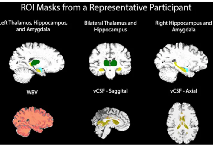
Fig 1illustrates brain regions investigated in our study mapped onto a participant’s brain.
Fig 1. T1-weighted MRI ROI FIRST masks.
doi:10.1371/journal.pone.0168100.g001
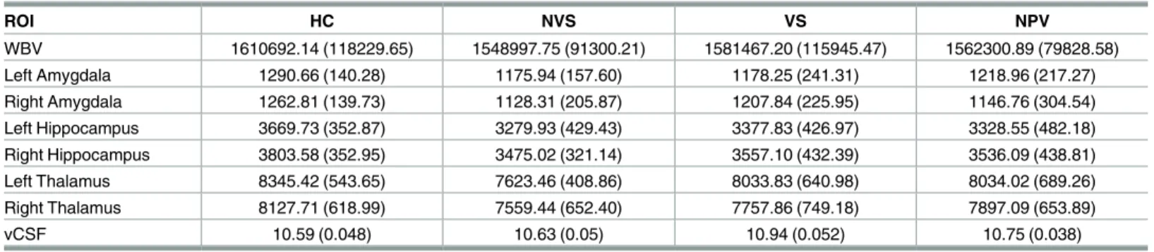
Table 3lists mean volumes of investigated brain regions for each of the four groups.
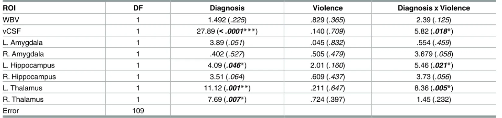
Whole Brain Volume. The ANOVA for WBV revealed no significant main and interac- tive effects.
Fig 2illustrates the effects of violence and diagnosis on brain region volumes.
Left and Right Amygdala. The ANOVA for left amygdala indicated a trend towards a main effect for diagnosis (F1,109= 3.89, p = .051). For the right amygdala, a significant interac- tion between diagnosis and violence was found (F1,109= 5.3, p = .023). However, only a trend towards an interaction was observed after including substance abuse as a covariate (F1,109= 3.679,p= .05). The interaction appeared to be driven by a double dissociation between factors, with decreased volume in NPV relative to HC and increased volume in VS relative to NVS.
Bonferroni corrected post hoc pairwise comparison using independent samples t-test revealed no differences between groups.
Table 4lists results obtained by analysis of variance.
Left and Right Hippocampus. For the left hippocampus volume, there was a main effect of diagnosis (F1,109= 4.08,p= 0.04) with reduction seen for patients with schizophrenia. There was also an interaction between diagnosis and violence (F1,109= 5.46,p= 0.02). Corrected post hoc pairwise comparisons revealed a group difference between NVS and HC (p= .026) for left hippocampal volume. No effects were found for the right hippocampus.
Left and Right Thalamus. For both left and right thalamus, ANOVA revealed a main effect for diagnosis (LT:F1,109= 11.12,p= 0.001; RT:F1,109= 7.69,p= 0.007) with reduction seen in patients with schizophrenia. Furthermore, a significant interaction between diagnosis and violence was observed for the left thalamus (F1,109= 8.35,p= 0.005). Post hoc pairwise comparisons revealed a group difference between NVS and HC (p<.001).
Ventricular Cerebrospinal Fluid. Due to issues of skewness, vCSF was transformed using a natural log to approximate a Gaussian distribution. Analysis indicated a main effect for diagnosis (F1,109= 27.88, p = 0.001) with increased vCSF volume for patients with schizophre- nia. Furthermore, there was a significant interaction between diagnosis and violence (F1,109= 5.82, p = 0.01). Post hoc pairwise comparisons revealed increased volume in NVS (p<.001) and VS (p = .008) relative to HC, as well as increased volume in NVS relative to NPV (p= .002).
The Role of Negative Symptoms and Medication. Here the analysis was limited to patients with a diagnosis of schizophrenia to investigate the potential role of several factors, such as medication, negative symptoms, and illness duration on gray matter volume loss. As indicated inTable 2, there was no group difference for CPZ equivalent. Further, CPZ equiva- lent did not correlate with WBV (p= .79), vCSF (p= .26), left amygdala (p= .43), left
Table 3. Adjusted means and standard deviations of the means for ROI.
ROI HC NVS VS NPV
WBV 1610692.14 (118229.65) 1548997.75 (91300.21) 1581467.20 (115945.47) 1562300.89 (79828.58)
Left Amygdala 1290.66 (140.28) 1175.94 (157.60) 1178.25 (241.31) 1218.96 (217.27)
Right Amygdala 1262.81 (139.73) 1128.31 (205.87) 1207.84 (225.95) 1146.76 (304.54)
Left Hippocampus 3669.73 (352.87) 3279.93 (429.43) 3377.83 (426.97) 3328.55 (482.18)
Right Hippocampus 3803.58 (352.95) 3475.02 (321.14) 3557.10 (432.39) 3536.09 (438.81)
Left Thalamus 8345.42 (543.65) 7623.46 (408.86) 8033.83 (640.98) 8034.02 (689.26)
Right Thalamus 8127.71 (618.99) 7559.44 (652.40) 7757.86 (749.18) 7897.09 (653.89)
vCSF 10.59 (0.048) 10.63 (0.05) 10.94 (0.052) 10.75 (0.038)
Adjusted means (S.D.) reported in mm3. All ROIs were adjusted for age, substance abuse, and WRAT-4 T-scores.
doi:10.1371/journal.pone.0168100.t003
Fig 2. ROI mean volumes for each group. Vertical error bars indicate standard error of the mean.
doi:10.1371/journal.pone.0168100.g002
hippocampus (p= .71), left thalamus (p= .84), right amygdala (p= .93), right hippocampus (p= .94), or right thalamus (p= .87) volumes. CPZ equivalent values were therefore not added as a covariate. Groups did differ on age, academic achievement, substance use, as well as nega- tive symptoms, and these variables were therefore included as covariates in our analysis. For this subsample analysis including only patients with schizophrenia, significant differences between patients with and without a history of violence were only found for vCSF with increased volume seen for NVS (F1,59= 4.63,p= 0.03).
Discussion
We set out to investigate structural characteristics in schizophrenia patients with and without a history of violence, and compare them to non-psychotic violent and healthy control individ- uals. Our goals were two-fold, to probe for volumetric abnormalities linked to aggression and to determine possible differences of such abnormalities between aggressive individuals with and without psychotic disorder. One particular interest pertained to amygdala volume as there are somewhat conflicting results implicating this region in violence. Out of four studies [24, 26,30,31], only one reported an association between amygdala reduction and violence in patients with schizophrenia. At the same time, decreased amygdala volume has been linked to a variety of mental disorders characterized by high aggression, including conduct disorder [59]; for a recent meta-analysis see [60] and psychopathy [61,62].
In our study, we found several regions with structural abnormalities in patients with schizo- phrenia. Patients showed elevated vCSF volume and smaller left hippocampus as well as left thalamus volumes, particularly in individuals without a history of violence [63,64]. Further- more, patients had reduction in right thalamus size and a strong trend towards smaller left amygdala volume was found. With regard to right amygdala, our findings indicated an interac- tion between violence and diagnosis. The effect appeared to be driven by a double dissociation.
That is, reductions associated with violence were seen for non-psychotic individuals, while for psychotic patient’s reductions were observed in non-violent individuals. Importantly, after accounting for substance abuse in our analysis only a trend towards an interaction was observed.
Overall, our findings in patients are in line with a recent large-scale study of subcortical brain regions in 2028 individuals with schizophrenia [65]. Among other structures,
Table 4. ANOVA; f/p values are listed for main and interactive effects for each ROI.
ROI DF Diagnosis Violence Diagnosis x Violence
WBV 1 1.492 (.225) .829 (.365) 2.39 (.125)
vCSF 1 27.89 (<.0001***) .140 (.709) 5.82 (.018*)
L. Amygdala 1 3.89 (.051) .045 (.832) .554 (.459)
R. Amygdala 1 .402 (.527) .505 (.479) 3.679 (.058)
L. Hippocampus 1 4.09 (.046*) 2.01 (.160) 5.46 (.021*)
R. Hippocampus 1 3.51 (.064) .609 (.437) 3.73 (.056)
L. Thalamus 1 11.12 (.001**) .211 (.647) 8.36 (.005*)
R. Thalamus 1 7.69 (.007*) .724 (.397) 1.45 (.232)
Error 109
ANOVAs using WRAT-4 T-scores, Age, and SUD as covariates.
*P0.05
**P0.001
***P.0001. Columns includes degree of freedom (DF), F-scores and p-values (italicized).
doi:10.1371/journal.pone.0168100.t004
abnormalities were found for hippocampus, amygdala, thalamus, and lateral ventricle volumes.
Evidence by us and others underscore widespread morphometric abnormalities in subcortical regions possible related to aberrant integration and information transfer processes in schizo- phrenia. Also, recent efforts to establish temporal lobe abnormalities as confirmatory biomark- ers among youth at high-risk for development of psychosis have produced promising results [66]. One open question concerns stronger abnormalities in non-violent patients relative to their violent peers in our data.
With regard to our substance abuse finding it is important to note that a recent meta-analy- sis of studies probing violence in individuals on the psychosis spectrum concluded that most of the elevated risk was driven by patients with substance abuse comorbidity [67]. Here we report, for the first time according to our knowledge, that substance abuse might play a medi- ating role in the association between amygdala reduction and violence in non-psychotic vio- lent individuals.
Considering all aggressive individuals in both the VS and NPV group, we found no evi- dence for shared structural abnormalities underlying violence. However, there might be other morphological markers and/or other regions of interest that might be important to consider to determine common structural abnormalities linked to violence. Furthermore, we found no evidence for amygdala volume reduction in violent patients with schizophrenia. Previous stud- ies also reported no reductions in violent patients [24–26], but questions remained as to whether these studies were sufficiently powered to detect differences. Our sample was between two and three times larger than the ones in previous schizophrenia studies with a focus on vio- lence [24–26], which provides a measure of confidence that amygdala size might not be part of the structural abnormalities commonly associated with violence, at least in schizophrenia.
A limitation of the current study is the use of a 1.5T scanner, which does not provide the same level of spatial resolution as more modern 3T scanners. Our sample is comprised of chronic patients with schizophrenia and therefore generalizations to patients in their early stages of illness might not be appropriate. Additionally, the self-report nature of the Buss- Perry, which has been found to be influenced by factors of social desirability [43] could possi- bly lead to lower scores, although the LHA scale is not based on self-report and is immune from concerns of social desirability. Utilizing the LHA scale for grouping purposes is a strength of the current report because of this consideration. Another strength of the study is the use of an automated approach to segmentation (FSL’s FIRST), which has been reported to be a reliable and valid method of subcortical segmentation [68], and is widely used by researchers in various clinical populations [37,69,70]. A final, and important strength, is the relatively large sample of clinical patients and control participants used in this study, with the former demonstrating aberrant processing of emotionally salient images [29].
Future research should focus on other areas of grey matter that could be related to the ele- vated rates of violence in patients with schizophrenia. In this study, due to selecta priori regions of interest, and limited regions within the FIRST library, the following regions were not investigated. One such region is the superior temporal sulcus (STS), a region associated with facial recognition [71,72] which has direct projections to the amygdala [73], and which has been found to be impaired in the schizophrenia population [74,75]. Another region of interest is the striatum, which includes the putamen, previously found to be enlarged in the VS group [24]. The putamen is part of the FIRST library but was not included in this analysis because ofa prioriROI selection. OFC and PFC regions have been previously implicated in aggression and schizophrenia and should, in concert with subcortical regions, be considered in future research. Additionally, large scale neuroimaging and electrophysiology studies involving genetics (e.g. MAO-A and COM-T) and their role in brain structure and function could be helpful in delineating differences between the VS and NVS groups from NPV
individuals. Both MAO-A and COM-T genes have been previously associated with violence and aggression [76–81], and both have been linked with schizophrenia [82–89].
In summary, our ROI analysis of temporal and subcortical regions confirmed widespread morphometric brain abnormalities in patients with schizophrenia. We found no evidence for smaller amygdala size in violent patients with schizophrenia. Finally, smaller amygdala size in violent non-psychotic individuals was mostly accounted by substance abuse problems within this group. The latter results might provide indication that substance abuse is a potential medi- ator in the relationship between brain abnormalities and aggression in non-psychotic individ- uals. Overall, our results underscore the importance of volumetric abnormalities in aggression and provide some indication for distinct brain regions in the characterization of psychotic and non-psychotic violent individuals.
Supporting Information S1 Table. Anonymized data.
(XLSX)
Acknowledgments
Sincere thanks go to Eileen Zenz and Melissa Brady for help with recruitment, clinical inter- viewing and neuropsychological testing, Karen Nolan (PhD) and Constance Shope (PhD) for clinical interviewing.
Author Contributions
Conceptualization: MK PDS JJF PC.
Data curation: LR PC.
Formal analysis: VDB LR PC.
Funding acquisition: MK.
Methodology: LR PC.
Resources: MK.
Supervision: PDS MK LR JJF.
Writing – original draft: VDB.
Writing – review & editing: PDS MK JJF.
References
1. Swanson JW, Swartz MS, Van Dorn RA, Elbogen EB, Wagner HR, Rosenheck RA, et al. A national study of violent behavior in persons with schizophrenia. Archives of general psychiatry. 2006; 63 (5):490–9. doi:10.1001/archpsyc.63.5.490PMID:16651506
2. Mullen PE, Burgess P, Wallace C, Palmer S, Ruschena D. Community care and criminal offending in schizophrenia. The Lancet. 2000; 355(9204):614–7.
3. Nederlof AF, Muris P, Hovens JE. The epidemiology of violent behavior in patients with a psychotic dis- order: A systematic review of studies since 1980. Aggression and violent behavior. 2013; 18(1):183–9.
4. Krakowski MI, Czobor P. Depression and impulsivity as pathways to violence: implications for antiag- gressive treatment. Schizophrenia bulletin. 2014; 40(4):886–94. PubMed Central PMCID:
PMC4059442. doi:10.1093/schbul/sbt117PMID:23943412
5. Davidson RJ, Putnam KM, Larson CL. Dysfunction in the neural circuitry of emotion regulation—a pos- sible prelude to violence. Science. 2000; 289(5479):591–4. Epub 2000/08/01. PMID:10915615
6. Terri Roberton MD, Romola S. Bucks. Emotion regulation and aggression. Aggression and Violent Behavior. 2012; 17(1):72–82.
7. Van Elst LT, Woermann F, Lemieux L, Thompson P, Trimble M. Affective aggression in patients with temporal lobe epilepsy. Brain: a journal of neurology. 2000; 123(2):234–43.
8. Bufkin JL, Luttrell VR. Neuroimaging studies of aggressive and violent behavior current findings and implications for criminology and criminal justice. Trauma, Violence, & Abuse. 2005; 6(2):176–91.
9. Coccaro EF. Neurotransmitter correlates of impulsive aggression in humans. Annals of the New York Academy of Sciences. 1996; 794(1):82–9.
10. Krakowski M. Violence and serotonin: influence of impulse control, affect regulation, and social function- ing. The Journal of neuropsychiatry and clinical neurosciences. 2003.
11. Coccaro EF, Fanning JR, Phan KL, Lee R. Serotonin and impulsive aggression. CNS spectrums. 2015;
20(03):295–302.
12. Blair RJ. Applying a cognitive neuroscience perspective to the disorder of psychopathy. Development and psychopathology. 2005; 17(3):865–91. doi:10.1017/S0954579405050418PMID:16262996 13. Kiehl KA. A cognitive neuroscience perspective on psychopathy: evidence for paralimbic system dys-
function. Psychiatry research. 2006; 142(2–3):107–28. PubMed Central PMCID: PMC2765815. doi:10.
1016/j.psychres.2005.09.013PMID:16712954
14. Siever LJ. Neurobiology of aggression and violence. The American journal of psychiatry. 2008; 165 (4):429–42. PubMed Central PMCID: PMC4176893. doi:10.1176/appi.ajp.2008.07111774PMID:
18346997
15. DeWall CN, Finkel EJ, Denson TF. Self-control inhibits aggression. Social and Personality Psychology Compass. 2011; 5(7):458–72.
16. Denson TF, DeWall CN, Finkel EJ. Self-control and aggression. Current Directions in Psychological Sci- ence. 2012; 21(1):20–5.
17. Hoptman MJ, Antonius D, Mauro CJ, Parker EM, Javitt DC. Cortical thinning, functional connectivity, and mood-related impulsivity in schizophrenia: relationship to aggressive attitudes and behavior. The American journal of psychiatry. 2014; 171(9):939–48. PubMed Central PMCID: PMC4178944. doi:10.
1176/appi.ajp.2014.13111553PMID:25073506
18. Heatherton TF. Neuroscience of self and self-regulation. Annual review of psychology. 2011; 62:363.
doi:10.1146/annurev.psych.121208.131616PMID:21126181
19. Niu L, Matsui M, Zhou S-Y, Hagino H, Takahashi T, Yoneyama E, et al. Volume reduction of the amyg- dala in patients with schizophrenia: a magnetic resonance imaging study. Psychiatry Research: Neuro- imaging. 2004; 132(1):41–51. doi:10.1016/j.pscychresns.2004.06.002PMID:15546702
20. Steen RG, Mull C, Mcclure R, Hamer RM, Lieberman JA. Brain volume in first-episode schizophrenia Systematic review and meta-analysis of magnetic resonance imaging studies. The British Journal of Psychiatry. 2006; 188(6):510–8.
21. Velakoulis D, Wood SJ, Wong MT, McGorry PD, Yung A, Phillips L, et al. Hippocampal and amygdala volumes according to psychosis stage and diagnosis: A magnetic resonance imaging study of chronic schizophrenia, first-episode psychosis, and ultra–high-risk individuals. Archives of general psychiatry.
2006; 63(2):139–49. doi:10.1001/archpsyc.63.2.139PMID:16461856
22. Bora E, Fornito A, Radua J, Walterfang M, Seal M, Wood SJ, et al. Neuroanatomical abnormalities in schizophrenia: a multimodal voxelwise meta-analysis and meta-regression analysis. Schizophrenia research. 2011; 127(1):46–57.
23. Shepherd AM, Laurens KR, Matheson SL, Carr VJ, Green MJ. Systematic meta-review and quality assessment of the structural brain alterations in schizophrenia. Neuroscience & Biobehavioral Reviews.
2012; 36(4):1342–56.
24. Barkataki I, Kumari V, Das M, Taylor P, Sharma T. Volumetric structural brain abnormalities in men with schizophrenia or antisocial personality disorder. Behavioural brain research. 2006; 169(2):239–47.
Epub 2006/02/10. doi:10.1016/j.bbr.2006.01.009PMID:16466814
25. Kumari V, Barkataki I, Goswami S, Flora S, Das M, Taylor P. Dysfunctional, but not functional, impulsiv- ity is associated with a history of seriously violent behaviour and reduced orbitofrontal and hippocampal volumes in schizophrenia. Psychiatry Research: Neuroimaging. 2009; 173(1):39–44. doi:10.1016/j.
pscychresns.2008.09.003PMID:19442493
26. Kumari V, Gudjonsson GH, Raghuvanshi S, Barkataki I, Taylor P, Sumich A, et al. Reduced thalamic volume in men with antisocial personality disorder or schizophrenia and a history of serious violence and childhood abuse. European psychiatry: the journal of the Association of European Psychiatrists.
2013; 28(4):225–34. Epub 2012/09/05.
27. Yang Y, Raine A, Han CB, Schug RA, Toga AW, Narr KL. Reduced hippocampal and parahippocampal volumes in murderers with schizophrenia. Psychiatry research. 2010; 182(1):9–13. PubMed Central PMCID: PMC2855857. doi:10.1016/j.pscychresns.2009.10.013PMID:20227253
28. Hoptman MJ, D’Angelo D, Catalano D, Mauro CJ, Shehzad ZE, Kelly AM, et al. Amygdalofrontal func- tional disconnectivity and aggression in schizophrenia. Schizophrenia bulletin. 2010; 36(5):1020–8.
Epub 2009/04/02. PubMed Central PMCID: PMC2930349. doi:10.1093/schbul/sbp012PMID:
19336392
29. De Sanctis P, Foxe JJ, Czobor P, Wylie GR, Kamiel SM, Huening J, et al. Early sensory-perceptual pro- cessing deficits for affectively valenced inputs are more pronounced in schizophrenia patients with a history of violence than in their non-violent peers. Social cognitive and affective neuroscience. 2012.
Epub 2012/05/09.
30. Wong M, Lumsden J, Fenton G, Fenwick P. Electroencephalography, computed tomography and vio- lence ratings of male patients in a maximum-security mental hospital. Acta Psychiatrica Scandinavica.
1994; 90(2):97–101. PMID:7976465
31. Kumari V, Das M, Taylor PJ, Barkataki I, Andrew C, Sumich A, et al. Neural and behavioural responses to threat in men with a history of serious violence and schizophrenia or antisocial personality disorder.
Schizophrenia research. 2009; 110(1):47–58.
32. Derogatis L, Lipman R. SCL-90. Administration, scoring and procedures manual-I for the R (revised) version and other instruments of the Psychopathology Rating Scales Series Chicago: Johns Hopkins University School of Medicine. 1977.
33. Matthies S, Rusch N, Weber M, Lieb K, Philipsen A, Tuescher O, et al. Small amygdala-high aggres- sion? The role of the amygdala in modulating aggression in healthy subjects. The world journal of bio- logical psychiatry: the official journal of the World Federation of Societies of Biological Psychiatry. 2012;
13(1):75–81.
34. Bobes Leon MA, Ostroski F, Diaz K, Romero C, Borja K, Santos Y, et al., editors. Functional and struc- tural impairment of the left amygdala in violent non-psychopath men. Psychophysiology; 2013.
35. Raine A, Dodge K, Loeber R, Gatzke-Kopp L, Lynam D, Reynolds C, et al. The Reactive-Proactive Aggression Questionnaire: Differential Correlates of Reactive and Proactive Aggression in Adolescent Boys. Aggressive behavior. 2006; 32(2):159–71. PubMed Central PMCID: PMC2927832. doi:10.1002/
ab.20115PMID:20798781
36. Salekin RT, Rogers R, Sewell KW. A review and meta-analysis of the psychopathy checklist and psy- chopathy checklist-revised: predictive validity of dangerousness. Clinical Psychology: Science and Practice. 1996; 3(3):203–15.
37. Pardini DA, Raine A, Erickson K, Loeber R. Lower amygdala volume in men is associated with child- hood aggression, early psychopathic traits, and future violence. Biological psychiatry. 2014; 75(1):73–
80. PubMed Central PMCID: PMC3751993. doi:10.1016/j.biopsych.2013.04.003PMID:23647988 38. Krakowski MI, De Sanctis P, Foxe JJ, Hoptman MJ, Nolan K, Kamiel S, et al. Disturbances in Response
Inhibition and Emotional Processing as Potential Pathways to Violence in Schizophrenia: A High-Den- sity Event-Related Potential Study. Schizophrenia bulletin. 2016; 42(4):963–74. PubMed Central PMCID: PMCPMC4903062. doi:10.1093/schbul/sbw005PMID:26895845
39. First MB, Spitzer RL, Gibbon M, Williams JB. User’s guide for the Structured clinical interview for DSM- IV axis I disorders SCID-I: clinician version: American Psychiatric Pub; 1997.
40. Association AP. Diagnostic and statistical manual-text revision (DSM-IV-TRim, 2000): American Psy- chiatric Association; 2000.
41. Coccaro EF, Berman ME, Kavoussi RJ. Assessment of life history of aggression: development and psy- chometric characteristics. Psychiatry research. 1997; 73(3):147–57. PMID:9481806
42. Buss AH, Perry M. The aggression questionnaire. Journal of personality and social psychology. 1992;
63(3):452–9. PMID:1403624
43. Harris JA. A further evaluation of the Aggression Questionnaire: Issues of validity and reliability. Behav- iour research and therapy. 1997; 35(11):1047–53. PMID:9431736
44. Patton JH, Stanford MS. Factor structure of the Barratt impulsiveness scale. Journal of clinical psychol- ogy. 1995; 51(6):768–74. PMID:8778124
45. Stanford MS, Mathias CW, Dougherty DM, Lake SL, Anderson NE, Patton JH. Fifty years of the Barratt Impulsiveness Scale: An update and review. Personality and individual differences. 2009; 47(5):385–
95.
46. Wilkinson GS, Robertson GJ. WRAT 4: Wide Range Achievement Test; professional manual: Psycho- logical Assessment Resources, Incorporated; 2006.
47. Kay SR, Flszbein A, Opfer LA. The positive and negative syndrome scale (PANSS) for schizophrenia.
Schizophrenia bulletin. 1987; 13(2):261. PMID:3616518
48. Peralta V, Cuesta MJ. Psychometric properties of the positive and negative syndrome scale (PANSS) in schizophrenia. Psychiatry research. 1994; 53(1):31–40. PMID:7991730
49. Morey RA, Petty CM, Xu Y, Hayes JP, Wagner HR 2nd, Lewis DV, et al. A comparison of automated segmentation and manual tracing for quantifying hippocampal and amygdala volumes. NeuroImage.
2009; 45(3):855–66. PubMed Central PMCID: PMCPMC2714773. doi:10.1016/j.neuroimage.2008.12.
033PMID:19162198
50. Patenaude B, Smith SM, Kennedy DN, Jenkinson M. A Bayesian model of shape and appearance for subcortical brain segmentation. NeuroImage. 2011; 56(3):907–22. doi:10.1016/j.neuroimage.2011.02.
046PMID:21352927
51. Smith SM, De Stefano N, Jenkinson M, Matthews PM. Normalized accurate measurement of longitudi- nal brain change. Journal of computer assisted tomography. 2001; 25(3):466–75. PMID:11351200 52. Smith SM, Jenkinson M, Woolrich MW, Beckmann CF, Behrens TE, Johansen-Berg H, et al. Advances
in functional and structural MR image analysis and implementation as FSL. NeuroImage. 2004; 23:
S208–S19. doi:10.1016/j.neuroimage.2004.07.051PMID:15501092
53. Jenkinson M, Smith S. A global optimisation method for robust affine registration of brain images. Medi- cal image analysis. 2001; 5(2):143–56. PMID:11516708
54. Jenkinson M, Bannister P, Brady M, Smith S. Improved optimization for the robust and accurate linear registration and motion correction of brain images. NeuroImage. 2002; 17(2):825–41. PMID:12377157 55. Smith SM, Rao A, De Stefano N, Jenkinson M, Schott JM, Matthews PM, et al. Longitudinal and cross-
sectional analysis of atrophy in Alzheimer’s disease: cross-validation of BSI, SIENA and SIENAX. Neu- roImage. 2007; 36(4):1200–6. doi:10.1016/j.neuroimage.2007.04.035PMID:17537648
56. Smith SM, Zhang Y, Jenkinson M, Chen J, Matthews P, Federico A, et al. Accurate, robust, and auto- mated longitudinal and cross-sectional brain change analysis. NeuroImage. 2002; 17(1):479–89. PMID:
12482100
57. Smith SM, Rao A, De Stefano N, Jenkinson M, Schott JM, Matthews PM, et al. Longitudinal and cross- sectional analysis of atrophy in Alzheimer’s disease: cross-validation of BSI, SIENA and SIENAX. Neu- roImage. 2007; 36(4):1200–6. doi:10.1016/j.neuroimage.2007.04.035PMID:17537648
58. Kane JM, Leucht S, Carpenter D, Docherty JP, Expert Consensus Panel for Optimizing Pharmacologic Treatment of Psychotic D. The expert consensus guideline series. Optimizing pharmacologic treatment of psychotic disorders. Introduction: methods, commentary, and summary. The Journal of clinical psy- chiatry. 2003; 64 Suppl 12:5–19.
59. Fairchild G, Passamonti L, Hurford G, Hagan CC, von dem Hagen EA, van Goozen SH, et al. Brain structure abnormalities in early-onset and adolescent-onset conduct disorder. The American journal of psychiatry. 2011; 168(6):624–33. doi:10.1176/appi.ajp.2010.10081184PMID:21454920
60. Rogers JC, De Brito SA. Cortical and Subcortical Gray Matter Volume in Youths With Conduct Prob- lems: A Meta-analysis. JAMA Psychiatry. 2016; 73(1):64–72. doi:10.1001/jamapsychiatry.2015.2423 PMID:26650724
61. Yang Y, Raine A, Narr KL, Colletti P, Toga AW. Localization of deformations within the amygdala in indi- viduals with psychopathy. Archives of general psychiatry. 2009; 66(9):986–94. PubMed Central PMCID: PMC3192811. doi:10.1001/archgenpsychiatry.2009.110PMID:19736355
62. Ermer E, Cope LM, Nyalakanti PK, Calhoun VD, Kiehl KA. Aberrant paralimbic gray matter in criminal psychopathy. Journal of abnormal psychology. 2012; 121(3):649–58. PubMed Central PMCID:
PMC4039408. doi:10.1037/a0026371PMID:22149911
63. Davatzikos C, Shen D, Gur RC, Wu X, Liu D, Fan Y, et al. Whole-brain morphometric study of schizo- phrenia revealing a spatially complex set of focal abnormalities. Archives of general psychiatry. 2005;
62(11):1218–27. doi:10.1001/archpsyc.62.11.1218PMID:16275809
64. Tanskanen P, Haapea M, Veijola J, Miettunen J, Jarvelin MR, Pyhtinen J, et al. Volumes of brain, grey and white matter and cerebrospinal fluid in schizophrenia in the Northern Finland 1966 Birth Cohort: an epidemiological approach to analysis. Psychiatry research. 2009; 174(2):116–20. doi:10.1016/j.
pscychresns.2009.04.009PMID:19853416
65. van Erp TG, Hibar DP, Rasmussen JM, Glahn DC, Pearlson GD, Andreassen OA, et al. Subcortical brain volume abnormalities in 2028 individuals with schizophrenia and 2540 healthy controls via the ENIGMA consortium. Molecular psychiatry. 2016; 21(4):547–53. PubMed Central PMCID:
PMCPMC4668237. doi:10.1038/mp.2015.63PMID:26033243
66. Roalf DR, Quarmley M, Calkins ME, Satterthwaite TD, Ruparel K, Elliott MA, et al. Temporal Lobe Vol- ume Decrements in Psychosis Spectrum Youths. Schizophrenia bulletin. 2016.
67. Fazel S, Gulati G, Linsell L, Geddes JR, Grann M. Schizophrenia and violence: systematic review and meta-analysis. PLoS Med. 2009; 6(8):e1000120. PubMed Central PMCID: PMCPMC2718581. doi:10.
1371/journal.pmed.1000120PMID:19668362
68. Morey RA, Selgrade ES, Wagner HR, Huettel SA, Wang L, McCarthy G. Scan–rescan reliability of sub- cortical brain volumes derived from automated segmentation. Human brain mapping. 2010; 31 (11):1751–62. doi:10.1002/hbm.20973PMID:20162602
69. De Jong L, Van der Hiele K, Veer I, Houwing J, Westendorp R, Bollen E, et al. Strongly reduced vol- umes of putamen and thalamus in Alzheimer’s disease: an MRI study. Brain: a journal of neurology.
2008; 131(12):3277–85.
70. Sexton CE, Mackay CE, Lonie JA, Bastin ME, Terrière E, O’Carroll RE, et al. MRI correlates of episodic memory in Alzheimer’s disease, mild cognitive impairment, and healthy aging. Psychiatry Research:
Neuroimaging. 2010; 184(1):57–62. doi:10.1016/j.pscychresns.2010.07.005PMID:20832251 71. Baylis G, Rolls ET, Leonard C. Selectivity between faces in the responses of a population of neurons in
the cortex in the superior temporal sulcus of the monkey. Brain research. 1985; 342(1):91–102. PMID:
4041820
72. Iidaka T. Role of the fusiform gyrus and superior temporal sulcus in face perception and recognition: An empirical review. Japanese Psychological Research. 2014; 56(1):33–45.
73. Aggleton J, Burton M, Passingham R. Cortical and subcortical afferents to the amygdala of the rhesus monkey (Macaca mulatta). Brain research. 1980; 190(2):347–68. PMID:6768425
74. Rajarethinam R, DeQuardo J, Nalepa R, Tandon R. Superior temporal gyrus in schizophrenia: a volu- metric magnetic resonance imaging study. Schizophrenia research. 2000; 41(2):303–12. PMID:
10708339
75. Kim J-J, Crespo-Facorro B, Andreasen NC, O’Leary DS, Magnotta V, Nopoulos P. Morphology of the lateral superior temporal gyrus in neuroleptic naıve patients with schizophrenia: relationship to symp- toms. Schizophrenia research. 2003; 60(2):173–81.
76. Hygen BW, Belsky J, Stenseng F, Lydersen S, Guzey IC, Wichstrøm L. Child exposure to serious life events, COMT, and aggression: Testing differential susceptibility theory. Developmental psychology.
2015; 51(8):1098. doi:10.1037/dev0000020PMID:26053146
77. Veroude K, Zhang-James Y, Fernàndez-Castillo N, Bakker MJ, Cormand B, Faraone SV. Genetics of aggressive behavior: An overview. American Journal of Medical Genetics Part B: Neuropsychiatric Genetics. 2015.
78. Kotler M, Barak P, Cohen H, Averbuch IE, Grinshpoon A, Gritsenko I, et al. Homicidal behavior in schizophrenia associated with a genetic polymorphism determining low catechol O-methyltransferase (COMT) activity. American journal of medical genetics. 1999; 88(6):628–33. PMID:10581481 79. Widom CS, Brzustowicz LM. MAOA and the “cycle of violence:” childhood abuse and neglect, MAOA
genotype, and risk for violent and antisocial behavior. Biological psychiatry. 2006; 60(7):684–9. doi:10.
1016/j.biopsych.2006.03.039PMID:16814261
80. Buckholtz JW, Meyer-Lindenberg A. MAOA and the neurogenetic architecture of human aggression.
Trends in neurosciences. 2008; 31(3):120–9. doi:10.1016/j.tins.2007.12.006PMID:18258310 81. Frazzetto G, Di Lorenzo G, Carola V, Proietti L, Sokolowska E, Siracusano A, et al. Early trauma and
increased risk for physical aggression during adulthood: the moderating role of MAOA genotype. PloS one. 2007; 2(5):e486. doi:10.1371/journal.pone.0000486PMID:17534436
82. Shifman S, Bronstein M, Sternfeld M, Pisante´-Shalom A, Lev-Lehman E, Weizman A, et al. A highly sig- nificant association between a COMT haplotype and schizophrenia. The American Journal of Human Genetics. 2002; 71(6):1296–302. doi:10.1086/344514PMID:12402217
83. Shifman S, Bronstein M, Sternfeld M, Pisante´ A, Weizman A, Reznik I, et al. COMT: a common suscep- tibility gene in bipolar disorder and schizophrenia. American Journal of Medical Genetics Part B: Neuro- psychiatric Genetics. 2004; 128(1):61–4.
84. Li T, Sham P, Vallada H, Xie T, Tang X, Murray R, et al. Preferential transmission of the high activity allele of COMT in schizophrenia. Psychiatric genetics. 1996; 6(3):131–4. PMID:8902889
85. Bilder RM, Volavka J, a´l Czobor P, Malhotra AK, Kennedy JL, Ni X, et al. Neurocognitive correlates of the COMT Val 158 Met polymorphism in chronic schizophrenia. Biological psychiatry. 2002; 52(7):701–
7. PMID:12372660
86. Zammit S, Jones G, Jones SJ, Norton N, Sanders RD, Milham C, et al. Polymorphisms in the MAOA, MAOB, and COMT genes and aggressive behavior in schizophrenia. American Journal of Medical Genetics Part B: Neuropsychiatric Genetics. 2004; 128(1):19–20.
87. De Luca V, Tharmalingam S, Mu¨ller DJ, Wong G, de Bartolomeis A, Kennedy JL. Gene–gene interac- tion between MAOA and COMT in suicidal behavior: Analysis in schizophrenia. Brain research. 2006;
1097(1):26–30. doi:10.1016/j.brainres.2006.04.053PMID:16725119
88. Chen Y, Zhang J, Zhang L, Shen Y, Xu Q. Effects of MAOA promoter methylation on susceptibility to paranoid schizophrenia. Human genetics. 2012; 131(7):1081–7. doi:10.1007/s00439-011-1131-5 PMID:22198720
89. Jo¨nsson EG, Norton N, Forslund K, Mattila-Evenden M, Rylander G,Åsberg M, et al. Association between a promoter variant in the monoamine oxidase A gene and schizophrenia. Schizophrenia research. 2003; 61(1):31–7. PMID:12648733