Clinical relevance of quantitative assessment of genetic background in molecular and protein level of Philadelphia
chromosome negative myeloprolipherative neoplasms.
Doctoral thesises
Réka Mózes MD
Semmelweis University
Doctoral School of Pathological Sciences
Supervisor: Csaba Bödör, PhD
Opponents: András Masszi MD, PhD
Zsófia Simon MD, PhD Chair of the examining committe: Janina Kulka MD, PhD, DSc
Members of the examining committe: Laura Horváth MD, PhD Erika Tóth MD, PhD
Budapest
2019
2 1. Introduction
Philadelphia (Ph) negative myeloproliferative neoplasms (MPNs) are clonal hematopoietic stem cell disorders characterized by excessive production of mature and fully functional blood cells as well as increased risk of thrombotic events and leukemic transformation. The classical Ph-negative MPNs comprise polycythemia vera (PV), essential thrombocythemia (ET) and primary myelofibrosis (PMF). Our understanding of the pathogenesis of MPNs massively evolved over the past 13 years. Since the discovery of the JAK2 V617F mutation in 2005, further key driver aberrations have been identified, such as recurrent alterations of the thrombopoietin receptor (MPL) gene, exon 12 mutations in the JAK2 gene and most recently, distinct mutation types of the CALR gene. These mutations are considered pactically mutually exclusive.
Gain-of-function mutations of JAK2 are present in the majority of MPNs. V617F is the driver aberration in 98% of PV patients, and in 50–60% of ET and PMF patients, while exon 12 mutations occur in 3% of PV patients and are absent in ET and PMF. 20–30% of ET and PMF patients are characterized by CALR and 3–5% by MPL mutations. The remaining 5–10%
of ET and PMF cases harbor none of these mutations, thus comprising the so-called ‘triple- negative’ subgroup. JAK2, MPL and CALR mutations confer a constitutive activation of the JAK-STAT or TPO-R oncogenic pathways. Due to their pathogenetic role, influence on the clinical course as well as on the therapeutical approach, these mutations are of major role in the diagnostic algorithm.
In 2013, mutations of a multifunctional protein, calreticulin (CALR), were described by two independent studies in the majority of JAK2 and MPL negative MPNs, with CALR mutations detected in 25–35% of the entire ET and PMF patient cohorts. Since 2013, more than 50 different types of frameshift mutations in exon 9 of CALR have been described, with type 1, a 52 bp deletion (c.1092_1143del), and type 2, a 5 bp insertion (c.1154_1155insTTGTC) representing approximately 65-75% of all CALR mutations. The vast majority of the non-type 1/2 mutations can be categorised as type 1-like, type 2-like variants based on the predicted helix propensity of the mutant amino acid sequence.
In both ET and PMF, CALR mutated patients exhibit lower white blood cell and higher platelet counts compared to JAK2 V617F mutated patients. Despite the higher platelet counts,
3
major thrombotic events occur less frequently among CALR mutated MPN cases. In PMF, CALR mutations define the subgroup with the most favourable prognosis. In contrast, neither the CALR nor the JAK2 mutational status seems to affect the overall survival of ET patients.
Testing of CALR mutations has become an integral part of the routine diagnostic algorithm of MPNs as a major diagnostic criterion, and is usually performed at a molecular level together with analysing JAK2 V617F and MPL mutations using qualitative as well as quantitative methods. Since the mutation load of these driver mutations appears to be associated with various clinical features of the patients, the importance of quantitative assessment of mutations in ET and PMF is being increasingly recognised. More recently, antibodies against mutated CALR emerged, and based on the novel C-terminus of the mutant protein, these enable detection of the CALR mutations at protein level, using immunohistochemistry (IHC). CAL2, a commercially available antibody specific for mutated CALR has demonstrated high sensitivity and specificity in detecting various CALR mutations in three smaller Ph– MPN cohorts, with the staining almost exclusively localised to the megakaryocytes. However, these studies included only a very few cases with rare CALR mutations without precise annotations.
Besides the mutational status, the importance of mutational load is being increasingly recognised. Higher JAK2 V617F mutational load is associated with older age, higher risk of thrombotic events, myelofibrotic transformation and splenomegaly in ET. In PMF, these associations are not well defined, mainly due to the relatively lower number of PMF cases compared to ET involved in the reported studies. While JAK2 V617F mutational load and its phenotypical correlations are well known, our knowledge about the clinical role of CALR mutational load is limited.
4 2. Objectives
to assess the genetic composition of Hungarian ET and PMF patients based on a large multi-centric cohort, and analysed the effect of driver mutational profile on the clinical phenotype
to assess the mutated CALR and JAK2 V617F variant allele frequencies (VAFs) of the abovementioned cohort.
to determine the influental role of variant allelefrequency on various clinical features and laboratory paramethers.
The quantitative analysis of CALR mutations in protein level using the mutation specific CAL2 antibody and testing the appicability of the method in the daily diagnostic routine.
to asses the correlation between the abundance of CALR mutations at protein level and CALR mutation load detected in the corresponding peripheral blood and various clinical features.
5 3. Methods
3.1 Molecular studies 3.1.1 Study population
Our cohort included 652 patients diagnosed with Philadelphia negative myeloproliferative neoplasms at the Semmelweis University, University of Szeged and University of Pécs, Hungary between 1976 and 2016. Before sample collection, written informed consent were received from all patients and the study was conducted in accordance with the Declaration of Helsinki. According to the World Health Organization 2008 criteria, 425 out of 652 patients had ET (131 males, 294 females; median age: 59, range: 4–97 years; median follow-up: 62 months, range: 13–444 months) and 227 patients had PMF (112 males, 115 females; median age: 67, range: 23–93 years; median follow-up: 42 months, range: 12–276 months).
Laboratory parameters as registered at the time of diagnosis were collected retrospectively.
Other clinical features, e.g. leukemic or myelofibrotic transformation, need for cytoreductive therapy, thrombotic events, etc., were collected during the follow-up period. Major thrombotic events included arterial thromboses (ischaemic stroke, transient ischaemic attack, acute myocardial infarction and angina pectoris) and venous thromboses (concerning cerebral sinuses, splanchnic veins and pulmonary embolism).
3.1.2 Molecular methods
JAK2 V617F, CALR and MPL mutation analyses Mutation analyses were carried out on genomic DNA extracted from bone marrow or peripheral blood samples at the time of diagnosis using the High Pure Template Preparation Kit (Roche, Pleasanton, CA, USA) according to the manufacturer’s intructions.
JAK2 V617F mutational status and mutant allele burden were determined by a TaqMan-based qPCR assay as published previously by Larsen et al.,eactions were completed in a Quantstudio 3 Real-Time PCR System (Life Technologies). JAK2 V617F mutational load was assessed using the ΔCt method.
6
CALR exon 9 mutations were screened by high-resolution fragment size profiling of fluorescence-labeled PCR products using capillary electrophoresis on an ABI3500 genetic analyser (Life-Technologies) with the following primers: CALR forward primer – 5′-6- FAM- AGT TTG GCA ACG AGA CGT G-3′ and CALR reverse primer – 5′-GAG TCT CAC AGA GAC ATT ATT TGG-3′. CALR mutational load (CALRmut) was calculated as the relative ratio of peak heights: CALRmut/(CALRmut+ CALRwild−type). Sanger sequencing was performed to determine the CALR mutation types. Bidirectional Sanger sequencing was performed to determine the exact type of the CALR mutations.
MPL S505N and W515 mutation analysis was performed by Sanger sequencing using the following primers: MPL forward primer – 5′-CCG AAG TCT GAC CCT TTT TG-3′ and MPL reverse primer – 5′-ACA GAG CGA ACC AAG AAT GC-3′.
3.2 Immunohistochemisry 3.2.1 Study population
Our cohort included 117 patients diagnosed with Ph– MPNs at the Semmelweis University and University of Pécs, Hungary, between 2002 and 2016. Before sample collection, written informed consent was received from all patients. According to the World Health Organization (WHO) 2008 criteria, 69 of 117 patients had ET (28 males, 41 females; median age 58.9 years, range 4–84 years), and 48 patients had PMF (24 males, 24 females; median age 64.5 years, range 31–90 years). Ninety-one of these patients carried a CALR mutation, while the remaining 26 cases with JAK2 V617F or MPL mutation, or triple-negative were used as controls. The study was conducted in accordance with the Declaration of Helsinki.
3.2.2 CAL2 Immunohistochemistry
7
Detection of CALR mutations using immunohistochemistry Formalin fixed, paraffin embedded (FFPE) bone marrow samples from 91 MPN patients with different CALR mutations were selected for IHC based CALR mutation analysis using CAL2, an antibody specific for the mutant CALR protein. Bone marrow samples from 26 patients with known JAK2 V617F and MPL mutation status or triple-negative genotype were included as negative controls.
Four-mm sections were cut from decalcinated, FFPE bone marrow biopsy samples.
Immunohistochemical reaction using a commercially available monoclonal antibody recognisingmutated calreticulin (CAL2 clone, DIA-CAL- 250; Dianova, Germany) was performed on dewaxed sections following endogenous peroxidase blocking with hydrogen peroxide pre-treatment for 20 min and antigen retrieval (at 100_C for 40 min in an electric cooker using a buffer of Tris-EDTA (pH 9.0). The antibody dilution was 1:100. Sections were stained using a biotin-free anti-mouse IgG polymer-peroxidase conjugate system (Dako Cytomation, Denmark). Immunoreactions were revealed using a diaminobenzidine (DAB) chromogen-hydrogen peroxide substrate for 5 min. Immunostained slides were digitalised using a Pannoramic 250 Flash III scan instrument (3DHISTECH, Hungary).
3.2.3 Quantification of the mutant CALR protein expression
The expression of the mutated calreticulin protein as detected by immunohistochemistry was assessed both manually and using automated image analysis. The intensity of calreticulin expression was scored manually as 0, 1, 2 or 3 when no, mild, moderate or strong expression was detected in individual cells, respectively (figure 1). One hundred megakaryocytes in each slide were analysed by two investigators independently. The individual scores of cells were added together to formulate the manual H-score (Hman score) ranging from 0 to 300. The ratio of non-megakaryocytic cells showing mutated calreticulin expression was also determined. An automated image analysis application (DensitoQuant module; 3DHISTECH) was used to assess CAL2 labelling. The cytoplasms of one hundred megakaryocytes were manually highlighted as region of interest. Intensity of immune labelling was assessed for each pixel within the region of interest. A value from 0 to 3 was assigned to each pixel in a similar fashion that was used manually. The Hauto score was determined using the following formula: (ratio of weak positive pixels) + (ratio of moderate positive pixels) × 2 + (ratio of strong positive pixels) × 3 = Hauto score. The Hauto score ranged from 0 to 300 and was comparable to the Hman score.
8
Figure. 1 Demostration of the intensity scale of CAL2 reaction.
3.3 Statistical analysis
Continuous variables are shown by their median and range, and dichotomous variables by count and frequency (%). Numerical variables were compared using Mann-Whitney U tests, while Fisher exact tests were carried out for categorical variables. All statistical analyses were performed using GraphPad Prism 5.0 software. P values were considered significant if lower than 0.05.
9 4. Results
4.1. Driver mutation status and clinical correlations
In ET, the results were as follows: 55.8% JAK2 V617Fmut (237/425), 24.5% CALRmut (104/425), 1.6% MPLmut (7/425), 0.2% JAK2 V617FmutCALRmut (1/425) and 17.9% triple- negative (76/425) (Table 1). In PMF, the frequency of driver mutations was as follows: 58.6%
JAK2 V617Fmut (133/227), 21.6% CALRmut (49/227), 7.0% MPLmut (16/227), 0.9%
CALRmutMPLmut (2/227) and 11.9% triple-negative (27/227) (Table 1.). We detected the following MPL mutations: W515A/K/L/R and S505N, with W515L being the most frequent.
24 different types of CALR aberrations were detected with 12 previously unreported novel mutations. Type 1/type 2 ratio proved to be higher in PMF compared to ET (66/12% vs. 49/34%, P=0.007). The distribution of type 1-like and type 2-like mutations, as defined by Tefferi et al., was similar to that of type 1 and type 2 mutations between PMF (75% vs. 18%) and ET (60%
vs. 37%, P=0.02). Due to an early stop codon, in one PMF patient we observed the lack of the common amino acid sequence (c.1110_1141del, E371fs*6). We identified rare in-frame CALR mutations in two cases (c.1142_1144del – E381del and c.1191_1199del – E398_D400del).
Table 1. MPN cohort according to the driver mutational status.
We scrutinized the relationships between laboratory and clinical features of JAK2mut, CALRmut and triple-negative subgroups in ET and PMF. Among other, previously published significant correlations, higher white blood cell counts (10.5 vs. 8.5×10⁹/L) and lower platelet counts (755 vs. 951×109/L) were observed in the JAK2 mutated ET subgroup compared to their CALR mutated counterpart. No correlations were observed between type 1-like and type 2-like subgroups and clinical features in ET (data not shown).
Myeloproliferative neoplasm (n=652) Occurrence (no. /total no.) Frequency (%)
JAK2 V617F mutated 237/425 55.8
CALR mutated 104/425 24.5
MPL mutated 7/425 1.6
JAK2 V617F - CALR double mutated 1/425 0.2
Triple negative 76/425 17.9
JAK2 V617F mutated 133/227 58.6
CALR mutated 49/227 21.6
MPL mutated 16/227 7.0
CALR -MPL double mutated 2/227 0.9
Triple negative 27/227 11.9
Essential thrombocythemia (n=425)
Primary myelofibrosis (n=227)
10
4.2. Assessment of CALR and JAK2 V617F mutant allele burden and their clinical correlations We compared the mutational loads to the main clinical features in ET and PMF. Different median values for the individual parameters refer to the number of cases with available data.
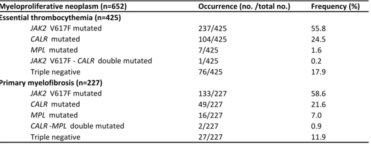
JAK2 V617F mutational load proved to be higher in PMF patients than in ET patients (31.4%
vs. 9.7%, P < 0.001) (Fig. 2A). Similar to JAK2mut load, CALRmut load was also found to be significantly higher in PMF cases (49.6%, range: 13.4–93.5%) compared to ET cases (42.5%, range: 0.7–89.7%; P < 0.001) Both in ET and PMF, the CALRmut load was significantly higher compared to the JAK2mut load (P < 0.001 in both subgroups). (Fig. 2B)
Figure 2. Distribution of the (A) JAK2 V617F and (B) CALR mutant allele burdens detected in ET and PMF patients.
JAK2 mutated ET cases with a mutant allele burden exceeding the median value had major thrombotic events more frequently than those with lower JAK2mut loads (32.3% vs.
13.8%, P=0.02). In ET, higher JAK2mut load was associated with elevated serum LDH levels (403 vs. 360 IU/L, P=0.03), whereas no similar correlation was seen in PMF. White blood cell counts (10.8 vs. 9.6×10⁹/L, P=0.04) and platelet counts (814 vs. 695.5×10⁹/L, P=0.004) also proved to be higher among the ET patients with higher mutational load. We found that patients presenting with splenomegaly had higher JAK2mut load compared to those with normal spleen size (13.2%, range: 1.5–84.9% vs. 8%, range: 0.1–86.6%, P=0.03 in ET and 40%, range: 0.6–
95.9% vs. 18%, range: 0.6–58%, P=0.06 in PMF).
11
We observed higher CALRmut load in type 1 compared to type 2 mutated cases in both PMF (49.6%, range: 13.4–64.3% vs. 36%, range: 27.2–90.9%, P=0.18) and ET (45.8%, range: 8.1–
67.9% vs. 35.6%, range: 1.2–89.7%, P=0.002), with only the latter being statistically different though. The low number of type 2 mutated PMF cases (n=6) may potentially be responsible for the non-significant correlation. In ET, higher CALRmut load was associated with elevated serum LDH levels (510 vs. 351 IU/L, P=0.01), and a clear trend was observed between higher CALRmut load and decreased hemoglobin value (12.0 vs. 13.6 g/dL, P=0.07) as well as increased risk of myelofibrotic transformation (19% vs. 5%, P=0.08). Major thrombotic events occurred more frequently during follow-up in the high CALRmut load cohort (25% vs. 13% in ET) albeit being statistically not different (P=0.18). In ET, patients presenting with splenomegaly exhibited higher CALRmut loads compared to those without splenomegaly (50%, range: 21.6–89.7% vs. 43.5%, range: 0.7–67.3%, P=0.09). In PMF, no difference was observed between these two subgroups.
4.3 Immunohistochemistry
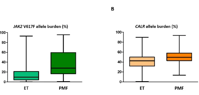
Cases without CALR mutation (n=26) showed no or only minimal labelling with the CAL2 antibody. H-scores evaluated manually (Hman score) were between 0 and 54 (8.6±3.0 mean value), the cases only showed megakaryocytes with dim expression. Automated image analysis yielded Hauto scores between 0 and 91 (10.0±5.0 mean value). All these cases were scored CALR wild-type by the two independent observers as well. CALR mutation positive cases (n=91) showed Hman scores between 62 and 276 (144.5±4.9 mean value), with an average of 45.7 (±2.6) % megakaryocytes demonstrating moderate to strong CALR expression. Automated image analysis resulted in Hauto scores between 98 and 287 withmean values of 186.5 (±2.9) and 68.5 (±1.28) % for the Hauto scores and CALR expressing megakaryocytes, respectively.
Using cut-off values of 58 and 95 for Hman and Hauto scores, respectively, resulted in accurate discrimination of CALR mutation negative and positive cases (figure 3).
12
Figure 3. Demonstration of the discriminatory power of the Hauto and Hman scores using box plots. Cases with CALR mutation have signicantly higher Hauto score (p<0.0001) (A) and Hman score (p<0.0001) (B) compared to the cases without CALR mutation. For comparison t-test was used in both cases.
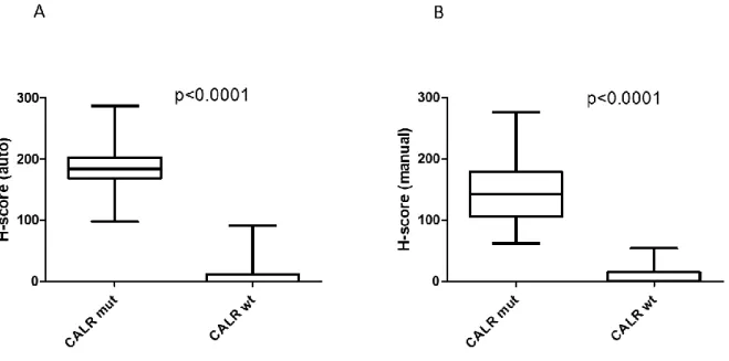
Immunohistochemistry revealed no significant difference in CALR staining pattern between ET and PMF cases or different types of CALR mutations. All 18 different types of CALR mutations analysed in our study were associated with CAL2 labelling. Sixteen of these mutations had not been analysed using the CAL2 antibody before, demonstrating the applicability of this IHC approach to identify the rare CALR variants (Fig. 4).
13
Figure 4. Positive staining of megakaryocytes in MPN cases with eighteen different CALR mutations using the CAL2 monoclonal antibody. JAK2 V617F or MPL mutation positive cases and samples from patients with triple-negative genotype were used as negative controls. The red and yellow arrows indicate a CALR mutation positive and a CALR mutation negative megakaryocyte in a case with a concomitant CALR and JAK2 V617F mutation, respectively.
14
Megakaryocytes with CALR expression showed no distinctive morphological features when compared to CALR negative cells within a sample, although immature cells with high nucleus to cytoplasmic ratio tended to have more intensive labelling. Forty-six percent (42/91) of the cases (27 ET and 15 PMF) with CALR mutation showed labelling of non-megakaryocytic cells as well. The range of positive cells was between 0 and 65%, with these cells appearing as granulocytic precursors.
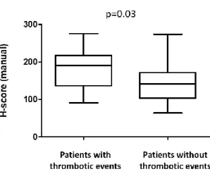
Next, we tested whether the CALR mutation load as determined by immunohistochemistry showed any correlation with the clinical and laboratory parameters of the patients. The Hman scores did not show a correlation with age of the patients, lactate dehydrogenase levels, platelet and white blood cell count, haemoglobin and haematocrit values (data not shown). However, patients with major thrombotic events presented with significantly higher CALR mutation load (p=0.03) compared to the patients without thrombotic events (Fig. 5).
Figure 5. Patients with major thrombotic events (n=9), including arterial thromboses (ischemic stroke, acute myocardial infarction, popliteal artery occlusion, transient ischemic attack and angina pectoris) and venous thromboses (pulmonary embolization, femoral vein thrombosis, portal vein thrombosis and hepatic vein thrombosis) characterized with significantly higher CALR mutation load as defined by H scores derived from quantitative analysis of the CAL2 immunohistochemisty staining when compared to the patients without thrombotic events (n=43), (p=0.03; t-test).
15
4.4 Correlation of molecular and immunohistochemical studies
Manual and automated evaluation of CALR staining showed significant correlation (p<0.001).
Although the percentage of megakaryocytes with moderate to strong labelling as well as the proportion of CAL2 positive non-megakaryocytic cells tended to be higher in cases with higher CALR mutation loads, statistical significance was not reached, and neither the Hman nor the Hauto score demonstrated any significant association with mutation loads determined by molecular studies.
16 5. Conclusions
In summary, in genetic level we performed mutation analysis of a large, multicentric Hungarian MPN cohort, described 12 novel CALR mutations, and provided the first in-depth analysis of the CALRmut load and its influence on clinical phenotype, demonstrating a novel correlation between high CALRmut load and a more proliferative phenotype in ET.
Considering th protein-level results, in summary, we performed a quantitative CALR mutation analysis using a CALR mutation specific antibody on a cohort of 91 MPN patients with CALR mutation. We demonstrate 100% concordance with molecular assays and confirm the applicability of this approach to identify CALR mutations in FFPE tissue material. Further, we extend these findings to 16 rare mutations previously not analysed by this approach, and demonstrate a correlation between the occurrence of thrombotic events and CALR protein mutation load.
17 6. Bibliography
6.1 Publication in the field of the dissertation
1. Mózes R, Gángó A, Sulák A, Vida L, Reiniger L, Timár B, Krenács T, Alizadeh H, Masszi T, Gaál-Weisinger J, Demeter J, Csomor J, Matolcsy A, Kajtár B and Bödör C. Calreticulin mutation specific cal2 immunohistochemistry accurately identifies rare calreticulin mutations in myeloproliferative neoplasms. Pathology 2018.
DOI: 10.1016/j.pathol.2018.11.007. IF: 3.068.
2. Gángó A, Mózes R, Boha Z, Kajtár B, Timár B, Király PA, Kiss R, Fésüs V, Nagy N, Demeter J, Körösmezey G, Borbényi Z, Marton I, Szőke A, Masszi T, Farkas P, Várkonyi J, Plander M, Pósfai É, Egyed M, Pál K, Radványi G, Hamed A, Csomor J, Matolcsy A, Alpár D, Bödör C. Quantitative assessment of JAK2 V617F and CALR mutations in Philadelphia negative myeloproliferative neoplasms. Leukemia Research 2018, 65:42-48.
IF: 2.319.
3. Mózes R, Gángó A, Boha Z, Csomor J, Bödör C. [The role of driver and subclonal mutations in pathogenesis of primary myelofibrosis]. Magyar Onkológia. 2017, 61 :36-45.
6.2 Publication in other fields
1. Réger B, Losonczy H, Nagy Á, Péterfalvi Á, Mózes R, Pótó L, Farkas N, Kovács GL, Miseta A, Hussain A, Tóth O. Detection of high-risk thrombophilia with an automated, global test:
the Coagulation Inhibitor Potential assay. Blood Coagul Fibrinolysis. 2018, 29: 435-441.
IF: 1.119.
2. Réger B, Péterfalvi A, Litter I, Pótó L, Mózes R, Tóth O, Kovács GL, Losonczy H.
Challenges in the evaluation of D-dimer and fibrinogen levels in pregnant women. Thromb
Res. 2013, 131: e183-7. IF: 2.427.