Role of aquaporin-1 expression in the metastatic progression of cutaneous melanoma
Doctoral thesis
Eleonóra Imrédi MD
Semmelweis University
Doctoral School of Pathological Sciences
Supervisor: József Tímár MD, DSc Official reviewers: Gábor Méhes MD, DSc
Sarolta Kárpáti MD, DSc Chair of comprehensive examination committee:
Norbert Wikonkál MD, DSc Members of comprehensive examination committee:
Péter Lakatos MD, DSc Márta Jäckel MD, PhD
Budapest 2019
1 Introduction
Malignant melanoma (MM) is a skin tumor characterized by bad clinical prognosis and an increasing incidence worldwide. Melanomas originate from pigment cells, and multiple environmental and genetic risk factors of the disease have been identified, while the onset of the disease is determined by the interaction of these individual factors in the different ethnical groups and geographical areas. Age, male gender, Breslow thickness, ulceration, the density of tumor infiltrating lymphocytes, the presence of microsatellites, the mitotic rate and the vascular or/and lymphatic invasion are all considered prognostic markers in malignant melanoma.
The prognosis of melanoma patients in stage IV is very poor, and novel treatment options would be essential to overcome the increasing burden of the disease. In the past few years two new therapeutic strategies have fundamentally changed the treatment of melanoma: the immune-checkpoint inhibitors and the targeted therapy have significantly increased both the progression free survival and the overall survival of melanoma patients.
To identify further therapeutic targets, extensive
2
experimental research is carried out in our days to clarify the tumor biology of melanoma.
AQPs carry important physiological function as regulators of water transport in normal tissues, and they are strongly expressed in tumor cells of different origin, particularly in aggressive tumors. For some malignancies– biliary duct carcinoma, glioma, lung adenocarcinoma, colorectal carcinoma and breast cancer - positive correlations have been established between histological tumor grade and the amount of AQP expression.
In human melanoma experimental data suggested the significance of APQ1 expression in the progression of the disease. The B16F10 melanoma tumor cells transfected with AQP1 showed 2- to 3-fold accelerated migration potential in vitro; while reduced tumor growth and angiogenesis was observed after subcutaneous injection of the original B16F10 cell line into AQP1 deficient mice in vivo. Furthermore, the intra-tumoral injection of inhibitory AQP1 siRNAs resulted in impaired tumor growth in a murine model of cutaneous melanoma. Despite the experimental findings suggesting the potential involvement of AQP1 in melanoma
3
progression, no clinical data has been available on the prognostic value of AQP1 expression in human melanoma so far.
Objectives
Our aim was to study the expression of AQP1 in human melanoma specimens and correlate our histological findings with the clinical data obtained from long term prospective clinical follow up. We have investigated the following key questions:
1.) We have determined the AQP1 protein expression level in formalin-fixed, paraffine-embedded human cutaneous melanoma samples by immunohistochemistry.
2.) We have correlated the level of AQP1 protein expression with standard histological parameters and BRAF mutational status.
3.) We have correlated of AQP1 protein expression with long-term clinical follow-up.
4.) We have performed comparative analysis of the AQP1 protein expression in primary cutaneous melanomas with cerebral and extracranial metastases.
5.) We have compared the expression of AQP1 protein in the corresponding cerebral metastasis and primary lesions of cutaneous melanoma patients.
4 Methods
Patients and clinical follow-up
Our studies were based on altogether 121 cases of cutaneous melanoma with a long-term clinical follow-up between 2003 and 2014 at the Department of Dermatooncology, in National Institute of Oncology and at the Department of Dermatology, Venerology and Dermatooncology of Semmelweis University.
The prognostic significance of aquaporin 1 expression’s study was based on two groups of consecutive patients altogether representing 78 cases of cutaneous melanoma with a clinical follow-up between 2003 and 2013.
Patients were diagnosed and operated with curative intent at the Department of Dermatology, Venerology and Dermato-oncology of Semmelweis University between 2003 and 2004. The ‘low-risk’ group included consecutive patients without disease progression before the inclusion, whereas the patients in the ‘high-risk’
group developed early metastasis before enrollment.
In the other study focusing to intracranial metastases, we studied the AQP1 protein expression in 67 metastatic melanoma patients using immunohistochemistry. Primary tumor samples were acquired from patients with brain
5
(BR) (n = 44) and extra- cranial (EC) (n = 23) metastases, while brain metastatic samples were collected during neurosurgical resection at National Institute of Clinical Neuroscience (n = 5).
Formalin-fixed and paraffin-embedded tumor tissue blocks were cut into 5 micrometer thick sections. All slides were deparaffinized and immune-stained with the 7D11 anti-AQP1 primary, mouse monoclonal antibody.
Detection was performed using a secondary antibody for 60 min at room temperature following the protocols of Novolink Polymer Detection Systems. Reaction products were developed using the AEC Peroxidase Substrate kit according to general protocols. After initial manual calibration of the optimal dose of the primary antibody, an automatic immunostainer was used for the serial immunolabeling.
We used the H-score system for quantifying the immunoreactions. The marked cells were scored low (1+), medium (2+), and high (3+) positive categories by subjective way, then the percentages of each categories included cells were defined compared to the whole tumor cells, finally the results were multiplied with the category number and added the numbers to get the final results.
6
The maximum value was 300, if every tumor cells were marked.
Formalin-fixed paraffin-embedded melanoma specimens from the patient included in the histological study were also tested for the presence of the BRAF V600E mutation using PCR and restriction fragment length polymorphism analyses. DNA extraction of melanoma samples was performed using the High Pure PCR Template Preparation Kit. The PCR reaction was carried out according to standardized protocols using commercially available BRAF V600 primers. The PCR reaction was carried out by Swift Max Pro Thermal Cycler machine.
PCR products were separated on a 3% agarose gel and detected with Gel Doc 2000 after ethidium bromide staining. PCR amplification yielded a 224 bp product that was then digested with TspRI restriction endonuclease.
The TspRI restriction enzyme digests the 224 bp product;
therefore, the reaction results in a band of 212 bp from the mutant allele and residual bands from the wild type.
All mutations were confirmed by the Sanger sequencing technique.
7
Statistical analysis was carried out using Statistica 10 Software. Mann–Whitney U-test, χ2-test, and Fischer’s exact test, as appropriate, were used to compare clinical and pathological findings of the different patient cohorts.
Correlations between parameters were analyzed using Spearman’s rank correlation test. Survival probabilities were estimated using the Kaplan–Meier method;
differences were assessed using the Log-rank test. Cox’s proportional hazards model was used to determine the independent prognostic value of AQP1 receptor expression on survival and metastatic progression. All tests were two-tailed, with a confidence interval of 95%;
significance was defined at a P value less than 0.05.
Results
The low-risk group (n=50) did not show progression in 5 years after surgery, while early metastases were detected in the high-risk group within the first 5 years after resection (n=28). According to patients age there was no significant difference between the two groups (p=0,65).
We have detected significant differences in the standard histopathological parameters of the two risk groups. The mitotic index, the Clark level, the ulceration rate and the
8
Breslow index were also equally significantly higher in the high-risk cohort.
The overall survival rate of high-risk patients was significantly lower, mainly due to the metastatic progression involving the central nervous system (21%), visceral organs (21%) and lungs (19%).
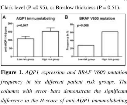
AQP1-protein expression was significantly higher in high-risk melanomas. The immune-histochemical expression of AQP1 was evaluated successfully in 78 patients with cutaneous melanoma, representing two predefined patient cohorts with a distinct estimated prognosis. Positive AQP1 labeling appeared as a red membranous staining on endothelial cells, and cytoplasmic and membrane labeling of tumor cells.
AQP1-labeled melanoma cells were documented in 52 cases (66.7%), with a median H-score of 124.24 (0–300) for the entire study population. Importantly, we found a significantly higher AQP1 H-score (P= 0.047) in the
‘high-risk’ group compared with the ‘low-risk’ patients.
(Figure 1.).
We also tested the Spearman rank order correlations of AQP1 H-score with the established prognostic factors of tumor progression in melanoma; however, no significant
9
correlation was found with the mitotic index (P =0.42), Clark level (P =0.95), or Breslow thickness (P = 0.51).
Figure 1. AQP1 expression and BRAF V600 mutation frequency in the different patient risk groups. The columns with error bars demonstrate the significant difference in the H-score of anti-AQP1 immunolabeling in the low risk compared to the high-risk patients. Error bars represent the standard error of mean (A). Columns on the left show the individual BRAF V600 mutation frequencies found in each risk cohort (B).
Successful BRAF V600 mutation analyses could be carried out from 70 samples, representing 89.74% of the cases included in the immunolabeling study. A mutant allele was found in 31 samples, adding up to an average 44.29% BRAF V600 mutation frequency in our study population. The abundance of the mutant allele was
10
significantly lower (p = 0.008) in the ‘low-risk’ cohort (n
=14/45; 31%) compared with the ‘high-risk’ group (n=
16/25; 64%) (Figure 1B).
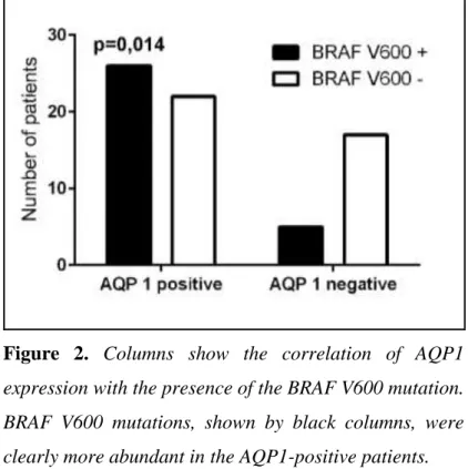
Importantly, a significant correlation of the AQP1 expression and the presence of the BRAF V600 mutation was documented (P =0.014) (Figure 2).
Figure 2. Columns show the correlation of AQP1 expression with the presence of the BRAF V600 mutation.
BRAF V600 mutations, shown by black columns, were clearly more abundant in the AQP1-positive patients.
11
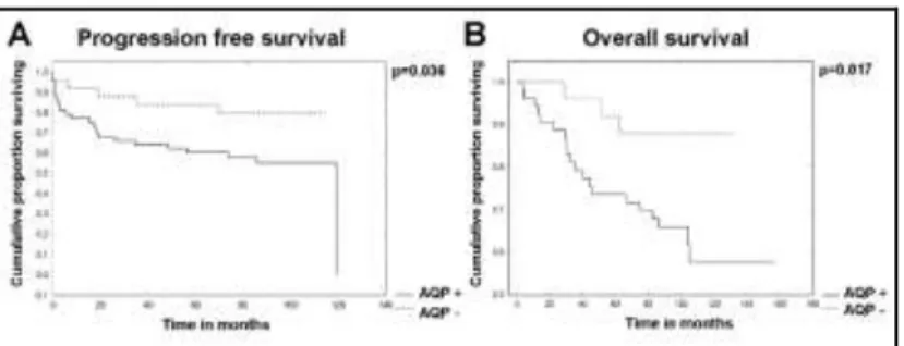
To estimate the impact of AQP1 expression on metastatic progression and overall survival, the Kaplan–Meier method and Log-rank tests were used. We found that the progression-free survival (P= 0.036) and overall survival (P =0.017) of AQP1-positive cutaneous melanoma cases were both significantly decreased (Figure 3 A, B).
Figure 3. Kaplan-Meier curves showing the comparison of progression free and overall survival of cutaneous melanoma patients in case of anti-APQ1 positive (AQP+) and anti-AQP1 negative (AQP-) immunolabeling.
Significant difference can be observed both in the progression free survival (p=0.038) (A), and overall survival (p=0.017) (B) favoring the AQP- cases.
To determine whether AQP1 carried an independent prognostic value in our patients, we carried out a Cox proportional hazard analysis with stepwise logistic
12
regression. We included the ulceration, mitotic index, Breslow thickness, age, and male sex in our model, besides AQP1 expression. According to our analysis, the AQP1 did not prove to be an independent predictor for progression-free survival or overall survival.
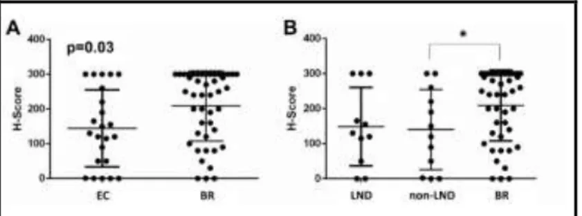
The correlation between AQP1 expression and intracranial progression was based on two groups of consecutive patients, extracranial (EC) (n=23) and brain (BR) (n=44) metastatic groups. We did not find any significant difference baseline comorbidities and standard histological evaluation of two patient groups.
Detailed clinical follow-up was carried out for a median time of 66,5 months (minimum 3,8 months; maximum 186,7 months) in the extracranial group, while it was only 40,4 months (minimum 4 months, maximum 227 months) in the intracranial group.
The extracranial metastases were categorized according to the new version of AJCC (American Joint Committee on Cancer): M1a category included 48% of patients (n=11) , because they had subcutaneous or lymph node metastases, while 26% of patients (n=6) had lung
13
metastases (M1b) and 26% of patients (n=6) had visceral metastases (M1c).
We found that the overall survival of the patients with intracranial progression (M1d) was indeed worse, compared to the patients developing extracranial metastasis exclusively (55,1 vs 83,0 months from the discovery of the primary tumor (p = 0.03).
AQP1 expression of primary melanoma with intracranial metastases is significantly higher than AQP1 expression of melanoma with extracranial metastases. Positive AQP1 labeling appeared as a red membranous staining on endothelial cells and cytoplasmic and membrane labeling of tumor cells in the primary cutaneous samples.
The capillary endothelial cells in the brain tissue did not express the AQP1 protein, similarly to previous report.
AQP1 positive melanoma cells were found in 60 primary tumors (89.56%), with a mean H-score of 187.00 (range 0; 300) for the entire study population. We found significantl y higher AQP1 H-score in the intracranial group compared to the extracranial gr o u p ( p = 0 . 019 ) , des pi t e no s i gni fi cant di f f er enc e i n t he Br esl o w s co r e.
14
Figure 4. AQP1 expression in the BR and EC metastatic patient groups. Each case is represented by a dot, while the bars represent the mean and standard deviation. On the left side (A) primary tumors are divided in two groups based on the extra-cranial vs. cerebral progression, while on the right side patient are further divided into lymph nodal (LND) and non-lymph nodal subgroups. Primary tumors with progression to the brain (BR) showed higher AQP1 expression compared to both EC subgroups. (B).
Since the EC group was heterogenous concerning visceral and LND metastasis we have compared LND metastatic cases to visceral metastatic cases as well as BR metastatic cases. Our statistical analysis indicated that there was no difference in AQP1 expression between the LND and visceral metastasis subgroups, but the visceral (non-LND) metastatic group remained still
15
statistically significantly different from the BR metastatic cases (p=0,048) (Figure 4B).
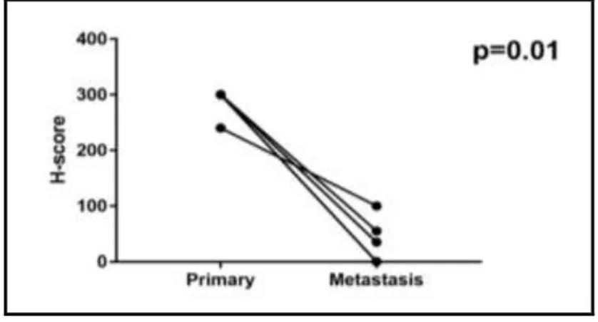
We found significantly lower AQP1 H-score both in the individual cases and by comparing the two groups (p=0.01): while the primary tumors were characterized by a median H-score of 300.0, in the brain metastatic group the median H-score was only 35.0 (Figure 5).
Figure 5. Comparison of AQP1 expression in primary and corresponding brain metastatic tumors. A significant reduction of AQP1 expression during the cerebral progression of cutaneous primary melanoma can be demonstrated (p=0.01).
Looking for a potential cause of reduced expression in the brain metastases we have found that the AQP1 positive tumor cells mainly found in areas distal to the
16
micro-vessels, while the melanoma cells adjacent to capillaries were mostly negative for AQP1.
Conclusions
1. We showed the AQP1 expression by immunohistochemistry in primary cutaneous melanoma in two predefined patient cohorts with a distinct estimated prognosis. Significantly higher AQP1 H-score was detected in high risk group compared with low risk melanomas.
2. The presence of BRAF V600 mutation was significantly lower in the low risk group compared to high risk melanomas. We have found significant correlation between AQP1 expression and BRAF V 600 mutation.
3. We have found that the progression-free survival and overall survival of AQP1-positive cutaneous melanoma cases were both significantly decreased compared with the patients treated for AQP1-negative cutaneous melanoma.
4. The overall survival of the patients with intracranial progression (M1d) was indeed worse, compared to the patients developing extracranial metastasis exclusively.
17
5. The distribution of AQP1 positive melanoma cells was heterogenous in intracranial metastases. The AQP1 positive tumor cells mainly found in areas distal to the micro-vessels, while the melanoma cells adjacent to capillaries were mostly negative for AQP1.
6. AQP1 expression of primary melanoma with intracranial metastases is significantly higher than AQP1 expression of melanoma with extracranial metastases
7. We have compared the AQP1 expression in primary and corresponding brain metastatic tumors and we documented a significant reduction of AQP1 expression during the cerebral progression of primary melanoma.
18 List of own publications
Publications included in the dissertation
Imrédi E, Liszkay G, Kenessey I, Plotár V, Gödény M ; Tóth B, Fedorcsák I, Tímár J.
Aquaporin-1 Protein Expression of the Primary Tumor May Predict Cerebral Progression of Cutaneous Melanoma.
PATHOLOGY AND ONCOLOGY RESEARCH DOI: 10.1007/s12253-018-0513-6 6p. (2018) IF:1,935
Imrédi E, Toth B, Doma V, Barbai T, Raso E, Kenessey I, Timar, J
Aquaporin 1 protein expression is associated with BRAF V600 mutation and adverse prognosis in cutaneous melanoma.
MELANOMA RESEARCH 26:3pp. 254-260. (2016) IF:2,615
Other publications
Imrédi E, Plotár V, Szavcsúr P, Pánczél G, Melegh K, Schlachter K, Liszkay G.
Metasztatikus progresszió kezelése primer cutan és ocularis melanoma szinkrón előfordulását követően.
ORVOSI HETILAP 159: 16 pp. 642-647. 6 p. (2018) IF:0,322