Introduction
The characterization of the genetic background of BCR- ABL1-negative chronic myeloproliferative neoplasms (MPN) was greatly advanced by the discovery that the V617F muta- tion of Janus kinase 2 (JAK2) is very common in the three clas- sic MPN, occurring in 90-95% of cases of polycythemia vera (PV), and in 40-60% of cases of essential thrombocythemia (ET) and primary myelofibrosis (PMF).1-4This led to the inclu- sion of V617F mutation screening in the diagnostic criteria for MPN.5JAK2exon 12 mutations occur in rare cases of V617F- negative PV allowing a close to perfect coverage by specific genetic alterations in PV.6,7On the other hand, clinicians con- tinue to face challenges during the diagnosis of JAK2muta- tion (JAK2mut)-negative ET and PMF. Moving the field very close to full coverage, in two parallel seminal discoveries, Klampfl et al.8and Nangalia et al.9recently described somatic, recurrent insertions/deletions exclusively affecting exon 9 of the calreticulin (CALR) gene. Affecting the same driver path- way, CALR mutations were mutually exclusive with JAK2 V617F or myeloproliferative leukemia virus oncogene gene (MPL) mutations. In a plethora of subsequent studies pub- lished within 3 months, the initial findings were confirmed
and extended focusing on the clinical correlates.10-13 Several additional studies described low frequencies of CALRmuta- tions in different MPN-related diseases, but not in other hematologic diseases.8,9,14-18
As a result of the discovery of CALR mutations, definite molecular diagnostics have become available for 75-90% of clonal MPN. However, for the introduction of CALRmutation screening into routine clinical practice, it is essential to char- acterize the potential effects of CALR mutations on disease phenotype and progression in several independent cohorts.
The aim of our study was to apply a complex array of molec- ular techniques to identify driver mutations. We aimed to confirm previous associations between the presence of acquired genetic alterations and clinical characteristics in a large, independent cohort of patients with MPN. An addition- al purpose was to systematically analyze the roles of particu- lar CALRmutation types and load.
Methods
Subjects
Our study population consisted of 603 patients with BCR-ABL1- negative MPN (260 males, 343 females; median age: 60, range: 10-94
Distinct clinical characteristics of myeloproliferative neoplasms with calreticulin mutations
Hajnalka Andrikovics,1Tunde Krahling,1Katalin Balassa,1Gabriella Halm,2Andras Bors,1Magdalena Koszarska,1Arpad Batai,2Janos Dolgos,2Judit Csomor,3Miklos Egyed,4Andrea Sipos,2Peter Remenyi,2Attila Tordai1, and Tamas Masszi2,5
1Laboratory of Molecular Diagnostics, Hungarian National Blood Transfusion Service, Budapest; 2Department of Hematology and Stem Cell Transplantation, St. Istvan and St. Laszlo Hospital, Budapest; 3Department of Pathology, St. István and St. Lászlo Hospital, Budapest; 4Department of Haematology, Kaposi Mor Hospital, Kaposvar; and 53rdDepartment of Internal Medicine, Semmelweis University, Budapest, Hungary
©2014 Ferrata Storti Foundation. This is an open-access paper. doi:10.3324/haematol.2014.107482 The online version of this article has a Supplementary Appendix.
Manuscript received on March 19, 2014. Manuscript accepted on May 28, 2014.
Correspondence: andrikovics.hajnalka@ovsz.hu
Somatic insertions/deletions in the calreticulin gene have recently been discovered to be causative alterations in myeloproliferative neoplasms. A combination of qualitative and quantitative allele-specific polymerase chain reac- tion, fragment-sizing, high resolution melting and Sanger-sequencing was applied for the detection of three driver mutations (in Janus kinase 2, calreticulin and myeloproliferative leukemia virus oncogene genes) in 289 cases of essen- tial thrombocythemia and 99 cases of primary myelofibrosis. In essential thrombocythemia, 154 (53%) Janus kinase 2 V617F, 96 (33%) calreticulin, 9 (3%) myeloproliferative leukemia virus oncogene gene mutation-positive and 30 triple-negative (11%) cases were identified, while in primary myelofibrosis 56 (57%) Janus kinase 2 V617F, 25 (25%) calreticulin, 7 (7%) myeloproliferative leukemia virus oncogene gene mutation-positive and 11 (11%) triple-negative cases were identified. Patients positive for the calreticulin mutation were younger and had higher platelet counts compared to Janus kinase 2 mutation-positive counterparts. Calreticulin mutation-positive patients with essential thrombocythemia showed a lower risk of developing venous thrombosis, but no difference in overall survival.
Calreticulin mutation-positive patients with primary myelofibrosis had a better overall survival compared to that of the Janus kinase 2 mutation-positive (P=0.04) or triple-negative cases (P=0.01). Type 2 calreticulin mutation occurred more frequently in essential thrombocythemia than in primary myelofibrosis (P=0.049). In essential thrombo- cythemia, the calreticulin mutational load was higher than the Janus kinase 2 mutational load (P<0.001), and increased gradually in advanced stages. Calreticulin mutational load influenced blood counts even at the time point of diagnosis in essential thrombocythemia. We confirm that calreticulin mutation is associated with distinct clinical characteristics and explored relationships between mutation type, load and clinical outcome.
ABSTRACT
years) diagnosed between 1974 and 2013, as an extension of those published earlier.19,20 According to World Health Organization 2008 criteria, 215 patients had JAK2V617F positive PV, 289 had ET and 99 had PMF. Laboratory parameters and clinical features at the time of diagnosis were collected retrospectively. Coagulation com- plications and myelofibrotic or acute leukemic transformation were recorded if they were present at diagnosis or occurred during the follow-up. Coagulation complications included venous throm- botic events (deep vein thrombosis, pulmonary embolism, splanchnic or cerebral venous thrombosis), arterial thrombosis (transient ischemic attack, ischemic stroke, acute myocardial infarction, or peripheral arterial vascular complications) and hem- orrhagic problems (gastrointestinal bleeding, hemorrhagic stroke, hematuria, severe bleeding during surgery or dental procedures).
The median follow-up was 5.7 years (range, 0-40 years). All par- ticipants signed informed consent. The study was approved by the Hungarian National Ethics Committee.
Detection of driver somatic mutations
All analyses were performed using genomic DNA isolated from peripheral blood or bone marrow. In a subset of patients, sampling and diagnostic ascertainment occurred within 1 year. In some analyses, these cases were considered separately as the ones best reflecting the patients’ condition at diagnosis.
All MPN patients were screened for JAK2V617F (c.1849G>T) by allele-specific polymerase chain reaction (PCR).1 In JAK2mut cases, real-time quantitative PCR was performed to determine the V617F load.21 The JAK2mut load was calculated as follows:
JAK2mut/(JAK2mut+JAK2wild-type).
JAK2mut-negative ET and PMF patients were screened for CALR mutations by high resolution sizing of fluorescence-labeled PCR products by capillary electrophoresis (fragment analysis).8 In CALRmut cases, the precise mutation was identified by Sanger- sequencing.8 To determine the mutant load, the ratio of peak heights was calculated using an analogous formula:
CALRmut/(CALRmut+CALRwild-type). Since fragment analysis was a semi-quantitative approach and the final amounts of the PCR products were influenced by the preferential amplification of shorter amplicons, we calculated the load after PCR with 25 cycles, which was a reduced cycling condition compared to the screening condition (35 cycles).
In JAK2mutand CALRmutnegative ET and PMF patients, screening for MPLS505N and W515 codon mutations was performed by
high resolution melting analysis.22 In cases that were positive in this screening analysis, the exact type of MPLmutation was deter- mined by allele-specific PCR and sequencing.23
Statistics
Continuous variables are presented as medians with 25thand 75thpercentiles. Mann-Whitney or Kruskal-Wallis tests were used to compare continuous variables, while χ2 or Fisher exact tests were used for dichotomous variables. A log-rank test was per- formed to compare overall survival between subgroups according to driver mutations. In the case of hematopoietic stem cell trans- plantation (n=13), the follow-up period was terminated at the date of the transplant. For multivariate analysis, age was considered in a Cox proportional hazards model beside driver mutations.
Hazard ratios (HR) and 95% confidence interval (CI) values were computed. The analyses were conducted using the SPSS (version 20.0) software package.
Results
Distribution of different types of driver mutations and comparisons of clinical parameters in subgroups according to driver mutation status
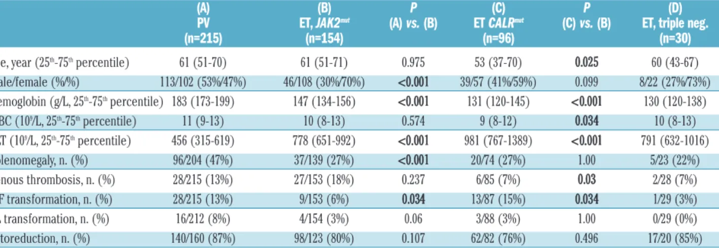
Only JAK2V617F positive (JAK2mut) PV patients (n=215) were included in this study. In patients with PV, qualita- tive and quantitative JAK2V617F tests, but no CALRand MPLmolecular tests, were performed. For ET, the distri- bution of driver mutations was as follows: JAK2mut: 154/289 (53%), CALRmut: 96/289 (33%), MPLmut: 9/289 (3%), triple-negative: 30/289 (11%) cases (Table 1). For PMF, the corresponding figures were: JAK2mut: 56/99 (57%), CALRmut: 25/99 (25%), MPLmut: 7/99 (7%), triple- negative: 11/99 (11%) cases (Table 2).
The patients’ clinical and laboratory parameters were systematically compared according to diagnosis and driver mutation. Given the low number of cases, patients with MPLmutation were omitted from all subsequent analyses.
Compared to the JAK2mutPV cohort, the JAK2mutET cohort contained more females (P<0.001), had lower hemoglobin levels (P<0.001), higher platelet counts (P<0.001), less fre- quent splenomegaly (P<0.001) and less frequent myelofi-
Table 1.Clinical and laboratory characteristics of PV and ET patients according to the presence of a driver somatic mutation.
(A) (B) P (C) P (D)
PV ET, JAK2mut (A) vs.(B) ET CALRmut (C) vs.(B) ET, triple neg.
(n=215) (n=154) (n=96) (n=30)
Age, year (25th-75thpercentile) 61 (51-70) 61 (51-71) 0.975 53 (37-70) 0.025 60 (43-67)
Male/female (%/%) 113/102 (53%/47%) 46/108 (30%/70%) <0.001 39/57 (41%/59%) 0.099 8/22 (27%/73%)
Hemoglobin (g/L, 25th-75thpercentile) 183 (173-199) 147 (134-156) <0.001 131 (120-145) <0.001 130 (120-138)
WBC (109/L, 25th-75thpercentile) 11 (9-13) 10 (8-13) 0.574 9 (8-12) 0.034 10 (8-13)
PLT (109/L, 25th-75thpercentile) 456 (315-619) 778 (651-992) <0.001 981 (767-1389) <0.001 791 (632-1016)
Splenomegaly, n. (%) 96/204 (47%) 37/139 (27%) <0.001 20/74 (27%) 1.00 5/23 (22%)
Venous thrombosis, n. (%) 28/215 (13%) 27/153 (18%) 0.237 6/85 (7%) 0.03 2/28 (7%)
MF transformation, n. (%) 28/215 (13%) 9/153 (6%) 0.034 13/87 (15%) 0.034 1/29 (3%)
AL transformation, n. (%) 16/212 (8%) 4/154 (3%) 0.06 3/88 (3%) 1.00 0/29 (0%)
Cytoreduction, n. (%) 140/160 (87%) 98/123 (80%) 0.107 62/82 (76%) 0.496 17/20 (85%)
ET-patients qualified for triple-negative status (D) if no JAK2mutor CALRmutor MPLmutwas present. Clinical and laboratory data apply at the time of first presentation. Cytoreduction was defined positive if hydroxyurea or anagrelide treatment was given for more than 6 months. Pvalues below 0.05 were considered statistically significant and are indicated in bold. AL: acute leukemia; ET: essential thrombocythemia; Hb: hemoglobin concentration; MF: myelofibrotic, PLT: platelet count; PMF: primary myelofibrosis; PV: polycythemia vera;
WBC: white blood cell count.
brotic transformation (P=0.034) (Table 1). Compared to the JAK2mut ET cohort, in the CALRmut ET patients we found a tendency toward less pronounced female predom- inance (P=0.099), younger age at diagnosis (P=0.025), lower hemoglobin levels (P<0.001), lower white blood cell counts (P=0.034) and higher platelet counts (P<0.001).
Coagulation complications (venous and arterial throm- boses, plus hemorrhages, taken together) were more fre- quent in JAK2mutET patients (36%, 55/153) than in CALRmut ET patients (18%, 15/85; P=0.003). Venous thrombosis was more frequent in JAK2mut ET than in CALRmut ET (P=0.03). Arterial thrombosis occurred in 14% (22/153) of JAK2mutand in 9% (8/85) of CALRmutET (P=0.3), and hem- orrhage in 9% (14/153) of JAK2mut and in 5% (4/85) of CALRmut ET (P=0.3). Myelofibrotic transformation occurred more frequently in the CALRmutcohort (P=0.03).
We did not find any significant differences comparing triple-negative ET patients to other ET subgroups: the sample size (n=30) was, however, small.
Similar analyses in the PMF cohort (Table 2) showed
younger age at presentation (P=0.002) and higher platelet counts (P=0.001) in the CALRmut subgroup than in the JAK2mutPMF subgroup. Other variables were not different, nor were any of the characteristics of the triple-negative PMF patients except for an increased rate of acute leukemic transformation in triple-negative cases (36% ver- sus 9% in JAK2mut, P=0.038 or versus 14% in CALRmut, P=0.19).
Regarding outcome parameters, overall survival was analyzed, initially by a univariate Kaplan-Meier approach, in patients with ET (Figure 1A) and PMF (Figure 1B) strat- ified according to different driver mutations. Out of the 289 ET patients, 261 cases had appropriate follow-up information [JAK2mut (n=150), CALRmut (n=85) and triple- negative (n=26) subgroups]. As shown in Figure 1A, no differences were detected in overall survival by univariate analyses (P=0.846).
Among PMF patients (n=87), the subgroups were as fol- lows: JAK2mut (n=55), CALRmut (n=21) and triple-negative (n=11). In contrast to ET, the overall comparison resulted
Table 2.Clinical and laboratory characteristics of PMF patients according to the presence of a driver somatic mutation.
(A) (B) P (D) PMF, JAK2mut PMF, CALRmut (A) vs.(B) PMF, Triple neg.
(n=56) (n=25) (n=11) Age, year (25th-75thpercentile) 68 (57-73) 56 (39-65) 0.002 69 (53-79) Male/female (%/%) 28/28 (50%/50%) 12/13 (48%/52%) 1.00 8/3 (72%/27%) Hemoglobin (g/L, 25th-75thpercentile) 116 (58-183) 112 (97-124) 0.484 95 (79-121) White cell count (109/L, 25th-75thpercentile) 12 (8-18) 9 (5-13) 0.147 12 (7-25) Platelet count (109/L, 25th-75thpercentile) 250 (132-500) 552 (320-712) 0.001 156 (54-452) Splenomegaly, n. (%) 47/56 (84%) 14/21 (67%) 0.120 9/11 (82%) Venous thrombosis, n. (%) 9/54 (17%) 1/21 (5%) 0.266 1/11 (9%) AL-transformation, n. (%) 5/54 (9%) 3/22 (14%) 0.684 4/11 (36%) Cytoreduction, n. (%) 30/43 (70%) 9/17 (53%) 0.243 6/8 (75%)
For explanation and abbreviations see Table 1.
Figure 1.Kaplan-Meier analysis of overall survival in patients with ET (panel A, n=261) and PMF (panel B, n=87) according to the presence of different driver mutations. Patients qualified for triple-negative status if JAK2mutand CALRmutand MPLmutwere all absent. For a subset of patients (ET: n=2, PMF: n=11) treated by hematopoietic stem cell transplantation, the follow-up was censored at the date of this intervention.
In ET, univariate analyses resulted in an overall Pvalue of 0.846 (A). In PMF, the same comparison gave a Pvalue of 0.023 (B). Upon pairwise univariate comparisons, the CALRmutsubgroup showed significantly better survival compared to JAK2mut(P=0.04) and triple-negative (P=0.01) PMF patients while JAK2mutpatients showed only a tendency towards better overall survival compared to triple-negative patients (P=0.076).
ET: essential thrombocythemia; PMF: primary myelofibrosis.
A B
Triple neg. ET n=26
JAK2mutET n=150
Triple neg. PMF n=11
JAK2mutPMF n=55 CALRmutPMF n=21 CALRmutET n=85
0 10 20 30 40
Time (years from diagnosis)
0 10 20 30 40
Time (years from diagnosis) 1.0
0.8
0.6
0.4
0.2
0.0 1.0
0.8
0.6
0.4
0.2
0.0
Probability of survival Probability of survival
P=0.04
P=0.846
P=0.023
in P=0.023 (Figure 1B) with the best overall survival in the CALRmutsubgroup and the worst in the triple-negative sub- group. This was further confirmed by pairwise univariate comparisons, namely CALRmutversus JAK2mut(P=0.04) and CALRmut versus triple-negative (P=0.01). The JAK2mut PMF patients showed only a tendency towards having a better overall survival than the triple-negative patients (P=0.076).
To further analyze the potential effect of driver mutations on survival, we utilized a Cox proportional hazard model choosing the triple-negative PMF subgroup as the refer- ence. In this model, age was a factor that significantly affected survival (HR 1.082, 95% CI: 1.023-1.144;
P=0.006), while the presence of a driver mutation showed an overall tendency (P=0.059). Performing pairwise com- parisons, the CALRmut subgroup was characterized by a HR=0.131 (95% CI: 0.023-0.739; P=0.021) indicating a sig- nificantly better survival than that of triple-negative patients. Comparing the CALRmut patients to the JAK2mut subgroup, only a tendency towards better survival was observed in the presence of CALR exon 9 mutation (HR=0.159, 95% CI: 0.018-1.391; P=0.097).
Subgroup analyses in patients with CALRmutaccording to type of mutation
Following fluorescence PCR and fragment analyses by capillary electrophoresis, the presence of CALR exon 9 mutation was confirmed by Sanger-sequencing. In the entire cohort of 121 CALR-positive cases, we found 64 (53%) with a type 1 mutation (52 base pair deletion, c.1092_1143del), 34 (28%) with a type 2 mutation (5 base pair insertion, c.1154_1155insTTGTC) and 23 (19%) with other types of mutations. These other mutations com- prised the following types: 3 (c.1095_1140del), 4 (c.1102_1135del), 5 (c.1091_1142del), 19 (c.1110_1140del), 22 (c.1120_1123del), 24 (c.1120_1138del), 29 (c.1135_1152delinsCCTCCTCTTTGTCT), 33 (c.1154_1155insATGTC), 34 (c.1154_delins CTTGTC), 35 (c.1154delinsTTTGTC) and potentially novel variants (n=8). The occurrence of these variants according to diag- nosis was as follows: ET: type 1: 48 (50%), type 2: 31 (32%) and other: 17 (18%); PMF: type 1: 16 (64%), type 2:
3 (12%) and other: 6 (24%). Comparing patients with ET and PMF and only considering types 1 and 2, we found a tendency to a different distribution with 48 type 1 (61%) and 31 type 2 (39%) mutations in patients with EF versus 16 type 1 (84%) and 3 type 2 (16%) in patients with PMF (P=0.064). An increased frequency of type 2 mutations (versusnon-type 2 mutations) was observed in ET (32%) compared to PMF (12%, P=0.049, Online Supplementary Table S1).
Next, we compared demographic and clinical parame- ters within patients with CALRmutET according to muta- tion types. We found a tendency towards older age among type 2 carriers compared to type 1 carriers (median age at diagnosis: 59 years versus51 years, P=0.06) or compared to non-type 2 carriers (median age at diagnosis: 59 years ver- sus51 years, P=0.09). Similarly, platelet count at diagnosis tended to be higher in the subgroup of type 2 mutation carriers than in patients with the type 1 mutation:
1237x109/L (25th-75th percentile: 884-1472) versus 946 x109/L (25th-75th percentile: 764-1280) (P=0.081); or com- pared to those with non-type 2 mutation: 865x109/L (25th- 75th percentile: 755-1218) (P=0.041). The frequency of cytoreductive therapy was higher among type 2 versus type 1 ET patients (89% versus67% P=0.05). No further
differences in any demographic or clinical characteristics (including overall survival) were found upon comparing the two ET subgroups with different types of mutations (Online Supplementary Table S2). Similar analyses were not feasible for the PMF cohort because of the low number (n=3) of patients with the type 2 CALRmutation.
Subgroup analyses in patients with a positive driver mutation (JAK2mutor CALRmut) according to mutational load
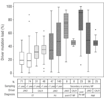
A systematic comparison was performed between MPN subgroups in relation to the quantity of driver mutation (Figure 2). The JAK2V617F load was determined by real- time quantitative allele-specific PCR using the TaqMan detection system,21while the semi-quantitative estimation of the CALR mutation was achieved by high resolution sizing of fluorescence-labeled PCR products.8 The muta- tional load in JAK2 V617F and CALR mutation-positive patients was calculated with a similar formula i.e. the quantity of mutant was expressed as a percentage fraction of total gene copies allowing the comparison of loads across MPN subgroups with different mutations.
Performing pairwise comparisons, we made the following observations (Figure 2). (i) NeitherJAK2mut, nor CALRmutET patients without myelofibrotic transformation at the sam- pling time showed significantly different driver mutation- al loads comparing the subgroup of patients whose sam- ples were taken within 1 year of diagnosis and the sub-
Figure 2. Comparisons of relative quantities of driver mutations (JAK2V617F or CALR) in different MPN subgroups according to diag- nosis and the relation of time of sampling and time of diagnosis. The first row under the x axis shows the number of patients (n) in the respective subgroups. If no primary or secondary myelofibrosis was present at the time of sampling, sampling within 1 year of diagnosis qualified, as seen in the second lines, as “<1 year”; all other situa- tions were allocated into the “>1 year” subgroup. If primary or sec- ondary myelofibrosis was present at the time of sampling, no such subgrouping was performed (“secondary or primary MF”).
Quantification of the respective driver mutation was performed by real-time quantitative allele-specific PCR (JAK2V617F) or fragment analysis (CALRexon 9). Pairwise comparisons were performed with the Mann-Whitney test. ET: essential thrombocythemia; PMF: pri- mary myelofibrosis; MF: myelofibrosis; PV: polycythemia vera.
100
80
60
40
20
0
Driver mutation load (%)
71 74 31 48 47 140 8 8 23 56 25
<1 year >1 year <1 year >1 year <1 year >1 year Secondary or primary MF
JAK2 CALR JAK2 JAK2 CALR JAK2 CALR
ET PV PMF
post- PV MF post-ET MF N.
Sampling Driver Diagnosis
group of patients whose samples were taken at a later time point. On the other hand, the two PV-subgroups (44 patients with sampling within 1 year of diagnosis and 140 patients with sampling at later time points but without myelofibrotic transformation at this later time point) showed a tendency toward increasing allele burden (P=0.066). (ii) JAK2 loads increased gradually in parallel with the appearance of more advanced stages of MPN (JAK2mutET versusPV: P<0.001, PV versuspost-PV myelofi- brosis: P<0.001). (iii) Within patients with ET, the CALR load was significantly higher than the JAK2 load (P<0.001). (iv) CALRload showed a steady, significant, but less steep increase corresponding to the appearance of more advanced MPN stages (CALRmut ET versus CALRmut post-ET MF: P=0.01; CALRmut ET versus CALRmut PMF:
P<0.001). (v) In contrast to JAK2mutload, the CALRmutload only rarely exceeded 50% (in 11 cases between 51-75%
and in 1 case above 75%).
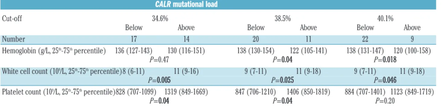
To investigate the potential effects of CALRmutational load, we divided CALRmutET patients with samples avail- able within 1 year after diagnosis (n=31) into two sub- groups according to CALRmut load, with the cut-off value being 38.5%, which was the median CALR mutational load of all patients with ET in the study (n=20 below the cut off and n=11 above the cut off; Table 3). White blood cell counts (9 versus11x109/L, P=0.025) and platelet counts (848 versus 1406x109/L, P=0.04) were lower among patients with low CALRmut load, while hemoglobin con- centration was higher in the same comparison (138 versus 122 g/dL,P=0.04). Applying different cut-off values to dis- criminate between groups with lower and higher CALRmut burden, white blood cell counts were always lower in sub- groups with lower CALRmut burden, although the differ- ences shrank between the lower and the higher CALRmut burden subgroups applying higher cut-offs. While the dif- ferences in hemoglobin levels between patients with lower and higher CALRmutload were only detectable above the cut-off value of 38.5% or higher, the differences in platelet counts diminished above this cut-off. We observed higher CALRmut burden in type 1 (n=14, 41%) compared to type 2 ET (n=15, 27%, P=0.023). However, this difference may have been influenced by the preferen- tial amplification of the shorter fragment in the presence of the type 1 mutant (52 base pair deletion) during PCR, thus this observation needs to be confirmed with an alter- native technique of quantification.
Discussion
We set up sequential application of different molecular techniques to identify the driver mutations in a large cohort of MPN patients, noting that JAK2V617F, CALR and MPLmutations were described as mutually exclusive driver mutations in the first studies.8,9 Concurrent JAK2 and CALRmutations were reported in only two individu- als in subsequent studies.13,14Confirming earlier observa- tions in different MPN cohorts,8-10,13we found a similar fre- quency of CALR mutations in an independent group of MPN patients, with the frequency of these mutations being high (121/162, 75%) among JAK2V617F and MPL- negative ET patients, thus leaving only a small cohort of triple-negative patients lacking a disease-causing genetic marker.
In our previous study comparing the clinical characteris- tics of JAK2mut and JAK2neg MPN patients (n=328),19 we already noted a female predominance, older age at presen- tation, higher hemoglobin values and a higher incidence of coagulation complications (thrombotic and hemorrhagic) in JAK2V617F-positive MPN patients versusV617F-nega- tive counterparts (including CALRmutpatients). The identi- fication of the CALRmutation allowed a more straight for- ward comparison of MPN patients with a homogeneous genetic background. Our present data, showing distinct clinical characteristics of CALRmutET subgroups in compar- ison with JAK2mut cohorts, confirmed previous observa- tions regarding younger age, sex distribution with less prominent female abundance (only in the ET subgroup), lower hemoglobin concentration, lower white blood cell counts (not significant in our PMF cohort), and higher platelet counts.8-12
Considering venous thrombosis and other coagulation complications, our observations confirmed previous find- ings describing a lower risk of thrombosis in patients with CALRmutET than in those with JAK2mutET.8In their extend- ed cohort, Rumiet al.12found that the rates of thrombosis at diagnosis in the CALRmutand JAK2mutgroups were 2.8%
and 7.1%, respectively (P=0.059), and observed a reduced cumulative incidence of thrombosis (25 versus10 events per 1000 person-years) in CALRmutpatients (P=0.001). In an independent series of patients with ET, Rotunno et al.
found that the incidence of thrombosis (in the 2 years pre- ceding diagnosis and during follow-up, combined) was 13.5% in their CALRmutcohort compared to 30.1% among
Table 3.Laboratory characteristics of ET patients according to the CALRmutational load.
CALRmutational load
Cut-off 34.6% 38.5% 40.1%
Below Above Below Above Below Above
Number 17 14 20 11 22 9
Hemoglobin (g/L, 25th-75thpercentile) 136 (127-143) 130 (116-151) 138 (130-154) 122 (105-141) 138 (131-147) 120 (100-158)
P=0.47 P=0.04 P=0.018
White cell count (109/L, 25th-75thpercentile)8 (6-11) 11 (9-16) 9 (7-11) 11 (9-18) 9 (7-11) 11 (9-18)
P=0.005 P=0.025 P=0.046
Platelet count (109/L, 25th-75thpercentile)828 (707-1099) 1319 (849-1669) 847 (706-1210) 1406 (850-1819) 884 (707-1401) 1123 (849-1719)
P=0.04 P=0.04 P=0.20
In all three pairwise comparisons, the same 31 ET patients with available sampling within 1 year of diagnosis were divided into dichotomous subgroups, according to CALRmuta- tional load. The following CALRmutational load cut-off values were chosen: (i) 34.6%: median CALRmut load of all ET patients excluding cases with myelofibrotic-transformation;
(ii) 38.5%: median CALRmutload of all ET patients; (iii) median CALRmutload of all MPN (ET and PMF all included) patients in the present study. Pvalues represent pairwise com- parisons between dichotomously divided subgroups of patients (Mann-Whitney test). Pvalues below 0.05 are considered statistically significant and are indicated in bold. ET: essen- tial thrombocythemia.
JAK2mutpatients (P=0.011).
With respect to transformation of ET to myelofibrosis, we observed a more than 2-fold higher transformation rate among CALRmut patients (15%) compared to JAK2mut patients (6%, P=0.034), the rate in the former being similar to that in the cohort of patients with PV (13%). Nangalia et al. also found an increased risk of transformation to myelofibrosis (19% in CALRmut versus 2% in JAK2mut patients; P=0.03) in a smaller cohort of patients with ET.9 In a large study, the incidence of myelofibrotic transforma- tion was 7% (95% CI 3-13) in CALRmutversus5% (95% CI 3-8) in JAK2mutET versus8% (95% CI 5-12) inJAK2mutPV, while the 15-year cumulative incidence of myelofibrotic transformation was 13.4% (CI 95% 5.4-25.2%) in CALRmut versus 8.4% (CI 95% 3.9-15.3%) in JAK2mut ET versus 13.6% (CI 95% 7.3-21.9%) in JAK2mut PV (P=not signifi- cant).12In our cohort, the median follow-up tended to be longer in the CALRmutET group (8 years in CALRmutversus4 years in JAK2mut; P=0.09), which might have resulted in a higher frequency of myelofibrotic transformation.
Among patients with ET, we did not find differences in overall survival according to the presence of a specific driver mutation. In contrast, Klampfl et al. observed better survival in patients with CALRmut than in those with JAK2mut (P=0.04).8 Interestingly, analyzing a larger ET cohort with substantial overlap with that of Klampfl et al., in univariate analysis, Rumi et al. found only a trend (P=0.085) towards a better overall survival at 15 years for CALRmut patients compared to JAK2mut patients, and the trend disappeared when age was considered as a covari- ate.12Similarly, Rotunno et al. did not find any difference in overall survival among patients with ET.11
As for patients with ET and in previous reports,8,13 younger age at disease onset and elevated platelet counts were found to characterize our CALRmutPMF cohort. Our observations in PMF were in good agreement with previ- ous reports,8,13indicating a significant beneficial effect of the presence of CALRmut, compared to JAK2mut, on overall survival. In our dataset, the differences were further sub- stantiated by pairwise comparisons in a multivariate (with age added as a covariate) Cox model indicating a trend towards better survival in CALRmut patients compared to JAK2mut ones and a significantly better survival between CALRmutand triple-negative PMF patients. In addition, the decreased leukemia-free survival in triple-negative PMF patients reported by Tefferi et al.13 was in line with the increased rate of acute leukemic transformation in our triple-negative PMF cases (Table 2). Recently, a set of gene mutations were described as detrimental in PMF. The presence of any “high molecular risk” mutations affecting ASXL1, EZH2, SRSF2, and IDH1/2genes predicted shorter overall survival and increased risk of leukemic transforma- tion. A CALRmutation proved to be an independent pro- tective factor for survival in PMF.24 ASXL1mutations, as the most frequent detrimental mutations, were reported to occur with similar frequency in JAK2mut, CALRmut and triple-negative PMF patients;13 thus, in our PMF-cohort, the survival benefit observed in the group of CALRmut patients was unlikely to result from an uneven distribution of ASXL1mutations. The presence of CALRmutin PMF may affect prognostication and should be incorporated in the scoring system, as recently suggested.25,26
Our data regarding the frequencies of type 1 and type 2 CALRmutin ET and in PMF were in good agreement with those of previous cohorts of patients with ET8-10,12 and
PMF.8,13The asymmetric distribution of different types of CALRmutin ET and PMF, i.e. the relatively lower frequency of type 2 CALRmut in PMF, was in line with data from Klampfl et al.8They also observed a lower (21/105, 20%) relative proportion of type 2 mutant cases among PMF patients than among those with ET (74/195, 38%). The profound differences in the CALR protein structure may explain this asymmetric distribution and may imply slight differences in the pathogenic effects of the respective vari- ants. The observed asymmetric distribution prompted us to systematically analyze clinical and outcome differences in ET patients according to CALRmut type, with the key findings being older age at diagnosis, higher platelet counts and more frequent use of cytoreductive therapy in patients with type 2 CALRmutcompared to type 1 or non- type 2 CALRmut. To our knowledge, similar data in ET have not been published yet. In line with our data in ET, very recently Tefferi et al.27observed distinct characteristics in PMF in the context of CALRtype 2 mutations and EZH2, IDHmutations, leukocytosis, and peripheral blast percent- age. Univariate survival analysis suggested a less favorable outcome in the presence of type 2 CALR mutations.27 These differences may indicate a distinct pathomech- anism related to insertion or deletion type CALR muta- tions.
The systematic analyses of CALRmutload in several MPN subgroups indicated higher CALRmut load compared to JAK2mut load in ET. In contrast to the JAK2mut load, the CALRmutload rarely exceeded 50%, indicating a decreased tendency of the CALRmutlocus to undergo mitotic recom- bination. Uniparental disomy was reported to be less fre- quent at the CALRlocus (19p13.3-p13.2) than at the JAK2 locus (9p24). Using an alternative quantitative PCR tech- nique, we are in the process of addressing the observation that CALRmut load may be higher at diagnosis in type 1 CALRmutcarriers than in type 2 carriers. Our results indi- cate that the mutational load of CALRmutresembled that of JAK2mutin that there was a clear trend towards increased load in parallel with the appearance of more severe phe- notypes of MPN. Campbell et al.28 suggested that JAK2 V617F mutated ET, PV and post-ET/PV-myelofibrosis or PMF form a biological continuum, in which the different MPN phenotypes are determined by JAK2 V617F allele burden modified by other environmental and inherited factors. The increasing JAK2V617F allele burden through ET-PV-myelofibrosis phenotypes was later confirmed in several studies,29-31and also in a transgenic mouse model.32 Our study shows, for the first time, that hematologic lab- oratory parameters (white blood cell count, hemoglobin concentration, platelet count) were directly influenced by CALR mutational burden in ET patients with samples available within 1 year of diagnosis. Similarly to JAK2mut MPN, CALRmutET and PMF also show overlapping clinical features, suggesting that the common molecular basis rep- resents a biological continuum between CALRmut ET and CALRmutmyelofibrosis, in which the clinical phenotype is modified by the actual CALRmutational load.12
In summary, analyzing a relatively large, independent cohort of MPN patients, we confirmed recent observa- tions that the presence of CALRmutations is responsible for the development of a distinct clinical phenotype in both ET and PMF. These distinctive features manifest as different clinical characteristics at diagnosis, different pat- terns of complications as well as significant differences in overall survival. Our data supplement and extend previous
observations regarding the distribution of various types of CALRmut and associations with different quantities of CALRmut. Our study shows, for the first time, that hemato- logic laboratory parameters are directly influenced by CALRmutational burden. The recent discovery of somatic CALRmutations not only improves the precision of non- invasive diagnostics of patients with MPN, but may influ- ence prognosis and therapy.
Acknowledgments
The authors thank Csehné Bánhidi Klára, Haluska Brigitta, Horváth Csongorné, Mezibroczky Martina, Pfundt Antalné, and
Petró Péterné for their technical assistance. This work was sup- ported by grants from OTKA (K104903). HA is a recipient of the Janos Bolyai Research Scholarship from the Hungarian Academy of Sciences, and benefited from a scholarship for the Third Training School of MPN&MPNr-EuroNet (COST Action BM0902) in May 2012. HA and AT are members of MPN&MPNr-EuroNet.
Authorship and Disclosures
Information on authorship, contributions, and financial & other disclosures was provided by the authors and is available with the online version of this article at www.haematologica.org.
References
1. Baxter EJ, Scott LM, Campbell PJ, East C, Fourouclas N, Swanton S, et al. Acquired mutation of the tyrosine kinase JAK2 in human myeloproliferative disorders. Lancet.
2005;365(9464):1054-61.
2. James C, Ugo V, Le Couedic JP, Staerk J, Delhommeau F, Lacout C, et al. A unique clonal JAK2 mutation leading to constitutive signalling causes polycythaemia vera.
Nature. 2005;434(7037):1144-8.
3. Kralovics R, Passamonti F, Buser AS, Teo SS, Tiedt R, Passweg JR, et al. A gain-of-function mutation of JAK2 in myeloproliferative dis- orders. N Engl J Med. 2005;352(17):1779-90.
4. Levine RL, Wadleigh M, Cools J, Ebert BL, Wernig G, Huntly BJ, et al. Activating muta- tion in the tyrosine kinase JAK2 in poly- cythemia vera, essential thrombocythemia, and myeloid metaplasia with myelofibrosis.
Cancer Cell. 2005;7(4):387-97.
5. Tefferi A, Vainchenker W.
Myeloproliferative neoplasms: molecular pathophysiology, essential clinical under- standing, and treatment strategies. J Clin Oncol. 2011;29(5):573-82.
6. Scott LM. The JAK2 exon 12 mutations: a comprehensive review. Am J Hematol.
2011;86(8):668-76.
7. Scott LM, Tong W, Levine RL, Scott MA, Beer PA, Stratton MR, et al. JAK2 exon 12 mutations in polycythemia vera and idio- pathic erythrocytosis. N Engl J Med. 2007;
356(5):459-68.
8. Klampfl T, Gisslinger H, Harutyunyan AS, Nivarthi H, Rumi E, Milosevic JD, et al.
Somatic mutations of calreticulin in myelo- proliferative neoplasms. N Engl J Med.
2013;369(25):2379-90.
9. Nangalia J, Massie CE, Baxter EJ, Nice FL, Gundem G, Wedge DC, et al. Somatic CALR mutations in myeloproliferative neo- plasms with nonmutated JAK2. N Engl J Med. 2013;369(25):2391-405.
10. Chi J, Nicolaou KA, Nicolaidou V, Koumas L, Mitsidou A, Pierides C, et al. Calreticulin gene exon 9 frameshift mutations in patients with thrombocytosis. Leukemia. 2014;28(5):
1152-4.
11. Rotunno G, Mannarelli C, Guglielmelli P, Pacilli A, Pancrazzi A, Pieri L, et al. Impact of calreticulin mutations on clinical and hema- tological phenotype and outcome in essen- tial thrombocythemia. Blood. 2014;123(10):
1552-5.
12. Rumi E, Pietra D, Ferretti V, Klampfl T, Harutyunyan AS, Milosevic JD, et al. JAK2
or CALR mutation status defines subtypes of essential thrombocythemia with substan- tially different clinical course and outcomes.
Blood. 2014;123(10):1544-51.
13. Tefferi A, Lasho TL, Finke CM, Knudson RA, Ketterling R, Hanson CH, et al. CALR vs JAK2 vs MPL-mutated or triple-negative myelofibrosis: clinical, cytogenetic and molecular comparisons. Leukemia. 2014 Jan 9. doi: 10.1038/leu.2014.3. [Epub ahead of print]
14. Broseus J, Lippert E, Harutyunyan AS, Jeromin S, Zipperer E, Florensa L, et al.
Low rate of calreticulin mutations in refractory anaemia with ring sideroblasts and marked thrombocytosis. Leukemia.
2014;28(6):1374-6.
15. Hou HA, Kuo YY, Chou WC, Chen PH, Tien HF. Calreticulin mutation was rarely detect- ed in patients with myelodysplastic syn- drome. Leukemia. 2014 Feb 17. doi:
10.1038/leu.2014.71. [Epub ahead of print]
16. Lasho TL, Elliott MA, Pardanani A, Tefferi A. CALR mutation studies in chronic neu- trophilic leukemia. Am J Hematol. 2014;89 (4):450.
17. Maffioli M, Genoni A, Caramazza D, Mora B, Bussini A, Merli M, et al. Looking for CALR mutations in familial myeloprolifera- tive neoplasms. Leukemia. 2014;28(6):1357- 60.
18. Patnaik MM, Belachew A, Finke C, Lasho TL, Hanson CA, Tefferi A. CALR mutations are infrequent in WHO-defined refractory anemia with ring sideroblasts. Leukemia.
2014;28(6):1370-1.
19. Andrikovics H, Meggyesi N, Szilvasi A, Tamaska J, Halm G, Lueff S, et al. HFE C282Y mutation as a genetic modifier influ- encing disease susceptibility for chronic myeloproliferative disease. Cancer Epidemiol Biomarkers Prev. 2009;18(3):929- 34.
20. Andrikovics H, Nahajevszky S, Koszarska M, Meggyesi N, Bors A, Halm G, et al. JAK2 46/1 haplotype analysis in myeloprolifera- tive neoplasms and acute myeloid leukemia.
Leukemia. 2010;24(10):1809-13.
21. Larsen TS, Christensen JH, Hasselbalch HC, Pallisgaard N. The JAK2 V617F mutation involves B- and T-lymphocyte lineages in a subgroup of patients with Philadelphia- chromosome negative chronic myeloprolif- erative disorders. Br J Haematol. 2007;136 (5):745-51.
22. Boyd EM, Bench AJ, Goday-Fernandez A, Anand S, Vaghela KJ, Beer P, et al. Clinical utility of routine MPL exon 10 analysis in the diagnosis of essential thrombo-
cythaemia and primary myelofibrosis. Br J Haematol. 2010;149(2):250-7.
23. Bergamaschi GM, Primignani M, Barosi G, Fabris FM, Villani L, Reati R, et al. MPL and JAK2 exon 12 mutations in patients with the Budd-Chiari syndrome or extrahepatic portal vein obstruction. Blood. 2008;111(8): 4418.
24. Guglielmelli P, Lasho TL, Rotunno G, Score J, Mannarelli C, Pancrazzi A, et al. The num- ber of prognostically detrimental mutations and prognosis in primary myelofibrosis: an international study of 797 patients.
Leukemia. 2014 Feb 19. doi:
10.1038/leu.2014.76. [Epub ahead of print]
25. Guglielmelli P, Nangalia J, Green AR, Vannucchi AM. CALR mutations in myelo- proliferative neoplasms: hidden behind the reticulum. Am J Hematol. 2014;89(5):453-6.
26. Tefferi A, Guglielmelli P, Lasho TL, Rotunno G, Finke C, Mannarelli C, et al. CALR and ASXL1 mutations-based molecular prognos- tication in primary myelofibrosis: an inter- national study of 570 patients. Leukemia.
2014;89(5):453-6.
27. Tefferi A, Lasho TL, Finke C, Belachew AA, Wassie EA, Ketterling RP, et al. Type 1 vs type 2 calreticulin mutations in primary myelofibrosis: differences in phenotype and prognostic impact. Leukemia. 2014 Feb 26.
doi: 10.1038/leu.2014.83. [Epub ahead of print]
28. Campbell PJ, Scott LM, Buck G, Wheatley K, East CL, Marsden JT, et al. Definition of sub- types of essential thrombocythaemia and relation to polycythaemia vera based on JAK2 V617F mutation status: a prospective study. Lancet. 2005;366(9501):1945-53.
29. Antonioli E, Guglielmelli P, Poli G, Bogani C, Pancrazzi A, Longo G, et al. Influence of JAK2V617F allele burden on phenotype in essential thrombocythemia. Haematologica.
2008;93(1):41-8.
30. Passamonti F, Rumi E, Pietra D, Della Porta MG, Boveri E, Pascutto C, et al. Relation between JAK2 (V617F) mutation status, granulocyte activation, and constitutive mobilization of CD34+ cells into peripheral blood in myeloproliferative disorders.
Blood. 2006;107(9):3676-82.
31. Wang J, Xu Z, Liu L, Gale RP, Cross NC, Jones AV, et al. JAK2V617F allele burden, JAK2 46/1 haplotype and clinical features of Chinese with myeloproliferative neoplasms.
Leukemia. 2014;27(8):1763-7.
32. Tiedt R, Hao-Shen H, Sobas MA, Looser R, Dirnhofer S, Schwaller J, et al. Ratio of mutant JAK2-V617F to wild-type Jak2 deter- mines the MPD phenotypes in transgenic mice. Blood. 2008;111(8):3931-40.