December 1-2, 2020 - Szeged, Hungary
INTERNATIONAL WORKSHOP ON LASER- INDUCED BREAKDOWN SPECTROSCOPY
PROCEEDINGS ISBN 978-963-306-765-9
Organizers:
Department of Inorganic and Analytical Chemistry, University of Szeged, Hungary
Spectrochemical Working Committee of the Hungarian Academy of Sciences ELI-ALPS Research Institute, Szeged, Hungary
Ultrafast physical processes in atoms, molecules, nanostructures and biological systems, EFOP 3.6.2-16-2017-00005 project
Editors:
Gábor Galbács Albert Kéri
2020
PREFACE
This workshop was originally conceptualized as a one-day meeting, with the support of several scientific organizations, but especially from a consortial research project being conducted at the University of Szeged (Ultrafast physical processes in atoms, molecules, nanostructures and biological systems, EFOP 3.6.2-16-2017-00005). The intention behind the meeting was that it would facilitate the sharing of new scientific results in the LIBS field and could boost the building of new cooperative projects between laser ablation/spectroscopy research groups operating in Hungary and worldwide. We also have a shiny, new, large laser facility (Extreme Light Infrastructure Attosecond Light Pulse Source, ELI-ALPS), which has much to offer for the field of LIBS research in terms of ultra- short, tunable, high repetition rate and high intensity laser pulses. We originally intended to organize a classic workshop, with meeting in person, laboratory visits, fine dining, etc.
Unfortunately, due to the covid-19 pandemic, we can now only have an online event (http://www2.sci.u-szeged.hu/libsconf/), which format imposes some limitations.
We are thankful for the support of the international participants, illustrious invited scientists – chemists, physicists and engineers – and we are sure that all participants will have a great time listening to the exciting, altogether 29 presentations from ten countries around the world. We wish everyone excellent health and we hope that this online event, now expanded to two days, will be able to come close to reaching our original, ambitious goals.
This volume contains the papers of all presentations, oral or poster, as well as associated vendor materials.
Prof. Gábor Galbács (University of Szeged), conference-chair Prof. Sándor Szatmári (University of Szeged), conference co-chair Prof. Katalin Varjú (ELI-ALPS), conference co-chair
Dr. Albert Kéri (University of Szeged), conference secretary November 30, 2020.
Szeged, Hungary
SCIENTIFIC PROGRAM
December 1, 2020
(Budapest time, GMT +1)
9:00 - 9:10 Opening address by Gábor Galbács (University of Szeged) 9:10 - 9:15 Opening address by Sándor Szatmári (University of Szeged) 9:15 - 9:30 Opening address by Katalin Varjú (ELI-ALPS, Szeged)
9:30 - 10:10 Igor B. Gornushkin (Federal Institute for Materials Research and Testing, Germany): Laser induced gas breakdown in reactive mixtures containing halides of boron and silicon: diagnostics and modeling 10:10 - 10:50 Alessandro De Giacomo (University of Bari, Italy): Nanoparticle
enhanced laser induced breakdown spectroscopy: fundamentals and perspectives
10:50 - 11:30 Pavel Veis (Comenius University, Slovak Republic): Why is LIBS the future online analytical technique for plasma-facing materials of thermonuclear reactors?
11:30 - 13:00 Lunch break
13:00 - 13:40 Andreas Limbeck (Vienna University of Technology, Austria):
Assessment of polymer degradation by the combined use of LIBS and LA- ICP-MS
13:40 - 14:20 Timur A. Labutin (Lomonosov Moscow State University, Russia):
Non-analytical applications of laser-induced breakdown spectrometry 14:20 - 15:00 Tamás Smausz (University of Szeged, Hungary): Study of the
fragmentation of solid drug particles during ablation with different pulse length lasers
15:00 - 15:40 Pavel Pořízka (Brno University of Technology, Czech Republic):
Recent advances in the imaging of biological tissues at the Brno University of Technology
15:40 - 16:00 Coffee break
16:00 - 16:40 Vincent Motto-Ros (University of Lyon, France), Benoit Busser (Grenoble Alpes University Hospital, France): Elemental imaging with laser spectroscopy is entering the clinic as a new diagnostic tool
16:40 - 17:20 Viktor Chikán (Kansas State University, USA/ELI-ALPS Research Institute, Hungary): Characterization of the CN(B) and CH(A) radical formation in plasma generated by femtosecond laser pulses via step-scan FT-UV-VIS spectroscopy
17:20 - 18:00 Jhanis Gonzalez (Lawrence Berkeley National Laboratory/ Applied Spectra Inc., USA): Laser Ablation Molecular Isotopic Spectrometry (LAMIS) - overview
18:00 - 18:40 Vincenzo Palleschi (University of Pisa, Italy): About the use of laser- induced breakdown spectroscopy for the determination of fundamental spectroscopic parameters
18:40 - 18:50 Closing of the oral session
December 2, 2020
(Budapest time, GMT +1)
10:00 - 12:00 Poster session I.
Instrumental, theoretical and numerical approaches Dávid J. Palásti (University of Szeged, Hungary):
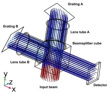
Optical and numerical modeling of a spatial heterodyne laser-induced breakdown spectrometer
Lajos P. Villy (University of Szeged, Hungary):
Signal enhancement of gaseous samples in the presence of nanoaerosols generated by a spark discharge
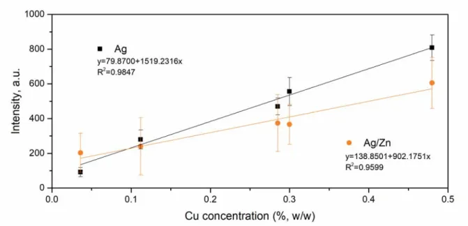
Jelena Petrović (University of Belgrade, Serbia):
Enhancement of the TEA CO2 LIBS emission using Ag and Ag/Zn nanostructures
Eszter Nagy (University of Szeged, Hungary):
Laser ablation of meloxicam in liquid environment
Jakub Buday (Brno University of Technology, Czech Republic):
Computation of blast wave energy in LIBS using shadowgraphy Roman Holomb (Uzhhorod National University, Ukraine/Wigner Research Centre for Physics, Hungary):
Reversible laser-assisted structural modification and laser-driven mass transport at the surface of chalcogenide nanolayers
István Rigó (Wigner Research Centre for Physics, Hungary): Effect of surface roughness on SERS enhancement of patterned gold substrates Aurélien Favre (Normandy University, France):
MERLIN, an RT-LTE software supporting the diagnostic application of LIBS to H-isotope measurements
12:00-14:00 Lunch break
14:00 - 16:00 Poster session II.
Analytical methodology
Patrick Janovszky (University of Szeged, Hungary):
Identification and Be, Li content assessment of minerals in granitoid rock samples by LIBS
Sára Střítežská (Brno University of Technology, Czech Republic):
Toxicity assessment of photon-upconversion nanoparticles and their bioimaging by using laser-induced breakdown spectroscopy in Brassica oleracea L. plant
Anna Šindelářová (Brno University of Technology, Czech Republic):
Implementation of laser-induced breakdown spectroscopy elemental imaging into the histopathological analysis of soft tissues
Patrick Janovszky (University of Szeged, Hungary):
Investigation of size and exposure time dependent bioaccumulation of silver nanoparticles in plants by LIBS
Nikolai Sushkov (Lomonosov Moscow State University, Russia):
Exploratory analysis of zooplankton spectra using matrix decomposition techniques
Dávid J. Palásti (University of Szeged, Hungary):
LIBS-based approaches for the classification of glass microfragment samples
Daniel Holub (Brno University of Technology, Czech Republic):
Classification of plastics using LIBS and Raman spectroscopy data fusion Lukas Brunnbauer (Vienna University of Technology, Austria): Matrix effects in quantitative polymer analysis: a comparison of ns and fs laser systems
Patrick Janovszky (University of Szeged, Hungary):
Analysis of uranium-bearing materials by laser-induced breakdown spectroscopy
Ádám Bélteki (University of Szeged, Hungary):
On-line and off-line LIBS detection of nanoaerosols generated by electrical discharges
ORAL
PRESENTATIONS
LASER INDUCED GAS BREAKDOWN IN REACTIVE MIXTURES CONTAINING HALIDES OF BORON AND SILICON:
DIAGNOSTICS AND MODELING
I. B. Gornushkin1*, P. G. Sennikov21BAM Federal Institute for Materials Research and Testing, Richard-Willstatter-street 11, 12489 Berlin, Germany
2Institute of Chemistry of High-Purity Substances of RAS, Tropinin street 49, 603600, Nizhny Novgorod, Russia
*e-mail: igor.gornushkin@bam.de
1. I
NTRODUCTIONPlasma-chemical approach is used for synthesis of various gaseous, liquid, and solid substances since 1960th [Vurzel 1970]. Nowadays, the method of plasma enhanced chemical vapor deposition (PECVD) is widely used for production of thin films, protective coatings, carbon-based nanostructures, high purity isotopic materials, biomaterials, and many other products. Plasma for PECVD is typically created in various electrical discharges; e.g. DC and AC glow discharges or discharges operated at audio (10-20 kHz), radio (13.56 MHz), and microwave (2.45 GHz) frequencies. Laser induced plasma (LIP) is rarely used to deposit materials from the gas phase and this work aims at reviving interest to this latter technology and showing its potential.
We run two pilot LIP experiments in reactive gas mixtures. First, LIP is excited in BCl3 or BF3 plus H2 or CH4 to evaluate the efficiency of deposition of solid boron and boron carbide, materials that are largely used for refractory coatings. Second, we investigate a possibility of synthesis of fluorochlorosilanes SiFxCl4-x (x = 1, 2, 3) by LIP induced in SiF4+SiCl4 gas mixtures. Using fluorochlorosilanes with different combinations of F and Cl in the SiFxCly molecule may add flexibility in processes of silicon deposition and etching.
The gases used and solid deposits are analyzed by optical emission spectroscopy (OES) and IR and mass spectrometry (MS). We also model the laser induced plasma by performing static equilibrium chemistry calculations to see whether desired reaction products are thermodynamically favorable and dynamic calculations of the expanding plasma plume to see how and where the products form.
2. E
XPERIMENTALA sketch of the experimental set-up is shown in Figure 1. A Nd:YAG laser (1064 nm, 15 ns pulse width, 800 mJ pulse energy, 5 Hz repetition rate) is focused inside a reactor to create a plasma in the reactive gas mixture. The reactor consists of two coaxial quartz cylinders;
it is loaded with gases shown in Table 1.
Figure 1. Experimental setup for PECVD with LIP and corresponding diagnostics.
The plasma is analyzed by OES while the gas mixture inside the reactor is analyzed by IR and MS both before and after the laser action. Solid residues that are deposited on the walls of the inner cylinder are studied by the reflectance FTIR.
BCl3 BF3 SiCl4
Н2: BCl3=10:1 Н2: BF3=3:1 SiF4
H2: Ar: BCl3=10:10:1 H2: Ar: BF3=3:4:1 SiCl4: SiF4=1:2.65 H2: BCl3: CH4=9:1.5:1 H2: BF3: CH4=9:1.5:1 SiCl4: SiF4=2.65:1
Table 1. Gases used in LIP experiments.
A numerical experiment on the gas mixtures was performed by first calculating the plasma equilibrium composition as a function of its temperature using open source software [CEARUN] and second, calculating plasma dynamic parameters using a hydrodynamic code [Shabanov 2018] and the same open source software embedded in this code.
3. R
ESULTS AND DISCUSSION 3.1. Halides of boronThe amount of the deposit enough for further analysis is collected with mixture BCl3+Н2+СН4. The deposit is identified as boron carbide and carbon (soot) by the reflectance FTIR technique. No deposit except soot (in methane) is observed for mixtures with BF3. The OES spectra of gas plasma show efficient formation of BH, BX (X=Cl, F), and C2 (in methane). The IR spectra of the reaction products after the laser action show the presence of the molecules already identified by OES plus precursors (BCl3, BF3, CH4),
radicals (HBCl2), and new derivatives (C2H2, B2H6). MS analysis confirms the presence of these molecules.
The results of experiment well agree with the predictions of both static and dynamic simulations. The model does not predict formation of solid boron or boron carbide from mixtures of BF3 with hydrogen or methane, and same is observed in experiment. For mixtures of BCl3 with the same gases, condensed phases of B, C, and B4C form that is detected in experiment and predicted by the model. Some results of laboratory and numerical experiments are reproduced in Figure 2.
Figure 2. Left panel: IR absorption spectrum of solid deposit from the mixture H2:BCl3:CH4=9:1.5:1. Right panel: dynamic 1D simulation of breakdown in 50% (CH4+Ar) + 50% (BCl3+H2) gas mixture; snapshot of
species concentrations at 1 μs of the plasma plume propagation time.
3.2. Halides of silicon
Generation of fluorochlorosilanes in the mixture of SiF4 and SiCl4 via the reactions 3SiF4 + SiCl4 = 4 SiF3Cl (1160 kJ/mole); SiF4 + SiCl4 = 2SiF2Cl2 (307 kJ/mole); and SiF4 + 3SiCl4 = 4SiFCl3 (71 kJ/mole) is thermodynamically unfavorable due to the positive Gibbs formation energy ΔG298 (in parentheses). These reaction products can easily be obtained in LIP after the plasma cools down and formerly dissociated atoms reassemble back into molecules. This is confirmed both experimentally and theoretically. As before, the plasma is analyzed by OES why the reactants and products by IR and MS. Optical emission spectra show the formation of SiCl, SiF, and SiCl2 along with all expected elemental species and their ions. The IR spectra of plasma products reveal strong absorption bands and, hence, efficient formation of sought-after fluorochlorosilanes SiF2Cl, SiFCl2, SiFCl3, SiF3Cl, and SiF2Cl2 (Figure 3., left panel); the MS measurements convincingly confirm this finding.
From IR absorption measurements, a 60% maximum yield of fluorochlorosilanes is estimated for the mixture SiF4:SiCl4=1:1; the dominant specie is SiF2Cl2. The same high yield for SiF2Cl2 is predicted theoretically based on data generated by ab initio calculations of thermodynamic properties of fluorochlorosilanes. The result of the dynamic simulation for the mixture SiF4:SiCl4=1:1is exemplarily given in Figure 3., right panel.
Figure 3. Left panel: IR absorption spectra of mixture SiCl4:SiF4=1:1 before (black) and after (red) the 40 min laser irradiation. Right panel: dynamic 1D simulation of breakdown in SiF4: SiCl4 = 1:1 gas mixture;
snapshots of the plasma composition after the 5.0μs propagation time.
4. C
ONCLUSIONSFor the BX3-containing systems (X=Cl, F), the creation of solid deposits of B, BH3, and C (in mixtures with methane) is observed by LIP excited in the reactive gas mixtures BX3+H2
and BX3+H2+CH4. The dynamic calculations of the expanding plasma plume predict coexisting condensed phases of boron, boron carbide, and graphite in mixtures with BCl3. The maximum concentration of the condensed species is reached in peripheral plasma zones. Overall, the calculations and experimental results imply that PECVD-LIP can be a promising technique for efficient conversion of gaseous precursors into solid elemental constituents and their compounds.
For the SiX4-containing systems (X=Cl, F), gaseous fluorochlorosilanes SiF3Cl, SiF2Cl2, SiFCl3 can efficiently be synthesized by LIP induced in SiF4+SiCl4 precursor gas mixtures. It is found that the total yield of fluorochlorosilanes in LIDB plasma comprises 60%, with ~30% of SiF2Cl2. The equilibrium chemical model adequately predicts the composition of LIP. The dynamic calculations of the expanding plasma plume also agree with experiment and show that fluorochlorosilanes form in peripheral plasma zone and show high sensitivity toward the mixture stoichiometry and plasma temperature.
5. A
CKNOWLEDGEMENTSThe authors thank V. S. Polyakov, A. A. Ermakov, R. A. Kornev, and V. E. Shkruninfor calculations and measurements and Melsytech LTD (D. Stepanov, A. Stepanov, O. Yeremeykin) for technical support. P. S. acknowledges the DAAD/2019 grant 9165134 and RSF grant No 20-13-00035 and Russian Ministry of Education and Science grant
0095-2019-0008. The authors also thank Prof. U. Panne and Dr. K. Rurack for the support of this project.
6. R
EFERENCES[Vurzel 1970] F. B. Vurzel, L. S. Polak, Ind. Eng. Chem., 62 (1970) 8.
[CEARUN] https://cearun.grc.nasa.gov (accessed on 15 October 2020).
[Shabanov 2018] S. V. Shabanov, I. B. Gornushkin, Appl. Phys. A, 124 (2018) 716.
NANOPARTICLE ENHANCED LASER INDUCED BREAKDOWN SPECTROSCOPY: FUNDAMENTALS AND PERSPECTIVES
Vincent Gardette1, Zita Salaikova1,2, Elena Vaníčková2, Marcella Dell’Aglio3,Alessandro De Giacomo1,3*
1University of Bari, Department of Chemistry, Via Orabona 4, 70126 Bari-Italy
2(CEITEC, Brno University of Technology, Purkyňova 656/123, 612 00 Brno, Czech Republic
3CNR-NANOTEC c/o University of Bari, Department of Chemistry, Via Orabona 4, 70126 Bari, Italy
*e-mail: alessandro.degiacomo@uniba.it
1. I
NTRODUCTIONIt has been recently proposed to use metallic nanoparticles for improving the sensitivity and the capabilities of laser ablation analytical techniques, such as LA ICP MS and LIBS (named respectively NE-LA-ICP-MS and NE-LIBS, NE stands for Nanoparticles Enhanced).
Metallic NPs allow modulation and enhancement of the incoming laser pulse with the ignition of a surface plasmon resonance (SPR). These enhanced techniques present different advantages: the ablation occurs mainly on the NPs deposited on the surface of the sample which leads to few damages on this one and thus is interesting for numerous domain of applications [Dell’Aglio 2018]. In addition, sensitivity is hugely increased in the case of LIBS allowing the detection of ultra-traces in the range of ppb level [De Giacomo 2013, De Giacomo 2016] whereas, signal from LA-ICP-MS mediated by NPs is also increased, but in a more moderate way, achieving enhancement between 2 to 10 [Mangone 2020]. Mechanisms that relies behind this enhancement are not yet completely understood. In addition, it seems that, mainly, the NPs play a role on the plasma emission in the case of NE-LIBS [De Giacomo 2020], and on laser ablation and elemental fractionating in the case of NE-LA-ICP-MS [Holá 2018], which are two complete different processes.
2. E
XPERIMENTALNE-LIBS experiments do not need any tuning setup, and in that way, experimental setup is a classical homemade LIBS setup with a pulsed Nd:YAG laser, 6 ns pulse duration, of fundamental wavelength 1064 nm, (Q-SMART 850, Quantel). Laser pulse is focalized on the target using a focal lens of various focal distance depending on the desired spot size and can be replaced by a microscope objective for conducting µ-LIBS experiment. Plasma emission light is collected with a fiber into a spectrometer (TRIAX 500, Andor) coupled with a CCD camera. Additional laser module can be used to change the laser wavelength into the 2nd and 3rd harmonics, respectively 532 and 354 nm, in order to fit perfectly the resonance band of the NPs system.
Figure 1. Experimental setup for typical LIBS and NE-LIBS measurement.
Various kind of NPs can be employed, of all different shapes, even if platinum, gold and silver spherical NPs are most commonly used. Sample preparation is quite fast and easy: µL drops of NPs colloidal solution are deposited on the sample surface and gently dried. The NPs size effect will be discussed below. It is important to notice that NE-LIBS effect can only occurs under specific conditions. First, the spot size must be large enough to ablate a large amount of NPs. Secondly, the laser fluence should be relatively low, so the NPs are not destroyed during the ablation. Finally, NPs should not aggregate during the deposition or inside the colloidal solution. This latter point is crucial in order to have a stable and reproducible signal. If all these conditions are fulfilled, NE-LIBS effect can occur and signal enhancement of some orders of magnitude can be reached.
3. R
ESULTS AND DISCUSSION 3.1. Analytical performanceBriefly, NE-LIBS can achieve a sensitivity close to the ppb level, and an enhancement of some order of magnitude between LIBS and NE-LIBS signal can be observed, especially on metallic target.
Figure 2. Typical NE-LIBS spectra obtained on a titanium target.
Even if NE-LIBS provides astonishing results on metallic target as shown in Figure 2, it can be used for a wide area of application, as seen in Table 1, including analysis of biological sample, liquids or even crystal and precious sample.
Table 1. Analytical performance of NE-LIBS taken from [Dell’Aglio 2018].
3.2. Understanding the NE-LIBS effect
NPs play a double role during laser-induced plasma experiment: first during the ablation and then on the plasma phase. During the ablation, if the laser wavelength matches the absorption band of the NPs system, plasmonic effects will occur : a coherent and collective oscillation of the NPs system will be created and NPs electrons will be able to move freely between the NPs system and the plasma phase. In addition, NPs will be shielded during the ablation, and the laser pulse will be focused in the space between themselves, leading to multiple ignition point and thus to a more efficient ablation.
Target Elements NPs Enhancement/LOD
Titanium Ti Ag, Au En > 100
Steel Fe Ag En = 5
Cu-based alloys Pb, Sn,Mn Au, Ag, Pt,
Cu/CuO En ∼ 50 Al-based alloys AlO, Ti,Fe Au En ∼10
CuSO4 (aq) Cu Au ppt level in 100 µl
CuSO4 (aq), NaF (aq) Cu, F Au En = 3.5
For F LOD ∼ 0.1% ppm in 20 µl
AgNO4 (aq)
PbCl2 (aq), PbSO4(aq)
Ag, Pb Au
Au LOD <ppb in 2 µl En >10
Blood Pb Au LOD ∼10 ppb in 2 µl
RC Protein solution
Li Au En ∼ 20
CuSO4 (aq), Cr(NO3)2 (aq)
Pb(NO3)2 (aq)
Cu, Cr, Pb ultrafine fibers +Au NPs
En ∼ 4
Leaf Fe, Mn, K, Ca, Mo Au, Ag En = 5
SiO2 crystal Si Au En = 30
ZnO pressed
pellets of NPs No plasmonic
enhancement ZnO 2<En<120
Tourmaline Mn, Fe Au En ∼10
Figure 3. Laser-NPs interaction during laser ablation experiment.
After the laser pulse, NPs will survive the ablation and will be ejected at the top of the plasma. During its expansion, the plasma will slowly take in the NPs and vaporize them. It was shown that the NPs size was not affecting the global enhancement, but rather the temporal evolution of the signal instead.
Figure 4. Temporal evolution of signal enhancement and gold (NPs) signal.
Finally, the NPs role on the plasma during its expansion is not yet fully understood.
Only a small portion of the ablated mass will take part into the emission processes and it is supposed that the NPs can convert a part of the “sleeping mass” into emissive mass, and thus leading to a higher emission efficiency.
4. C
ONCLUSIONSNE-LIBS has shown astonishing capabilities in term of emission efficiency, signal enhancement and sensitivity. NPs can be used for all kind of experiments and their use is not time consuming and no setup tuning is needed. In order to fully optimize the NE-LIBS
effect, there is a crucial need to understand all the fundamental processes which relies behind this enhancement. So far, the NPs are acknowledged to play a role during the ablation and on the plasma phase.
6. R
EFERENCES[Dell’Aglio 2018] M. Dell’Aglio, R. Alrifai, A. De Giacomo, Spectrochim. Acta B, 148 (2018) 105.
[De Giacomo 2013] A. De Giacomo, R. Gaudiuso, C. Koral, M. Dell’Aglio, O. De Pascale, Anal. Chem., 85 (2013) 10180.
[De Giacomo 2016] A. De Giacomo, C. Koral, G. Valenza, R. Gaudiuso, M. Dellaglio, Anal. Chem., 88 (2016) 5251.
[A. Mangone 2020] A. Mangone, F. Mastrorocco, L. C. Giannossa, R. Comparelli, M. Dell’Aglio, A. De Giacomo, Spectrochim. Acta B, 163 (2020) 105731.
[De Giacomo 2020] A. De Giacomo, R. Alrifai, V. Gardette, Z. Salajková M. Dell’Aglio, Spectrochim. Acta B, 166 (2020) 105794.
[Holá 2018] M. Holá, Z. Salajková, A. Hrdlička, P. Pořízka, K. Novotný, L. Čelko, P. Šperka, D. Prochazka, J. Novotný, P. Modlitbová, V. Kanický, J. Kaiser, Anal. Chem., 90 (2018) 11820.
WHY IS LIBS THE FUTURE ONLINE ANALYTICAL TECHNIQUE FOR PLASMA-FACING MATERIALS OF THERMONUCLEAR REACTORS?
Pavel Veis1*, Vishal Dwivedi1, Alicia Marín Roldán1, Gulab Singh Maurya1, Sahithya Atikukke1, Matej Veis1, Matej Pisarčík1 Milan Držík2
1Department of Experimental Physics, Comenius University, FMPI, Mlynská dol. F2, 842 48, Bratislava, Slovakia
2International Laser Center, Ilkovičova 3, 841 04 Bratislava, Slovakia
*e-mail: veis@fmph.uniba.sk
1. I
NTRODUCTIONCurrently, a global tendency of reducing carbon footprint is pursued by leading institutions and governments. Unlike other alternatives to fossil fuel burning, such as wind and solar sources, nuclear reactors provide a stable high energy output that can match the ever-rising demand. Even though nuclear fission plants have been well established (a total of 450 power plants in 31 counties), much of the research is being focused to nuclear fusion instead. An advantage over a fission reactor includes heavily reduced operational radioactivity and drastic reduction of radioactive waste. In fusion reactors deuterium and tritium are used as fuels. Deuterium is available in the seawater and tritium can be generated from lithium. For initiating the fusion reaction, a stable and very high temperature plasma is required. These conditions can be only achieved in magnetic confinement devices e.g. torus shaped tokamaks. One of such projects is The International Thermonuclear Experimental Reactor (ITER) that aims to generate fusion energy on a commercial scale in the range of 500 MW.
This research is focused on elemental analysis of plasma-facing components (PFCs) of the aforementioned ITER by means of laser-induced breakdown spectroscopy (LIBS).
PFCs of a fusion device face harsh operating environment of high heat, radiation and neutron flux that lead to irreversible mechanical changes of the PFCs surface like cracking, blistering and erosion [Li 2016, Maury 2020, Merca 2011, Hai 2014]. During the high temperature plasma operation in tokamaks (despite of magnetic field confinement), the plasma interaction with walls - especially in the divertor and limiter zones - leads to the erosion of the PFCs. The eroded material migrates away and forms new mixed materials on the surface of the first wall or deposits in the form of flakes and dust in the cooler parts of tokamak.
Certain re-deposited materials exhibit higher fuel retention than original wall material and that is a serious problem for safety in the case of T fuel. In the case of ITER- like wall (tungsten divertor and beryllium principal wall), the re-deposition of Be/W layers together with O impurities leads to the more efficient trapping of D/T fuel than in the case of pure Be/W layers. In ITER, The maximum amount of permitted fuel within the
ITER reactor is up to 1 kg, so the retained T fuel in the first wall must be observed regularly to make sure its reliable operation with a complete fuel cycle [Oelma 2018].
Also due to the re-deposition of the impurities, the visibility of the mirrors and optical windows (essential components for the plasma diagnosis, imaging, and spectroscopy) is decreasing and their spectral transmittance and sensitivity is also changing. Their maintenance and protection is a serious issue for the operation of fusion devices [Mukhi 2009].
The study of material erosion and migration, the formation, release, and re- deposition of the dust are important issues. So, for the reliable and safe operation of the future fusion reactor, it is necessary to measure elemental compositions of all key PFCs periodically, for monitoring of impurities, its deposition pattern, and retained fuel.
The main advantages of laser-induced breakdown spectroscopy (LIBS) are in-situ and standalone analysis, no pre-treatment of the samples. LIBS analysis could be performed under vacuum, low-pressure, or up to atmospheric pressure environments and moreover magnetic field doesn’t affect its performance. LIBS can be eventually combined with laser ablation (LA) based technique for the necessary cleaning process of the fusion device first wall [Fanto 2013, Schwe 2009].
2. R
ESULTS AND DISCUSSIONLIBS has been applied for the depth profile analysis of ITER-relevant materials, time- resolved and space-resolved analysis of PFCs, heating diagnostics of different PFCs, the study of multi-layered and calibrated samples, monitoring the features of the impurity layers deposited on the PFMs, in-situ and real-time diagnostics of the PFCs, etc [Brezi 2017, Maury 2017]. LIBS is an emission spectroscopic technique with the merits e.g., its speed, suitable for almost all kind of the samples, simultaneous multi-elemental analysis, minimal destructive nature (~1 µg material is required), analysis without sample preparation, the ability of contactless or remote analysis/sensing, immune to harsh environments, does not provide any interference itself, etc. Owing to its applicability and versatility, A robotic arm-based LIBS is being considered to monitor different parts of the reactor walls.
2.1. LIBS experimental set-up
Based on requirements in different fields, various LIBS set-ups such as conventional LIBS, back-collection LIBS, double pulse LIBS, etc. is being used for the elemental analysis and depth profile analysis of the samples [Maury 2020].
2.2. Calibration-free (CF) approach
For the CF-LIBS analysis [Ciucc 1999], more spectral lines for neutral and first ionization degree of the same element are necessary, if possible, emitted from different upper levels.
Otherwise, the evaluation of the electron temperature from the Boltzmann plot is doubtful. The transition probabilities for these lines should have good precision, and the spectral peaks should not be too weak, in addition, not to be in interference with other possible lines and not affected by self-absorption. We have performed a quantitative and depth profile analysis of the Be-W-D-based simulated layers (thickness 1.5–2 μm) on the Mo substrates for the H/D content. D content of the samples has been calculated using the CF-LIBS approach ∼4.7% ± 2.9%, which is in good agreement with the other spectroscopic techniques for the same samples. We have also characterized Li-based coatings deposited on Ni-Cr based screws obtained after liquid metal experiments from the COMPASS vacuum chamber. After evaluation of plasma parameters using the multi elemental Boltzmann plot (MEBP) for different elements, impurities (e.g. Li, C, B) were quantified and depth profiling of Li content has also been verified.
Figure 1. MEBP for the elements Li I, Ca I-II, and Fe I, and Boltzmann plot for Li I, B I, C I, Na I, Mg I, Si I, K I, Ca I, and Fe I spectral lines using the evaluated Te from the MEBP (for one of the screws from COMPASS Li
campaign) [Veis 2020a].
CF-LIBS approach is also important for the control of impurities and dust pollution in tokamaks or linear fusion devices. Finally, this LIBS based monitoring is important for the knowledge of the pollution level caused by previous specific campaigns (e.g. previous carbon-based first wall, special limiter, or test with liquid metals, etc.). More precise Te
evaluation from W SB plots by enlarging LIBS analysis down to VUV range was recently proposed [Pribu 2016].
2.3. Molecular bands and LAMIS
Emission of molecular bands of diatomic radicals appears significantly later in the decay of laser induce plasmas, at the moment, when atomic lines start to disappear. So, the molecular emission measurements should have to be done separately from the atomic emission measurements. The perspective of the molecular emission spectroscopy in the fusion reactors wall diagnostics is the ability of an efficient isotopic analysis, due to the fact that the molecular lines isotopic shift is several order of magnitude higher than the atomic line isotopic shift. This idea of so-called Laser Ablation Molecular Isotopic
Spectrometry (LAMIS) was proposed in 2011 by Russo et al. [Russo 2011] and a review paper about the LAMIS method was published in [Bolsh 2016].
A possible application in LIBS fusion research is for the efficient quantification of relevant isotopes (e.g. D/H [Sarka 2013, Bolsh 2017], B [Mao 2011], C [Dong 2013], N, etc.) in laser-induced plasma after the conversion to molecular radicals as OH/ OD, BO, C2, CN and others. For separation of hydrogen isotopes H/D (and in the future also T), the proposed molecular OH/OD (A-X, Δv=+1) transitions at 281 nm and 287 nm respectively have large separation (6 nm) of both isotopes bands [Russo 2011].
2.4. Depth profile analysis
LIBS has been found promising for the depth profile analysis by recording a successive number of LIBS spectra at the same position of the samples (especially of the analysis of thin layers and deposited impurities). Variation of the spectral signal with the number of laser shots represents depth profiling of the sample and provides information about thickness of the sample layer and ablation rate. LIBS depth profile analysis has been applied for different materials from fusion devices (JET, WEST, COMPASS, W7-X etc.). Veis et al. [Veis 2020b, Sucho 2017a] have applied depth profile analysis and quantification of the elements in Be/W(D) and Dwivedi et al. [Dwivedi 2020] have applied the same method for Be-O-C-D mixture samples and found in good agreement with other established technique like SIMS, NRA and TDS for D retention. Depth profile analysis of LiSn alloy by CF-LIBS was proposed in [Sucho 2017b].
Quantitative LIBS elemental and depth profile analysis of prepared mixed fusion relevant layers or post-mortem analysis of samples exposed in linear machines (Magnum- PSI, PSI-2) or tokamaks (JET, WEST, ASDEX, Compass….) were or could be used for validation by several off-line techniques like AAS, ICP-MS, spark discharge AES, NAA, RBS, NRA, TDS, SIMS, XRF and others. Most of the techniques are time-consuming and need special sample preparation, which is a drawback for in-situ, and online experiments.
2.5. Fuel retention and separation of the hydrogen isotope lines
For safety and control purposes of fusion devices, in-situ analysis of fuel retention is very important. In ITER, A remote-LIBS has been considered the most promising technique for the detection of fuel retention in plasma-facing walls and divertor materials regularly.
Paris et al. [22, 23] have studied erosion, deposition, and fuel retention in fusion-related materials using LIBS and compared with other postmortem analysis techniques like NRA, RBS, and SIMS.
At high pressure, in laser-induced plasma, Hα (656.3 nm) and Dα (656.1 nm) lines are partly overlapped due to the Stark broadening effect. Large gate delay can reduce this overlapping, but spectral lines are with a low S/N ratio. Suitable conditions for resolving these lines has been optimized. At atmospheric pressure, suitable gate delay was optimized >2 µs, while vacuum conditions provide well-resolved Dα and Hα lines.
3. C
ONCLUSIONOwing to its inherent advantages (in-situ and standalone analysis, no pre-treatment of the samples, possible combination of laser diagnostics and laser cleaning process), LIBS, LIBS- based techniques are useful to diagnose and/or quantify the fuel retention, the impurities and the depth profiling of coatings and redeposited mixed materials related to the fusion devices.
4. A
CKNOWLEDGEMENTSThe authors acknowledge the Scientific Grant Agency of the Slovak Republic (contract number VEGA-1/0903/17) and the Slovak Research and Development Agency (APVV-16- 0612) for financial support. GSM would like to acknowledge the National Scholarship Programme of the Slovak Republic to provide research mobility.
5. R
EFERENCES[Bolsh 2016] A. A. Bol'shakov, X. Mao, J. J. Gonzalez, R: E. Russo, J. Anal. At. Spectrom., 31 (2016) 119.
[Bolsh 2017] A. A. Bol'shakov, X. Mao, R. E. Russo, J. Anal. At. Spectrom., 32 (2017) 657.
[Brezi 2017] S. Brezinsek, J. W. Coenen, T. Schwarz-Selinger, et al., Nucl. Fusion, 57 (2017) 116041.
[Ciucc 1999] A. Ciucci, M. Corsi, V. Palleschi, S. Rastelli, A. Salvetti, E. Tognoni, Appl. Spectrosc., 53 (1999) 960.
[Dong 2013] M. Dong, X. Mao, J. Gonzalez, J. Lu, R. E. Russo, Anal. Chem., 85 (2013) 2899.
[Dwivedi 2020] V. Dwivedi, A. Marín-Roldán, J. Karhunen, P. Paris, P. Veis et al., submitted to Nucl. Mater. Energy
[Fanto 2013] R. Fantoni, S. Almaviva, L. Caneve, F. Colao, A. M. Popov, G. Maddaluno, Spectrochim. Acta B, 87 (2013) 153.
[Hai 2014] R. Hai, P. Liu, D. Wu, H. Ding, J. Wu, G. N. Luo, Fusion Eng. Des., 89 (2014) 2435.
[Li 2016] C. Li, C.-L. Feng, H. Y. Oderji, G.-N. Luo, H.-B. Ding, Front. Phys., 11 (2016) 114214.
[Mao 2011] X. Mao, A. A. Bol’shakov, D. L. Perry, O. Sorkhabi, R. E. Russo, Spectrochim. Acta B, 66 (2011) 604.
[Maury 2017] G. S. Maurya, K. Belešová, M. Anguš, J.Miškovičová, M. Suchoňová, P. Veis, Proceedings of ELITECH ’17, 2017.
[Maury 2020] G. S. Maurya, A. Marín-Roldan, P. Veis, A. K. Pathak, P. Sen, J. Nucl. Mater., 541 (2020) 152417.
[Merca 2011] L. Mercadier, A. Semerok, P. A. Kizub, A. V. Leontyev, J. Hermann, C. Grisolia, P.-Y. Thro, J. Nucl. Mater., 414 (2011) 485.
[Mukhi 2009] E. Mukhin, K. Vukolov, V. Semenov, S. Tolstyakov, M. Kochergin, G. Kurskiev, K. Podushnikova, A. Razdobarin, A. Gorodetsky, R. Zalavutdinov, V. Bukhovets, A. Zakharov, S. Bulovich, V. Veiko, E. Shakshno, Nucl. Fusion, 49 (2009) 085032.
[Oelma 2018] J. Oelmann, N. Gierse, C. Li, S. Brezinsek, M. Zlobinski, B. Turan, S. Haas, Ch. Linsmeier, Spectrochim. Acta B, 144 (2018) 38.
[Paris 2015] P. Paris, K. Piip, A. Hakola, M. Laan, M. Aints, S. Koivuranta, J. Likonen, A. Lissovski, M. Mayer, R. Neu, V. Rohde, K. Sugiyama, ASDEX Upgrade Team, Fusion Eng. Des., 98-99 (2015) 1349.
[Paris 2017] P. Paris, J. Butikova, M. Laan, M. Aints, A. Hakola, K. Piip, I. Tufail, P. Veis, Phys. Scr., T170 (2017) 014003.
[Pribu 2016] M. Pribula, J. Krištof, M. Suchoňová, M. Horňáčková, J. Plavčan, A. Hakola, P. Veis, Phys. Scr., T167 (2016) 014045.
[Russo 2011] R. E. Russo, A. A. Bol’shakov, X. Mao, C. P. McKay, D. L. Perry, O. Sorkhabi, Spectrochim Acta B, 66 (2011) 99.
[Sarka 2013] A. Sarkar X. Mao, G. C.-Y. Chan, R. E. Russo, Spectrochim. Acta B, 88 (2013) 46.
[Schwe 2009 B. Schweer, G. Beyene, S. Brezinsek, N. Gierse, A. Huber, F. Irrek, V. Kotov, V. Philipps, U. Samm, M. Zlobinski, Phys. Scr., T138 (2009) 014008.
[Sucho 2017a] M. Suchoňová, P. Veis, J. Karhunen, P. Paris, M. Pribula, K. Piip, M. Laan, C. Porosnicu, C. Lungu, A.Hakola, Nucl. Mater. Ener., 12 (2017) 611.
[Sucho 2017b] M. Suchoňová, J. Krištof, M. Pribula, M. Veis, F. L. Tabarés, P. Veis, Fusion Eng. Des., 117 (2017) 175.
[Veis 2020a] P. Veis, S. Atikkuke, A. M. Roldan, V. Dwivedi, M. Veis, P. Barton, M. Jerab, R. Dejarnac, Nucl. Mater. Ener., 25 (2020) 100809.
[Veis 2020b] P. Veis, A. Marín-Roldán, V. Dwivedi, J. Karhunen, P. Paris, I. Jõgi,
C. Porosnicu, C. P. Lungu, V. Nemanic, A. Hakola, Phys. Scr., T171 (2020) 014073.
ASSESSMENT OF POLYMER DEGRADATION BY THE COMBINED USE OF LIBS AND LA-ICP-MS
Andreas Limbeck1*, Laura Pagnin2, Rita Wiesinger2, Manfred Schreiner1,2, Lukas Brunnbauer2
1TU Wien, Institute of Chemical Technologies and Analytics, Getreidemarkt 9/164, 1060 Vienna, Austria
2Academy of Fine Arts, Schillerplatz 3, 1090, Vienna, Austria
*e-mail: andreas.limbeck@tuwien.ac.at
1. I
NTRODUCTIONSynthetic polymers are nowadays widely used in a variety of applications, ranging from food packaging, construction materials to the housing of electronic devices. In general, the applied synthetic polymers are composed of an organic-carbon-chain polymer and different additives that give the materials the intended chemical and physical properties.
Commonly applied additives include plasticizers, antioxidants, antistatic agents, lubricants, flame retardants or inorganic pigments.
During application, polymers are often exposed to harmful environmental conditions, causing changes in their chemical composition. In this context, the negative influences of sunlight but also contact with ambient gases and environmental liquids have to be mentioned. Whereas UV light and oxidative gases are known to promote degradation, corrosive gases or metals dissolved in rain, snow and river or sea-water are susceptible for uptake into the polymer network, resulting in increased concentrations of inorganic constituents in aged materials. All of these possible interactions contribute to unwanted changes in the polymer composition, which finally lead to altered material properties (e.g. bleaching of colors, reduced thermal stability, increased brittleness, etc.).
Thus, analysis of synthetic polymers is experiencing growing interest due to increased environmental regulations and the need for sustainable polymer recycling strategies.
Traditional methods for the investigation of polymer degradation are FT-IR and Raman spectroscopy. Furthermore, Pyrolytic-GC-MS, MALDI-ToF–MS, thermogravimetric analysis (TGA) and differential thermal analysis (DTA) are frequently used techniques.
Although well established, these techniques cannot provide the entire information required for thorough material analysis. In particular, with the aforementioned techniques sample analysis is limited to bulk investigations or surface near regions only.
Additionally, detection of metal content within polymers with a high sensitivity is not feasible. These shortcomings could be circumvented with the use of LIBS and LA-ICP-MS.
In the field of polymer analysis, broadband LIBS spectra have already been used for the identification [Sattmann 1998] and classification [Haddad 2014] of different polymer types. Additional benefits of LIBS are the possibility to perform spatially resolved analysis such as depth profile measurements.
In this contribution, we report the use of LIBS for assessment of polymer degradation. Moreover, the uptake of contaminations from the surrounding environment has been determined using LA-ICP-MS. The developed Tandem LA-ICP-MS/LIBS approach for polymer analysis has been applied for two different research tasks:
• Objects of art and cultural heritage are sometimes exposed to harsh
environmental conditions. The permanent exposure to humidity, UV-radiation and corrosive gases results in the degradation of valuable artwork. As the fundamentals of the degradation process are still not completely understood, knowledge about durability and aging properties of applied art materials is of particular interest.
• At the end of their life-cycle, polymers often end up in the environment, for example in the form of microplastics which pose a significant threat to various ecosystems. Accordingly, the composition and metal contents of the degraded polymers should be monitored, to better estimate the adverse health effects of microplastics in the environment.
2. E
XPERIMENTALA LIBS system (Model J200) equipped with a 266 nm Nd: YAG laser by Applied Spectra, Inc. (West Sacramento, California) was used for LIBS analysis. For collection and spectroscopic analysis of the radiation emitted by the laser-induced plasma, an optical fiber system connected to a Czerny-Turner spectrometer with six-channel CCD detection was employed. ICP-MS analysis of the generated aerosol was performed using a Thermo iCAP Qc quadrupole ICP-MS device (ThermoFisher Scientific, Bremen, Germany).
Connection of the tandem LA/LIBS system to the mass spectrometer was achieved using PTFE tubing.
Modern paints composed of inorganic pigments and organic binders were prepared by mixing Alkyd Medium 4 (Lukas®, Germany) with 9 inorganic pigments (Kremer Pigmente, Germany). The pigment/binder ratio chosen was 1:3, prepared paint mixtures were cast on glass slides with a wet film thickness of approx. 150 μm. Sample drying was conducted at ambient conditions for 1 week. Accelerated stress tests were performed to cause degradation of the polymers as well as uptake of sulfur within the investigated samples. Therefore, samples were exposed to synthetic air in combination with corrosive gases (SO2, H2S and O3) or UV light in a weathering chamber [Wiesinger 2010].
Degradation of commercial polystyrene thin films obtained from Goodfellow Inc.
(Hamburg, Germany) was conducted by sample exposure to UV radiation and the concurrent treatment with oxidizing agents (H2O2 and HNO3). Careful optimization of the process parameters ensured successful and reproducible degradation of the investigated micro-plastics. The degree of sample degradation was determined using FTIR spectroscopy and LIBS analysis. Subsequently sorption experiments with the aged
polystyrene thin films were performed, with the aim to investigate the temporal behavior of trace metal uptake. Aged polymer samples as well as unaged blank samples were exposed for different times to artificial seawater containing defined concentrations of selected trace metals.
3. R
ESULTS AND DISCUSSION3.1. Degradation of modern art materials
Aim of this study was to test the feasibility of LIBS for measurement of polymer degradation and to confirm the uptake of sulfur with LA-ICP-MS of modern art materials consisting of a polymeric binder and inorganic pigments. Unlike most currently employed techniques for polymer characterization, these two are not limited to the surface but also enable depth profiling.
Using LIBS in combination with advanced data evaluation procedures, changes in the polymer composition due to weathering with corrosive gases and UV light was observed. Performing depth profiles on various samples exposed to different corrosive conditions, the influence and severity of these conditions on the degradation of the modern art material was assessed.
LA-ICP-MS parameters were optimized for the qualitative measurement of gas permeability into the investigated modern art material. The acquisition of depth profiles was successful. Again, the influence of different corrosive conditions on the sulfur uptake was investigated. These experiments revealed a high variation of the degree of sulfur uptake depending on the aging conditions. Additionally, sulfur uptake was not only observed in surface near regions of the investigated modern art material but also diffusion profiles into the bulk material were observed.
3.2. Artificial micro-plastic samples
FTIR measurements of the aged polystyrene samples revealed significant changes in the main polymer absorbance bands, in particular when compared to unaged reference samples. This outcome could be confirmed with LIBS, showing a significant increase in the intensity of the oxygen emission line at 777 nm, indicating partial oxidation of the polymer samples.
Samples derived from the sorption experiments were analyzed using tandem LA/LIBS-system, particular attention has been paid to determine the distribution of the absorbed metals within the polymer films. Depth profiles reveal that uptake of trace metals did not only occur on the sample surface, even in the underlying bulk material significant differences to the reference samples were observed. Thus, measurement of the sample surface is insufficient for assessment of total metal contents in aged polymer films, instead analysis over the whole film thickness is required. Moreover, the measured
uptake of the toxic trace metal Cd could be directly correlated with the information obtained for degradation and oxidation of the polymer sample.
4. C
ONCLUSIONSA Tandem LA/LIBS approach has been successfully employed for assessment of polymer degradation and measurement of gas or metal uptake from ambient environment. In recent studies the occurrence of molecular emission lines in broadband LIBS spectra has been successfully used for identification and classification of polymers, here we demonstrated that measurement of these lines enables also the detection of polymer degradation. Analysis of the generated aerosol with ICP-MS has been shown to provide the sensitivity required for the determination of sample constituents present in traces only, such as corrosive gases used in the weathering experiments with modern art materials or the trace metals used in the sorption experiments performed with aged micro-plastics.
Analysis of aged modern art materials suggest that UV exposure or pretreatment with O3 significantly influences the polymer permeability for SO2 and leads to observable degradation of the polymeric binder material. As the capabilities and limitations of LIBS and LA-ICP-MS have been assessed, a multitude of additional experiments would be of interest to retrieve more comprehensive insights into the aging process of the investigated material.
Application of the proposed tandem LA/LIBS procedure for the analysis of aged micro-plastics has been found to be beneficial. This approach provides information about the extent of polymer degradation but also data about the distribution of the metals within the investigated polymer thin films. Thus, current knowledge about the interaction of trace metals with micro-plastics could be improved. In particular it could be shown that increased degradation of micro-plastics results in elevated trace metal uptake. Moreover, metal uptake is not limited to the sample surface only, even for the bulk material enhanced concentration levels were found.
5. A
CKNOWLEDGEMENTSPresented work was financially supported by the Austrian Research Promotion Agency (FFG) under project number 874907.
6. R
EFERENCES[Sattmann 1998] R. Sattmann, I. Monch, H. Krause, R. Noll, S. Couris,
A. Hatziapostolou, A. Mavromanolakis, C. Fotakis, E. Larrauri, R. Miguel, Appl. Spectr., 52 (1998) 456.
[Haddad 2014] J. El Haddad, L. Canioni, B. Bouscquet, Spectrochim. Acta B, 101 (2014) 171.
[Wiesinger 2010] R. W. Siesinger, M. Schreiner, C. Kleber, Appl. Surf. Sci., 256 (2010) 2741.
NON-ANALYTICAL APPLICATIONS OF
LASER-INDUCED BREAKDOWN SPECTROMETRY
Timur A. Labutin1*, Andrey M. Popov11Department of Laser Chemistry, Lomonosov Moscow State University 119234 Moscow, Leninskie gory 1b3, Russia
*e-mail: timurla@laser.chem.msu.ru
1. I
NTRODUCTIONIn laser-induced breakdown spectrometry (LIBS) a pulsed laser generates a plasma that vaporizes a small amount of a solid, gaseous or liquid sample. The emission spectrum of the excited species in the laser-induced plasma usually used for qualitative or quantitative analysis of the sample. The last 20 years witnessed a sharp growth of the number of publications devoted to the application of LIBS for analytical purposes. Indeed, LIBS started to be used for determining the elemental composition of various samples from molten steels [Noll 2001] to zooplankton [Sushkov 2020]. LIBS instruments were successfully implemented for on-line control of the raw materials [Gaft 2007], steel or sludge during production as well as finished products on the conveyor belt [Legnaioli 2020]. An interesting application of this method is elemental mapping, which was performed for minerals, plant tissues and histological examination of animal organs [Jolivet 2019].
Protocols and instrumentations for analysis of samples located in a harsh environment were designed and developed to perform measurements on the bottom of a sea up to 3000 m deep [Thornton 2015], to examine underwater garbage in the nuclear reactor core [Saeki 2014], and to study the Martian surface by means of the ChemCam and SuperCam [Meslin 2013, Nelson 2020]. Numerous papers are devoted to applications of LIBS for solving analytical problems like biomedical research, pharmaceutical products [Gaudiuso 2018], food [Markiewicz-K. 2017], analysis of aerosols [Diaz 2020], cultural heritage objects [Botto 2019] and so on. Several handheld LIBS instruments appeared on the market in recent years aiming at providing fast sorting ofscrap, plastics or geological samples [Senesi 2021]. Thus, LIBS instruments with pulsed solid state ns-lasers became a “work horse” in various analytical applications. Besides this, there are other application areas where the emission of laser-induced plasma can be employed to solve specific, mostly theoretical tasks. We aim to focus on such non-analytical applications, namely the fundamental studies of laser ablation itself, determination of atomic line parameters (mostly Stark broadening parameters) and imitation of radiation from cosmic objects.
2. R
ESULTS AND DISCUSSIONThe model of the evolution of a laser plasma is based on numerical simulation of ongoing processes [Shabanov 2014], relying on data obtained by optical tomography of plasma.
Spectra of laser-induced plasma is also used to support the modeling of the processes occurring during laser–solid interaction [Bogaerts 2005]. This model was then applied to study the influence of laser irradiance, pulse duration and wavelength on laser ablation.
In case of fs-plasma the competing processes lead to formation of several fractions in plasma plume. The LIBS helps to distinguish three different velocity populations during the plasma expansion: ions with high kinetic energy, neutrals with a velocity comparable to the nanosecond regime, and lastly by nanoscale clusters.
The next possibility is the determination of transition probabilities in the laser- induced plasma. In this case, branching ratios are determined by measuring the relative emission-line intensities for lines arising from the same upper levels in an optically thin laser-produced plasma. Following these relative values of transition probabilities are put on an absolute scale [Ferrero 1997]. Laser-induced plasmas are also quite extensively used for the determination of Stark parameters [Konjević 2002, Popov 2016], relatively easy to work with, have considerable electron number density and allow selecting different working conditions merely by changing temporal parameters of signal acquisition. Generating of a long plasma (“long spark”) instead of a spherical plasma can provide the following advantages for Stark parameter measurements: i) illumination of the whole available focal plane of spectrograph, thus increasing the vertical dimension of plasma image on the detector 5–7-fold, which leads to an enhancement of signal-to-noise ratio; ii) a long spark is more homogeneous than a spherical one, as we demonstrated earlier, resulting in lesser optical thickness and lower experimental errors (<10%). This allowed us to accurately estimate an impact of hyperfine splitting on the profile shapes of the copper lines taking also into account the isotope shifts. We have shown that both effects considerably influence shift and width of Cu I line at 510.554 nm, and shifts of Cu I lines at 515.324 and 521.820 nm. Hyperfine structure and isotope shift additionally broad and shift the profile of the Cu I 510.554 nm line. This observation helps to resolve observed discrepancies between existing theoretical results and experimental data.
Last, we consider the time-evolution of the spectra of laser-induced plasma of high- purity iron in air, which is used to mimic the FeO pseudo-continuum emission. The iron oxide “orange arc” bands are unambiguously detected in persistent meteor trains, meteor wakes, and clouds, as well as in the terrestrial airglow. In contrast to the majority of other astronomically important diatomic molecules, theoretical simulation of the FeO rovibronic spectra is not feasible due to the extremely condensed and strongly perturbed multiplet structure of its excited states. The LIBS spectra were convolved with Gaussian profile with the FWHM equal to 0.38 nm which is provided a good agreement between the observed profiles of the Fe I isolated line at 537.15 nm in LIBS and Benesov meteor spectra. For comparison, the bolide spectrum at 39 km altitude was chosen, since it contains both persistent Fe I lines and FeO peaks. The significant variations of the intensity of the band at 625 nm should be noted. Such strong variations cannot be
explained by changes in the level population due to changes in temperature. At the same time, this can be easily explained if one assumes a significant contribution to the 625 nm band from FeO2 emissions as the laser plume cools, which is confirmed by the thermodynamic calculations. Although current experiments have been performed in air at atmospheric pressure, they provide high resolution spectra, which are similar to bolide spectra. Moreover, it seems possible to complete a simultaneous fit for both atomic and molecular emissions of meteor spectra in order to avoid errors due to the erroneous exclusion of atomic lines. The environment of FeO airglow emission looks to be quite different from that of meteor events and laser experiments. Considering that the cold FeO molecules in the upper Earth’s atmosphere are excited by solar radiation during the day, while FeO molecules produced during bolide events and laser experiments are hot, LIBS experiments seem to be more suitable for modeling FeO emission in meteor spectra at low altitudes.
3. R
EFERENCES[Noll 2001] R. Noll, H. Bette, A. Brysch, M. Kraushaar, I. Mönch, L. Peter, V. Sturm, Spectrochim. Acta B, 56 (2001) 637.
[Sushkov 2020] N. I. Sushkov, N. V. Lobus, I. V. Seliverstova, T. A. Labutin, Opt. and Spectrosc., 128 (2020) 1343.
[Gaft 2007] M. Gaft, I. Sapir-Sofer, H. Modiano, R. Stana, Spectrochim. Acta B, 62 (2007) 1496.
[Legnaioli 2020] S. Legnaioli, B. Campanella, F. Poggialini, S. Pagnotta, M. A. Harith, Z. A. Abdel-Salam, V. Palleschi, Anal. Methods, 12 (2020) 1014.
[Jolivet 2019] L. Jolivet, M. Leprince, S. Moncayo, L. Sorbier, C.-P. Lienemann, V. Motto-Ros, Spectrochim. Acta B, 151 (2019) 41.
[Thornton 2015] B. Thornton, T. Takahashi, T. Sato, T. Sakka, A. Tamura,
A. Matsumoto, T. Nozaki, T. Ohki, K. Ohki, Deep Sea Res. Part I, 95 (2015) 20.
[Saeki 2014] M. Saeki, A. Iwanade, C. Ito, I. Wakaida, B. Thornton, T. Sakka, H. Ohba, J. Nucl. Sci. Technol., 51 (2014) 930.
[Meslin 2013] P.-J. Meslin, Science, 341 (2013) 6153.
[Nelson 2020] T. Nelson, et al., 2020 IEEE Aerospace Conference, 1-12, 2020.
[Markiewicz-K. 2017] M. Markiewicz-Keszycka, X. Cama-Moncunill, M. P. Casado- Gavalda, Y. Dixit, R. Cama-Mouncunill, P. J. Cullen, C. Sullivan, Trends Food Sci. Technol., 65 (2017) 80.
[Botto 2019] A. Botto, B. Campanella, S. Legnaioli, M. Lezzerini, G. Lorenzetti, S. Pagnotta, F. Poggialini, V. Palleschi, J. Anal. At. Spectrom., 34 (2019) 81.
[Diaz 2020] D. Diaz, D. W. Hahn, U. Panne: LIBS for aerosol analysis in: Laser- Induced Breakdown Spectroscopy, Elsevier, 2020.
[Gaudiuso 2018] R. Gaudiuso, E. Ewusi-Annan, N. Melikechi, X. Sun, B. Liu, L. F. Campesato, T. Merghoub, Spectrochim. Acta B, 146 (2018) 106.
[Senesi 2021] G. S. Senesi, R. S: Harmon, R. R: Hark, Spectrochim. Acta B, 175 (2021) 106013.
[Shabanov 2014] S. V. Shabanov, I. B. Gornushkin, Spectrochim. Acta B, 100 (2014) 147.
[Bogaerts 2005] A. Bogaerts, Z, Chen., Spectrochim. Acta B, 60 (2005) 1280.
[Ferrero 1997] F. S. Ferrero, J. Manrique, M. Zwegers, J. Campos, J. Phys. B, 30 (1997) 893.
[Konjević 2002] N. Konjević, A. Lesage, J. R. Fuhr, W. L. Wiese, J. Phys. Chem., 31 (2002), 819.
[Popov 2016] A. M. Popov, T. F. Akhmetzhanov, T. A. Labutin, S. M. Zaytsev, N. B. Zorov, N. V. Chekalin, Spectrochim. Acta B, 125 (2016) 43.
STUDY OF THE FRAGMENTATION OF SOLID DRUG PARTICLES DURING ABLATION WITH DIFFERENT PULSE LENGTH LASERS
Tamás Smausz1*, Eszter Nagy1, Tamás Gera1, Zsolt Homik1, Judit Kopniczky1, Tibor Ajtai1, Rita Ambrus2, Piroska Szabó-Révész2, Martin Ehrhardt3,
Klaus Zimmer3, Pierre Lorenz3, Béla Hopp1,4
1Department of Optics and Quantum Electronics, University of Szeged, 6720 Szeged, Dóm square 9, Hungary
2Institute of Pharmaceutical Technology and Regulatory Affairs, University of Szeged, 6720 Szeged, Eötvös street 6, Hungary
3Leibniz-Institut für Oberflächenmodifizierung e. V., Permoserstraße 15, 04318 Leipzig, Germany
4Department of Materials Science, Interdisciplinary Excellence Centre, University of Szeged, 6720 Szeged, Dugonics square 13, Hungary
*e-mail: tomi@physx.u-szeged.hu
1. I
NTRODUCTIONA large number of pharmaceutical drugs are poorly water-soluble and have relatively low bioavailability, therefore significant efforts have been made to develop methods for improving their solubility and dissolution rate. Due to its high efficiency, the drug particle size reduction is one of the frequently used methods, since the increase of specific surface (surface to volume ratio) leads to enhanced dissolution rate and a higher bioavailability during the administration [Mosharraf 1995, Shegokar 2010]. Besides the different conventional approaches applied for size reduction, such as grinding, wet milling, forming of solid dispersions for which the smallest attainable sizes remain in the micrometer regime, in the last decade several studies aimed the fragmentation of drug particles by means of pulsed laser ablation. While laser ablation in gas or liquid environments has been widely used for nanoparticle production from inorganic target materials, there are much fewer studies on the production of sub-micrometer sized particles of organic or pharmaceutical compounds with this method [Sylvestre 2011, Ding 2014, Hopp 2018, Gera 2020, Ambrus 2020].
In this study we compare the main properties of the particles produced by pulsed laser ablation of three poorly water-soluble nonsteroidal anti-inflammatory drugs (NSAID) (ibuprofen, meloxicam and niflumic acid) tablets when using of various laser types with pulse lengths in nanosecond, picosecond and femtosecond range.
2. E
XPERIMENTALThe target tablets were produced using a hydraulic compactor at 175 MPa pressure from
• Ibuprofen (α-Methyl-4-(isobutyl) phenylacetic acid): white color, particle size of 15.3 μm (d(0.5)), 75-77 °C melting point, 157 °C boiling point,
230 to 250 °C decomposition temperature;
• Meloxicam (4-hydroxy-2-methyl-N-(5-methyl-2-thiazolyl)-2H-benzothiazine- 3-car-boxamide-1,1-dioxide): yellow color, particle size of 3.78 μm (d(0.5)), 100% crystalline, 254 °C melting/decomposition temperature;
• Niflumic acid (NIF) (2-(3-(trifluoromethyl)-phenyl)-amino)-3- pyridinecarboxylic acid, white color, 18.85 μm (d(0.5)) particle size, 203 °C melting/decomposition temperature.
Irradiation of the targets was performed in ambient pressure using the following lasers:
• nanosecond range, with 1.5–12 J/cm2 fluence:
o KrF excimer laser: FWHM= 18 ns, λ= 248 nm, 10 Hz repetition rate o Nd:YAG laser: FWHM= 6 ns, λ= 532/1064 nm, 10 Hz repetition rate
• picosecond range, 0.45–4.35 J/cm2 fluence
o Nd:YAG laser: FWHM= 20 ps, λ= 355/532/1064 nm, 80 kHz repetition rate
• femtosecond range, 0.7–1.5 J/cm2 fluence
o Ti:sapphire: FWHM=135 fs, λ=800 nm, 10 Hz repetition rate
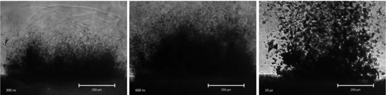
The ablated aerosol particles were transported by continuous air/N2 flow and collected on a filter membrane. The collected particles were analyzed by FTIR spectrometry and scanning electron microscopy (SEM). In some cases the size distribution of the produced aerosol particles was analyzed by Scanning Mobility Particle Sizer (SMPS) and Optical Particle Counter (OPC). For the nanosecond ablation the material removal process was studied with fast photographic method.
Figure 1. Experimental setup.