Development of novel poly(aspartamide) based drug conjugates and gels for biomedical and
pharmaceutical applications
PhD ThesisDavid Juriga
Semmelweis UniversityDoctoral School of Pharmaceutical Sciences
Supervisor: Miklós Zrínyi, HAS member Official reviewers: Péter Nemes, DSc.
Marianna Budai, PhD.
Head of the Final
Examination Committee: Tamás Török, DSc.
Member of the Final
Examination Committee: Imre Klebovich, DSc.
Péter Huszthy, DSc.
Budapest 2018
1 Introduction
In the medical and pharmaceutical field, polymers can be used for several applications such as packaging, implantation and drug formulation or to prepare modern stents or drug delivery systems. Therefore polymer based materials are in the focus of modern biomedical and pharmaceutical researches. The two principally researched applications of polymer based materials are focusing on drug delivery systems in pharmaceutics and on scaffolds in the field of tissue engineering. For these applications, it is crucial that, the polymer used is biocompatible and biodegradable namely it is a biopolymer.
Biopolymer based drug delivery systems can increase the absorption as well as the water solubility or lipophilicity of drug molecules in the human body and deliver the drug into a specific organ or tissue, where it is released. Due to these features, the amount of the applied drug can be reduced, resulting in decrease of side-effects as well as significantly increased therapeutic effect. Polymer-drug conjugates are novel class of polymer based drug delivery systems, in which drug molecules are attached covalently to the polymer main chain. These macromolecular prodrugs are inactive until they reach the targeted tissue, where drug release can be induced by different metabolic processes or as a response to the change of environmental parameters (such as pH, temperature, etc.). By using electrospinning, nanofibrous meshes can be prepared from the conjugates, which can increase the effectiveness of the drug therapy.
In the medical field, biopolymer based materials can be used as a scaffold in for tissue engineering. In this process an artificial tissue can be made by isolating specific cells from the patient and seeding them into an artificial scaffold material, which supports 3 dimensional cell proliferation and
migration. Such scaffold has to possess similar properties to the native extracellular matrix (ECM), which is a 3 dimensional network, constructed from collagen and different polymers, such as fibronectin or proteoglycans. ECM is permeable for both small molecules or cells and one of its main roles is to support cell proliferation. Polymer hydrogels are similar to the native ECM because they are constructed from a 3 dimensional polymer matrix, filled with water based liquids. Due to their structure, they possess the properties of liquids (e.g.: permeability for small molecules), as well as the properties of solid materials (e.g.: deformability). The properties of hydrogels are determined by the polymer molecule, therefore the selection of the appropriate polymer is crucial for the successful application.
The chemical structure of the poly(aminoacid)s is similar to native peptides, therefore they are promising candidates for conjugates and scaffold materials. The main disadvantage of these polymers is their relatively high cost and difficult synthesis. Therefore, during my PhD research, I have focused on biomedical and pharmaceutical applications of easily synthesized poly(aspartamide) and its derivatives.
2 Objectives
The main aim of my research was the investigation of poly(aspartamide) and its derivatives for biomedical and pharmaceutical applications. The research revolved around the following major tasks:
1. Synthesis and characterization of different poly(aspartamide)-dopamine conjugates. Investigation of dopamine release in different environments;
2. Preparation of nano-fibrous implants from the conjugates by electrospinning. Investigation of the effect of the
formulation on dopamine release and monitoring the cytotoxicity of the fibrous conjugates in vitro;
3. Application of different poly(aspartamide) hydrogels as scaffold materials for in vitro cultivation of MG-63 osteosarcoma cells. Investigation of the stability and the degradation of the hydrogels in the experimental conditions;
4. Applicability of the previously developed hydrogels for cultivation of human periodontal ligament stem cells (PDLSCs). Optimization of the crucial properties of the hydrogels to improve the cell viability;
5. Applicability of dopamine modified poly(aspartamide) hydrogels for PDLSC cultivation;
3 Methods
3.1 Synthesis of poly(succinimide) (PSI), poly(aspartamide) based conjugates and polymers
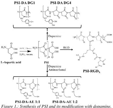
Poly(succinimide) can be synthesized by thermal polycondensation of L-aspartic acid in the presence of phosphoric acid, in vacuum, at 180 ºC (Figure 1). After the reaction the raw polymer can be dissolved in dimethylformamide then precipitated and washed with water until neutral pH. The average molecular weight of the polymer varied between 26 and 31 kDa, according our previous result [1-3].
Figure 1.: Synthesis of PSI and its modification with dopamine, dopamine-aminoethanol mixture and RGD tripeptide
The succinimide rings in the PSI can be easily modified with primer amines at room temperature. Due to this property, poly(aspartamide) drug conjugates can be prepared by using amino group containing drug molecules. I have synthesized dopamine and dopamine-aminoethanol containing conjugates with different chemical constitutions (Figure 1).
The DG (degree of grafting) represents the molar ratio of the monomers to the dopamine. In the other hand, different Arg- Gly-Asp (RGD) modified polymers were synthesized which were used to prepare poly(aspartic acid) hydrogels for tissues engineering application (Figure 1). The chemical structure of the modified polymers was proven by 1H-NMR, FTIR-ATR and 1H-1H COSY NMR spectroscopy.
3.2 Preparation of the conjugate fibers by electrospinning
For the preparation of fibrous meshes, a home-made electrospinning instrument was used consisting of a plastic syringe with a metal Hamilton tip and a syringe pump. The positive electrode was attached to the metal tip whereas the negative electrode (ground) was attached to the collector, made of aluminum foil in front of the needle in a well determined distance of 15 cm. The voltage applied between 13 and 15 kV was provided by a DC power supply [3]. For electrospinning, different solvent or solvent mixtures with different concentrations were used depending on the chemical structure and solubility of the samples. The structure and the size of the fibers were measured by scanning electron (SEM), two-photon and atomic force microscopy. The chemical structure of the fibers was determined by FTIR-ATR spectroscopy.
3.3 Investigation of the physical properties, such as solubility, solubility kinetics, lipophilicity and membrane permeability, of the bulk and fibrous conjugates
To investigate the solubility of the bulk conjugates, 0.1 g sample was mixed with 11 mL ultra-pure water, and the absorbance at 280 nm of the liquid phase was measured by spectrophotometry after 24 and 48 hours. To measure the lipophilicity of the conjugates, concentrated solutions were prepared in water and mixed with the same volume of n-octanol. The mixtures were shaken and after 24 and 48 hours the concentration in both phases were determined by spectrophotometry. The lipophilicity can be identified as the ratio of the concentrations in the two phases. As the solubility of the PSI-DA conjugates depends on the time, the dissolution
kinetics of these samples were determined in water and phosphate buffer solution (PBS. pH=7.5). For these measurements, 0.3 g bulk or 0.05 g fibrous conjugate was mixed with the solvents and the change in the absorbance was measured by spectrophotometry until 48 hours. The membrane permeability of the conjugates was determined by parallel artificial membrane permeability assay (PAMPA). 0.5 mg conjugate was placed into a 96 well plate. As model membrane isolecitine solution in dodecane was used. The concentration of the conjugates was determined by spectrophotometry in both sides of the model membrane after 4 and 28 hours. The membrane permeability can be given by the ratio of the two concentrations.
3.4 Determination of the dopamine release from bulk and fibrous conjugates
To determine the dopamine release from the different conjugates, samples were placed into a dialysis membrane with 3.5 kDa cut-off. 6 mL PBS or different enzyme solutions (Bromelain or α-Chymotrypsin) with 3 mg/mL concentration were injected in to the dialysis membrane, while 60 mL PBS was placed in to the outer side of the membrane. The concentration of the released dopamine was determined by measuring the absorbance at 280 nm wavelength in the outer side of the membrane. The release kinetics was followed for 48 hours.
3.5 Cytotoxicity of the fibrous meshes
For cytotoxicity measurements human PDLSCs were used. The cells were seeded in to the 96 well-plate with 10000 cell/cm2 density. In the first series of the experiments, the effect of free dopamine and conjugated dopamine on PSI-DA-AE 1:2 conjugate on cell viability was measured between 1 µM - 1 mM concentration. The cell viability was
measured by WST-1 reagent while the morphology of the cells was monitored by phase contrast microscopy. In the second series of experiments, the different fibrous conjugates were used with 250 µM concentration, but the cell morphology was investigated by two photon microscopy.
3.6 Preparation of poly(aspartic) acid hydrogels
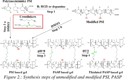
Since PSI reacts easily with primer amines, bi- or multifunctional amines can be used to crosslink the polymer chains resulting in polymer gels. To prepare PSI and modified PSI based gels, two different diamines, 1,4-diaminobutane (DAB) and cystamine (CYS) were used. To prepare poly(aspartic acid) (PASP) based hydrogels the PSI gels were immersed in to alkaline buffer solution. Furthermore, the simultaneously crosslinked (CYS and DAB) gels were treated with dithiotreitol (DTT), to turn disulphide bonds in to thiol groups. During my work, I have prepared PASP hydrogels with dopamine, RGD or thiol sidechains (Figure 2).Figure 2.: Synthesis steps of unmodified and modified PSI, PASP and thiolated PASP gels
3.7 Degradation of PASP based hydrogels
The degradation experiments of differently crosslinked PASP based hydrogels were carried out in two different measurements. In the first measurement a series of hydrogel discs were immersed in to different enzyme solutions (Dispase, Trypsin-EDTA, and Collagenase II). The change of the mass of the gel discs was followed for 48 days. In the second series of experiments, the change of the elastic modulus and mass of the different gel cylinders were monitored until 18 days in cell medium.
3.8 Investigation of the applicability of the PASP hydrogels as scaffold
For this application, two different, human MG-63 osteosarcoma and human PDLSC, cell types were used. 20000 cells in 200 µL suspension were seeded onto the different PASP based hydrogel discs. The viability was measured after 1, 3, and 6 days in case of MG-63 cells, while in case of PDLSC the viability was determined after 1, 3, 7, and 14 days.
The morphology of the cells was determined by phase contrast microscopy and the 2 and 3 dimensional distribution of the cells were measured by two photon microscopy.
4 Results
4.1 Description of the dopamine release from powder conjugates (T1)
To describe the dopamine release the conjugates were divided into two groups: samples which contain both aminoetanol and dopamine dissolve immediately in water, while the conjugates, containing only dopamine, has time dependent water solubility. Due to the fact, that dopamine releases from dissolved conjugates, in the first case a 1 step release can be considered, while in the second case,
consecutive reactions (diffusion and release) happen. Thus, the first case can be used to determine the reaction order of the dopamine release which can be used to determine the limiting factor of the release in the second case. However, dopamine degrades in the reaction environments, therefore at first, I had to determine the reaction kinetics of dopamine degradation. It can be seen on the Figure 3 that the decrease of dopamine concentration linearly depends on time, so the dopamine degradation follows 0 order kinetics (Figure 3 a). Due to the fact, that the dissolution of PSI-DA conjugates depends linearly on the square root of time, it can be described by Higuchi dissolution kinetics (Figure 3 b). In case of the PSI-DA-AE conjugates, if the reaction rate is linearly proportional to the concentration of the released dopamine, the reaction can be described by first order kinetics. The differential equation on Figure 3 c represents this case, and it can be seen on the graph (Figure 3 c), within an experimental accuracy this equation satisfactorily describes the experimental data. Since PSI-DA conjugates dissolve slowly in water, this situation can be described with a diffusion followed with the first order release kinetics. If we consider an infinitely long measurement time, the kinetic model is resulting in the equation on Figure 3 d. Since, the logarithmic part of the equation is the linear function of time, it proves the correctness of the kinetical model (Figure 3 d)
3.ábra: a) degradation kinetics of dopamine, b) dependency of the dissolution of PSI-DA conjugates on the square root of time, c)
release kinetics of dopamine from PSI-DA-AE conjugates, d) determination of the reaction constant in case of slowly dissolving
conjugates
4.2 Investigation of the fibrous conjugates (T2)
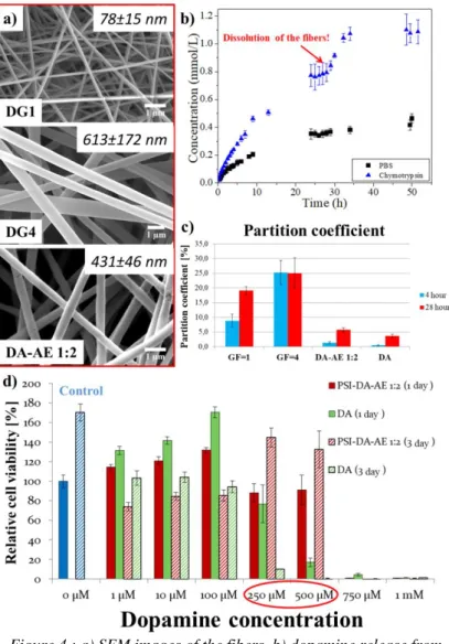
It can be seen on the SEM images, that the conjugate fibers were produced with a diameter in nanometer scale, without any defects (Figure 4 a). The formulation might have influence on the dissolution kinetics of the conjugates, which has high influence on the dopamine release (see in the previous section). The dopamine release results prove, that the dopamine releases significantly faster, than before formulation (Figure 4 b). It can be seen, that the released amount of dopamine abruptly increased after 26 hours especially in the presence of α-Chymotrypsin. This abrupt increase can be caused by the macroscopical disintegration of the fibrous mesh (Figure 4 b). The presence of the α-Chymotrypsin is resulting in higher and faster release of dopamine (Figure 4 b). Themembrane permeability has high influence on the utilization of the drug in the human body. According to the PAMPA measurement, the PSI-DA conjugates significantly increase dopamine permeability, compared to free dopamine and PSI- DA-AE conjugate. This can be resulted from the lower amount of hydroxyl groups in these conjugates. The difference between DG1 and DG4 conjugates can be attributed to the same reason (Figure 4 c). According to the cytotoxicity measurements, neither the free dopamine nor the PSI-DA-AE 1:2 conjugates are cytotoxic in low concentration (1-100 µM).
However, between 250 and 500 µM concentration the free dopamine proved to be cytotoxic as opposed to the conjugated dopamine, which did not decrease cell viability. At higher concentration either free dopamine or the conjugate proved to be cytotoxic (Figure 4 d).
Figure 4.: a) SEM images of the fibers, b) dopamine release from DG1 fibrous conjugate, c) partition coefficient of the conjugates, and
d) cytotoxicity data
4.3 MG-63 cell cultivation on different PASP hydrogels and stability of the hydrogels (T3)
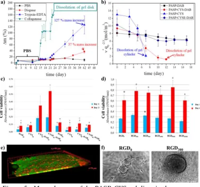
As it can be seen on Figure 5 a, the PASP-CYS hydrogels are completely stable for 42 days in PBS, due to the
fact that their mass haven’t changed. The mass of the hydrogels shows monotonous increase in the presence of Dispase and Trypsin-EDTA during the observation time, which is the result of small degradation in the polymer matrix.
In the presence of Collagenase II, the mass of the gel discs abruptly increased and the samples were completely dissolved after a few days (Figure 5 a). By measuring the elastic modulus and the swelling degree of the gels, the degradation can be followed. The concentration of the elastically active chains (ν*) includes both values and gives a better representation of the mechanical properties. The concentration of the elastically active chains in PASP-DAB gels have not changed in cell medium, while the PASP-CYS gels completely dissolved after 5 days. The concentration of the elastically active chains in the simultaneously crosslinked PASP-CYS- DAB gels decreased, but the gels did not dissolve (Figure 5.
b). Due to these results the dissolution of the gels caused by the cleavage of the disulphide bonds in the polymer matrix.
The presence of the thiol groups in the polymer matrix as well as the increased elastic modulus (PASP-CYSE20-DAB20), increase the adhesion of the MG-63 cells on the gel surface (Figure 5 c). The viability of the cells increased to day 3 on every gel sample (Figure 5 c). According to two photon microscopic images, the cells were able to incorporate into the polymer matrix, by digesting themselves into it (Figure 5 e).
The presence and the amount of the RGD had no significant effect on cell viability neither at day 1 nor at day 6 (Figure 5 d). However, on the phase contrast microscopic images rounded cluster formation of the cells can be seen, on RGD containing gels, which hallmarks the osteogenic behavior of the cells (Figure 5 f)
Figure 5.: Mass changes of the PASP-CYS gel discs in the presence of different enzymes (a), degradation of the gel cylinders in cell medium (b), Cell viability on different PASP (c) and RGD containing
gels (d), 3 dimensional distribution of the cells on gel discs (e), and cell clusters on RGD containing gels (f)
4.4 PDLSCs cultivation on differently thiolated PASP based hydrogels (T4)
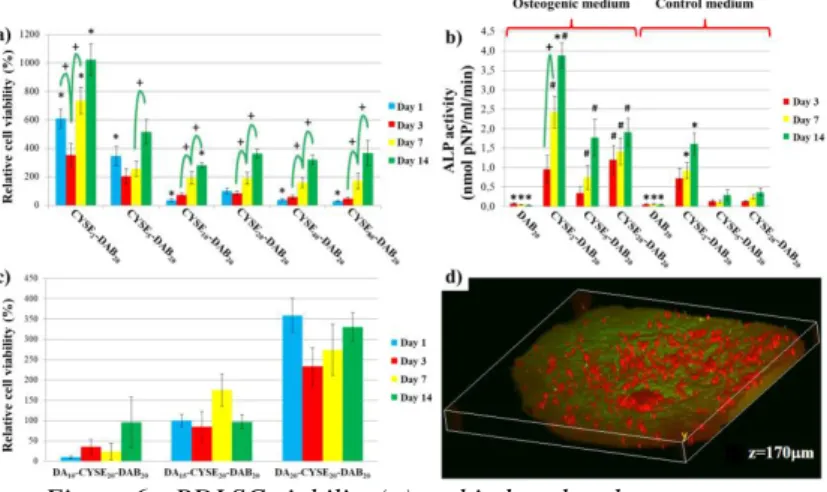
By increasing the amount of thiol groups in PASP hydrogels an enhanced cell adhesion and proliferation can be achieved (Figure 6 a). Cell viability shows an increasing tendency on every sample until 14 days. The increased amount of thiol groups can induce the spontaneous differentiation of the PDLSCs, according to the alkaline phosphatase activity results (Figure 6 b).
Figure 6.: PDLSC viability (a) and induced and spontaneous differentiation of PDLSCs on differently thiolated PASP gels (b), PDLSCs viability on different dopamine modified PASP gels (c) and
3 dimensional distribution of the cells on dopamine modified PASP gel.
4.5 PDLSCs cultivation on dopamine modified PASP gels (T5)
The highest viable cell population was measured on the hydrogels which contained less dopamine (every 20.
monomer was modified) (Figure 6 c). The higher concentration resulting in lower cell viability, due to the concentration of the dopamine reached the cytotoxic concentration. The presence of dopamine induced the 3 dimensional migration and proliferation of the cells which was proved by two photon microscopy. The cells were able to digest themselves into 170 µm depth, which is equal to the thickness of 5-6 cell layers.
5 Conclusion
Biopolymer based polymer-drug conjugates and hydrogels have been in the main focus of pharmaceutical and biomedical researches in the past few years. For these applications, poly(aminoacid)s would be a promising
candidates, due to the fact, that their chemical structure is similar to natural peptides. During my PhD research, I have successfully synthesized poly(aspartic acid) based conjugates with dopamine and I have investigated to applicability of poly(aspartic acid) hydrogels as scaffolds for cell cultivation.
The dopamine release was investigated from different conjugates in several environments, which significantly depends on dopamine content on the polymer, on solubility and on presence of different enzymes. To describe the release kinetics a detailed kinetical description has been developed, for both poorly and highly soluble conjugates. In case of highly soluble conjugates, the experimental data and the kinetical model showed high correlation (Figure 3 c). The release kinetics coupled with Higuchi diffusion can be used to describe dopamine release from poorly soluble conjugates (Figure 3 d). Nano-fibrous meshes with homogenous size distribution, can be produced from the conjugates by electrospinning (Figure 4 a). By this formulation the dopamine release can be increased due to the enhanced specific surface of the conjugates. The macroscopic disintegration of the meshes after 26 hours resulted in abrupt dopamine release and a two steps drug release kinetics (Figure 4 b). According to the PAMPA experiments, the conjugates are able to increase membrane permeability of the dopamine due to their higher lipophilicity (Figure 4. c). The conjugated dopamine caused lower cytotoxic effect than the free dopamine in the same concentration (Figure 4 d).
The mechanical properties and chemical structure of the PASP based hydrogels have high impact on the viability and on the 3 dimensional distribution of MG-63 and PDL stem cells. By increasing the elastic modulus and the amount of thiol groups in the hydrogel matrix an enhanced MG-63 cell adhesion and proliferation can be achieved (Figure 5 c).
Because the MG-63 is an osteosarcoma cell line originated from hard tissue, it prefers scaffolds with higher elastic modulus. The cells were able the incorporate into the polymer matrix (Figure 5 e), while the presence of RGD in the hydrogels induced the cluster formation of the cells, which indicates the osteogenic behavior of the cells (Figure 5 d and f). According to the degradation experiments, the hydrogels containing only disulphide crosslinks are instable in the presence of collagenase II and in the cell medium (Figure 5 a and b), due to the cleavage of the disulphide bonds. By increasing the thiol groups in the hydrogels an enhanced PDLSC adhesion can be achieved (Figure 6 a). Since cell viability increased until 14 days the thiol groups improved cell proliferation as well. The presence of dopamine has beneficial effect on cell viability (Figure 6 c), if the concentration of the dopamine does not exceed the cytotoxic concentration. The presence of dopamine induced the vertical migration of PDLSCs, thus the cells were able to incorporate into the polymer matrix with 5-6 cell layers (Figure 6 d).
The new scientific accomplishments are collected in the following points:
1. I have successfully synthesized different poly(aspartamide)-dopamine conjugates, which provide prolonged dopamine release (T1)
2. Detailed release kinetic models were developed for covalent polymer-drug conjugates, which can be used for either good or poor water soluble conjugates. (T1)
3. Homogenous fibrous meshes were prepared from the conjugates with a diameter in nanometer scale by electrospinning. The formulation has high impact on the drug release kinetics, which was compared with the drug release kinetics from bulk conjugates at first in the literature (T2)
4. The biocompatibility of the conjugates was proved by using human derived PDLSCs. (T2)
5. The crucial parameters of the PASP gels, which increase the stability and decrease the degradability in the environment of in vitro cell cultivation, were determined (T3)
6. The higher elastic modulus and the presence of thiol groups in PASP matrix has good influence on the viability and cell morphology of MG-63 as well as PDLSCs. By incorporating RGD tripeptide in to the hydrogels, either increased cell adhesion and cluster formation of the cells can be achieved (T3)
7. By increasing the amount of the thiol groups in the polymer hydrogel matrix, a spontaneous osteogenic formation of the PDLSCs can be induced. (T4)
8. The presence of the dopamine in the hydrogel can induce a vertical migration of the PDLSCs by increasing the cell caused biodegradability of the polymer matrix (T5)
6 Publications
6.1 Publications related to the thesis
[1] Juriga D, László I, Ludányi K, Klebovich I, Chae CH, Zrínyi M, (2018) Kinetics of dopamine release from poly(aspartamide)-based prodrugs. Acta Biomater, Available online, doi: 10.1016/j.actbio.2018.06.030, IF2017: 6,319
[2] Juriga D, Nagy K, Jedlovszky-Hajdú A, Perczel-Kovách K, Chen YM, Varga G, Zrínyi M (2016) Biodegradation and Osteosarcoma Cell Cultivation on Poly(aspartic acid) Based Hydrogels. ACS Appl Mater Interfaces 8:23463–23476. IF2016: 7,504
6.2 Publications not related to the thesis
[3] Molnar K, Juriga D, Nagy PM, Sinko K, Jedlovszky- Hajdu A, Zrinyi M (2014) Electrospun poly(aspartic acid) gel scaffolds for artificial extracellular matrix.
Polym Int 63:1608–1615. IF2014: 2.409
[4] Zrinyi M, Gyenes T, Juriga D, Kim J-H (2013) Volume change of double cross-linked poly(aspartic acid) hydrogels induced by cleavage of one of the crosslinks.
Acta Biomater 9:5122–31. IF2013: 5,684