Long-term results after single event multilevel surgery for the correction of gait disorders in spastic diplegic cerebral
palsy
PhD dissertation
Dr. Dóra Végvári
Semmelweis University
Clinical Medicine of Doctoral Studies
Consultant: György Szőke MD, DSc
Reviewers: Rita Kiss, PhD, DSc
Zsuzsanna Miklós, MD, PhD
President of examiners’ committee: Imre Oláh MD, DSc
Members of examiners’ committee: Kinga Karlinger MD, CSc Jenő Kiss MD, CSc
Budapest
2015
2
Table of contents
1. Introduction ... 8
1.1. Definition ... 8
1.2. Etiology ... 8
1.3. Pathology ... 9
1.4. Gross Motor Function Classification Scale (GMFCS) ... 11
1.5. Diagnosis ... 12
1.6. Therapy ... 13
1.6.1. Physical, occupational and speech therapy... 13
1.6.2. Medical equipment ... 14
1.6.3. Orthosis ... 14
1.6.4. Drugs ... 15
1.6.5. Surgery ... 16
1.6.5.1. Single event multilevel surgery (SEMLS), muscle tendon lengthening (MTL) 17 1.6.5.1.1. Hamstring lengthening ... 18
1.6.5.1.2. Distal rectus femoris transfer ... 19
1.6.5.2. Surgical approaches in the central nervous system ... 21
1.6.5.3. Surgical and chemical approaches in the peripheral nervous system 21 1.7. Gait ... 22
1.7.1. Three dimensional gait analysis ... 25
1.7.1.1. Kinematics ... 25
1.7.1.2. Time-distance parameters ... 28
1.7.1.3. Kinetics... 29
1.7.1.4. Electromyography ... 30
1.7.1.5. Pedobarograph ... 31
1.7.1.6. GGI ... 32
1.7.2. Normal gait ... 33
1.7.2.1. Pelvis ... 33
1.7.2.2. Hip ... 33
1.7.2.3. Knee ... 34
3
1.7.2.4. Ankle ... 35
1.7.3. Spastic diplegic gait ... 36
1.7.3.1. Crouch knee... 36
1.7.3.2. Stiff knee ... 37
1.7.3.3. Jump knee ... 38
1.7.3.4. Recurvatum knee ... 39
2. Purpose ... 40
2.1. Development of knee function after hamstring lengthening as a part of multilevel surgery in children with spastic diplegia ... 40
2.2. Long-term results after distal rectus femoris transfer as a part of multilevel surgery for the correction of stiff knee gait in spastic diplegic cerebral palsy ... 41
2.3. Does proximal rectus femoris release influence kinematics in patients with cerebral palsy and stiff knee gait? ... 42
2.4. Long-term effects after conversion of biarticular to monoarticular muscles compared with musculotendinous lengthening in children with spastic diplegia ... 43
3. Methods ... 44
3.1. Overview ... 44
3.1.1. Clinical examination ... 44
3.1.2. 3D gait analysis ... 45
3.1.3. Statistics ... 46
3.2. Development of knee function after hamstring lengthening as a part of multilevel surgery in children with spastic diplegia ... 47
3.3. Long-term results after distal rectus femoris transfer as a part of multilevel surgery for the correction of stiff knee gait in spastic diplegic cerebral palsy ... 50
3.4. Does proximal rectus femoris release influence kinematics in patients with cerebral palsy and stiff knee gait? ... 53
3.5. Long-term effects after conversion of biarticular to monoarticular muscles compared with musculotendinous lengthening in children with spastic diplegia ... 55
4. Results ... 59
4.1. Development of knee function after hamstring lengthening as a part of multilevel surgery in children with spastic diplegia ... 59
4.1.1. Clinical examinations ... 59
4.1.2. Global gait variables ... 60
4.1.3. 3D gait analysis ... 61
4
4.1.3.1. Temporal parameters ... 61
4.1.3.2. Kinematics ... 62
4.1.3.3. Kinetics... 66
4.1.4. Adverse effects ... 67
4.1.5. Correlation between static and dynamic parameters ... 68
4.2. Long-term results after distal rectus femoris transfer as a part of multilevel surgery for the correction of stiff knee gait in spastic diplegic cerebral palsy ... 69
4.2.1. Clinical examination ... 69
4.2.2. Global gait variables ... 70
4.2.3. 3D gait analysis ... 72
4.2.3.1. Temporal parameters ... 72
4.2.3.2. Kinematics ... 73
4.2.4. Response and recurrence ... 75
4.2.5. Correlations ... 76
4.3. Does proximal rectus femoris release influence kinematics in patients with cerebral palsy and stiff knee gait? ... 77
4.3.1. Global gait variables ... 77
4.3.2. 3D gait analysis ... 78
4.3.2.1. Temporal parameters ... 78
4.3.2.2. Kinematics ... 79
4.4. Long-term effects after conversion of biarticular to monoarticular muscles compared with musculotendinous lengthening in children with spastic diplegia ... 83
4.4.1. Clinical examinations ... 83
4.4.2. Global gait variables ... 84
4.4.3. 3D gait analysis ... 85
4.4.3.1. Temporal parameters ... 85
4.4.3.2. Kinematics ... 85
4.4.4. Adverse effects and recurrence of flexed knee gait ... 88
5. Discussion ... 91
5.1. Development of knee function after hamstring lengthening as a part of multilevel surgery in children with spastic diplegia ... 91
5.2. Long-term results after distal rectus femoris transfer as a part of multilevel surgery for the correction of stiff knee gait in spastic diplegic cerebral palsy ... 95
5
5.3. Does proximal rectus femoris release influence kinematics in patients with
cerebral palsy and stiff knee gait? ... 99
5.4. Long-term effects after conversion of biarticular to monoarticular muscles compared with musculotendinous lengthening in children with spastic diplegia .... 102
6. Conclusion ... 105
6.1. Development of knee function after hamstring lengthening as a part of multilevel surgery in children with spastic diplegia ... 105
6.2. Long-term results after distal rectus femoris transfer as a part of multilevel surgery for the correction of stiff knee gait in spastic diplegic cerebral palsy ... 105
6.3. Does proximal rectus femoris release influence kinematics in patients with cerebral palsy and stiff knee gait? ... 106
6.4. Long-term effects after conversion of biarticular to monoarticular muscles compared with musculotendinous lengthening in children with spastic diplegia .... 106
7. Summary ... 107
8. Összefoglalás ... 108
9. Literature ... 109
10. Publications ... 119
10.1. Publications directly related to the thesis ... 119
10.1.1. Full articles ... 119
10.2. Publications not directly related to the thesis ... 120
10.2.1. Full articles ... 120
10.2.2. Abstracts ... 120
11. Acknowledgement ... 121
6
Abbreviations
2D: two dimensional 3D: three dimensional
A/D converter: analog digital converter ANOVA: analysis of variance
APT: anterior pelvic tilt BMI: body mass index BTX: botulinum toxin type A
CBM: conversion of biarticular to monoarticular muscles CC-Distraction: calcaneo-cuboid-joint distraction arthrodesis
C-DRFT: group that had distal rectus femoris transfer to correct decreased peak knee flexion in swing phase
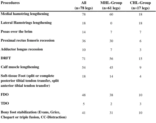
CHL: combined medial and lateral hamstring lengthening CNS: central nervous system
CP: cerebral palsy DF: dorsiflexion
DRFT: distal rectus femoris transfer E0: preoperative examination
E1: examination 1 year postoperatively E2: second examination after surgery E3: third examination after surgery EMG: electromyography
FDO: femoral derotation osteotomy GC: gait cycle
GGI: Gillette Gait Index
7
GMFCS: Gross Motor Function Classification Scale GRF: ground reaction force
MHL: medial hamstring lengthening MRC: medical research council MTL: muscle tendon lengthening PASW: public art south west
P-DRFT: group that had prophylactic distal rectus femoris transfer PF: plantar flexion
pKFSw: peak knee flexion in swing ROM: range of motion
SD: standard deviation
SEMLS: single event multilevel surgery
SENIAM: surface EMG for a non-invasive assessment of muscles SPSS: statistics statistical procedures companion
TDO: tibial derotation osteotomy UK: United Kingdom
USA: United States of America
8
1. Introduction
1.1. Definition
Cerebral palsy (CP) is a childhood condition in which there is a motor disability (palsy) caused by a static, non-progressive lesion in the brain (cerebral). The causative event has to occur in early childhood usually defined as less than 2 years of age. Children with CP have a condition that is stable and non-progressive; therefore, they are in most ways normal children with special needs (1).
The prevalence of cerebral palsy is 2-3 children from 1000 neonate (2). The most commonly reasons are premature and low birth weight (<1500g).
1.2. Etiology
Etiological factors can be separated into a time period as to when these insults occurred:
congenital, neonatal and postnatal. (1. Table, (3)) Congenital deformities result from defects that occur in normal development and follow patterns based on failures of normal formation. In neonatal deformities the newborn suffers some injury during the birth.
Postnatal causes may overlap somewhat with the prenatal and neonatal group. Most of infections are prenatal and neonatal viral infections (CMV, VZV), but postnatal deformities can be caused by bacterial meningitis. Children, who survive this infection have a 30-50% chance to become CP.
1. Table: Etiology of cerebral palsy
Congenital Neonatal Postnatal
defect in the development of neural tube or brain
infection alcohol, nicotine
hypoxic events cerebral hemorrhages
neonatal stroke Kernicterus
postnatal trauma (shaken baby syndrome, blunt head trauma) metabolic encephalopathy
infections toxicins
9
A specific region of brain injury can cause variation in the impairments because the initial injury also hinders normal development. Because all these injuries occur in the young and immature brain, growth and development over time affects the impairment. During healthy development, the early primitive reflexes (Moro reflex, sucking reflex, tonic labyrinth reflex, asymmetric tonic neck reflex, step reflex, parachute reaction etc.) should disappear. The preservation of these could indicate the presence of brain injury.
1.3. Pathology
Cerebral palsy requires a pathologic lesion in the brain, in the central motor system (3).
In most cases the spinal cord has a normal function, but there are some children who have a primary lesion there. The control of motion is either volitional or automatic. The automatic response is a relatively simple neuronal reflex at the spinal cord level. All volitional motion initiates in the cerebral cortex and transmitted to the peripheral motor nerves through the cortical spinal tracts traversing the internal capsule and the spinal cord.
The peripheral motor system includes the nerves and the musculoskeletal system. In CP children there are no primary lesion in any of peripheral systems. The impairment of the central motor system causes secondary abnormal development on the periphery.
Patterns of CP can be categorized further by clinical presence quality or by topography of palsy (4). The quality can be spasticity, athetosis, dystonia and rigid quality of palsy.
Spasticity is the most frequent form (more than 80%). It is caused by co-contraction by agonist and antagonist muscle. This is a velocity dependent increase in resistance to motion. Athetosis (6%) is large, involuntary, slow cramped motions of the more proximal joints, which can be exacerbated by mental strain. Dystonia is a slow motion with a torsional element, which may be localized to one limb or involve the whole body (9%).
The appearance of rigidity is under 5%.
Balance is the maintenance of posture, when impaired children overcompensate for a movement and unable to stand in one place. Ataxia (15%) is the term used to mean abnormal balance.
10
Topographically CP can be hemiplegic, diplegic and quadriplegic. Hemiplegic involves one half of the body. The flexion is typical in upper extremity and the extension in lower extremity. These children can usually learn to walk. Diplegia involves primarly the lower extremity with extension and have a mild upper extremity involvement with flexion. They require frequently orthosis and other aids to walk. The quadriplegic patients are usually mentally retarded (75%) and have highly spasticity in all four limbs. Only 10% can learn to walk.
Normal muscle tone is composed of stretch reflex, central control of stretch reflex and reflex activity of spine cord. The opposite of spasticity is hypotonia, which means decreased muscle tension when the joint is moved. Spasticity includes an increased sensitivity of the normal stretch reflex in addition to a velocity-dependent increase in resistance, which initiates a muscle contraction to resist the motion (5). To measure the muscle tone most commonly the Ashworth scale or the modified Ashworth scale is used, however it is a very subjective measurement (2. Table) (6, 7).
2. Table: Modified Ashworth scale (6)
0 normale tone
1 slight increase in tone, a catch and release at the end of the range of motion (ROM)
1,5 slight increase in tone, a catch followed by minimal resistance in remainder of range
2 more marked increase in muscle tone through most of
the ROM, but affected part(s) easily moved
3 considerable increase in tone, passive movement
difficult
4 affected parts rigid in flexion or extension
The effects of spasticity on the skeletal muscle include shorter fiber length, decreased volume of the muscle, change in the fiber type and neuromotor junction type. These changes result in weaker muscle and decreased joint range of motion (8). The muscle and tendon growth can not follow the bone growth resulting motion constriction. In the long term, spasticity cause muscle shortening in agonist and muscle elongation in antagonist, resulting in malposition. Especially the multijoint muscles tend to spastic shortening (9, 10). The untreated deformity becomes more severe with growing. First it is still able to
11
change dynamically, but it can be fixed with time resulting in constrictions. These constrictions are followed by the bony malformations. The efficiency of therapy diminishes with growing, but it is hard to say the appropriate time for it. Some surgeons prefer to wait until patients are 8-10 years and perform all of their surgical interventions in one sitting. According to Sussman and Aiona different deformities should be operated in different time points (11).
1.4. Gross Motor Function Classification Scale (GMFCS)
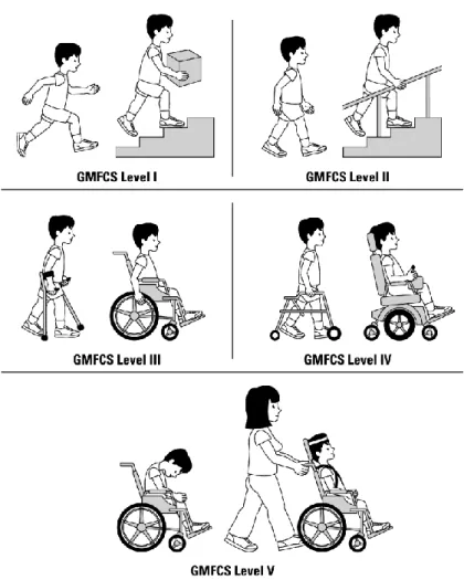
The Gross Motor Function Classification Scale (GMFCS) is a simple five-level system to describe gross motor function in cerebral palsy patients (1.Fig.). The GMFCS I children walk indoors and outdoors and climb stairs without any limitation. Children perform gross motor skills including running and jumping, but speed, balance and coordination are impaired. The GMFCS II children are limited in walking on uneven surfaces and inclines and walking in crowds or confined spaces. They have only minimal ability to run and jump. GMFCS III children can walk only with an assistive mobility device. They may climb stairs holding onto a railing. To take a long distance these children may need a wheelchair. GMFCS IV children may walk for a short distance on a walker. They may achieve self-mobility using a power wheelchair. GMFCS V children are unable for independent mobility. All areas of motor function are limited (12, 13).
12
1. Figure: Gross motor function classification scale (13)
1.5. Diagnosis
The maturation of neonates take place from infancy to one year of age rapidly from proximal to distal. Children have first head control, then develop the ability to bear weight on the arms, followed by the trunk control, then develop the ability to stand. There are no agreed-upon diagnostic criteria to make a diagnosis of CP. When a child is not meeting developmental milestones, has persistent primitive reflexes, or has significant abnormalities in the elements of motor function, a diagnosis of CP can be made. The history should clearly demonstrate that this is a nonprogressive and is nonfamilial (3).
The lack of balance and abnormalities in motor tone are the most common abnormalities that occur in children with CP. But there is a well-recognized phenomenon of children occasionally outgrowing CP. For this reason, it is preferred to make the diagnosis in
13
young children only when it is clear and without doubt, but wait until least age 2 years for children who have more mild and questionable signs.
1.6. Therapy
Most of the CP children receive therapy and go to the school (14). Therapy will be ordered by physicians and medical doctors. It begins already in infancy. Early therapy is provided in a medically-based construct. As the children get older, the main intervention shifts to the educational system. In addition to the standard therapy treatment (physical, occupational, speech therapy, orthotics, pharmacological therapy, surgery), there are many treatment modalities that are promoted as beneficial for CP treatment:
hippotherapy, hyperbaric oxygen therapy, hydrotherapy, acupuncture etc. The goal of different therapeutic methods is to decrease the increased muscle tone and to preserve the length of the muscles. The first aim is to prevent those provocative factors that give rise to the problems. The second aim is to prevent the consequences of spasticity (15).
1.6.1. Physical, occupational and speech therapy
Physical therapy focuses on gross motor function, such as walking, running, jumping and joint range of motion (14). It prevents some spasticity from developing and worsening or complications from arising. Its first aim is the muscle lengthening (15). In occupational therapy the main focus is on fine motor skills, specifically upper extremity function and activities of daily living such as dressing, toileting and bathing. The focus depends on the age and functional ability of a child. The therapist uses a learning approach based on specific task as the goal. Speech therapy focuses on oral motor activities such as speech, chewing and swallowing. A major focus of all therapy is to maximize the individual’s independence.
14 1.6.2. Medical equipment
Durable medical equipment is the category of devices that are prescribed to ameliorate the disabilities from the motor impairments (16). Each of devices has a very specific indications and contraindications. The medical equipment has a very wide range of application. It assists a limb positioning with orthosis or sitting with seating system. To prevent a contracture the patient needs daily physical therapy and stretching the joints with orthosis. Most children with CP, at some time during their growth and development, use a walking device. It makes the parents and caretakers very happy, if their child became ambulatory with a walking aids. To assist walking there are many devices: forward- based walker, posterior walker, crutches etc.
1.6.3. Orthosis
Orthosis is externally used medical equipment, which supports the structure and function of neuromuscular system. Its goal is to restore the functions of the joints (17). The orthosis is named for the joints that are crossed by the orthotic (16). The basic aim is always to influence the muscle length and muscle tone. In the literature, a minimum time of 6 hours daily is recommended (18). The more influence the pathological forces have, the more beneficial the orthosis treatment applies. Each orthosis maintenance can be assisted by other conservative measures. An orthosis can only be effective if the related deformity at least partially still can be corrected passively. There are three different orthosis types:
orthosis for upper extremities, for spine and for under extremities. Orthosis have different types depending on the anatomical location they applied, for example in the case of the ankle: solid ankle-foot orthosis (AFO), ground reaction AFO, articulated ground reaction AFO, half-height AFO.
While in the past the emphasis was put on the elimination of the consequences of spasticity of the musculoskeletal system, nowadays the modulation of spasticity itself became an important role. The improvement of function and enhancement of the quality of life are also available for this therapeutic approach (19).
15 1.6.4. Drugs
Pharmacological treatments are available for spasticity of both patterns (15). Oral agents can be used for generalized spasticity and botulinum toxin or phenol injections for focal spasticity. There is no evidence to suggest the best time to introduce pharmacological treatments, but there is evidence for the effectiveness of a variety of oral agents. These include baclofen, dantrolene, tizanidine and benzodiazepines. When physical therapy does not afford sufficient control, supplementary intervention is necessary.
Intrathecal baclofen is frequently mentioned in relation to the treatment of spasticity.
However, the numbers of patients who require this therapy will always be small, because of the costs of it. It is designed particularly for people who cannot tolerate oral antispastic agents and is extremely effective in reducing the tone and disability due to lower limb spasticity.
Nerve blockade with phenol or alcohol are also used to treat regional spasticity. Phenol causes fewer problems than alcohol and is a useful adjunct to other forms of focal spasticity treatment, like botulinum toxin A. It is cheap and very effective, but the procedure itself is time consuming.
When focal spasticity is considered, botulinum toxin A is the most widely used treatment.
It is indicated where the spasticity is harming the spasticity. It is not used to treat contracture, which is treated using physical means alone. In determining when to intervene, this drug should be given when the spasticity causes functional problem. Some clinicians are also finding that this treatment can be given as a prophylactic agent in certain patients soon after brain injury. There is evidence that the outcomes of physiotherapy improve when used in conjunction with botulinum toxin A treatment.
16 1.6.5. Surgery
In cerebral palsy the primary disorders of the central nervous system (CNS) can hardly be influenced by surgery (20). Spasticity itself can only be influenced secondarily by reducing the stretch reflex after muscle and tendon lengthening (6). The secondary disorders display the primary target of surgical treatment. Tertiary problems, which represent compensatory mechanisms, normally should vanish after correction of secondary problems and therefore need rarely to be treated. Only if tertiary problems become fixed treatment may be needed. Based on the indications the surgical interventions can be grouped: prophylactic indication, therapeutic indication and palliative indication. Surgical treatment can be done on soft tissue, bones, joints or on central or peripheral nervous system.
Operations on soft tissue are the myotomy, muscle tendon lengthening, tenotomy, tenodese, tendon transfer, capsulotomy and capsulodesis. The definition of myotomy is a partial or a full ablation in the near of the muscle origin. This technique will be done if a tendon or intramuscular lengthening cannot be allowed. It is typical on the legs by the head of gastrocnemius (operation after Silverskjöld). Tendon lengthening is an incision on the tendon intramuscular or Z-form lengthening and suture on the tendon. It can be done on one or multilevel. On the lower extremity most of the muscle can lengthened:
psoas muscle, ischiocrurale muscle, hamstrings etc. It is called tenotomy if the whole tendon will be cut. Typical indication field is on the proximal rectus muscle or on iliopsoas muscle. If a tendon will be sectioned at the end of muscle and over a joint it will be fixed on the bone, it is called tenodese. After this operation the joint will be blocked.
Tendon transfer is more common used, then a tenodese. The tendon will be transferred on another muscle to improve the effect of the muscle. The distal end of rectus muscle is often transferred to gracilis or semitendinosus muscle. By cutting the joint capsule to correct the malformation of a joint is called capsulotomy. Under capsulodesis the displacement and a new fixation of a joint capsule in a corrected place is meant. To correct fixed contractures, bony mal-alignment or joint instability, osteotomies or arthrodesis may be needed The transection of the bone and fixation in desired position called osteotomy.
17
1.6.5.1. Single event multilevel surgery (SEMLS), muscle tendon lengthening (MTL)
Patients with diplegic cerebral palsy show a variety of gait disorders, of which crouch knee gait is one of the most frequent (21-23). Abnormal muscle tone is accused to be a primary factor for gait disorders in the presence of cerebral palsy (24-27). These gait problems are commonly treated by single event multilevel surgery (SEMLS) (26, 28-30).
SEMLS refers to the correction of all orthopedic deformities in one session and can be defined as at least two orthopedic procedures at different anatomical sites in each limb (i.e. a minimum of four procedures) (29). The bony and soft tissue operations are combined to adjust joints (hip, knee, ankle) in one session. Various operative principles are available to correct the deformities (lengthening of tendons, transfer of tendons, muscle lengthening, derotation osteotomy, extension osteotomy, stabilization) (6, 31-35).
A component of SEMLS is the MTL, which is commonly used to treat flexed knee gait.
It can be performed on intramuscular, aponeurotic and tendon. MTL does not directly treat spasticity but only addresses the secondary effects of decreased muscle growth.
The Blix curve, which represents the muscle fiber length-tension relation, consist of an active and a passive part (36). The muscle excursion corresponds to the resting muscle length, which is achieved by passive stretch. At resting length the muscle has the ability to generate the highest amount of active force. As the muscle shortens, this ability to generate force decreases to zero at approximately 60% of rest length. As the muscle lengthens, the active force-generating ability also decreases and reaches zero at approximately 170% of rest length. However, as the muscle lengthens, the passive collagen elements provide a passive restraint to further lengthening, thereby increasing tension as the muscle is lengthened. This increases until approximately 200% of resting length, when the muscle starts to physically fail (Figure 2A). Muscle shortening seen in children with spastic CP leads to the frequently observed decreased joint range of motion.
This shortening of the muscle fiber also leads to significant changes in the length-tension response of the muscle. The effect of decreased muscle fiber length causes a great narrowing of the length-tension curve, meaning that the muscle can generate effective force over a much shorter range. This change concentrates the muscle force-generating
18
ability into a very narrow range of joint (Figure 2B). In addition, many children have decreased muscle diameters, causing muscle weakness defined as having a decreased ability to generate maximum force. This atrophy or weakness causes the peak tension of the length-tension curve to be decreased (Figure 2C).
A B
C
2. Figure: Blix curve. Fig. 2A: Normal, Fig. 2B: Impact of decreasing muscle fiber length by 50%; Fig. 2C: Atrophy (37)
1.6.5.1.1. Hamstring lengthening
Hamstring lengthening is one type of the MTL, which can be performed by medial and lateral hamstrings to correct flexed knee gait (38, 39). During the operation the patient lies supine and a 3-4 cm long incision is done first medial between the proximal and distal
19
half of the thigh, above the popliteal crease. The semitendinosus and gracilis tendons are then elongated by tenotomy of the proximal intramuscular tendon (Fig.3A). The fractional intramuscular lengthening of the semimembranosus muscle is achieved by making transverse incisions on the aponeurosis of the semimembranosus in single or several levels (Fig.3B). In our studies the gracilis tendon was often used for rectus transfer. If the popliteal angle was greater than 20° after medial hamstring lengthening, biceps femoris muscle was lengthened by an intramuscular aponeurotic lengthening technique.
A B
3. Figure: Medial intramuscular lengthening; Fig 3A Semitendinosus muscle, Fig 3B Semimembranosus muscle
1.6.5.1.2. Distal rectus femoris transfer
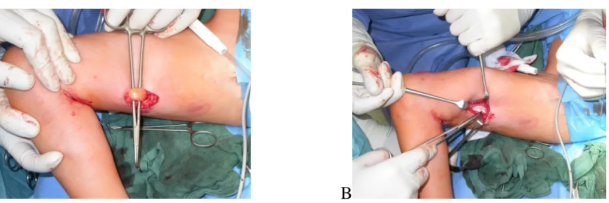
The treatment of stiff knee gait standardly is the distal rectus femoris transfer (40-44)..
(Fig. 4A) The patient lies in the supine position and a 2 cm anterior approach is carried out at the distal part of the thigh, 3 to 4 cm above the proximal patellar pole. After incision of the fascia, the distal rectus tendon is identified and separated from the underlying remaining parts of quadriceps femoris. The tendon is tagged and released as distally as possible (red dotted line in left image). The rectus tendon is then mobilized and further separated from the remaining parts of the quadriceps femoris by digital preparation.
Stronger adhesions are released by scissors or scalpel (red dotted line in right image). The fascia is dissected enough to avoid angular deviation of the transfer distally (red dotted
20
line in right image). (Fig. 4B) A medial thigh incision is made to isolate the gracilis muscle. The intramuscular tendon is exposed, tagged, and released proximally (red line) and is successively separated from its muscle belly distally while the knee is flexed. (Fig.
4C) A mini-incision (red dotted line) at the posteromedial knee is used to expose and pull out the palpable distal gracilis tendon. (Fig. 4D) Through the initial anterior approach, a long clamp is passed under the fascia and below the sartorius muscle belly to the mini- incision at the posteromedial aspect of the knee. The gracilis tendon is grasped by the tagging sutures and is transferred anteriorly, where it is sutured to the rectus femoris tendon with moderate tension while the knee is flexed 20 º (45).
4. Figure: The surgical technique of distal rectus femoris transfer (45). For detailed explanation please refer to the relevant parts of the text. Fig.4A Preparation of rectus femoris muscle, Fig.4B Isolation of gracilis muscle, Fig.4C Mini incision to pull out the distal gracilis tendon, Fig.4D Anteriorly transfer of gracilis tendon.
21
1.6.5.2. Surgical approaches in the central nervous system
Surgical approaches of the central nervous system (CNS) are most commonly done at spinal cord level, with posterior dorsal rhizotomy. It means cutting the dorsal sensory nerve rootlets. There are two types of it: the Peacock approach involves a laminectomy from L1 to L5 with separation of the rootlets as they exit the spinal canal. The consequence of the Peacock technique is a progressive lumbar lordosis (46). The other method is called Fasano technique. It involves a laminectomy from T12-L1 in which the rootlets are separated at the end of the conus. The long-term effect of the Fasano approach is the thoracolumbar kyphosis (47). There is no apparent difference between the outcomes of the two procedures. Although there may be less need of orthopedic surgery after a dorsal rhyzotomy has been performed, others have shown that there definitely is still significant skeletal deformity occurring throughout development possibly necessitating more orthopedic surgery (48). Complications may include hip dysplasia and spine deformities including kypohosis, lordosis, spondylolisthesis and spondylosis (49, 50).
1.6.5.3. Surgical and chemical approaches in the peripheral nervous system
Another way to decrease spasticity is to intervence at the level of the peripheral nerves (48). A lesion can be made either chemically or by physical transection. The chemical agents can be short-acting or long acting anesthetics, as alcohol and phenol. The most commonly sectioned nerve is the obturator to decrease adductor spasticity at the hip. But it should be done only in nonambulatory children, and only the anterior branch of the obturator nerve should be sectioned. Overall the control of spasticity and peripheral neurectomy has a minimal role in the management of spasticity in children with CP.
22
1.7. Gait
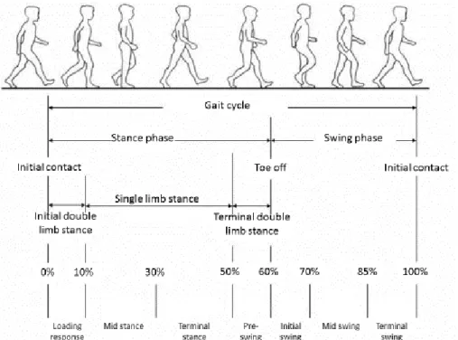
Human gait is a complex interaction between the CNS and the peripheral musculoskeletal system. The gait is a periodically repeating sequence of the lower extremities to move the body forward (51). According to velocity it can be walking, sprinting and running. The stability during walking is kept and the movement of limbs repeated in cycles. The gait cycle is the smallest unit of the gait. This is a single sequence of events between two sequential initial contacts by the same limb.
One gait cycle during walking can be divided into two phases: stance phase (60%) and swing phase (40%) (Fig.5). These phases represent specific functional patterns. There is no specific starting or ending point, with one action flowing smoothly into the next. But because of the best defined event is the floor contact, it has been selected as the start of gait cycle and it is called initial contact. The stance phase begins with initial contact of one limb. A healthy person initiates floor contact with his heel. During stance phase the foot is on the ground. Swing phase follows the stance phase. The foot is in the air for limb advancement. It begins with toe-off that means the foot is lifted from the ground.
Stance phase subdivided into three subgroups according to the sequence of floor contact by the this two feet. At the initial double stance both feet are on the floor after initial contact. The body weight is shared is equally by the two feet. When the opposite limb is lifted for swing begins the single stance. The whole body weight is resting on one limb.
It is followed by the third subdivision; the terminal double stance. It begins with floor contact by the other foot and continues until the original stance leg is lifted for swing.
Each gait cycle contains eight functional patterns (52). These are subphases of the gait cycle. The gait cycle (GC) begins with initial contact and takes about 0-2% of GC. It is followed by loading response, namely the initial double stance period that is 0-10% of GC. The single stance period involves two phases: mid stance and terminal stance. Mid stance (10-30%) begins as the other foot is lifted and continues until body weight is aligned over the forefoot, follows the terminal stance. This takes 30-50% GC until the other foot strikes the ground. Stance phase closes with pre-swing (50-60% GC), in other words the terminal double stance. The opposite limb has initial contact on the ground and ipsilateral limb has toe-off. Swing phase subdivides into three phases. Initial swing (60-
23
73% GC) begins with toe-off and ends when the swinging foot is opposite the stance foot.
It follows the mid swing (73-87% GC) till the swinging limb is forward and the tibia is vertical. The final phase of GC is the terminal swing (87-100% GC). It begins with vertical tibia and ends when the foot touches the floor.
5. Figure: Gait cycle (53)
During walking the body functionally divides itself into two units: passenger and locomotor (54). The passenger unit is composed of the head, neck, trunk and arms. It takes 70% of the body weight. The locomotor system is formed by the two lower extremities and the pelvis. Locomotor unit is responsible to move forward the passenger unit (propulsion), stance stability, shock absorption and energy conservation. Stability means a functional balance between the alignment of the body and muscle activity at each joint. There is a point, the center of gravity, that is representative of the weight of that mass. There is a passive stability when the center of gravity of the upper segment is aligned directly over the center of the supporting joint. Its security depends on the quality of the supporting surface and the nature of any external forces. During standing and walking the effect of body weight is identified by the ground reaction force vector. The body weight falls toward to the floor, it creates a force in the floor of equal magnitude but
24
opposite in direction. It can be captured by three dimensional gait analyses. There are three forces that act on the joints: falling body weight, ligamentous tension and muscular activity. The hip and knee can achieve a passive stability when the joints are hyperextended. It is balanced by ligamentous tension and body vector. At the knee there is the posterior oblique ligament. The hip is limited anteriorly by the iliofemoral ligament.
In this position the joint are locked by two opposing forces: the body weight vector on one side of the joint and ligamentosus tension on the other. At the ankle there is no similar source of passive stability. The ankle joint is not located at the middle of the foot, but posterior to the center of the foot. The heel lever is much shorter than the forefoot lever, which extends to the metatarsal heads. During walking, the area of support changes from the heel to flat foot and then the forefoot. The body’s passive stability changes to dynamic stability. The dynamic stability is modified by continual realignment of the vector to the joints. The center of gravity of the passenger unit is aligned medial to the supporting limb, and the connecting link is highly mobile hip joint. Two preparatory actions need to keep the balance: lateral shift of the body mass and local muscular stabilization of the hip joint.
It is important to establish a clear and common understanding of the terms present. They are founding in the Table 3.
3. Table: Description of biomechanical terms (37)
Term Description
Temporal spatial characteristics Changes in the body or body segments related to the GC
Gait velocity Change in distance per unit time of the whole body
during gait
Step The GC of one limb; the distance one foot moves with
each GC
Cadence The number of GC’s per unit time
Stride GC of the whole body that equals two steps
Step width The distance in the transverse plane of how far the feet
are separated during double support.
25 1.7.1. Three dimensional gait analysis
The origins of the science of gait analysis began in Europe in the 17th century and continued through the early 20th century (55). Today the three dimensional (3D) gait analysis is the gold standard of refined diagnosis and treatment for planning spastic gait disorders. With appropriate systems can be analyzed the kinematic, kinetic, dynamic electromyography and dynamic foot pressure. In modern laboratories the subject reflective markers are fixed on the subject and they walk on a calibrated walkway, while the markers transmit the data from the moving joints to the computers. The resultant joint angles can be viewed within minutes from the end of collection of the data.
However there are still some difficulties to overcome. One of them is the accurate timing of toe-off, while the other is the marker movement over the under lying skeleton on the skin during walking.
Since the inception of the 3D gait analysis it has gained an emerging role in the diagnosis, to plan operations and it is able to give an objective recording after surgery. By now 3D gait analysis´ importance can be evaluated focusing on the relevance of the 3D gait analysis to decision making by the treatment of gait disorders in cerebral palsy patients (56). Some studies showed, that the results are better, if the references of gait analysis are taken into consideration (57, 58). The 3D gait analysis is established as a crucial component of multimodal planning unit (clinic and radiologic examinations, 3D gait analysis).
1.7.1.1.Kinematics
This is a measurement of the displacement of the body segments during gait, usually defined as angular change of the distal segment relative to its proximal articulated segment, or motion relative to a global coordinate system. It is based on dividing the body into 8 segments: trunk, pelvis, two thighs, two shanks and two feet. The principia exist, that reflective or active markers are placed on specific anatomic segments and their
26
motion will be captured by many cameras (Table 4, Fig. 6 and 7). Kinematic motion can be captured by two dimensional (2D) or 3D system. The 2D system is prone to more errors, because it is analyze only the sagittal motion, the frontal and transversal plane will be lost. Because of the loss of information in 2D the 3D gait analysis system is the standard method. Each segment must be defined by a minimum of 3 markers. Each of these markers is imaged by a minimum of two cameras simultaneously. Only this way a marker can be defined in three-dimensional space. The cameras are focused on a fixed place in the room, which is assigned a room coordinate system. These are synchronized to take images at the rate of 60 frames per second. The markers are identified by separate identifiers based on the body segments. These motions can be calculated into clinically defined joint range of motion with a specific software (Plug-in, Vicon®, Oxford Metrics, UK).
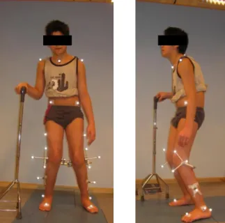
The static capture completed with a special device to measure the correct position of the knee (Fig. 7). It is positioned on the medial and lateral femur condyle. Its position gives us the horizontal, sagittal and transversal plane of the knee and the torque of the femur.
27
4. Table: Position of the reflective markers in Plug-in Trunk
C7 Processus spinosus of C7
LSHO Articulation acromioclaviculare left
RSHO Articulation acromioclaviculare right
CLAV Incisura jugularis
Pelvis
SACR
The half way between two spina iliaca posterior superior
LASI Spina iliaca anterior superior left
RASI Spina iliaca anterior superior right
Thigh
LTHI
Between Trochanter major and Condyle lateralis femoris left
RTHI
Between Trochanter major and Condyle lateralis femoris right
LKNE Lateralis condyle of femur left
RKNE Lateralis condyle of femur right
Shank
LTIB
On the lateral side of the tibia between LANK and LKNE
RTIB
On the lateral side of the tibia between RANK and RKNE
LANK Malleolus lateralis left
RANK Malleolus lateralis right
Foot
LHEE Tuber calcanei left
RHEE Tuber calcanei right
LTOE Second metatarsal head left
RTOE Second metatarsal head right
28
6. Figure: Position of the reflective markers on the body
7. Figure: Positioning of the knee in static capture
1.7.1.2.Time-distance parameters
The time-distance parameters were elevated from kinematics data and they give an advice from distance measurement in the entire GC. These parameters are the cadence (number of steps/min), speed (m/s), step length (m) and timing of a step (s). Stride is the interval between two sequential initial floor contacts by the same limb.
29 1.7.1.3.Kinetics
The measurement of forces at each joint is called kinetic evaluation (37). Net joint forces are indirectly measured as the opposite of the force required to counteract the momentum and ground reaction force (Fig.8). Momentum is measured by assigning each segment a weight and a center of weight, and by the velocity and acceleration of the mass through the use of kinematic measurement. The ground reaction force (GRF) is measured with sensitive and accurate force plates fixed to the floor, over which children walk. At the corner of the force plate there are three sensors measuring the three directions: vertical horizontal and side-ways, as well as moments about each of these axes.
8. Figure: Ground reaction force (37)
Each joint has a direction and distance from the defined center of the joint. By knowing where the joint’s center is in space and the direction of the GRF vector, the moment arm can be calculated. With the knowledge of the moment arm and the GRF vector, the moment generated by the GRF vector can be calculated. The GRF vector cross the force plate, where is the center of the pressure. During stance phase the center of pressure is changing, which can be followed on the foot (Fig.9).
30
9. Figure: The center change of pressure during stance phase
The moment from the GRF vector is then added to the moment of momentum and the total external joint moment is measured. The muscle, ligaments and bones must create an equal opposite internal force because of the system stability. The joint moment is calculated by the magnitude and direction of the GRF measured from the force plate combined with the momentum component calculated from the kinematic motions of the joint segment.
Moments are typically measured in units of Newtonian meter (Nm), which are then divided by a child’s body weight (Nm/kg). Joint powers have units of watts and divided with a child’s body weight: W/kg.
1.7.1.4.Electromyography
Electromyography (EMG) is an electrical recording of muscle activity, summation of all muscle fiber action potentials (37). Muscles are stimulated by motor neurons. This stimulation causes electrical activity in the muscle, which can be detected by an electrode.
The signal of the electrical activity is diminished due to subcutaneous fat and skin. The EMG has to be correlated to the gait cycle by synchronizing the EMG to the kinematic measurements (Tabl.5).
31
5. Table: Clinical definitions of EMG activity
Terminology Definition
Early onset Activity of the muscle begins before the normal onset
Prolonged Muscle activity continues past the normal cessation
Continuous The muscle is always on with no turn-off time (constant activity may be hard to distinguish from no activity that generates background noise).
Early off Early termination of the muscle activity
Delayed Onset of muscle activity is later than normal
Absent No muscle activity, which can be hard to separate from continuous activity.
Out of phase The muscle is active primarily during the time it would normally be silent and is silent when it should be active.
Utilizing EMG besides the 3D gait analysis we are able to address the normal muscle pattern or its deviations from the physiological standard. Frequently analyzed muscles are: rectus femoris, biceps femoris, vastus lateralis, semimembranosus, gastrocnemius lateralis, soleus, tibialis anterior.
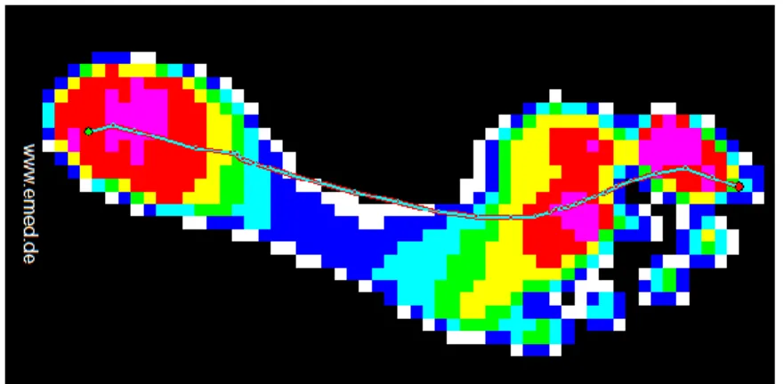
1.7.1.5.Pedobarograph
The measurement of the pressure distribution on the sole of the foot is called a pedobarograph (Fig.10). This allow us to diagnose planovalgus or equinovalgus foot deformity in CP children. The children have to walk over the measurement plate without targeting the plate. The information will be reliable and it is the best way currently to monitor childhood foot deformities. The test is quick and easy to obtain information.
32
10. Figure: Pedobarograph
1.7.1.6.GGI
The Gillette Gait Index (GGI) was defined by Schutte et al. in 2000 (59). This index is used to measure pathologic gait severity and assess therapeutic outcomes in children with CP. It incorporates 16 clinically important spatial, kinematic and temporal parameters (Tabl.6), and estimates the deviation of a patient’s gait from a normal gait pattern. It was found, that the mean index for the control group is the lowest (15.7) and the quadriplegic patients have the highest (491.0). There was no overlap between index values for the control subjects and the individuals with gait abnormalities. With this index the surgical outcomes and postoperative changes can be easy quantified.
6. Table: 16 parameters of GGI
Time of toe off Peak abduction in swing
Walking speed Mean hip rotation in stance
Cadence Knee flexion at initial contact
Mean pelvic tilt Time of peak knee flexion Range of pelvic tilt Range of knee flexion Mean pelvic rotation Peak dorsiflexion in stance Minimum hip flexion Peak dorsiflexion in swing Range of hip flexion Mean foot progression angle
33 1.7.2. Normal gait
1.7.2.1.Pelvis
Pelvis belongs to passenger unit, but its function is special: it connects the two hips with each other (60). Throughout the GC the pelvis deviates in all three planes: sagittal, coronal and transverse. The site of action is the supporting hip joint. All motions are very small (Fig.11): anterior/posterior tilt 4°, contralateral drop/rise 4°and posterior/anterior rotation 10°. But it increases with increasing speed of walking. Two muscle groups were identified, which are responsible to control the pelvic movement: abductors and extensors of the hip. From the abductors two muscles are specifically involved: gluteus maximus and medius.
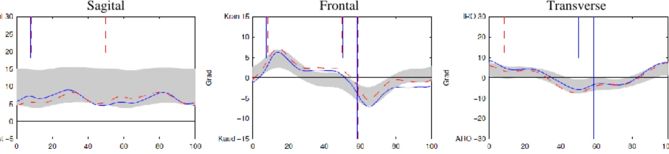
Sagital Frontal Transverse
11. Figure: Pelvismotion. Grey area represents the median and the standard error of the mean of control group. The blue continuous and red dashed line is the left and the right leg of a representative subject respectively.
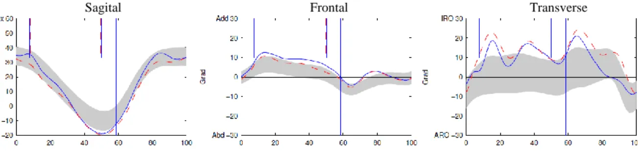
1.7.2.2.Hip
Hip is the junction between the passenger and locomotor units (61). During a normal stride the hip moves through only two arcs of motion: extension and flexion (Fig.12). Its average takes out 40° (62). The peaks of hip movements on sagittal plane are 10° for extension and 30°for flexion. There is a minimal movement on the coronal plane as the unloaded side of the pelvis follows the swinging limb. At initial contact, the hip adducted about 10°and it decrease to 5°during loading response. At initial swing a relative hip abduction occurs with 5° (63). The total arc of transverse hip motion averages 8°. If it is added to pelvic rotation, the total thigh rotation is 15°. In stance the primary muscles which control the hip, are the extensors and abductors (biceps femoris, semimembranosus, semitendinosus, adductor magnus, gluteus maximus, gluteus medius,
34
gluteus maximus and tensor fascia lata), while during swing it is the flexors (adductor longus, rectus femoris, gracilis, Sartorius and iliacus) .
Sagital Frontal Transverse
12. Figure: Hipmotion. Grey area represents the median and the standard error of the mean of control group. The blue continuous and red dashed line is the left and the right leg of a representative subject respectively.
1.7.2.3.Knee
The flexibility and stability of the knee are major factors in the normal pattern of walking (64). In stance the knee gives the basic stability of the limb and during swing it is responsible for the leg’s freedom to advance. There are some two joint muscles that also control the moving of ankle and hip.
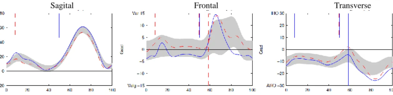
The normal knee motion has a big range in sagittal plane (0-70°) during normal walking (Fig.13). The differences between studies of knee motion are related to variations of walking speed, subject individuality and the landmarks. At initial contact the knee is flexed about 5°. At the first half of the stance phase the knee is flexed to 20-25°. This is the time when the flexed knee is under maximum weight-bearing load. At the second part of the mid stance, the knee extends. Shortly before the knee slowly begins to flex again in terminal stance, the knee procure the maximal extension of it with about 3°. During terminal stance the knee begins its second flexion. At mid-swing the knee is flexed to the maximum (60-70°). In terminal swing the knee begins to extend rapidly. It continues till full extension (5°) is gained (65). In the transverse plane the knee shows about 9° rotation during the GC. It begins with external rotation at the end of stance, and from the toe-off it continuous with internal rotation during swing and loading response. In the coronal plane there are abduction and adduction. During stance phase the motion is abduction and
35
during swing the knee returns to adduction with 8°(66). The thirteen muscles contributing to knee control contract at selected intervals within the GC. Only five muscle are unique to the knee. The extensors are the three vasti heads, and two flexors are the popliteus and short head of the biceps femoris. All the other muscles control either hip or ankle motion.
One of the two joint muscles is rectus femoris which contributes to knee extension. The gluteus maximus provides a knee extensor force through its iliotibial band insertion. The three hamstring muscles (semimembranosus, semitendinosus and biceps femoris long head) are two-joint muscles (10). These are known as knee flexors. The gastrocnemius participates in knee flexion, while principally acting on the ankle. The gracilis and sartorius also contribute to swing phase knee flexion.
Sagital Frontal Transverse
13. Figure: Kneemotion. Grey area represents the median and the standard error of the mean of control group. The blue continuous and red dashed line is the left and the right leg of a representative subject respectively.
1.7.2.4.Ankle
Ankle is the junction between tibia and foot. There are two single axis joints to provide three-dimensional mobility: subtalar and tibiotalar articulations. During a GC four arcs of ankle motion occur: two plantar flexions (PF) and then two dorsiflexions (DF) (Fig.14).
In stance PF, DF and PF follow each other. During swing the ankle only dorsiflexes (65, 66). The whole range of ankle motion extends about 30°. Initial contact by the heel occurs with the ankle at neutral or minimal plantar flexed (0-5°). The maximal dorsiflexion takes place in terminal swing with about 10°. The maximum of plantar flexion is at initial swing (20°).
The ankle controlling muscles function either dorsiflexiors (tibialis anterior, extensor halluces longus and extensor digitorum longus) or plantar flexors (soleus, gastrocnemius,
36
tibialis posterioir, flexor digitorum longus, flexor halluces longus, peroneus longus and peroneus brevis.
Sagital Transverse
14. Figure: Anklemotion. Grey area represents the median and the standard error of the mean of control group. The blue continuous and red dashed line is the left and the right leg of a representative subject respectively.
1.7.3. Spastic diplegic gait
The cerebral palsy patients have been found to exhibit several common gait abnormalities.
With respect to the knee, there are four common pathologic patterns seen in ambulatory patients with spastic diplegic-type cerebral palsy. Despite three dimensions of knee motion, the common gait abnormalities in cerebral palsy occur in the sagittal plane.
Coronal plane abnormalities appear less frequently and are usually attributable to abnormalities at other joints, such as the extreme femoral anteversion or valgus instability of subtalar joint. Following knee abnormalities are just secondary problems. Transverse plane abnormalities at the knee are also secondary phenomena, most often attributable to femoral or tibial torsional deformities (67). The gait abnormalities in cerebral palsy patients lead to contractures across joints and muscle spasticity. In 1993, Sutherland and Davids classified the common gait abnormalities of the knee in cerebral palsy into four types: jump, crouch, recurvatum, and stiff (67).
1.7.3.1.Crouch knee
Increased knee flexion during the stance phase of gait called flexed knee (crouch knee) gait, with variable alignment in swing phase (Fig.15) (68). Hamstring spasticity with resultant contracture has been identified as one of the main factors leading to a flexed
37
knee gait (69-74). Later investigations have shown that increased external tibial torsion, instability of the foot, and quadriceps weakness can cause or aggravate flexed knee gait (75-78). Knee kinematics on sagittal plane shows an increased knee flexion of at least 30°during stance phase. Decreased knee extension in terminal swing will be mostly presented. The peak knee flexion in swing (pKfSw) is over 50°, causing shortened stride length, resulting in a decrease in velocity. On the EMG in stance phase prolongation of quadriceps and hamstring activity are usually presented (67). During stance phase the increased knee flexion results a continuous flexion moment about the knee, leading to excessive demands on the quadriceps (79).
1.7.3.2.Stiff knee
Stiff-knee gait is a common gait abnormality in patients with spastic diplegic cerebral palsy (68). Decreased knee flexion during swing phase leads to foot clearance problems, reduces gait velocity, and reduces step length. According to Rodda et al. (80) stiff-knee gait shows a decreased knee excursion throughout the whole gait cycle of <30°, whereas
15. Figure: Crouch knee
38
Sutherland et al. described it as delayed and decreased pKFSw phase with diminished total knee motion (81).
The spasticity of the rectus femoris muscle is the primary cause of stiff-knee gait (42, 72, 82, 83). Rectus femoris is pathologically active during swing phase, counteracting the knee flexion necessary for foot clearance and step length (42, 67, 72, 82, 83).
1.7.3.3.Jump knee
The name is telling, because the subject is jumping up and down. The patients are usually younger than those with crouch knee. There is increased knee and hip flexion in early stance phase (at least 30°), and extend variable in mid and terminal stance (10-20°), but never reach normal knee extension. In late swing there is a decreased knee extension (67).
The ankle is in equinus, especially in late stance by increased tone in triceps surae. The movement of pelvis during GC is either normal or tilted anteriorly (80). Hamstrings show an increased tone, the hip adductors and in flexors mild contracture is present. Quadriceps weakness can be occasionally seen. During stance phase, hamstrings present overactivity.
In case of younger children whit minimal or no contracture present, selective dorsal rhizotomy may be considered (84). Multilevel botulinum toxin A injection to the psoas, hamstrings and calf can be suggested in children with jump knee (85). In the third level, when there are contractures, simultaneous bilateral single event multilevel surgery on the muscles can be considered: psoas, hamstrings and calf (85). The GRF is anterior to the knee, leading to overactivity in plantar flexors and knee extensors. A posterior leaf spring or hinged ankle foot orthosis restricts excessive plantar flexion and aids movement into dorsiflexion, thereby realigning the GRF and normalizing the plantar flexion knee- extension couple (86).
39 1.7.3.4.Recurvatum knee
The etiology of recurvatum knee can be triceps surae contracture or overactivity in mid or terminal stance, leading equinus in ankle and excessive lengthening or transfer of hamstrings (9). It is characterized by increased knee extension in mid or terminal stance with variable knee motion in swing phase. In physical examination a hyperextension in knee ROM can be present. This hyperextension can lead to varus-valgus deformity, especially in older children. The equinus contracture often presents significantly increased tone and clonus. The knee motion shows a hyperextension in mid and terminal stance phase, the pKFSw is delayed and diminished. The EMG demonstrates an overactivity of the triceps surae and elongated quadriceps activity in stance phase. The hyperextension leads decreased velocity by limiting stride length. In the kinetics, it exhibits an excessive flexor moments throughout stance phase.
40
2. Purpose
2.1. Development of knee function after hamstring lengthening as a part of multilevelsurgery in children with spastic diplegia
Lengthening of the hamstrings is widely considered to be the standard surgical procedure for the correction of increased knee flexion (26, 38, 87, 88). The hamstrings can be lengthened with use of an open or a percutaneous technique (88-91). Satisfactory short- term results with improved knee extension during stance phase have been shown in different studies (41, 87, 88, 91). However, outcomes have been inconsistent; many patients have had improvement, whereas other patients have had only little benefit or even worsening (41, 88-92). After short term follow-up, some authors have reported increased pelvic tilt and a high prevalence of genu recurvatum as a consequence of overcorrection (38, 88, 91). It is an interesting question, whether is there any different in the appearance of the anterior pelvic tilt between medial and combined medial and lateral hamstring lengthening in short and long-term, however it was already analyzed short-term by Kay et al. (88). Evidence for the effectiveness of hamstring lengthening to correct flexed knee gait in spastic diplegia is scant because of small and inhomogeneous case series, different surgical techniques, and short follow-up. As a result, hamstring lengthening currently is viewed as controversial. A major problem is that, to our knowledge, there have been no long-term studies investigating the effects of hamstring lengthening in skeletally mature patients who were managed in childhood. Therefore, the long-term results in adolescents and adults who had had hamstring lengthening as a part of multilevel surgery in childhood were investigated in one of our study. Our hypothesis was that correction of flexed knee gait seen at short-term follow-up is not maintained in the long term.
41
2.2. Long-term results after distal rectus femoris transfer as a part of
multilevel surgery for the correction of stiff knee gait in spastic diplegic cerebral palsy
The distal rectus femoris transfer (DRFT) is the standard surgical procedure for the treatment of stiff-knee gait. The tendon transfer can be occurred to medial hamstrings or to lateral hamstring or iliotibial tract (72, 83). It aims to improve knee flexion in swing and pKFSw for foot clearance. The criteria of DRFT in the literature are inconsistent (67, 72, 93-98). Some studies have noted good initial results, with an improvement in pKFSw and knee flexion in swing following DRFT (93-95). Nevertheless, some authors have suggested that DRFT does not generate a knee flexion moment, although the capacity of knee extension is diminished (96-98). Other authors have described no significant increase of pKFSw but an increase of total knee motion (39, 43, 99). Summarized, the results after DRFT are inconsistent. In addition, it is debatable whether patients with severe flexed knee gait who show highly increased knee flexion throughout stance phase and a normal or even increased pKFSw phase should receive a prophylactic DRFT to preserve pKFSw after correction of flexed knee contractures by concomitant procedures such as hamstring lengthening or femoral extension osteotomy. There are only a few studies, which have longer-term outcomes after DRFT. Saw et al. showed a significant improvement in pKFSw phase 1 year after surgery, but this improvement in knee flexion in swing decreased and was associated with a loss of knee extension during stance phase 4.6 years postoperatively (93). Moreau et al. also reported significant improvements three years after surgery (94). However, these studies were based on relatively small numbers, and both included patients with different types of cerebral palsy. Furthermore, recruitment was done from different treatment centers that used different surgical techniques of tendon transfer, and the time to the last follow-up was inconsistent, ranging from 0.9 to 6.7 years after surgery. Long-term studies with adequate patient numbers, homogeneous patient groups, and a follow-up interval of more than five years, when growth is expected to be finalized, are missing. The purpose of our study was the evaluation of long-term results (mean, 9 years) in adolescents and adults with diplegic cerebral palsy who were treated with DRFT as a part of single-event multilevel surgery in their childhood. Since the indication for DRFT differed between patients with decreased pKFSw and those with normal or increased pKFSw, in which the DRFT was