Characterizing cerebral small vessel disease with a focus on CADASIL as a genetic model
-an MRI based approach
PhD thesis Bence Barna Gunda Semmelweis University
János Szentágothai School of Neurosciences
Supervisor: Dániel Bereczki MD, DSc Consultant: Hugues Chabriat MD, PhD Reviewers: László Oláh MD, PhD
Péter Barsi MD, PhD
Head of Examination Committee: László Tringer MD, DSc
Members of Examination Committee:
Zoltán Benyó MD, DSc Attila Valikovics MD, PhD
Budapest, 2012
2
Introduction
Cerebral small vessel disease (cSVD) is a spectrum of clinical and imaging abnormalities linked to the local pathology (arteriolosclerosis and/or microatheromatosis) of small (50-500 μm) penetrating arteries of the brain irrigating subcortical structures. Its sporadic form related mostly to hypertension and diabetes manifesting in lacunar stroke, deep intracerebral hemorrhage and progressive vascular dementia affects a considerable proportion of the general population above 65 years of age and accounting for approximately 20% of all ischemic strokes. Accumulating data suggest that cSVD is the most prevalent neurological disorder in the ageing society of the developed world. Hereditary, monogenic variants affecting young individuals often as a part of a systemic disorder are increasingly identified CADASIL being to most prevalent and well-studied example.
The affection of cerebral microvessels irrigating subcortical structures can lead to their gradual stenosis, sudden occlusion, leakage or rupture resulting in chronic hypoperfusion/ischemia with progressive disintegration of the
3
white matter; acute ischemia with lacunar infarcts; and cerebral micro- or macrobleeds. In advanced stages of the disease cerebral atrophy invariably occurs as the consequence of diffuse tissue damage and secondary degeneration after lacunar infarcts.
The gradual ischemic tissue damage clinically manifests in progressive cognitive impairment mainly affecting executive function in the earliest stages and later leading to dementia of subcortical type; and disability characterized by gait disturbances and other motor impairments, pseudobulbar palsies, urinary incontinence etc. Acute focal ischemia presents –if it involves main sensorimotor pathways- with the so-called lacunar syndromes. Cerebral microbleeds are usually asymptomatic and their clinical significance is yet to be determined.
Magnetic resonance imaging (MRI) is the most appropriate tool to assess cSVD. Since the cerebral microvasculature cannot be currently visualized in vivo, the consequent parenchymal lesions (lacunar infarcts -LI, white matter hyperintensities -WMH, microbleeds -cMB and atrophy) have been adopted as markers of cSVD. New MRI sequences and postprocessing techniques have greatly helped to explore and better understand cSVD. Diffusion weighted imaging (DWI) proved to be particularly useful in visualizing hyperacute LIs
4
thus refining clinico-anatomical correlations; and guiding acute phase stroke therapy and etiologic diagnosis based on DWI patterns in routine clinical practice. Diffusion tensor imaging (DTI) is capable of detecting ultrastructural changes even in otherwise normal appearing WM, and quantifying the global burden of tissue damage in cSVD (whole brain DTI histograms).
Brain atrophy –a phenomenon previously considered to be related to cortical disease- is now recognised as a marker of cSVD as assessed with volumetric measures.
CADASIL (Cerebral Autosomal Dominant Arteriopathy with Subcortical Infarcts and Leukoencephalopathy) is a hereditary small vessel disease of the brain caused by mutations of the NOTCH 3 gene encoding a transmembrane receptor of vascular smooth muscle cells. It has recently gained great interest in vascular neurology as the most common heritable cause of stroke and vascular dementia in adults. This autosomal dominant cSVD –unlike the sporadic, hypertensive form- appears already in adult midlife in the absence of vascular risk factors with ischemic episodes and progressive dementia. Its first clinical manifestation can be migraine with aura, and is often associated with psychiatric disturbances. The MRI changes may precede symptoms by more than a decade. Apart from the well-
5
known radiologic manifestations of cSVD in general, CADASIL has a unique feature: WMH symmetrically involving the temporal poles and the external capsule. This characteristic pattern can be considered specific to the disease and is an important point in differential diagnosis. The special importance of this disease lies in the fact that CADASIL provides a pure genetic model for cSVD without the confounding factors of comorbidities and advanced age. Thus insights into CADASIL may help us better understand the more common sporadic forms as well.
We performed two studies in the world’s largest cohort of CADASIL patients from the Lariboisière Hospital in Paris where the disease was identified, and the term CADASIL coined. The first one (Study 1) aimed to evaluate gender related differences in the clinical and MRI characteristics of the disease.
The second study (Study 2) investigated the possible role of a DWI derived simple quantitative MRI method in disease monitoring as compared to the already established DTI based reference method.
6
Purpose
Study 1: Effects of gender on the phenotype of CADASIL
In the general population, migraine, cerebrovascular diseases and vascular dementia differ in many aspects between men and women. The prevalence of migraine appears to be 3-4 times higher in women than in men, whereas the age-adjusted prevalence and incidence of stroke are respectively 30 and 40%
higher in men than in women. A similar trend for male sex is observed for the prevalence and incidence of vascular dementia.
Accumulating evidence suggests that these differences may be related to female hormones that can both modify the excitability of the cortex and exert major neuroprotective effects. CADASIL is considered as a unique model to investigate migraine with aura, stroke and dementia related to ischemic small vessel disease. This study aimed to evaluate the effect of gender on the main clinical and neuroimaging characteristics of CADASIL.
7
Study 2: Whole brain ADC histogram in CADASIL
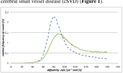
Diffusion tensor imaging (DTI) derived whole brain histogram metrics –as global measures of ultrastructural tissue integrity- have been widely reported to correlate with clinical parameters in various diffuse cerebral pathologies in research settings. The aim of our study was to evaluate whether histograms from simple diffusion weighted imaging (DWI) used in routine clinical practice without any postprocessing can be used similarly to DTI histograms in CADASIL –a model of cerebral small vessel disease (cSVD) (Figure 1).
Figure 1. ADC histogram of a severely affected CADASIL patient (green line) compared to a normal control (dashed blue line), diffusivity thresholded at 180x10 -5 mm2/s.
8
Methods
Study 1
Cross sectional data from 313 CADASIL patients including anamnestic data, various clinical and cognitive scores (Rankin, Barthel, NIHSS, MMSE, Mattis DRS) and MRI parameters (normalized WMH and LI volume, brain parenchymal fraction -BPF as indicator of atrophy and number of microbleeds) were compared (Student’s t, Wilcoxon, ANCOVA) between men and women, and under and over the median age of the population corresponding to the usual age of menopause (51 years).
Study 2
348 CADASIL patients were evaluated by DWI-DTI MRI (different 1,5 T scanners with updates) and clinical examination (Rankin, Barthel, NIHSS, MMSE, Mattis DRS).
We first compared DWI-derived apparent diffusion coefficient (ADC) histogram parameters (mean, peak, height) to those of
9
DTI-derived mean diffusivity (MD). Then we evaluated the effect of different image correction methods (CSF suppression, manual and automatic artefact removal) on ADC histogram results (both with linear regression analysis and Bland-Altmann plots). Finally a mixed effects model was used to evaluate the effect of difference /updates in scanners and clinical scores on ADC histogram parameters.
10
Results
Study 1
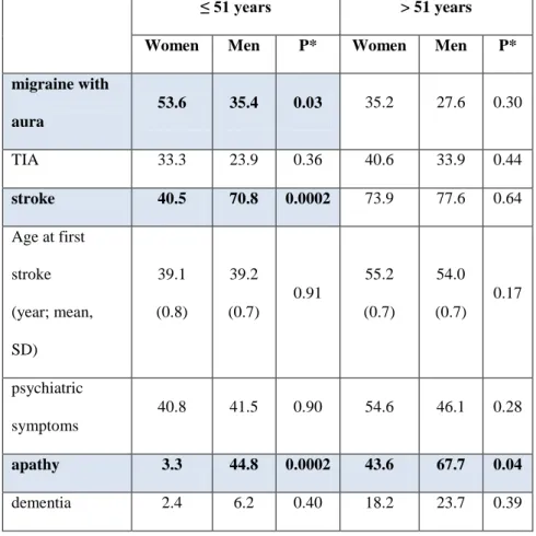
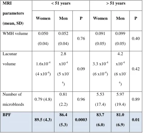
Under 51 years, migraine with aura was 50% more prevalent in women and stroke 75% more prevalent in men (Table 1). After the 5th decade, men had higher NIHSS and Rankin scores than women and more severe executive dysfunction, although global cognitive scores were similar (Table 2). Age at first stroke, the number of stroke events and the prevalence of dementia and psychiatric symptoms did not differ between men and women. Brain volume was lower in men with a trend for a larger volume of lacunar infarcts (Table 3).
11
Table 1:Main clinical manifestations according to gender, prevalence (%) in subgroups of patients according to age more or less than 51 years
≤ 51 years > 51 years
Women Men P* Women Men P*
migraine with aura
53.6 35.4 0.03 35.2 27.6 0.30
TIA 33.3 23.9 0.36 40.6 33.9 0.44
stroke 40.5 70.8 0.0002 73.9 77.6 0.64
Age at first stroke (year; mean, SD)
39.1 (0.8)
39.2 (0.7)
0.91
55.2 (0.7)
54.0 (0.7)
0.17
psychiatric symptoms
40.8 41.5 0.90 54.6 46.1 0.28
apathy 3.3 44.8 0.0002 43.6 67.7 0.04
dementia 2.4 6.2 0.40 18.2 23.7 0.39
12
Table 2: Main clinical scores according to gender in subgroups of
patients according to age more or less than 51 years Scores
(mean, SD)
< 51 years > 51 years
Women Men P* Women Men P*
Rankin 0.38
(0.17)
0.41 (0.18)
0.85
1.35 (0.30)
2.01 (0.29)
0.009
NIHSS 0.77
(0.41)
1.07 (0.45)
0.39 1.1 (0.5)
2.4 (0.5)
0.004
MMSE 27.9
(0.5)
28.0 (0.6)
0.91
24.8 (1.0)
24.2 (1.0)
0.51
Mattis DRS 138.2 (2.2)
137.2 (2.4)
0.57
129.9 (4.1)
125.6 (3.9)
0.21
Mattis initiation
35.3 (0.8)
34.5 (0.9)
0.23
32.5 (1.5)
29.9 (1.5)
0.05
13
Table 3: MRI parameters according to gender in subgroups of
patients according to age more or less than 51 years MRI
parameters (mean, SD)
< 51 years > 51 years
Women Men P Women Men P
WMH volume 0.050 (0.04)
0.052 (0.04)
0.76
0.091 (0.05)
0.099 (0.05)
0.40
Lacunar
volume 1.6x10⁻⁴ (4 x10⁻⁴)
2.8 x10⁻⁴ (5 x10-
4)
0.09
3.3 x10⁻⁴ (6 x10⁻⁴)
4.2 x10⁻⁴ (6 x10-
4)
0.42
Number of microbleeds
0.79 (4.8)
0.81 (2.2)
0.96
5.53 (17.4)
5.97 (19.4)
0.89
BPF
89.5 (4.3)
86.4 (5.3)
0.0003
83.7 (6.0)
81.0 (6.9)
0.01
14 Study 2
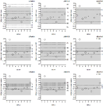
We found an excellent correlation between MD and ADC histogram parameters (Table 4). Image correction had a negligible effect on ADC measures (Table 5). In contrast, the magnitude of the scanner effect was high and larger than that of clinical scores, sex and age (Table 6).
Table 4: Correlation coefficients (R2) for ADC/MD parameters mean peak height with CSF suppression 0,964 0,795 0,982 without CSF
suppression
0,965 0,722 0,974
Table 5: Correlation coefficients (R2) for corrected/non-corrected ADC parameters
mean peak height with/without CSF
suppression
0,742 1,0 0,958
with/without artefact removal
0,998 0,967 0,997
with/without top- bottom slices
0,993 0,943 0,989
15
Table 6: Magnitude of MRI scanner effect in the adjusted analysis of DWI parameters and clinical scores. Circles surface is proportional to the number of patients evaluated by each scanner, and their vertical position reflects the random MRI scanner effect. The gray shaded area represents the expected effect of 95% of scanners. For purpose of comparison, the (fixed) effects of clinical score differences given in the right margin are displayed by dashed lines.
16
Conclusions
Study 1
In CADASIL, migraine with aura is more frequent in women and stroke in men before the age of menopause. This difference seems to vanish after this age limit but may result in a higher degree of cognitive impairment and cerebral atrophy in men at the late stage of the disease. The presumable role of ovarian hormones in these gender-related differences remains to be explored.
Study 2
Simple non-corrected whole brain ADC histograms could replace DTI histograms in assessing tissue damage in cSVD in the routine clinical practice. However, given the important scanner effect on histogram parameters, results from different scanners/updates should be normalised to be comparable across centres and in individual patients.
17
Perspectives
Quantitative MRI techniques such as whole brain diffusion histograms and volumetry are more sensitive to the full spectrum of cSVD expressions than other imaging parameters and clinical scores (also witnessed by our first study). Moreover our second study showed that simple, non-corrected DWI derived ADC histograms seem suitable for the routine clinical practice providing a promising tool for the quantified monitoring of the disease and surrogate markers for future therapeutic trials in cSVD. Thus the use of quantitative MRI methods is expected to increasingly enter clinical practice in the future.
18
Publications
Gunda B, Várallyay Gy, Rudas G, Bereczki D, Challenges in diagnosing cerebral lacunar infarcts, Curr. Med. Imaging Rev., 2009, 5, 75-84.
Gunda B, Chabriat H, Bereczki D. CADASIL and other hereditary small vessel diseases of the brain-increasingly diagnosed conditions underlying familial ischemic stroke and dementia. Ideggyogy Sz. 2011 Mar 30; 64 (3-4):88-100.
Gunda B, Hervé D, Godin O, Bruno M, Reyes S, Alili N, Opherk C, Jouvent E, Düring M, Bousser MG, Dichgans M, Chabriat H. Effects of Gender on the Phenotype of CADASIL.
Stroke, 2012 Jan; 43(1):137-41.
Gunda B, Várallyay Gy, Bereczki D. Multimodal MRI of cerebral small vessel disease. In: Peter Bright (ed.), Neuroimaging –Clinical Applications, ISBN 978-953-51-0200- 7, InTech, Mar 2012: 277-300.
19
Acknowledgements
First of all I would like to thank my supervisor professor Dániel Bereczki for his wise and inspiring guidance all through my studies. I am grateful both for his professional and human support in my scientific and clinical work. It is him to whom I owe the most in the making of this thesis.
I am also thankful to my colleagues at the Department of Neurology at Semmelweis University for helping my work wherever possible. I would also like to render thanks to the Head of the MR Research Centre, Gábor Rudas, and all the radiologist colleagues who introduced me into neuroimaging. My special thanks go to György Várallyay –a dedicated, patient and profoundly humane teacher.
This thesis could not have been written without my fortunate stay at the centre of rare cerebrovascular diseases (CERVCO) at Hôpital Lariboisière in Paris-an outstanding workshop of clinical neurology organised by Marie-Germaine Bousser and currently led by Hugues Chabriat. I am grateful to him and his team who were exceedingly helpful, friendly and open towards me. Their work as a clinical and scientific team
20
was exemplary to me. The scientific thinking, critical attitude and methodology that I learned from Professor Chabriat will accompany me in my whole professional career.
My work was financially supported by grants from the European Federation of Neurological Societies (EFNS) and Institut Servier in Paris and the Hungarian Government at my home institution (TÁMOP).
Finally and most of all I am thankful to my Family. Primarily to my Wife, Dóra Gunda-Szabó, who encouraged, accompanied, tolerated me and gave up her ways for mine. To my Parents who provided everything I needed to pursue my ambitions. Finally to my Daughter, Julka, who just happened to appear along the way and is a source of unprecedented happiness.