Experimental pericardial tamponade–translation of a clinical problem to its large animal model
Objectives: Pericardial tamponade is a life-threatening medical emergency, when the hemodynamic consequences of low cardiac output severely disturb the perfusion of the peripheral tissues. Our aim was to design a reliable large animal model to reproduce the clinical scenario with the relevant pathophysiological consequences of pericardial tamponade -induced cardiogenic shock.
Material and Methods: Anesthetized Vietnamese mini pigs were used (n=12). Following laparotomy, a cannula was fixed into the pericardium through the diaphragm without thoracotomy. A sham-operated group (n=6) ser- ved as control, while in the second group (n=6) pericardial tamponade was induced by intra-pericardial injection of heparinized own blood. Throughout the 60-min pericardial tamponade and the 180-min reperfusion, macro hemodynamics, renal circulation and the mesenteric macro- and micro-circulatory parameters were monitored.
Myeloperoxidase activity was measured to detect neutrophil leukocyte accumulation and in vivo histology was performed by confocal laser scanning endomicroscopy to observe the structural changes of the intestinal mucosa.
Results: PT increased the central venous pressure, heart rate, and decreased mean arterial pressure. The mesenteric artery flow (from 355.5±112.4 vs 182.0±59.1 mL/min) and renal arterial flow (from 159.63±50.7 vs 35.902±27.9 mL//min) and the micro-circulation of the ileum was reduced. The myeloperoxidase activity was elevated (from 3.66±1.6 to 7.01±1.44 mU/mg protein) and manifest injury of the ileal mucosa was present.
Conclusion: This experimental model suitably mimics the hemodynamics and the pathology of clinical pericardial tamponade situations, and on this basis, it provides an opportunity to study the adverse macro- and micro-circula- tory effects and biochemical consequences of human cardiogenic shock.
Keywords: Animal model, cardiogenic shock, mesenteric ischemia, mini pig, pericardial tamponade INTRODUCTION
This article will explore the line between patients and experimental subjects, in an attempt to provide a proper view on the role of classical surgical research in contemporary cardiac surgery. According to the proverbial quote of Francis D. Moore, a “surgical investigator must be a bridge tender, channeling knowl- edge from biological science to the patient’s bedside and back again” (1). This traditional approach empha- sizes the great potential of biomedical sciences to provide help for unsolved problems in surgery. An equally important aspect of this type of research is that it develops new data, or more comprehensible knowledge on a specific area of clinical practice. In this sense, the science of surgery, like any other area of biomedical sciences, has four main methodological steps: observation of a clinical problem, formula- tion of a hypothesis, and performance of test procedures to reach a conclusion, supporting or refuting the initial hypothesis. In this analytical circle, models are indispensable tools to check the validity of hypotheses and to generate clinically useful data. In pragmatic terms differences are always present between the clinical reality and the models and therefore these dissimilarities should be reduced by choosing the experimental setup as accurate as possible. Understandably, the demand is huge for de- veloping high fidelity models to decrease existing limitations and to obtain better applicable results for human medical science and healthcare (2).
A specific problem addressed by our study is cardiogenic shock due to cardiac tamponade. Pericar- dial tamponade is one of the most dreadful complication of cardiac surgery and invasive cardiology.
Tamponade can develop after myocardial infarction, it can be the corollary of intra- or post-operative complications, or the consequence of non-cardiac surgical procedures (NCSP) as well (3). During tam- ponade, fluid will accumulate within the pericardial space, which can be transudate or pus, however the most common cause is bleeding (4). The increasing pressure in the closed pericardial sac blocks the venous inflow and decreases stroke volume. The subsequently reduced cardiac output (CO) results in tissue hypoxia and multi-organ damage. The long-term systemic consequences of pericardial tampon- ade are pronounced and serious even after the primary cause has been diagnosed and the basic prob- lem was solved, i.e., the pericardial pressure was decreased via pericardiocentesis, re-operation or other intervention (5). The main reason for late complications is ischemia-reperfusion injury of distant organs, including the lungs, kidneys, and splanchnic tissues. This circulatory condition results in an antigen- Gábor Bari2 , Szilárd Szűcs1 , Dániel Érces1 , Mihály Boros1 , Gabriella Varga1
205
ABSTRACT
ORCID IDs of the authors:
G.B. 0000-0003-4167-5736;
S.S. 0000-0001-8478-6386;
D.E. 0000-0002-4283-2441;
M.B. 0000-0003-1410-1999;
G.V. 0000-0003-1888-8629.
1Institute of Surgical Research, Szeged University, Szeged, Hungary
2Department of Cardiac Surgery, Szeged University, Szeged, Hungary
This study was presented at the 17th Congress of the European Shock Society, 13-15/09/2017, Paris, France.
Corresponding Author Gabriella Varga e-mail:
varga.gabriella.1@med.u-szeged.hu Received: 06.04.2018 Accepted:18.06.2018
©Copyright 2018 by Turkish Surgical Association Available online at www.turkjsurg.com
Cite this paper as:
Bari G, Szűcs S, Érces D, Boros M, Varga G. Experimental pericardial tamponade–
translation of a clinical problem to its large animal model. Turk J Surg 2018 34 (3): 205-211.
independent inflammatory reaction with cytokine storm and increased endothelial permeability, which finally leads to an overwhelmingly severe systemic immune response (6).
According to previous experiences, cardiogenic shock can be modelled if an animal is anaesthetized, mechanically ventilat- ed, and the pericardial sac is accessible through thoracotomy (7). The disadvantage of the model is thoracotomy itself with significantly impaired lung function, extended wound surface with a high risk of bleeding, lung injury, and tissue damage.
Based on the above, our aim was to develop an experimental procedure where thoracotomy can be avoided and thus to de- sign a new model of iatrogenic cardiogenic shock, where the situation is realistic, similar to the clinical appearance.
MATERIAL AND METHODS Animals
The experiments were performed in accordance with National Institutes of Health guidelines on the handling and care of experimental animals and European Union Directive 2010/63 for the protection of animals used for scientific purposes. The study was approved by the Animal Welfare Committee of the University of Szeged (approval number: V/148/2013).
Surgical interventions
Anesthesia was induced by a mixture of ketamine (20 mg kg−1 im.) and xylazine (2 mg kg−1 im.) and maintained by a continu- ous infusion of propofol (6 mg kg−1h−1 iv.). The animals were placed on heating pad in a supine position; the body tempera- ture was kept between 36-37°C. After endotracheal intuba- tion, mechanical ventilation was started with a tidal volume of 10 mlkg−1. The left jugular vein was cannulated for fluid and drug administration and the left femoral artery for the mea- surement of mean arterial pressure (MAP), heart rate (HR) and cardiac output (CO) by transpulmonary thermodilution (PICCO Catheters; PULSION Medical Systems, Feldkirchen, Germany).
MAP, HR, CO and SMA flow (SMAF) data were recorded, pres-
Hungary) and superior mesenteric artery (SMA), renal artery (RA) flow signals (T206 Animal Research Flowmeter; Transonic Systems Inc., Ithaca, NY, USA) were measured continuously by surgically placing a urinary catheter in the bladder via the femoral incision. and registered with a computerized data-ac- quisition system (SPEL Advanced HAEMOSYS 1.17; Experime- tria Ltd, Budapest, Hungary). Ringer’s lactate was given at the rate of 10 mlkg−1h−1. After a median laparotomy, the SMA was dissected free and a flow probe was placed around the SMA (Transonic Systems Inc., Ithaca, NY, USA). The RA was dissected free and a flow probe was placed around it (Transonic Systems Inc., Ithaca, NY, USA). The wound of the abdominal wall was temporarily closed thereafter with clips. In addition, intra-vital examination of the intestinal microcirculation was carried out by an orthogonal polarization spectral imaging system (OPS;
Cytoscan A/R, Cytometrics, Philadelphia, PA, USA), and the extent of damage of the gastric mucosa was evaluated by in vivo histology (Five1, Optiscan Pty. Ltd., Melbourne, Victoria, Australia). The diaphragm was accessed through a median laparotomy and a 3-cm incision was made at the sternal part, avoiding the muscular region of the diaphragm. The pericar- dium was opened and a cannula was fixed into the pericar- dial cavity with a pledgeted purse string suture (Figure 1). The tamponade was induced for 60 min by intra-pericardial ad- ministration of heparinized own blood (100±50 mL), and the MAP was kept between 40–45 mmHg. During the observation period and between microcirculatory imaging the abdominal wall was closed by surgical clips.
Experimental Protocol
The animals were randomly allocated into two experimental groups. Group 1 (n=6) served as sham-operated control, with the same surgical interventions, time-frame, and sampling as in group 2 (n=6) but without the induction of a tamponade. In both groups, the diaphragm was accessed through a median mini-laparotomy. A 3-cm incision was performed at the sternal part, avoiding the muscular region of the diaphragm. The peri- cardium was opened and a cannula was fixed into the peri- cardial cavity with a pledgeted purse string suture. In Group 2, after the end of 60-min tamponade the blood was released from the pericardial sac and the animals were monitored for 180 min. Blood gas and hemodynamic parameters were mea- sured in every 30 min. In vivo histological examination on the ileal mucosa and determination of neutrophil granulocytes was performed at baseline, 30 min after the relief of tampon- ade (90 min) and at the end of the experiments (240 min). My- eloperoxidase enzyme activity was measured at the −5; 30; 60;
90 and 240 min of observation period.
Hemodynamic Measurements
Central venous pressure (CVP), mesenteric and renal artery blood flow (AMSF, ARF) signals were monitored continuously and registered with a computerized data acquisition system (SPELL Hemosys; Experimetria, Budapest, Hungary). The MAP, CO, and HR were measured with the PICCO Plus monitoring system (PULSION Medical Systems; Munich, Germany).
Myeloperoxidase enzyme activity
Blood samples (0.5 mL) were taken from the left jugular vein into precooled, heparizined (100 U mL−1) polypropylene tubes, centrifuged at 1.200 g at 4°C for 15 minutes, and stored at Figure 1. The insertion of the pericardial cannula via the
transphrenic route (1 diaphragm, 2 pericardium, 3 liver, arrow: the cannula fixed into the pericardium)
206
−70°C until assay. Plasma MPO enzyme activity, a marker of polymorphonuclear leukocyte activation, was determined by the modified method of Kaszaki et al. (7).
Microcirculatory measurement
An intra-vital orthogonal polarization spectral imaging tech- nique (Cytoscan A/R; Cytometrics, Philadelphia, PA) was used for non-invasive visualization of the mucosal microcirculation of ileum. This technique utilizes reflected polarized light at the wavelength of the isosbestic point of oxy- and deoxyhe- moglobin (548 nm). As polarization is preserved in reflection, only photons scattered from a depth of 200 to 300 μm con- tribute to image formation. A ×10 objective was placed onto the mucosal surface of the small intestine, and microscopic images were recorded by a S-VHS video recorder 1 (Panasonic AG-TL 700; Matsushita Electric Ind, Osaka, Japan). Quantitative assessment of the microcirculatory parameters was accom- plished off-line by frame-to-frame analysis of the videotaped
images. Red blood cell velocity (RBCV, μm/s) changes in the capillary were determined in three separate fields by means of a computer-assisted image analysis system (IVM Pictron, Bu- dapest, Hungary). All microcirculatory evaluations were per- formed by the same investigators (GB and GV).
In vivo detection of mucosal damage
The extent of damage of the ileal mucosa was evaluated by means of fluorescence confocal laser scanning endomicros- copy (CLSEM) developed for in vivo histology. The analysis was performed twice, separately by two investigators (GV and GB).
The mucosal surface of the ileum was surgically exposed and laid flat for examination. The ilaeal mucosal structure was re- corded after the topical application of the fluorescent dye ac- riflavin (Sigma-Aldrich Inc, St. Louis, MO, USA). The surplus dye was washed off the mucosal surface of the ileum with saline 2 min before imaging. The objective of the device was placed onto the mucosal surface of the ileum and confocal imaging
207
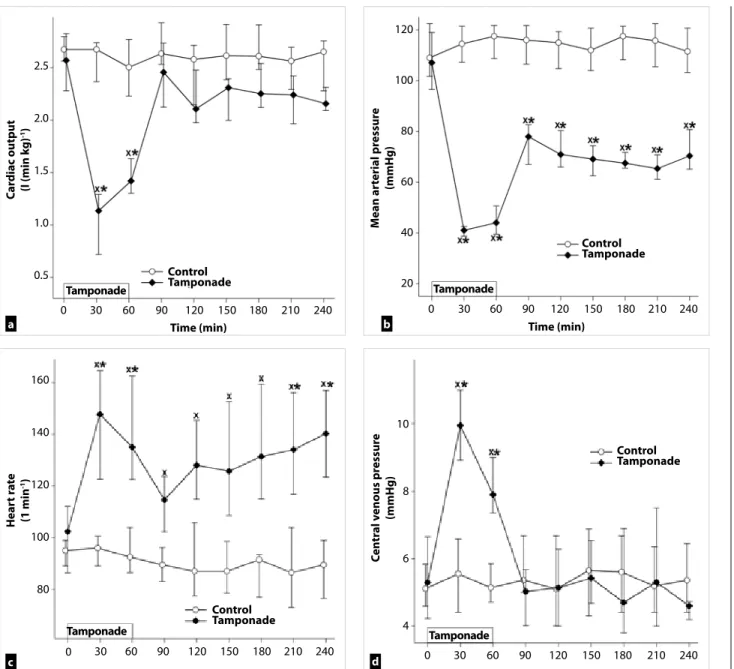
Figure 2. a-d. Changes in cardiac index (a), mean arterial pressure (b), heart rate (c), and central venous pressure (d) in control (white circle–solid line) and tamponade groups (black diamond–solid line). The plots demonstrate the median and the 25th (lower whisker) and 75th (upper whisker) percentiles
*p<0.05 for groups vs baseline values, xp<0.05 for cardiac tamponade vs control group values
Cardiac output (I (min kg)-1)Heart rate (1 min-1) Central venous pressure (mmHg)Mean arterial pressure (mmHg)
Time (min) Time (min)
Control Tamponade
Control Tamponade
Control Tamponade Control
Tamponade Tamponade
Tamponade Tamponade
Tamponade 2.5
2.0
1.5
1.0
0.5
160
140
120
100
80
10
8
6
4 120
100
80
60
40
20 0
0 30 60 90 120 150 180 210 240 0 30 60 90 120 150 180 210 240
30 60 90 120 150 180 210 240 0 30 60 90 120 150 180 210 240
a
c d
b
was performed 5 min after dye administration (1 scan/image, 1024x512 pixels and 475x475 μm per image). The changes in the mucosal architecture were examined by using a semi- quantitative scoring system as described previously (8). (I. oe- dema (0=no oedema, 1=moderate epithelial swelling, 2=se- vere oedema); and II. epithelial cell outlines (0=normal, clearly, well-defined outlines, 1=blurred outlines, 2=lack of normal cellular contours).
Statistical Analysis
Data analysis was performed with a statistical software package (SigmaStat for Windows, Jandel Scientific, Erkrath, Germany).
Friedman repeated measures analysis of variance on ranks was applied within groups. Time-dependent differences from the baseline for each group were assessed by Dunn’s method. The differences between groups were analyzed with Mann-Whitney probe. In the figures, median values and 75th and 25th percen- tiles are given; p<0.05 were considered significant.
RESULTS
Changes in Hemodynamic Parameters
The average duration of the surgical preparation phase was 70±20 min. The MAP remained at 40–45 mmHg as previously Figure 4. Changes in myeloperoxidase enzyme activity in
control (white box) and tamponade (gray striped box to the right side) groups. The plots demonstrate the median and the 25th (lower whisker) and 75th (upper whisker) percentiles
*p<0.05 for groups vs baseline values, xp<0.05 for cardiac tamponade vs control group values
8
6
4
2 Myeloperoxidase enzyme activity (mU (mg protein)-1)
Control Tamponade
0 30 60
Tamponade
90 240
Time (min)
Figure 5. Changes in red blood cell velocity in control (white box) and tamponade (gray striped box to the right side) groups. The plots demonstrate the median and the 25th (lower whisker) and 75th (upper whisker) percentiles.
*p< 0.05 for groups vs baseline values, xp<0.05 for cardiac tamponade vs control group values
Red blood cell velocity (um s-1) 600
500
400
300
200
Control Tamponade
Tamponade
0 90
Time (min)
240
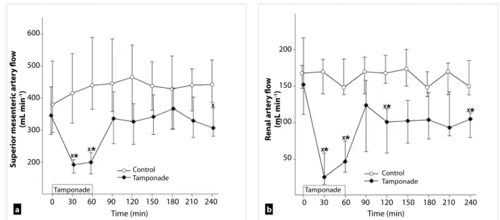
Figure 3. a-b. Changes in mesenteric artery (a) and renal artery flow (b) in control (white circle–solid line) and tamponade (black diamond–solid line) groups. The plots demonstrate the median and the 25th (lower whisker) and 75th (upper whisker) percentiles.
*p<0.05 for groups vs baseline values, xp<0.05 for cardiac tamponade vs control group values
Superior mesenteric artery flow (mL min-1) Renal artery flow (mL min-1)
600
500
400
300
200
200
150
100
50
0 30 60 90 120
Time (min) Time (min)
Control
Tamponade Control
Tamponade
Tamponade Tamponade
150 180 210 240 0 30 60 90 120 150 180 210 240
a b
208
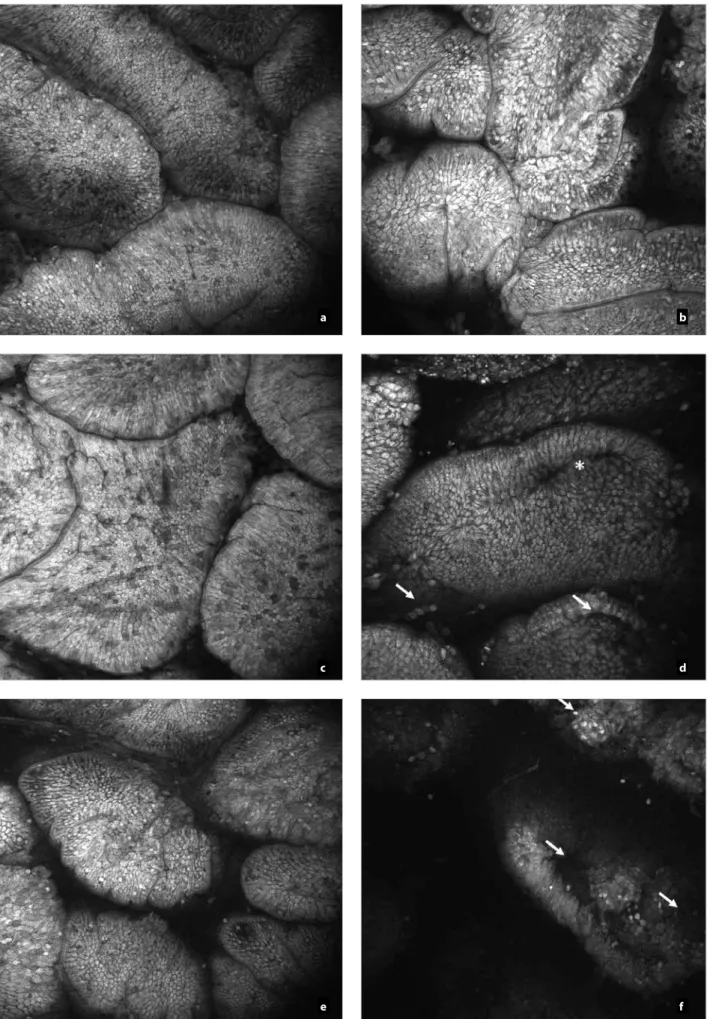
Figure 6. a-f. In vivo histology (CLSEM technique) showing the changes of the epithelial surface of the terminal ileum after acriflavin staining. Mucosal surface of control group (a, c, e); and structure of mucosal surface in tamponade group (b, d, f) are shown
a
c
e
b
d
f
209
accompanied by a concomitantly decreased CO. The CO in- creased to baseline levels after the reversal of tamponade (Fig- ure 2a) but the MAP remained significantly depressed (Figure 2b). The elevation of CO was accompanied by a compensatory elevation of HR (Figure 2c). The decreased venous return was evidenced by a significant elevation of CVP throughout the tamponade phase.
Significant deterioration of artery mesenteric flow was ob- served during tamponade, but no significant differences were found in the post–tamponade phase as compared to the base- line values and the control group (Figure 3a). The renal artery flow was significantly and permanently reduced during tam- ponade and the post-tamponade period as well (Figure 3b).
Leukocyte accumulation
The plasma MPO activity increased during the tamponade pe- riod and its peak was reached 30 minutes after tamponade. At the end of the observation period the MPO activity decreased towards the control level (Figure 4).
Changes in microcirculation
The RBCV did not change in the control group, while in the tamponade group, a significant decrease from the baseline and control group was detected but at the end of the observa- tion period (Figure 5).
In vivo Histology
The mucosal morphology was examined by intra-vital CLSEM technique in real time. The epithelial morphology of the small intestinal mucosa presented normal morphological patterns in sham-operated animals and in the control samples of the tamponade group while 30 min after tamponade induction longitudinal fissures appeared and partial epithelium defects were detected (Figure 6a-e). The lack of epithelium was ex- tended by the end of observation (Figure 6f).
DISCUSSION
Animal models have important, sometimes, decisive roles in today’s medical research. Large animal models have many ad- vantages for studying human pathophysiology and by their size they can provide additional benefits for the development of new surgical techniques and therapies. Experimental peri- cardial tamponade models using thoracotomy can be suitable to examine the complication of open cardiac surgical proce- dures or to monitor the hemodynamic changes (7, 9). Our pur- pose was to develop a suitable model based on experimental surgical standards for the investigation of cardiogenic shock, which evolves after NCSP where the duration, extent, char- acteristics, and outcome variables of impaired systemic cir- culation can be determined exactly. According to our results the presented experimental model can be used to study the acute pathophysiological consequences of low cardiac output states. Moreover, it may well reflect the signs of cardiogenic shock during NCSP. In addition, this pericardial tamponade setup can be useful for the practical training of surgical resi- dents for the real-life demonstration and management of peri- cardiocentesis or emergency thoracotomy.
Increased pressure in the pericardium effects the low-pressure areas, like the superior and inferior caval veins and the right
inside the atria and ventricles, thus the pericardial pressure blocks the right atrial inflow and consecutively the right ven- tricular diastolic filling. The high venous pressure leads to the distension of the jugular veins (Kussmaul-sign) the cardiac sounds are “muffled.” In addition, the peripherial pulse can- not be palpated while cardiac auscultation detects heartbeats, this is referred as pulsus paradoxus. The ECG signs reveal the lower amplitude of R waves due to the increased resistance of intra-pericardial liquids (5, 10). The development of cardio- genic shock activates numerous compensation mechanisms to maintain the perfusion pressure to the vital organs. Besides the enhanced contractility, the heart rate is increased to main- tain CO. Additionally, the body redistributes the circulating volume by systemic and local vasoactive responses, includ- ing the remarkable vasoconstriction of the muscle-, skin-, and mesenteric vessels (5, 10-12).
The decreasing diuresis could be the first sign of accumulat- ing pericardial fluid and thriving pericardial tamponade on cardiac surgical ICU. In our experiments, the renal blood flow remained significantly depressed despite the resolving CO.
The deteriorating renal function is a challenging problem in clinical practice, thus our porcine model could be an impor- tant tool for studies aiming to study the improvement of renal function.
CONCLUSION
Our model using anesthetized mini pigs reflects the local and systemic hemodynamic and inflammatory changes of the clin- ical picture correctly. In the absence of thoracotomy, the surgi- cal procedure is relatively easy and fast while at the same time the experimental setup allows for extensive hemodynamic monitoring, tissue biopsies, and biochemical measurements.
We tried to make the model as realistic as possible, thus the animal’s own blood is used to fill the pericardium. Leakage of blood was not detected and therefore the amount of intra- pericardial fluid could be standardized. The MAP level can be precisely controlled and the pressure response can be exactly modified throughout the experiments. Another advantage is that there is no explicit need for mechanical ventilation, and the lack of thoracotomy allows for longer duration or even chronic experiments with prolonged data collection. Besides, the transphrenic thoracal approach is definitely less stressful (refinement), and considering that the reduced invasiveness enables a reduction in animal numbers, this new model facili- tates the adherence to the 3R principles as well.
Ethics Committee Approval: Ethics committee approval was re- ceived for this study from the Ethics Committee of Szeged University (V/148/2013).
Informed Consent: N/A.
Peer-review: Externally peer-reviewed.
Author Contributions: Concept - G.B., D.É., M.B., G.V.; Design - B.G., S.S., É.D., G.V.; Supervision - M.B. Resource - M.B., G.V.; Materials - B.G., S.S., É.D., G.V.; Data Collection and/or Processing - B.G., S.S., É.D., G.V.;
Analysis and/or Interpretation B.G., S.S., É.D., G.V..; Literature Search - B.G., S.S., É.D., G.V.; Writing Manuscript - G.B., G,V.; Critical Reviews - D.É., M.B.
210
Acknowledgments The authors are grateful to Ágnes Fekete, Csilla Mester, Nikolett Beretka, Éva Nagyiván, Károly Tóth, and Péter Sárkány for their skillful assistance.
Conflict of Interest: The authors have no conflicts of interest to declare.
Financial Disclosure: This study was funded by Hungarian National Research, Development and Innovation Office NKFIH-K120232 and NKFIH-K116861 and GINOP-2.3.2-15-2016-00015 grants.
References
1. Moore FD. The university in American surgery. Surgery 1958; 44: 1-10.
2. Osuchowski MF, Remick DG, Lederer JA, Lang CH, Aasen AO, Aibiki M, et al. Abandon the Mouse Research Ship? Not Just Yet!.
Shock 2014; 41: 463-475. [CrossRef]
3. Orbach A, Schliamser JE, Flugelman MY, Zafrir B. Contemporary evaluation of the causes of cardiac tamponade: Acute and long- term outcomes. Cardiol J 2016; 23: 57-63. [CrossRef]
4. Bodson L, Bouferrache K, Vieillard-Baron A. Cardiac tamponade:
Curr Opin Crit Care 2011; 17: 416-424. [CrossRef]
5. Carmona P, Mateo E, Casanovas I, Peña JJ, Llagunes J, Aguar F, et al. Management of Cardiac Tamponade After Cardiac Surgery. J Cardiothorac Vasc Anesth 2012; 26: 302-311. [CrossRef]
6. Rivera-Nieves J, Gorfu G, Ley K. Leukocyte adhesion molecules in animal models of inflammatory bowel disease. Inflamm Bowel Dis 2008; 14: 1715-1735. [CrossRef]
7. Kaszaki J, Nagy S, Tárnoky K, Laczi F, Vecsernyés M, Boros M.
Humoral changes in shoc induced by cardiac tamponade. Circ Shock 1989; 29: 143-153.
8. Kovács T, Varga G, Erces D, Tőkés T, Tiszlavicz L, Ghyczy M, et al. Dietary Phosphatidylcholine Supplementation Attenuates Inflammatory Mucosal Damage in a Rat Model of Experimental Colitis. Shock 2012; 38: 177-185. [CrossRef]
9. Érces D, Nógrády M, Nagy E, Varga G, Vass A, Süveges G, et al.
Complement C5A Antagonist Treatment Improves the Acute Cir- culatory and Inflammatory Consequences of Experimental Car- diac Tamponade. Crit Care Med 2013; 41: e344-e351. [CrossRef]
10. Spodick DH. Acute cardiac tamponade. N Engl J Med 2003; 349:
684-690. [CrossRef]
11. Ashikhmina EA, Schaff HV, Sinak LJ, Li Z, Dearani JA, Suri RM, et al. Pericardial Effusion After Cardiac Surgery: Risk Factors, Pa- tient Profiles, and Contemporary Management. Ann Thorac Surg 2010; 89: 112-118. [CrossRef]
12. Kuvin JT, Harati NA, Pandian NG, Bojar RM, Khabbaz KR. Postoper- ative cardiac tamponade in the modern surgical era. Ann Thorac Surg 2002; 74: 1148-1153. [CrossRef]