Lancet Neurol 2018; 17: 782–89 Published Online July 24, 2018 http://dx.doi.org/10.1016/
S1474-4422(18)30231-X See Comment page 737
*Contributed equally Department of Emergency Medicine, University of Rochester School of Medicine and Dentistry, Rochester, NY, USA (J J Bazarian MD); Technical University of Munich, Klinikum rechts der Isar, Munich, Germany (P Biberthaler MD);
Department of Emergency Medicine, Wayne State University, Detroit Receiving Hospital, Detroit, MI, USA (R D Welch MD); Division of Emergency Medicine, Washington University, St Louis, MO, USA (L M Lewis MD); University of Szeged, Szeged, Hungary (P Barzo MD); Department of Trauma Surgery, Ludwig Maximilians University, Munich, Germany (V Bogner-Flatz MD); Carilion New River Valley Hospital, The Edward Via College of Osteopathic Medicine, Blacksburg, VA, USA (P Gunnar Brolinson DO);
Department of Neurosurgery, The MTA-PTE Clinical Neuroscience MR Research Group, János Szentágothai Research Center, Hungarian Brain Research Program, Medical School, University of Pecs, Pecs, Hungary (A Büki MD); Department of Radiology, VA San Diego Healthcare System/University of California, San Diego Health System, La Jolla, CA, USA (J Y Chen MD); Department of Pathology, University of Maryland School of Medicine, University of Maryland Medical Center, Baltimore, MD, USA (R H Christenson PhD); US Army Medical Research and Materiel Command, Fort Detrick, MD,
Serum GFAP and UCH-L1 for prediction of absence of
intracranial injuries on head CT (ALERT-TBI): a multicentre observational study
Jeffrey J Bazarian*, Peter Biberthaler*, Robert D Welch, Lawrence M Lewis, Pal Barzo, Viktoria Bogner-Flatz, P Gunnar Brolinson, Andras Büki, James Y Chen, Robert H Christenson, Dallas Hack, J Stephen Huff, Sandeep Johar, J Dedrick Jordan, Bernd A Leidel, Tobias Lindner,
Elizabeth Ludington, David O Okonkwo, Joseph Ornato, W Frank Peacock, Kara Schmidt, Joseph A Tyndall, Arastoo Vossough, Andy S Jagoda
Summary
Background More than 50 million people worldwide sustain a traumatic brain injury (TBI) annually. Detection of intracranial injuries relies on head CT, which is overused and resource intensive. Blood-based brain biomarkers hold the potential to predict absence of intracranial injury and thus reduce unnecessary head CT scanning. We sought to validate a test combining ubiquitin C-terminal hydrolase-L1 (UCH-L1) and glial fibrillary acidic protein (GFAP), at predetermined cutoff values, to predict traumatic intracranial injuries on head CT scan acutely after TBI.
Methods This prospective, multicentre observational trial included adults (≥18 years) presenting to participating emergency departments with suspected, non-penetrating TBI and a Glasgow Coma Scale score of 9–15. Patients were eligible if they had undergone head CT as part of standard emergency care and blood collection within 12 h of injury.
UCH-L1 and GFAP were measured in serum and analysed using prespecified cutoff values of 327 pg/mL and 22 pg/mL, respectively. UCH-L1 and GFAP assay results were combined into a single test result that was compared with head CT results. The primary study outcomes were the sensitivity and the negative predictive value (NPV) of the test result for the detection of traumatic intracranial injury on head CT.
Findings Between Dec 6, 2012, and March 20, 2014, 1977 patients were recruited, of whom 1959 had analysable data.
125 (6%) patients had CT-detected intracranial injuries and eight (<1%) had neurosurgically manageable injuries.
1288 (66%) patients had a positive UCH-L1 and GFAP test result and 671 (34%) had a negative test result. For detection of intracranial injury, the test had a sensitivity of 0·976 (95% CI 0·931–0·995) and an NPV of 0·996 (0·987–0·999).
In three (<1%) of 1959 patients, the CT scan was positive when the test was negative.
Interpretation These results show the high sensitivity and NPV of the UCH-L1 and GFAP test. This supports its potential clinical role for ruling out the need for a CT scan among patients with TBI presenting at emergency departments in whom a head CT is felt to be clinically indicated. Future studies to determine the value added by this biomarker test to head CT clinical decision rules could be warranted.
Funding Banyan Biomarkers and US Army Medical Research and Materiel Command.
Copyright © 2018 Elsevier Ltd. All rights reserved.
Introduction
An estimated 54–60 million people worldwide sustain a traumatic brain injury (TBI) each year.1 In the USA, TBI results in more than 2·5 million emergency department visits annually, most of these being mild TBI (also known as concussion).2 Head CT scan is the diagnostic modality of choice to evaluate patients for traumatic intracranial injuries,3 contributing to the approximately 20 million head CT scans performed annually in the USA.4 Although effective for detecting traumatic injuries that require observation or neurosurgical evacuation, the widespread use of head CT scanning has been questioned due to potential adverse effects of radiation exposure, unnecessary emergency department resource use, and cost.4–6 These factors take on added importance for patients with mild TBI in whom the prevalence of
CT-detected intracranial injury is typically less than 10%.6 Clinical decision rules (CDRs) have been developed with the goal of reducing unnecessary head CT scans, but they have had only modest impact on CT use.5,7
Prior studies have shown the potential for blood-based brain injury biomarkers to predict the absence of intracranial injury after TBI and aid in reducing unnecessary head CT use.8–11 Of the many different proteins, small molecules, and lipid products investigated in humans for this purpose, the S100 astroglial calcium- binding protein B (S100B) has been the most studied.
Although some guidelines have recommended this biomarker for clinical use to predict intracranial injury on CT,12 the 2008 American College of Emergency Physician guidelines for managing adult patients with mild TBI determined that use of S100B only had level C
evidence.3 Currently, no blood-based brain biomarker tests in the USA have been approved by the US Food and Drug Administration (FDA) for use outside a research setting. This gap in diagnostic capability has received much attention with many calls for advancing brain biomarker research.13
Several novel brain proteins have been identified that potentially address this gap.14 Two proteins, ubiquitin C-terminal hydrolase-L1 (UCH-L1) and glial fibrillary acidic protein (GFAP), have emerged as promising candidates for translation to clinical use. Preliminary evidence suggesting both proteins were accurate predictors of CT-detected intracranial injuries were limited by small cohort size, variability in the timing of blood sample acquisition, and retrospective determi- nation of cutoff values that probably biased the estimate of diagnostic accuracy.8–10,15,16
We therefore did a large, prospective, multicentre trial, entitled A Prospective Clinical Evaluation of Biomarkers of Traumatic Brain Injury (ALERT-TBI), aiming to validate the ability of a biomarker test combining UCH-L1 and GFAP, at predetermined cutoff values, to predict traumatic intracranial injuries on head CT scan within 12 h of TBI.
Methods
Study design and participants
The ALERT-TBI trial was designed to validate the ability of a biomarker test combining UCH-L1 and GFAP to predict CT-detected traumatic intracranial injuries within 12 h of TBI. This study was done at 22 investigational sites globally (15 in the USA and seven in Europe;
appendix). All sites provided approval from their institutional ethics committee or appropriate regulatory body.
Enrolled participants represented a convenience sample of patients aged 18 years or older presenting to participating emergency departments with a suspected, non-penetrating TBI resulting from an external force and a Glasgow Coma Scale (GCS) score of 9–15 at the time of informed consent. Because of the controversy regarding the clinical symptoms that define the lower bounds of TBI severity,17 neither loss of consciousness nor amnesia was a requirement for inclusion. However, patients were only eligible for inclusion if they underwent non-contrast head CT scanning within 12 h of injury as part of their clinical care. Additional inclusion criteria comprised blood sampling within 12 h of injury and provision of informed written consent.
USA (D Hack MD, K Schmidt PhD); University of Virginia, Charlottesville, VA, USA (J S Huff MD);
Neurosurgery, Orthopedics &
Spine Specialist, Waterbury, CT, USA (S Johar DO); University of North Carolina School of Medicine, Chapel Hill, NC, USA (J D Jordan MD); Charité Universitätsmedizin Berlin, Berlin, Germany (B A Leidel MD, T Lindner MD); Agility Clinical, Carlsbad, CA, USA
(E Ludington PhD); Department of Neurosurgical Science, University of Pittsburgh, Pittsburgh, PA, USA (D O Okonkwo MD);
Department of Emergency Medicine, Virginia Commonwealth University Health System, Richmond, VA, USA (J Ornato MD); Department of Emergency Medicine, Baylor College of Medicine, Houston, TX, USA (W F Peacock MD); US Army Medical Research and Material Command, Fort Detrick, MD, USA (K Schmidt PhD); Department of Emergency Medicine, The University of Florida, Gainesville, FL, USA (J A Tyndall MD); Department of Radiology, University of Pennsylvania, Philadelphia, PA, USA (Arastoo Vossough MD);
and Department of Emergency Medicine, Mount Sinai Health System, New York, NY, USA (A S Jagoda MD) Correspondence to:
Prof Robert D Welch, Department of Emergency Medicine, Wayne State University School of Medicine, UHC- 6G, Detroit, MI 48201, USA rwelch@med.wayne.edu See Online for appendix
Research in context Evidence before this study
Existing evidence suggests that head injuries formerly thought of as minor, such as concussion, might actually result in brain damage. This line of evidence comes principally from advanced neuroimaging studies (eg, diffusion tensor imaging, functional MRI, and magnetic resonance spectroscopy) over the past 10 years revealing traumatic axonal injury not detectable on standard CT or MRI. Although these advanced neuroimaging tests are sensitive, they are expensive, not readily available, are still considered suitable only for research, and have not yet entered standard clinical practice. Consequently, more than 20 different brain proteins have been studied, with seven— S100 astroglial calcium-binding protein B (S100B), neuron-specific enolase (NSE), ubiquitin C-terminal hydrolase-L1 (UCH-L1), glial fibrillary acidic protein (GFAP), alpha-II spectrin, and tau—having demonstrated diagnostic accuracy for either distinguishing concussion from non-concussion or predicting head CT results.
Several systematic reviews and meta-analyses of S100B have revealed its high sensitivity and negative predictive value (NPV) for acute head CT scan. The best reference standard for TBI-associated axonal injury, brain tissue histopathology, cannot be done in most patients with head injuries. However, head CT scans are frequently done in emergency departments to evaluate TBI, which made them a practical reference standard for analysing the diagnostic performance of a blood-based brain biomarker.
Added value of this study
Our study results showed that a serum biomarker test combining GFAP and UCH-L1 had a sensitivity of 97·6% and
NPV of 99·6% for the detection of acute intracranial injuries on head CT scan. These findings support its potential clinical role for ruling out the need for a CT scan among patients presenting to emergency departments with TBI in whom a head CT is felt to be clinically indicated. Because of the large sample size and the methodological rigour used to conduct this study, we believe our estimates of the diagnostic accuracy of a combined GFAP and UCH-L1 biomarker test are precise with minimal bias.
This methodological rigour was balanced by integrating elements of pragmatic clinical trials, which make our results broadly generalisable. These elements include a practical study definition of TBI and no strict criteria for head CT ordering among acute care clinicians.
Implications of all the available evidence
Combined with existing evidence, our results suggest that up to a third of head CT scans done in the acute setting of TBI could be avoided, with a very low false-negative rate. Clinical use of the GFAP and UCH-L1 biomarker test has the potential to reduce unnecessary head CT scans with their attendant radiation exposure and cost, but only if applied to appropriate patients. The results of our study pertain to the subset of patients with TBI thought to have a clinical need for head CT scanning, not to all patients with TBI. Future studies to compare this biomarker test to more sensitive indicators of traumatic axonal injury such as diffusion tensor imaging and to clinical recovery would bring us one step closer to the goal of developing improved, objective measures to aid in diagnosing TBI.
A GCS score was determined at the time of consent by study staff trained and certified by having successfully passed a GCS score examination (appendix). Site investigators assessed patients’ decisional capacity for informed consent in accordance with local institutional review boards and ethics committee guidance. Informed written consent was provided by legally authorised representatives for those patients without decisional capacity. Patients were excluded if the time of injury could not be determined, if head CT scanning was not performed, if venepuncture was not feasible, or if informed consent was not obtainable. Additional exclusion criteria are provided in the appendix.
Procedures
Venous blood was processed to serum, transferred to cryovials, and frozen at −80°C within 1 h of collection.
Samples were shipped on dry ice to a central repository and later sent to one of three core laboratories for analysis. Laboratory personnel were blinded to patients’
diagnosis and clinical status. Serum samples were analysed for UCH-L1 and GFAP concentration using chemiluminescent enzyme-linked immunosorbent assays. Assay results were not provided to treating health- care providers. Results from the GFAP and UCH-L1 assays were combined into a single qualitative test result.
A patient’s test result was considered valid if it satisfied the Boolean criteria for either positive or negative. A negative test result was defined as both markers falling
below their prespecified cutoff value, whereas a positive test result was defined as one or both markers falling above their prespecified cutoff value. A patient’s test result was invalid if the assay result for both markers was invalid, or if the result for one marker was invalid and the result for the other was below its predetermined cutoff value (see appendix for details).
The reportable ranges were 10–320 pg/mL for the GFAP assay and 80–2560 pg/mL for the UCH-L1 assay. The lower limit of quantification was less than 10 pg/mL for GFAP and less than 80 pg/mL for UCH-L1. Concentrations of UCH-L1 or GFAP above the reportable range were analysed as 2560 pg/mL and 320 pg/mL, respectively, whereas those below were analysed as 80 pg/mL and 10 pg/mL, respectively. The lower limit of detection was not determined for either assay because the lower limit of quantification was found to lie below the reportable ranges for each assay. For both assays, the intra-run and inter-run coefficient of variation was less than 10%. Prespecified biomarker cutoff values were derived in an independent group of 334 adults with mild TBI defined by a GCS 13–15, yielding cutoff concentration values of 327 pg/mL for UCH-L1 and 22 pg/mL for GFAP (appendix).
Head CT scans acquired at each site were transmitted to a central imaging laboratory (appendix). In the case of multiple CT scans, the images obtained closest to the blood draw were analysed. Two independent, board-certified neuroradiologists (JYC and AV), not affiliated with any enrolment site, reviewed each CT scan and determined if it was positive, negative, or inconclusive for an acute traumatic intracranial injury.
Radiologists were blinded to GFAP and UCH-L1 results and all clinical information except age and sex. If the two primary CT reviewers did not agree, the scan was adjudicated by a secondary reviewer (also a board- certified neuroradiologist). A head CT scan was considered valid if it could be interpreted as either positive or negative. Patients with unreadable (defined by the inability to fully assess the head CT scan) or inconclusive head CT scans were excluded from analysis.
Head CT scans were considered inconclusive if both primary readers thought the scan was inconclusive, or if one of the primary reviewers thought the scan was inconclusive and the secondary reviewer agreed.
CT-positive was defined as the presence of one or more of the following injuries: acute epidural haema- toma, acute subdural haematoma, indeterminate extra- axial haemorrhage, intraventricular haemorrhage, parenchymal haematoma, petechial haemorrhagic or bland sheer injury, subarachnoid haemorrhage, brain oedema, brain herniation, non-haemorrhagic contusion, ventricular compression, ventricular trapping, cranial fractures, depressed skull fractures, facial fractures, scalp injury, or skull base fractures. CT findings were also categorised as neurosurgically manageable, defined as an acute epidural haematoma greater than 30 cm³, acute subdural haematoma with a thickness greater than
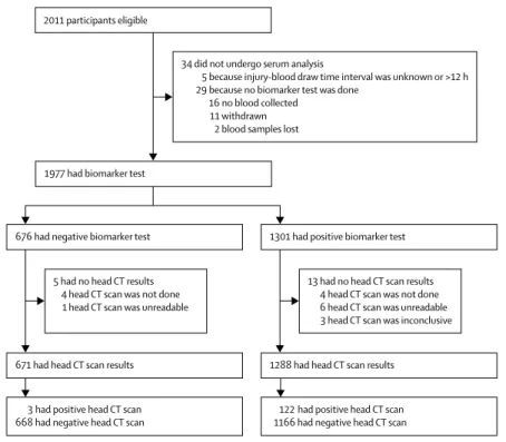
2011 participants eligible
34 did not undergo serum analysis
5 because injury-blood draw time interval was unknown or >12 h 29 because no biomarker test was done
16 no blood collected 11 withdrawn 2 blood samples lost 1977 had biomarker test
676 had negative biomarker test
671 had head CT scan results 1288 had head CT scan results
3 had positive head CT scan
668 had negative head CT scan 122 had positive head CT scan 1166 had negative head CT scan 1301 had positive biomarker test
5 had no head CT results 4 head CT scan was not done 1 head CT scan was unreadable
13 had no head CT scan results 4 head CT scan was not done 6 head CT scan was unreadable 3 head CT scan was inconclusive
Figure 1: Trial profile
No participants had invalid (ie, inconclusive) GFAP and UCH-1L biomarker test results.
10 mm or a midline shift greater than 5 mm, a parenchymal contusion greater than 50 cm³, or a frontal or temporal contusion greater than 20 cm³ with midline shift of at least 5 mm or cisternal compression.18 Inter- rater reliability for the two primary CT reviewers was estimated using the κ coefficient statistic.
Outcomes
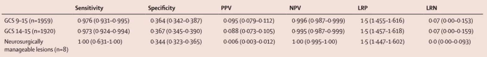
The primary study outcomes were the negative predictive value (NPV) and sensitivity of the UCH-L1 and GFAP test result for intracranial injury on head CT. Additionally, we determined UCH-L1 and GFAP test specificity, positive predictive value (PPV), and likelihood ratio positive (LRP) and likelihood ratio negative (LRN) values for the presence or absence of CT-detected intracranial injury and for neurosurgically manageable injury.
Statistical analysis
A minimum sample size of 107 CT-positive patients was required to estimate a test sensitivity of 0·95 with a lower 95% CI bound of 0·90. This sample size provided 80% power at p<0·05 to achieve a test sensitivity of at least 95% and an NPV of at least 99% with a misclassification rate of 5%. The total number of patients needed to accrue 107 CT-positive patients was estimated to be 2000 on the basis of the presumption that the prevalence of CT- detected injuries would be between 5% and 6%. To determine if test accuracy was influenced by sex, a stratified analysis was performed by comparing the diagnostic performance of the biomarker test in men and women. Sensitivity analyses were also carried out on the subset of patients with GCS 14–15, because this TBI severity range was the focus of the two most recently proposed CDRs.3,12 We also analysed the subset of patients with neurosurgically manageable lesions on head CT.
95% CIs for sensitivity, specificity, NPV, and PPV were estimated using the exact (Clopper-Pearson) methods for proportions, and for LRN and LRP using normal approximation methods. Statistical significance testing was performed using α=0·05. All analyses were done using SAS version 9.4 (SAS Institute, Cary, NC, USA).
This study is registered with ClinicalTrials.gov, number NCT01426919.
Role of the funding source
The study was designed by the Study Steering Committee, the sponsor, Banyan Biomarkers, and the US FDA. Data collection was coordinated by Pharmaceutical Research Associates and Perceptive Informatics. Data analysis was conducted by Agility Clinical following a prespecified statistical analysis plan. The authors had full access to study data and had final responsibility for the decision to publish.
Results
Between Dec 6, 2012, and March 20, 2014, 2011 participants were enrolled, 1354 (67%) from US sites and
657 (33%) from European sites. Of those enrolled, 34 did not undergo serum analysis for a variety of reasons (figure 1), yielding 1977 patients with biomarker test results. 18 of these participants did not have analysable head CT scans, resulting in 1959 participants with TBI with a valid head CT scan and test result for analysis. Of these, 1920 (98%) had a GCS of 14–15 (table 1). No
All patients
(GCS 9–15; n=1959) Patients with
GCS 14–15 (n=1920) Patients with neurosurgically manageable lesions (n=8) Age, years 48·9 (20·9, 18–98) 48·8 (20·9, 18–98) 64·3 (24·5, 21–90) Sex
Women 852 (43%) 833 (43%) 5 (63%)
Men 1107 (57%) 1087 (57%) 3 (38%)
Race or ethnicity*
White 1369 (70%) 1334 (69%) 8 (100%)
Black or African American 529 (27%) 527 (27%) 0
Hispanic 92 (5%) 89 (5%) 0
Other or unknown 72 (4%) 70 (4%) 0
GCS score
9 2 (<1%) ·· 2 (25%)
10 0 ·· 0
11 3 (<1%) ·· 0
12 12 (1%) ·· 1 (13%)
13 22 (1%) ·· 0
14 92 (5%) 92 (5%) 0
15 1828 (93%) 1828 (95%) 5 (63%)
Mechanism of injury†
Acceleration or
deceleration 413 (2%) 408 (21%) 3 (38%)
Motor vehicle accident 606 (31%) 598 (31%) 3 (38%)
Pedestrian struck by vehicle 69 (4%) 69 (4%) 0
Fall 1010 (52%) 981 (51%) 6 (75%)
Explosion 3 (<1%) 3 (<1%) 0
Assault 185 (9%) 183 (10%) 0
Sports Injury 48 (2%) 47 (2%) 0
Other 52 (3%) 52 (3%) 0
Unknown 126 (6%) 123 (6%) 0
Loss of consciousness
Yes 825 (42%) 803 (42%) 5 (63%)
No 1035 (53%) 1026 (53%) 3 (38%)
Unknown 99 (5%) 91 (5%) 0
Post-traumatic amnesia
Yes 645 (33%) 626 (33%) 3 (38%)
No 1282 (65%) 1271 (66%) 2 (25%)
Unknown 32 (2%) 23 (1%) 3 (38%)
Intoxicated with alcohol or drugs
Yes 416 (21%) 402 (21%) 2 (25%)
No 1528 (78%) 1504 (78%) 5 (63%)
Unknown 15 (1%) 14 (1%) 1 (13%)
Head CT scan
Traumatic injury 125 (6%) 113 (6%) 8 (100%)
No traumatic injury 1834 (94%) 1807 (94%) 0
(Table 1 continues on next page)
participants died during the study investigation period (time of consent until 24 h after injury or emergency department discharge, whichever came first).
125 (6%) participants with TBI had a traumatic intracranial injury on head CT (table 2), and
eight (<1%) had neurosurgically manageable lesions. The prevalence of intracranial injury was slightly lower among patients with a GCS of 14–15 (table 2). The most common head CT finding was subarachnoid haemorrhage, followed by acute subdural haematoma and parenchymal haematoma. The distribution of CT findings was similar in the subset of patients with a GCS of 14–15 (table 2). Among patients with neurosurgically manageable lesions, acute subdural haematoma was the most common finding followed by ventricular compression and subarachnoid haemorrhage. Inter-rater reliability for head CT scan interpretation was 0·73 (95% CI 0·67–0·80; appendix). The two primary reviewers were in agreement for 1899 (97%) of head CT scans; the secondary reviewer adjudicated the remaining 60 (3%) scans. Of these 60 scans, the secondary reviewer judged 37 to be CT positive, and 23 to be CT negative. A sensitivity analysis to estimate the potential effect of disagreement among the neuroradiology reviewers can be found in the appendix.
Patients underwent blood draw a median of 3·2 h after injury (table 1). Among all patients, both GFAP and UCH-L1 concentrations were significantly higher among those who were CT positive compared with those who were CT negative (median GFAP 135·0 pg/mL vs 22·2 pg/mL; p<0·0001; median UCH-L1 604·8 pg/mL vs 261·0 pg/mL; p<0·0001; figure 2). Among all patients with TBI, 1288 (66%) had a positive test and 671 (34%) had a negative test (appendix). Overall test sensitivity was 0·976 (95% CI 0·931–0·995) and NPV was 0·996 (0·987–0·999) for acute intracranial injury (table 3). Three patients had false-negative test results (appendix); however, one of these patients might have been a false CT positive, because a follow-up MRI showed an underlying cavernous malformation, a vascular defect that can mimic a focal haemorrhagic contusion on CT.
The test performed similarly among patients with GCS 14–15 (figure 2), and had slightly better performance among the 39 patients with a GCS 9–13 (sensitivity 1·00, 95% CI 0·74–1·0; NPV 1·00, 0·48–1·0). The performance of the test among men was numerically higher than that for women, although the difference was not significant (appendix). The test was 100% sensitive with a 100% NPV for detecting neurosurgically manage able lesions (table 3). A sensitivity analysis comparing the diagnostic accuracy of the biomarker test to each protein individually among a subset of 1790 patients having quantitative values for both GFAP and UCH-L1 proteins revealed that, with respect to the primary outcomes, the combination of both proteins outperformed each marker separately, but that the diagnostic improvement over GFAP alone was not significant (appendix).
Discussion
The development of a clinically validated brain biomarker test has the potential to substantially alter diagnostic approaches to patients with TBI in acute care settings. In
All patients
(GCS 9–15; n=1959) Patients with
GCS 14–15 (n=1920) Patients with neurosurgically manageable lesions (n=8) (Continued from previous page)
Assay
Time from injury to blood draw, h
Median 3·2 (2·3–4·0) 3·2 (2·3–4·0) 3·4 (2·6–5·2)
Range 0·3–11·9 0·3–11·9 1·1–7·8
GFAP, pg/mL‡
Median 24·3 (10·0–57·4) 24·1 (10·0–55·4) 105·3 (40·7–320·0)
Range 10·0–320·0 10·0–320·0 40·6–320·0
UCH-L1, pg/mL§
Median 273·1 (151·7-535·5) 270·1 (150·3–530·6) 963·6 (452·7–2560·0)
Range 80·0–2560·0 80·0–2560·0 208·1–2560·0
Data are n (%), mean (SD, range), and median (IQR), unless stated otherwise. GCS=Glasgow Coma Scale.
UCH-L1=ubiquitin C-terminal hydrolase-L1. GFAP= glial fibrillary acidic protein. *Patients could choose more than one race. †Patients could have more than one mechanism of injury. ‡Assessed in 1819 patients (1787 with GCS 14–15 and six with neurosurgically manageable lesions). §Assessed in 1915 patients (1876 with GCS 14–15 and eight with neurosurgically manageable lesions).
Table 1: Patient demographics and characteristics
All patients (GCS 9–15;
n=125*)
Patients with GCS 14–15 (n=113†)
Patients with neurosurgically manageable lesions (n=8) Haemorrhagic lesions
Acute epidural haematoma 12 (10%) 10 (9%) 2 (25%)
Acute subdural haematoma 60 (48%) 55 (49%) 8 (100%)
Indeterminate extra-axial haemorrhage 19 (15%) 17 (15%) 1 (13%)
Intraventricular haemorrhage 7 (6%) 6 (5%) 0
Parenchymal haematoma 27 (22%) 22 (19%) 3 (38%)
Petechial haemorrhagic or bland sheer injury 4 (3%) 3 (3%) 1 (13%)
Subarachnoid haemorrhage 76 (61%) 64 (57%) 5 (63%)
Non-haemorrhagic lesions
Brain oedema 12 (10%) 8 (7%) 3 (38%)
Brain herniation 4 (3%) 2 (2%) 3 (38%)
Non-haemorrhagic contusion 8 (6%) 6 (5%) 1 (12%)
Ventricular compression 13 (10%) 10 (9%) 6 (75%)
Ventricular trapping 1 (1%) 0 1 (13%)
External or skull injuries‡
Cranial fractures 35 (28%) 29 (26%) 3 (38%)
Depressed skull fractures 4 (3%) 3 (3%) 0
Facial fractures 24 (19%) 20 (18%) 2 (25%)
Scalp injury 121 (97%) 109 (96%) 6 (75%)
Skull base fractures 21 (17%) 16 (14%) 3 (38%)
CT-positive indicates presence of traumatic intracranial injury on head CT. GCS=Glasgow Coma Scale. *Includes eight patients with neurosurgically manageable lesions. †Includes five patients with neurosurgically manageable lesions. ‡External or skull injuries were not used to define a CT-positive result.
Table 2: CT findings in participants with traumatic intracranial injury, by Glasgow Coma Scale category
what is, to our knowledge, the largest TBI biomarker trial to date, we report the high sensitivity of a novel serum- based test for the detection of traumatic intracranial injuries on head CT within 12 h of injury. A test combining GFAP and UCH-L1 assays at prespecified cut- off concentrations had a sensitivity of 0·976 and an NPV of 0·996 with a specificity of 0·364 for prediction of acute CT-detected intracranial injury in our study population, with similar performance (sensitivity of 0·973, NPV of 0·995, and specificity of 0·367) among the subset with a GCS 14–15. Results from this study were used to support a request to the US FDA for commercialisation of this test, which was granted on Feb 14, 2018.
Currently, many acute health-care providers use CDRs to determine patients with head injuries requiring head CT scan. Similar to the GFAP and UCH-L1 test, these CDRs are also sensitive for detection of intracranial injury.19 However, studies suggest that CDRs have had little effect on reducing unnecessary head CT use5,7 and might result in increased use of CT scans.20 Obstacles to CDR use include time and effort required to properly administer them and a perception that they rely on subjective variables that might be difficult to reliably obtain from a broad range of patients.21
Despite widespread recognition that development of blood-based brain biomarkers is important,13 no clinically useable test is available in North America. S100B, which has comparable sensitivity and NPV11,22 and is in clinical use in several countries in Europe,12 has been shown to identify patients at low risk for traumatic injuries on CT.11 However, S100B has not been approved by the US FDA for this use, and neither the American College of Emergency Physicians3 nor the Eastern Association of Surgery of Trauma23 recommends its use as a pre-head CT screen.
The GFAP and UCH-L1 test performed as well among patients with a GCS of 14–15 as it did in the entire cohort (GCS of 9–15). Despite the favourable results for use of this test across GCS ranging from 9 to 15, clinicians might be reluctant to withhold head CT scanning in patients with TBI with a GCS less than 14 (indicating more severe TBI). Patients with a GCS of 14–15 represent a challenge to acute care clinicians. They might appear neurologically normal but most CDRs recommend scanning both those with a persistent GCS of 14 and a subset of those with a GCS of 15.19 It is in patients with GCS of 14–15 that the use of a biomarker test could be of greatest value. In the current study, 666 (35%) of 1920 patients with a GCS of 14–15 had a negative test result, representing the proportion of patients thought to be in clinical need of a head CT in whom scanning could potentially have been avoided.
Our results have several important implications. First, clinical use of the GFAP and UCH-L1 test could substantially reduce head CT use. If the GFAP and UCH-L1 test were used in patients with TBI in whom a head CT scan was felt to be clinically indicated, those with a negative test (34–35% among all patients with TBI
and those with GCS 14–15) would not undergo CT scan.
Thus, by imaging only those with a positive GFAP and UCH-L1 test, overall scanning could be reduced by about a third. The extent to which these results can be applied to patients with GCS 9–13 is uncertain. Although the point estimates for both sensitivity and NPV was 1·00, the small number of patients in this group produced wide 95% CIs. Until the precision of these point estimates can be confirmed by appropriately powered studies, caution should be used in applying these results to patients with TBI with GCS 9–13.
Second, the observation that there were ten times as many positive GFAP and UCH-L1 tests as positive CT scans among patients with head injuries suggests that these two proteins might be detecting more subtle degrees of injury not visible on CT scan. In support of this idea, a preliminary study24 of nine patients with mild TBI reported significantly elevated GFAP levels among those with haemorrhage on MRI, including one with a normal head CT scan. In a study of 38 patients with head injuries with normal CT scans, elevated plasma levels of
Patients MedianMean
All CT positive CT negative
22 pg/mL
327 pg/mL 1819
51·9 24·3
1787 51·1 24·1
105 171·7 135·0
98 167·4 132·1
1714 44·6 22·2
1689 44·3 22·0 GCS patient score All patients (GCS 9–15) Mild TBI (GCS 14–15)
0 300
200
GFAP assay result (pg/mL) 100
A
Patients Mean Median
All CT positive CT negative
451·81915 273·1
445·41876 270·1
890·7122 604·8
843·3110 601·3
422·01793 261·0
420·61766 258·5 0
2000 2500
1500 1000 UCH-L1 assay result (pg/mL) 500
B
Figure 2: (A) Serum GFAP and (B) UCH-L1 levels by head CT result
The upper and lower bounds of each box indicate the 75th and 25th percentiles, respectively. Within each box, the square marker symbol indicates the mean and the horizontal line indicates the median. Whiskers extend up to 1·5 × IQR (bounded by observed minimum and maximum). Small circles indicate actual datapoints, staggered by patient. Dotted horizontal lines represent prespecified cutoff value. UCH-L1=ubiquitin C-terminal hydrolase-L1.
GFAP=glial fibrillary acidic protein. GCS=Glasgow Coma Scale.
the neuronal protein spectrin N-terminal fragment correlated with fractional anisotropy changes on diffusion tensor imaging and with cognitive impairment persisting longer than 3 months.25 Future studies to compare GFAP and UCH-L1 profiles with more sensitive indicators of traumatic brain injury such as diffusion tensor imaging and with clinical recovery are probably warranted.
The current study has several limitations. We did not evaluate the GFAP and UCH-L1 test’s predictive ability for clinical outcomes such as prolonged post-concussive symptoms, cognitive impairment, or decreased functional status. Furthermore, although patients with TBI with extracranial injuries were eligible for study inclusion, we did not evaluate the impact of these injuries on test performance. Although trauma-related release from non- brain tissue is a well described problem for S100B,11 several studies have been unable to detect an effect of extracranial injuries on the performance of GFAP and UCH-L1.15,16 The advantage of including those with extracranial injuries in our study is the enhanced external generalisability of our results.
Although we examined the GFAP and UCH-L1 test performance among those with neurosurgically manage- able lesions, we did not evaluate its performance among those actually undergoing neurosurgery. Moreover, we did not compare the performance of the GFAP and UCH-1 test to CDRs. Although a number of clinical variables were collected that could potentially allow for a post-hoc determination of which patients did or did not meet CDR criteria, such a retrospective analysis would not meet accepted methodological standards performing CDR-related research and would be considered exploratory.26 These standards should be integrated in a future study with the express aim of comparing CDRs with biomarker tests. We did not attempt to improve the test’s diagnostic accuracy by adding other clinical variables, although performance did not appear to vary by sex. Because a methodological strength of this study was the use of prespecified marker cutoffs, we chose not to explore alternative cutoffs. Nor did we compare the performance of the GFAP and UCH-1 test to S100B, which has comparable sensitivity and NPV.11,22 This study did not look at turnaround time for test results, and it is axiomatic that for a pre-head CT screening test to be useful, the results need to be rapidly available. Finally, although the independent neuroradiologists interpreting the head CT scans agreed on approximately 97% of cases,
the κ was 0·73, which some might consider low. This might be due in part to the use of three interpretation categories (positive, negative, and inconclusive) rather than the more often used two categories (positive and negative). Moreover, a κ of 0·73 is actually higher than prior reports of agreement among reviewers interpreting head CT scans.27
Another final important study limitation involves the sensitivity analysis comparing the diagnostic perfor- mance of the biomarker test to its component proteins.
Because this analysis contained a subset of patients, it had reduced power to detect small differences in the primary outcomes and was probably biased due to dependence of participant inclusion on results of the test. In support of this, the CT-positive prevalence among the 179 participants excluded by this sensitivity analysis (10%) was considerably higher than among those included (6%). Although these results raise the possibility that GFAP alone might perform as well as the two proteins combined, they require further validation.
In summary, our results show the high sensitivity and NPV of a brain biomarker test combining UCH-L1 and GFAP for traumatic intracranial injuries on head CT scan. These results support the potential clinical role of this biomarker test for ruling out the need for a head CT scan among patients with TBI presenting at emergency departments in whom a head CT is felt to be clinically indicated.
Contributors
JJB, PB, ASJ, LML, and RDW conceived or designed the study. PB, JJB, PB, VB-F, PGB, AB, RHC, JSH, SJ, JDJ, LML, BAL, TL, DOO, JO, WFP, JAT, and RDW were responsible for data collection. EL was responsible for data analysis. JJB, PB, JYC, DH, ASJ, LML, KS, AV, and RDW were responsible for interpreting the data. All authors drafted or critically revised the manuscript and gave final approval of the version to be published.
Declaration of interests
PB, RDW, VB-F, and JAT received research funding from Banyan Biomarkers during the conduct of the study. JYC reports personal fees from Banyan Biomarkers during the conduct of the study. JJB received research funding from Banyan Biomarkers and research funding from BrainScope Company during the conduct of the study. RHC reports grants and personal fees from Roche Diagnostics, Siemens Healthcare Diagnostics, and Becton Dickinson during the conduct of the study.
DH reports personal fees from US Army Medical Research & Materiel Command, during the conduct of the study; and personal fees from Brainscope Company, Philips, and Abbott, outside of the submitted work. TL reports grants from Banyan Biomarkers during the conduct of the study, and personal fees from WMC Technologies GmbH, outside of the submitted work. EL reports other support from Banyan Biomarkers during the conduct of the study. WFP reports grants from
Sensitivity Specificity PPV NPV LRP LRN
GCS 9–15 (n=1959) 0·976 (0·931–0·995) 0·364 (0·342–0·387) 0·095 (0·079–0·112) 0·996 (0·987–0·999) 1·5 (1·455–1·616) 0·07 (0·00–0·153) GCS 14–15 (n=1920) 0·973 (0·924–0·994) 0·367 (0·345–0·390) 0·088 (0·073–0·105) 0·995 (0·987–0·999) 1·5 (1·457–1·618) 0·07 (0·00–0·159) Neurosurgically
manageable lesions (n=8) 1·00 (0·631–1·00) 0·344 (0·323–0·365) 0·006 (0·003–0·012) 1·00 (0·995–1·00) 1·5 (1·447–1·602) 0·0 (0·00–0·093) Data in parentheses are 95% CIs. PPV=positive predictive value. NPV=negative predictive value. LRP=likelihood ratio positive. LRN=likelihood ratio negative.
Table 3: Performance of UCH-L1 and GFAP assay for predicting intracranial injury on head CT scan
Banyan during the conduct of the study, and grants from Immunoarray, BrainCheck, and Gaia, outside of the submitted work. AV reports other support from Banyan during the conduct of the study. ASJ reports personal fees from Banyan Biomarkers and other support from Brain Trauma Foundations, outside of the submitted work. All other authors declare no competing interests.
Acknowledgments
The sponsor of this study is Banyan Biomarkers and this work is supported by the US Army Medical Research and Materiel Command under Contract No W81XWH-10-C-0251. The views, opinions, or findings contained in this report are those of the authors and should not be construed as an official Department of the Army position, policy, or decision unless so designated by other documentation. In the conduct of research where humans are the participants, the investigators adhered to the policies regarding the protection of human participants as prescribed by Code of Federal Regulations Title 45, Volume 1, Part 46; Title 32, Chapter 1, Part 219; and Title 21, Chapter 1, Part 50 (Protection of Human Subjects). The authors would also like to recognise the work of Neil B Quigley at Geneuity Clinical Research Services (Maryville, TN, USA) and Louis Ferland at ResearchDx (Irvina, CA, USA) for their contribution to the testing of study samples for the study, and to Robert Howard at Veridical Solutions (San Diego, CA, USA) for his contributions to the tables and figures presented herein. Additional study investigators not included as authors are Andreas Unterberg (Heidelberg, Germany), Robert L Sherwin (Detroit, MI, USA), Latha Ganti (Orlando, FL, USA), Robyn Hoelle (Gainesville, FL, USA), and Alex B Valadka (Richmond, VA, USA).
Data sharing
Data related to determination of sensitivity, specificity, NPV, PPV, and likelihood ratios will be placed in a publicly accessible online repository.
References
1 Feigin VL, Theadom A, Barker-Collo S, et al. Incidence of traumatic brain injury in New Zealand: a population-based study.
Lancet Neurol 2013; 12: 53–64.
2 Marin JR, Weaver MD, Yealy DM, Mannix RC. Trends in visits for traumatic brain injury to emergency departments in
the United States. JAMA 2014; 311: 1917–19.
3 Jagoda AS, Bazarian JJ, Bruns JJ Jr, et al. Clinical policy:
neuroimaging and decisionmaking in adult mild traumatic brain injury in the acute setting. Ann Emerg Med 2008; 52: 714–48.
4 Brenner DJ, Hall EJ. Computed tomography—an increasing source of radiation exposure. N Engl J Med 2007; 357: 2277–84.
5 Sharp AL, Nagaraj G, Rippberger EJ, et al. Computed tomography use for adults with head injury: describing likely avoidable emergency department imaging based on the Canadian CT head rule. Acad Emerg Med 2017; 24: 22–30.
6 Easter JS, Haukoos JS, Meehan WP, Novack V, Edlow JA.
Will neuroimaging reveal a severe intracranial injury in this adult with minor head trauma?: the rational clinical examination systematic review. JAMA 2015; 314: 2672–81.
7 Melnick ER, Szlezak CM, Bentley SK, Dziura JD, Kotlyar S, Post LA.
CT overuse for mild traumatic brain injury.
Jt Comm J Qual Patient Saf 2012; 38: 483–89.
8 Welch RD, Ayaz SI, Lewis LM, et al. Ability of serum glial fibrillary acidic protein, ubiquitin C-terminal hydrolase-L1, and S100B to differentiate normal and abnormal head computed tomography findings in patients with suspected mild or moderate traumatic brain injury. J Neurotrauma 2016; 33: 203–14.
9 Papa L, Brophy GM, Welch RD, et al. Time course and diagnostic accuracy of glial and neuronal blood biomarkers GFAP and UCH-L1 in a large cohort of trauma patients with and without mild traumatic brain injury. JAMA Neurology 2016; 73: 551–60.
10 Diaz-Arrastia R, Wang KK, Papa L, et al. Acute biomarkers of traumatic brain injury: relationship between plasma levels of ubiquitin C-terminal hydrolase-L1 and glial fibrillary acidic protein.
J Neurotrauma 2014; 31: 19–25.
11 Unden J, Romner B. Can low serum levels of S100B predict normal CT findings after minor head injury in adults?:
an evidence-based review and meta-analysis.
J Head Trauma Rehabil 2010; 25: 228–40.
12 Unden J, Ingebrigtsen T, Romner B, Scandinavian Neurotrauma C.
Scandinavian guidelines for initial management of minimal, mild and moderate head injuries in adults: an evidence and
consensus-based update. BMC Med 2013; 11: 50.
13 Tosetti P, Hicks RR, Theriault E, et al. Toward an international initiative for traumatic brain injury research. J Neurotrauma 2013;
30: 1211–22.
14 Ottens AK, Kobeissy FH, Fuller BF, et al. Novel neuroproteomic approaches to studying traumatic brain injury. Prog Brain Res 2007;
161: 401–18.
15 Papa L, Lewis LM, Silvestri S, et al. Serum levels of ubiquitin C-terminal hydrolase distinguish mild traumatic brain injury from trauma controls and are elevated in mild and moderate traumatic brain injury patients with intracranial lesions and neurosurgical intervention. J Trauma Acute Care Surg 2012;
72: 1335–44.
16 Papa L, Silvestri S, Brophy GM, et al. GFAP out-performs S100beta in detecting traumatic intracranial lesions on computed tomography in trauma patients with mild traumatic brain injury and those with extracranial lesions. J Neurotrauma 2014; 31: 1815–22.
17 Roozenbeek B, Maas AI, Menon DK. Changing patterns in the epidemiology of traumatic brain injury. Nat Rev Neurol 2013;
9: 231–36.
18 Bullock MR, Chesnut R, Ghajar J, et al. Surgical management of acute epidural hematomas, acute subdural hematomas, and traumatic parenchymal lesions. Neurosurgery 2006;
58 (suppl 3): S7–46.
19 Stein SC, Fabbri A, Servadei F, Glick HA. A critical comparison of clinical decision instruments for computed tomographic scanning in mild closed traumatic brain injury in adolescents and adults.
Ann Emerg Med 2009; 53: 180–88.
20 Stiell IG, Clement CM, Grimshaw JM, et al. A prospective cluster-randomized trial to implement the Canadian CT Head Rule in emergency departments. CMAJ 2010; 182: 1527–32.
21 Tavender EJ, Bosch M, Gruen RL, et al. Understanding practice:
the factors that influence management of mild traumatic brain injury in the emergency department—a qualitative study using the Theoretical Domains Framework. Implement Sci 2014; 9: 8.
22 Heidari K, Vafaee A, Rastekenari AM, et al. S100B protein as a screening tool for computed tomography findings after mild traumatic brain injury: systematic review and meta-analysis.
Brain Inj 2015; 29: 1146–57.
23 Barbosa RR, Jawa R, Watters JM, et al. Evaluation and management of mild traumatic brain injury: an Eastern Association for the Surgery of Trauma practice management guideline.
J Trauma Acute Care Surg 2012; 73 (suppl 4): S307–14.
24 Kou Z, Gattu R, Kobeissy F, et al. Combining biochemical and imaging markers to improve diagnosis and characterization of mild traumatic brain injury in the acute setting: results from a pilot study. PLoS One 2013; 8: e80296.
25 Siman R, Giovannone N, Hanten G, et al. Evidence that the blood biomarker SNTF predicts brain imaging changes and persistent cognitive dysfunction in mild TBI patients. Front Neurol 2013; 4: 1–8.
26 Stiell IG, Wells GA. Methodologic standards for the development of clinical decision rules in emergency medicine.
Ann Emerg Med 1999; 33: 437–47.
27 Huff JS, Jahar S. Differences in interpretation of cranial computed tomography in ED traumatic brain injury patients by expert neuroradiologists. Am J Emerg Med 2014; 32: 606–08.