C A S E R E P O R T Open Access
Primary alveolar rhabdomyosarcoma of the bone: two cases and review of the literature
Petra Balogh1, Rita Bánusz2, Monika Csóka2, Zsófia Váradi2, Edit Varga2,3and Zoltán Sápi1*
Abstract
Background:Rhabdomyosarcoma (RMS) is a malignant tumor of mesenchymal origin and comprises the largest category of soft-tissue sarcomas both in children and adolescents. From a pediatric oncology point of view, RMS has traditionally been classified into alveolar (ARMS) and embryonal (ERMS) subtypes. The anatomical localization of the tumor may vary, but commonly involve the head/neck regions, male and female urogenital tract or the trunk and extremities.
Case presentation:Here, we report two challenging cases involving 17- and 9-years-olds males where diffuse and multiplex bone lesions suggested either a hematological disease or a primary bone tumor (mesenchymal chondrosarcoma). Biopsies, proved a massive infiltration of the bone marrow cavity with rhabdomyosarcoma.
In both cases, the ARMS subtype was confirmed using FOXO1 break-apart probes (FISH). Radiological examination could not identify primary soft tissue component in any localization at the time of diagnosis in either cases.
Conclusions:Primary alveolar rhabdomyosarcoma of the bone as a subtype of ARMS, seems to be a distinct clinico- pathological entity with challenging diagnostic difficulties and different, yet better, biological behavior in comparison to soft tissue ARMS. However, it is difficult to be characterized or predict its prognosis and long-term survival as only sporadic cases (four) were reported so far.
Keywords:Alveolar rhabdomyosarcoma of bone, FOXO-1, Clinico-pathological entity
Background
Rhabdomyosarcoma (RMS) is among the most common soft tissue sarcomas in childhood and adolescence with 4.5 new cases/1 million person/year in the USA and in- cidences in Europe share similar numbers [1, 2]. It is a high-grade malignancy that primarily involves the head and neck region, the urogenital tract or may develop in soft tissues of the trunk or extremities. Histologically, RMS is comprised of four subtypes; among which em- bryonal and alveolar RMSs are the most common ones under the age of 20, while pleomorphic and spindle cell variants of the tumor may also occur in adults, with a peak at the 4th-5th and 6th -7th decades of lifetime, respectively. RMS is a high-grade malignancy and the subtype determines the prognosis of the disease. While
embryonal RMS has a better outcome (5-year survival rate of 82 %), the alveolar variant of the tumor has a worse prognosis (5-year survival rate of 65 %) which is presumably associated with the cytogenetic aberrations this latter subtype carry [3, 4]. Alveolar RMS can be characterized by a recurrent cytogenetic alteration in- volving FOXO-1 and PAX3 or PAX7 genes, and the con- secutive translocations (t(2;13) or t(1;13) respectively) lead to the excess synthesis of fusion proteins with onco- genic effects [5, 6].
Available data about primary bone ARMS is limited due to the fact that so far only four cases were found in literature reporting fusion-positive alveolar RMS con- fined to the bone marrow [7–10]. Thus, it is difficult to predict the disease course, the biological behavior and its characteristics. Nonetheless, according to these re- ports as well as our experiences, primary bone ARMS seems to have a better prognosis and survival rate com- pared to its soft tissue counterpart. Here we report two
* Correspondence:sapi.zoltan.dr@gmail.com
11st Department of Pathology and Experimental Cancer Research, Semmelweis University, Üllői út 26, Budapest H-1085, Hungary Full list of author information is available at the end of the article
© 2016 The Author(s).Open AccessThis article is distributed under the terms of the Creative Commons Attribution 4.0 International License (http://creativecommons.org/licenses/by/4.0/), which permits unrestricted use, distribution, and reproduction in any medium, provided you give appropriate credit to the original author(s) and the source, provide a link to the Creative Commons license, and indicate if changes were made. The Creative Commons Public Domain Dedication waiver (http://creativecommons.org/publicdomain/zero/1.0/) applies to the data made available in this article, unless otherwise stated.
further cases of primary ARMS of the bone that posed a diagnostic challenge both from a clinical as well as a pathological point of view.
Case presentations Clinical findings of Case 1
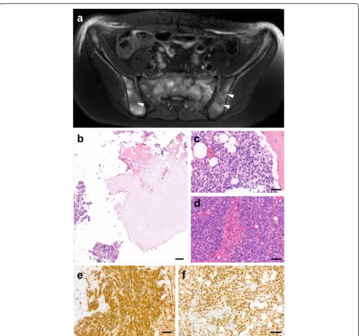
A 17-year-old male with Crohn’s disease in his medical history, presented with fever, weight loss and lower back pain; experienced over a period of 1–2 weeks. He was found to have elevated inflammatory markers and ser- ious hypercalcemia with impaired renal function. Bone scintigraphy, lumbar spine and pelvic MRI revealed dis- seminated, diffuse infiltration of the bone marrow which primarily raised the suspicion of lymphoma (Fig. 1a). Re- peated bone marrow biopsies (iliac crests) confirmed ARMS (Grade III). A primary soft tissue tumor was never found. During chemotherapy according to CWS- 2012 Protocol’s metastatic arm, dose reduction and modifications of cytostatic drugs, intensive care and hemodialysis were required several times due to serious arrhythmias and renal insufficiency caused by osteolysis- induced hypercalcemia. Despite the appropriate, aggres- sive chemotherapy, his disease showed progression that could be delayed temporarily by RANKL inhibitor deno- sumab monotherapy for a four month period. We lost him seven months after the initial symptoms.
Pathological findings of Case 1
Histological examination of the second bone marrow bi- opsy (iliac crests) revealed solid sheets of tumor cells in- filtrating the entire bone marrow replacing and expelling the normal hematopoietic cells. The highly cellular infil- trate showed no special arrangement, although fine fibro-vascular stroma could focally be identified. The monomorphic, poorly differentiated tumor cells had round, vesicular nuclei with fine chromatin content and were localized at the periphery of the cells, in an eccen- tric position (Fig. 1b). The cytoplasm of most of the
tumor cells possessed either an eosinophilic appearance or abundant intracytoplasmic vacuoles could be seen.
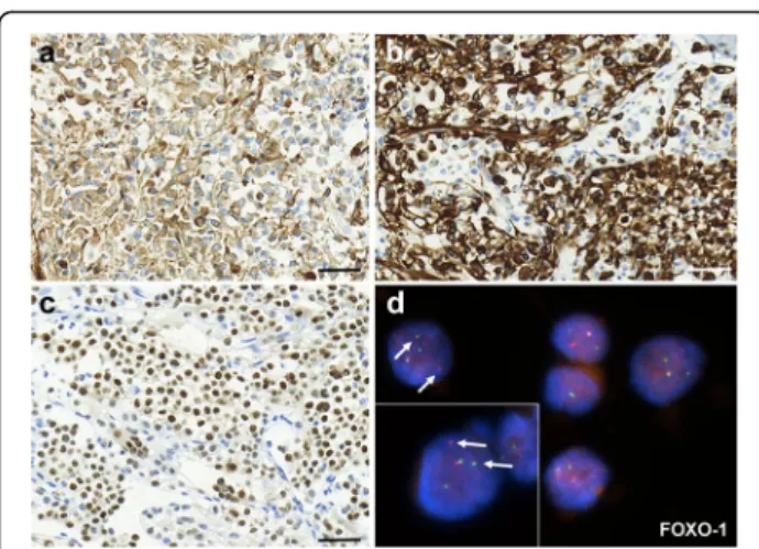
While tumor cells did not show striation, the overall morphology suggested rhabdomyoblast-like differenti- ation (Fig. 1b). Although, by examining a HE specimen, a hematological malignancy could be ruled out, further immunohistochemical (IHC) tests were needed to characterize the phenotype of the tumor cells. The re- sults of IHC showed diffuse vimentin positivity as well as the cells gave substantial cytoplasmic and nuclear la- belling with both rhabdomyogen markers, desmin and Myf-4, respectively (Fig. 2a–c). The pan-cytokeratin and TFE-3 reactions were negative as well as INI-1 was retrained; by which alveolar soft part sarcoma or rhab- doid tumor as a differential diagnostic possibility could be ruled out. As the overall pattern of the tumor was not typical for neither embryonal nor alveolar RMS, we further performed a FOXO-1 break-apart FISH probe as the aforementioned gene is known to be involved and is consistently associated with the alveolar subtype of RMS. Indeed, we detected the translocation and break- apart signals involving FOXO-1 (Fig. 2d). Based on the histological and molecular findings as well as extended radiological examinations not proving a primary soft tissue tumor, the diagnosis of primary ARMS (solid vari- ant) of the bone was made.
Clinical background of Case 2
A 9-year-old male was admitted to the hospital pre- sented with recurrent fever, lower back and right lower limb pain, experienced over the period of a month. He was found to have mild anemia and elevated inflamma- tory markers. Imaging studies revealed disseminated multiplex bone lesions involving the entire vertebral col- umn, pelvic bones, ribs, skull, the distal part of the right femur and the proximal part of the right tibia (Fig. 3a).
These findings and the lack of primary soft tissue mani- festation raised the possibility of Ewing-sarcoma or
Fig. 1Radiological image and hematoxylin-eosin stained specimen of the tumor.aAxial T2 SPAIR image of pelvis shows diffuse patchy infiltration (arrowheads) of the bone marrow. Some small necrotic areas are also visible.bThe HE stained biopsy sample shows highly cellular infiltrate among the bony trabeculae repelling the normal hematopoietic cells (insert image). With higher magnification, the monomorphic tumor cells have a characteristic eosinophilic cytoplasm, however tumor cells also show intracytoplasmic vacuolization (arrowheads). The nuclei of tumor cells are eccentric in position, but rather identical in size and own a finely granulated, basophilic nuclear structure (arrows). Note that the organization of tumor cells represent a somewhat nest-like pattern, but lack fine fibrovascular stroma, that is characteristic of the solid variant of alveolar RMS. Bars indicate 50μm, insert 100μm
malignant lymphoproliferative disease. Initially, the pa- tient required intensive therapy for serious hypercalce- mia and its complications due to osteolysis. The initial histological diagnosis was mesenchymal chondrosar- coma (Grade III), but the atypical clinical findings made histological revision necessary which, in turn, confirmed alveolar rhabdomyosarcoma with bone marrow involve- ment. A primary soft tissue tumor could not be identi- fied. The therapeutic response was excellent in relation to the first-line chemotherapy given according to CWS- 2009 Protocol’s metastatic arm as control MRI and PET/
CT revealed complete remission. Seven months after fin- ishing the first-line therapy, a relapse of the primary dis- ease was confirmed, localized to the distal femur and proximal tibia on the right side. Second-line therapy was given according to CWS-2012 relapse protocol and based on the proven ALK-positivity of the tumor, ALK inhibitor crizotinib was permitted as an off-label drug for maintenance therapy for 10 months. In the fourth month of crizotinib treatment multiplex metastases were confirmed. Based on the proven increased mTOR activ- ity of the previous biopsy specimen (iliac crests), mTOR inhibitor temsirolimus was given for 3 months. Due to disease progression, both targeted therapies were stopped and 30 months after the primary diagnosis, we lost the patient.
Pathological findings of Case 2
The biopsy sample that was taken from the tibia showed different morphological patterns and areas that made the overall histological picture misleading: among the
bony trabeculae, a cellular tumor infiltrate could be identified and the cells were arranged in solid sheets re- placing the normal hematopoiesis. Other foci of the tumor showed intramedullary cartilage islands around which spindle or ovoid shape tumor cells formed a cohe- sive structure; allowing mesenchymal chondrosarcoma diagnosis. Besides this, however, some areas of the tumor formed solid sheets of tumor islands that were divided by fine fibro-vascular stroma (Fig. 3b–d). The cytomorphology was identical with a so called “small round blue cell tumor”. Considering that neither the age nor the dissemination of the process (multiplex bony le- sions) were typical for mesenchymal chondrosarcoma, we further evaluated the phenotype of the tumor cells with several IHC tests. The cells showed cytoplasmic positivity with vimentin as well as intensive and diffuse cytoplasmic desmin and nuclear Myf-4 reactions being observed (Fig. 3e–f ). To further characterize the subtype of RMS without an identifiable soft tissue component, we performed a FOXO-1 break apart FISH DNA probe that evaluated the translocation involving FOXO-1. Con- cerning the clinical and pathological findings, the final diagnosis of primary alveolar rhabdomyosarcoma of the bone was made.
Discussion
Alveolar rhabdomyosarcoma is a high grade neoplasm that has the worst prognosis amongst other subtypes of RMSs (despite combined surgical and chemo/radiother- apy), especially in fusion-positive cases when FOXO-1 gene is involved. It is generally known that the overall outcomes for patients with soft tissue ARMS is worse than in patients with ERMS - even with aggressive multimodal therapy [4]. The prognostic factors defining the outcome of patients with RMS includes the follow- ing parameters: patient’s age, site of origin, tumor size, resectability, presence of metastases, number of meta- static sites or tissues involved, presence or absence of regional lymph node involvement, delivery of radiation therapy in selected cases, the unique biological charac- teristics of RMS tumor cells and, lastly the histological subtype. [11–14]. Regarding the histopathological sub- type, there is a significant difference between the 5-year survival with ERMS (82 %) and soft tissue ARMS (65 %) [4]. Besides this, patients with (soft tissue) ARMS who have regional lymph node involvement face a worse out- come (5-year failure-free survival: 43 %) as compared to patients lacking lymph node involvement (5-year failure- free survival: 73 %) [15].
Although the previously reported four cases of primary ARMS (as well as our current two cases) show a better survival rate compared to its soft tissue counterpart, it still causes difficulties in precisely characterizing this tumor type. One reason is the low number of reported
Fig. 2Immunohistochemical and molecular characterization of tumor cells.aTumor cells show diffuse vimentin positivity, as well as diffuse and strong cytoplasmic and nuclear labelling could be observed with myogenic markers desmin and Myf-4, respectively (b–c). The result of FOXO-1 gene break-apart FISH probe demonstrates divided green and red signals indicating translocation of the affected gene (d). Bars indicate: 50μm
cases, while a major problem alongside this is that even data contained within medical literature is confusing with regards to ARMS classification. It distinguishes fusion-positive and fusion-negative cases; however, there is a tendency that fusion-negative cases should be con- sidered in practical terms ERMS [16]. Until this ten- dency is not generalized and accepted in routine diagnostic pathology, there will be cases influencing and altering the results exhibited in statistics.
In the case of (soft tissue) alveolar RMS it is known that it commonly infiltrates the bone marrow [17], caus- ing a diagnostic challenge (both in childhood and adult cases), as it can mimic the symptoms of either a hematological malignancy or a primary bone tumor;
therefore, biopsy sampling is necessary in each and every case. The most common differential diagnostic problems (considering the localization and/or age) are as follows:
Ewing sarcoma, non-Hodgkin lymphoma, mesenchymal
Fig. 3Radiological image and histopathology of Case 2.aOn the axial T2 SPAIR image of the pelvic bone multiple, partially coalesced tumor nodules are visible in the bone marrow. Some of them (arrowheads) show central necrosis.bThere were foci of tumor islands with atypical chondrogenic differentiation surrounded by round/spindle shape tumor cells.c–dBesides that, expansive sheet like pattern with solid nests could also be identified that were divided by fine fibrovascular septa. The infiltrate displaced the normal hematopoiesis of the bone marrow.e–fThe diffuse and intensive cytoplasmic desmin and nuclear Myf-4 positivity proved rhabdomyosarcoma differentiation of tumor cells. Bars indicate: 50μm
chondrosarcoma and the small cell variant of osteosar- coma. While the morphology of tumor cells are similar (small, round cells), the pattern of infiltration or the ac- companying component of the tumor (neoplastic osteoid or hyaline cartilage in small cell variant of OS and mesen- chymal chondrosarcoma, respectively) as well as special cytomorphological features such as intracytoplasmic vacu- oles or striation of the tumor cells (like in RMS) may sometimes suggest the differentiation lineage. Besides the careful examination of HE stained samples and morpho- logical analysis, ancillary techniques are essential in these cases in order to give a definitive diagnosis. The combin- ation of IHC tests including LCA, vimentin, desmin and CD99 is useful to primarily assess the phenotype of the tumor cells. Although IHC evaluation is sufficient and may lead to a final diagnosis, in most of the cases further molecular examinations such as flow cytometry (especially in hematological diseases) or genetic analysis with regard to gene fusion status (e.g. in Ewing sarcoma and ARMS) are now part of the routine diagnostic panel [18, 19].
The natural history of primary alveolar RMS of bone may show individual variations, but our current cases,
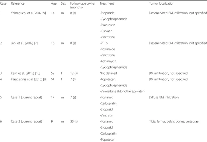
together with the other four reported ones [7–10], sug- gest a better overall prognosis as compared to soft tissue ARMS (Table 1). Primary alveolar rhabdomyosarcoma of the bone as a subtype of ARMS seems to be a distinct clinico-pathological entity. We wish to stimulate the sci- entific community into publishing and following-up similar cases. With this proposal, there might be more available data to predict not only the biological behavior and prognosis of the disease, but also to develop and set up further chemotherapeutical combinations that may increase the overall survival of the patients in the future.
Conclusion
Primary alveolar rhabdomyosarcoma of the bone as a subtype of ARMS, seems to be a distinct clinico- pathological entity with challenging diagnostic difficul- ties and different biological behavior when compared to soft tissue ARMS. More available data might be neces- sary to predict not only the course of the disease, but also to develop and set up further chemotherapeutical combinations that may increase the overall survival of the patients in the future.
Table 1Reported cases of primary alveolar rhabdomyosarcoma of the bone so far without identifiable soft tissue component
Case Reference Age Sex Follow-up/survival
(months)
Treatment Tumor localization
1 Yamaguchi et al. 2007 [9] 14 m 8 (s) -Etoposide Disseminated BM infiltration, not specified
-Cyclophosphamide -Pirarubicin -Cisplatin -Vincristine
2 Jani et al. (2009) [7] 16 m 8 (s) -VP16 Disseminated BM infiltration, not specified
-Ifosfamide -Vincristine -Adriamycin -Cyclophosphamide
3 Kern et al. (2015) [10] 52 f 12 (s) Not detailed BM infiltration, not specified
4 Karagiannis et al. (2015) [8] 61 f 7 (f) -Topotecan BM infiltration, not specified
-Cyclophosphamide
-Vinorelbine (Monotherapy-later)
5 Case 1 (current report) 17 m 7 (s) -Ifosfamid Diffuse BM infiltration
-Carboplatin -Etoposid -Vincristin
6 Case 2 (current report) 9 m 30 (s) -Ifosfamid Tibia, femur, pelvic bones, vertebrae
-Etoposid -Carboplatin -Topotecan
The subtype has been evaluated in each case with molecular diagnostic tools (FISH, Sanger sequencing, spectral karyotyping, cytogenetics) BMbone marrow,ssurvival,ffollow-up
Abbreviations
ALK:Anaplastic lymphoma receptor tyrosine kinase; ARMS: Alveolar rhabdomyosarcoma; CWS: Cooperative Soft Tissue Study Group;
DNA: Deoxyribonucleic acid; ERMS: Embryonal rhabdomyosarcoma;
FISH: Fluorescence in situ hybridization; FOXO-1: Forkhead box protein O1; HE: Hematoxylin-eosin; IHC: Immunohistochemistry; INI-1: Integrase interactor 1; LCA: Leukocyte common antigen; MRI: Magnetic resonance imaging; mTOR: Mammalian (mechanistis) target or rapamycin; Myf4: Myogenin factor 4; OS: Osteosarcoma; PAX-3/7: Paired box gene 3/7; PCR: Polymerase chain reaction; PET/CT: Positron emission tomography-computed tomography; RANKL: Receptor activator of nuclear factor kappa-B ligand;
RMS: Rhabdomyosarcoma; SKY: Spectral karyotyping; TFE3: Transcription factor E3
Acknowledgement None.
Funding
The authors declare that no funding was used.
Availability of data and material
The radiological and clinical data can be found in the archive of the Department of II. Pediatric Clinic, Semmelweis University. The histological specimens and the corresponding data are belonging to the archive of the 1stDepartment of Pathology and Experimental Cancer Research, Semmelweis University. The raw data (radiological, clinical, pathological) included in the current manuscript is available upon request for review by the Editor-in-Chief.
Authors’contributions
PB, ZS participated in the design of the study and histopathological evaluation, and drafted the manuscript. MCS assisted in drafting the manuscript and revised the manuscript. RB, ZSV and EV made contributions in analyzing the clinical background and radiological evaluation of the cases. ZS revised the manuscript and gave final approval of the manuscript as professor of the 1st Department of Pathology and Experimental Cancer Research, Semmelweis University. All authors have given approval for the final version to be published.
Competing interest
The authors declare that they have no competing interests.
Consent for publication
Consent was obtained from the next of kin of the patients (parents) for the publication of the cases and any accompanying images. A copy of the written consent is available for review by the Editor-in-Chief of this journal.
Ethical approval and consent to participate
The ethical approval and documentation for a case report was waived with approval of the Institutional Review Board at Semmelweis University.
Author details
11st Department of Pathology and Experimental Cancer Research, Semmelweis University, Üllői út 26, Budapest H-1085, Hungary.22nd Department of Pediatrics, Semmelweis University, Tűzoltó utca 7-9, Budapest H-1094, Hungary.3Magnetic Resonance Research Center, Semmelweis University, Tűzoltó utca 7-9, Budapest H-1094, Hungary.
Received: 5 May 2016 Accepted: 8 October 2016
References
1. Parkin DM, Stiller CA, Draper GJ, Bieber CA. The international incidence of childhood cancer. Int J Cancer. 1988;42(4):511–20.
2. Ognjanovic S, Olivier M, Bergermann TL, Hainaut P. Sarcomas in TP53 germline mutation carriers. Cancer. 2012;118(5):1387–96.
3. Raney RB, Anderson JR, Barr FG, Donaldson SS, Pappo AS, Qualman SJ, Wiener ES, Maurer HM, Crist WM. Rhabdomyosarcoma and undifferentiated sarcoma in the first two decades of life: a selective review of intergroup rhabdomyosarcoma study group experience and rationale for Intergroup Rhabdomyosarcoma Study V. J Pediatr Hematol Oncol. 2001;23(4):215–20.
4. Meza JL, Anderson J, Pappo AS, Meyer WH. Analysis of prognostic factors in patients with nonmetastatic rhabdomyosarcoma treated on Intergroup
Rhabdomyosarcoma Studies III and IV: The Children’s Oncology Group.
J Clin Oncol. 2006;24(24):3844–51.
5. Davis RJ, Barr FG. Fusion genes resulting from alternative chromosomal translocations are overexpressed by gene-specific mechanisms in alveolar rhabdomyosarcoma. Proc Natl Acad Sci U S A. 1997;94(15):8047–51.
6. Keller C, Arenkiel BR, Coffin CM, El-Bardeesy N, DePinho RA, Capecchi MR.
Alveolar rhabdomyosarcoma in conditional Pax3:Fkhr mice: cooperativity in Ink4a/ARF and Trp53 loss of function. Genes Dev. 2004;18(21):2614–26.
7. Jani P, Charles CY. Massive bone marrow involvement by clear cell variant of rhabdomyosarcoma. Ind J Pediatrics. 2009;76(2):224–8.
8. Karagiannis P, Guth N, Thoennissen GB, Bern C, Sperveslage J, Oschlies I, Bokemeyer C, Klapper W, Wardelmann E, Thoennissen NH. Alveolar rhabdomyosarcoma confined to the bone marrow with no identifiable primary tumour using FDG-PET/CT. Clin Sarcoma Res. 2015;5:24. doi:10.
1186/s13569-015-0039-6.
9. Yamaguchi K, Koda Y, Suminoe A, Saito Y, Matsuzaki A, Kanno S, Takimoto T, Suda M, Oda Y, Muto T, Takatsuki H, Hara T. Alveolar rhabdomyosarcoma of unknown origin mimicking acute leukemia at the initial presentation. Rinsho Ketsueki. 2007;48(4):315–20.
10. Kern JB, Hil A, Kruse MJ, Szabo Z, Argani P, Hibbard MK, Gladstone DE, Meyer C, Zheng R, Borowitz MJ, Duffield AS. A leukemic presentation of alveolar rhabdomyosarcoma in a 52-year-old woman without an identifiable primary tumor. Int J Surg Pathol. 2015;23(1):75–7.
11. Crist W, Gehan EA, Ragab AH, et al. The Third Intergroup Rhabdomyosarcoma Study. J Clin Oncol. 1995;13(3):610–30.
12. Maurer HM, Gehan EA, Beltangady M, Crist W, Dickman PS, Donaldson SS, Fryer C, Hammond D, Hays DM, Hermann J, et al. The Intergroup Rhabdomyosarcoma Study-II. Cancer. 1993;71(5):1904–22.
13. Breneman JC, Lyden E, Pappo AS, Link MP, Anderson JR, Parham DM, Qualman SJ, Wharam MD, Donaldson SS, Maurer HM, Meyer WH, Baker KS, Paidas CN, Crist WM. Prognostic factors and clinical outcomes in children and adolescents with metastatic rhabdomyosarcoma–a report from the Intergroup Rhabdomyosarcoma Study IV. J Clin Oncol. 2003;21(1):78–84.
14. Mandell L, Ghavimi F, LaQuaglia M, Exelby P. Prognostic significance of regional lymph node involvement in childhood extremity rhabdomyosarcoma. Med Pediatr Oncol. 1990;18(6):466–71.
15. Rodeberg DA, Garcia-Henriquez N, Lyden ER, et al. Prognostic significance and tumor biology of regional lymph node disease in patients with rhabdomyosarcoma: a report from the Children's Oncology Group.
J Clin Oncol. 2011;29(10):1304–11.
16. Rudzinski ER, Teot LA, Anderson JR, Moore J, Bridge JA, Barr FG, Gastier- Foster JM, Skapek SX, Hawkins DS, Parham DM. Dense pattern of embryonal rhabdomyosarcoma, a lesion easily confused with alveolar rhabdomyosarcoma.
Am J Clin Pathol. 2013;140(1):82–90.
17. Kuttesch Jr JF, Parham DM, Kaste SC, Rao BN, Douglass EC, Pratt CB. Embryonal malignancies of unknown primary origin in children. Cancer. 1995;75(1):115–21.
18. Rajwanshi A, Srinivas R, Upasana G. Malignant small round cell tumors. J Cytol.
2009;26(1):1–10.
19. Walther C, Mayrhofer M, Nilsson J, Hofvander J, Jonson T, Madahl N, Ora I, Gisselsson D, Mertens F. Genetic heterogeneity rhabdomyosarcoma revealed by SNP array analysis. Genes Chromosomes Cancer. 2015;55(1):3–15.
• We accept pre-submission inquiries
• Our selector tool helps you to find the most relevant journal
• We provide round the clock customer support
• Convenient online submission
• Thorough peer review
• Inclusion in PubMed and all major indexing services
• Maximum visibility for your research Submit your manuscript at
www.biomedcentral.com/submit
Submit your next manuscript to BioMed Central and we will help you at every step:


