Research Article
Surgery Treatment Improved the Overall Survival Rate in Locoregional Myxoid Leiomyosarcoma than Other
Myxosarcomas in the United States
Marwan Almoiliqy ,1,2Abdullah Al-danakh,3Mohammed Safi,4Mohammed Alradhi,5 Mahmoud AL-Azab ,6Salah Adlat,7Wanhai Zhou,1Aiman Saleh A. Mohammed,8 and Ahmed Al-maamari9
1Key Lab of Aromatic Plant Resources Exploitation and Utilization in Sichuan Higher Education, Yibin University, Yibin, 644000 Sichuan, China
2Department of Pharmacology, Pharmaceutical College, Dalian Medical University, Dalian 116044, China
3Department of Urology, First Affiliated Hospital of Dalian Medical University, Dalian 116044, China
4Department of Oncology, First Affiliated Hospital of Dalian Medical University, Dalian 116044, China
5Department of Urology, Second Affiliated Hospital of Dalian Medical University, Dalian 116044, China
6Department of Immunology, Guangzhou Institute of Pediatrics, Guangzhou Women and Children’s Medical Center, Guangzhou Medical University, Guangzhou 510623, China
7Guangdong Cardiovascular Institute, Guangdong Provincial People’s Hospital, Guangdong Academy of Medical Sciences, Guangzhou, Guangdong 510100, China
8Department of Pharmacology and Pharmacotherapy, Faculty of Medicine, Interdisciplinary Excellence Center, University of Szeged, Szeged 6720, Hungary
9Department of Pharmacology, School of Basic Medical Sciences, Xi’an Jiaotong University, Health Science Center, Xi’an 710061, China
Correspondence should be addressed to Marwan Almoiliqy; almoiliqy@yahoo.com Received 7 March 2021; Accepted 20 April 2021; Published 3 May 2021
Academic Editor: Vladimir Jakovljevic
Copyright © 2021 Marwan Almoiliqy et al. This is an open access article distributed under the Creative Commons Attribution License, which permits unrestricted use, distribution, and reproduction in any medium, provided the original work is properly cited.
Myxosarcomas are rare malignant tumors of soft connective tissues, classified into various subtypes, including myxoid liposarcoma, myxoid chondrosarcoma, and myxoid leiomyosarcoma. In this study, we proposed to study the demographic, tumor characteristics, and overall survival rate and compared the treatment modalities between these cancers. Patient data collected based on locoregional metastasis presentation of the abovementioned tumors with a cutoff study of survival duration up to 10 years were obtained from the SEER database during 1975-2016. Our results indicated that elderly patients and females were more in locoregional myxoid leiomyosarcoma than myxoid liposarcoma and myxoid chondrosarcoma with locoregional metastasis. The white race represented the most patients who suffered from these cancers than other races. The heart is the primary site for the abovementioned cancers, in addition to the female genitals to the myxoid leiomyosarcoma. Myxoid liposarcoma and myxoid chondrosarcoma patients with locoregional metastasis were suffering from grade II, while locoregional myxoid leiomyosarcoma patients with blank grading were due to missed data. Surgery was the most common treatment modality in this study compared with radiotherapy and chemotherapy.
Kaplan-Meier analysis showed a significant difference in survival time between the three subtypes by using histology, and myxoid leiomyosarcoma showed prolonged survival than others. Elderly, female, white, unknown grade, surgery, no radiation, and no chemotherapy variables were independent factors associated with overall survival among these cancers.
Volume 2021, Article ID 9999529, 12 pages https://doi.org/10.1155/2021/9999529
Multivariate analysis also showed significant differences in overall survival between the three tumors by histology, and myxoid leiomyosarcoma was with a better prognosis than others. Multivariate analysis of locoregional myxoid leiomyosarcoma showed the statistical significance of black race, grade, and radiotherapy, indicating them as independent prognostic factors of locoregional myxoid leiomyosarcoma. We conclude that surgery was the primary treatment modality against these cancers than radiotherapy and chemotherapy. And the locoregional myxoid leiomyosarcomas showed a better prognosis and higher survival rate than locoregional myxoid liposarcoma and locoregional myxoid chondrosarcoma.
1. Introduction
Myxosarcomas are rare malignant tumors of soft connective tissues [1]. Myxomatous areas indicated mucin deposition between the tumorous cells or in their exoplasm, including mesenchymal and epithelial cells [2]. Myxosarcoma mostly occurs in the heart [3, 4] but also may appear in other body locations including bone [5, 6], skin [7, 8], skeletal muscles [9], and ocular [10] and oral [11] cavities. Myxosarcomas are characterized histopathologically with stellate-shape and spare-spindle cells, neoplastic undifferentiated cells, mitotic figures, and mucoid stroma [12–14], and a low rate of myxo- sarcoma distant metastasis has been reported [15]. Myxosar- coma is mostly treated via surgery [16].
Myxosarcoma tumors are classified into various sub- types, including myxoid liposarcoma, myxoid chondrosar- coma, and myxoid leiomyosarcoma [17, 18]. Myxoid liposarcoma is a subtype of liposarcoma with a low risk of local recurrence than larger tumors [19]. Myxoid liposarco- mas represent 5% of soft tissue sarcoma and 10-20% of lipo- sarcoma [20]. However, myxoid liposarcomas may develop metastasis in almost 10% of myxoid liposarcoma patients and constitute 80% of the 5-year overall survival rate and 60% of the 10-year overall survival rate [21]. Myxoid chon- drosarcomas are rare in extremities’soft tissues with less than 3% of soft tissue sarcomas [22]. Despite the myxoid chondro- sarcomas characterized with a high risk of local recurrence and high spread rate of metastasis, several studies reported a prolonged survival rate of almost 70% of 10-year overall survival [22–24]. Myxoid leiomyosarcomas are a subtype of leiomyosarcomas characterized as rare, aggressive, and well-recognized uterus tumors [25]. Myxoid leiomyosarcoma showed a poor prognosis and a worse overall survival rate of 5 years, almost 11% [26].
Directed locoregional therapy on the metastasis pattern with additional treatment of the primary tumors may reduce the primary tumor burden, reduce metastasis prognosis, and improve the overall survival rate than systemic treatment to the distant metastasis [27–29]. It is reported that primary tumor resection could promote distant metastasis [30, 31]
while this hypothesis was drawn by recent studies and eluci- dated clinically the role of surgery in reducing the distant metastasis and improving the overall survival rate [32, 33].
The Surveillance, Epidemiology, and End Results (SEER) is a comprehensive program of incidence, prognosis, and cancer surveillance in the United States (U.S.). SEER is an authoritative program that provides information on cancer statistics in the U.S. to minimize the cancer prognosis on the U.S. population. SEER is supported by the Surveillance Research Program (SRP) in the National Cancer Institute’s (NCI’s) Division of Cancer Control and Population Sciences
(DCCPS). Among 19 registries around the U.S., cancer data has been collected through a coordinated system. These geo- graphic registries reflect about 35% of the U.S. population, which represents the entire U.S. demographics. Data collec- tion started with a few registries in 1973 and expanded to include more areas in the U.S. [34–36].
Myxoid liposarcomas, myxoid chondrosarcomas, and myxoid leiomyosarcomas are rare tumors with low incidence and survival data, so a comparison study has also been chal- lenging to clarify. So, we proposed to study the demographic, tumor characteristics, and overall survival rates and to com- pare the treatment modalities between locoregional metasta- tic myxoid liposarcomas, locoregional metastatic myxoid chondrosarcomas, and locoregional metastatic myxoid leio- myosarcomas during 1975-2017 in 18 registries of the United States based on SEER database analysis.
2. Methods 2.1. Data Collection
2.1.1. Study Cohort. Patient data of myxoid liposarcoma, myxoid chondrosarcoma, and myxoid leiomyosarcoma were obtained from the SEER database of cases diagnosed during 1975-2016 in 18 registries of the United States [37]. All patients were identified based on the international classifica- tion of disease for oncology, third edition (ICD-O-3), using codes of myxoid liposarcoma (8850-8889), myxoid chondro- sarcoma (9180-9249), and myxoid leiomyosarcoma (8890- 8929). The data extracted from the SEER database in this study was based only on the locoregional metastasis presen- tation of myxoid liposarcoma, myxoid chondrosarcoma, and myxoid leiomyosarcoma tumors. We obtained a total number of patients of about 1398 of locoregional myxoid liposarcoma, 356 of locoregional myxoid chondrosarcoma, and 129 of locoregional myxoid leiomyosarcoma. Cases with unknown or missing data are included in the descriptive and survival analysis.
2.1.2. Study Measurement. This study’s primary measure- ment is to identify the locoregional metastasis differences of demographic, tumor characteristics, and survival rates between these three rare myxoid tumors (myxoid liposar- coma, myxoid chondrosarcoma, and myxoid leiomyosar- coma) among 18 registries of the United States during1975- 2016. All variables included in this study are based on the cutoffstudy of survival duration up to 10 years [37].
2.1.3. Variable Study.All patient data are extracted from the SEER database. Demographic data were organized as age at diagnosis, gender, and race. The age variable was studied in two categories (less than 50 and greater than 50) to ease the
analysis. The race was categorized as white, black, and others (Asian and others). The anatomical locations were catego- rized into six variables, including soft tissue including heart, retroperitoneum, bones and joints, female genitals, digestive organs, and others. Oncology study includes grading and sur- vival analysis. The tumor grade in the SEER database was classified into grades I, II, III, IV, and unknown. Overall sur- vival rate analysis is performed based on the study cutoff selection of patient data from SEER. So, all cases in this study considered only the patients with 10-year survival (120 months). The SEER database’s treatment variables, including surgery, radiation, and chemotherapy, were included in this study. In surgery treatment, the patients were categorized into two variables: whether they performed the surgery or not (yes/no). Radiation and chemotherapy variables include the patients who received treatment or not (yes/no) [37].
2.2. Statistical Analysis.Data analysis was performed using SPSS software version 22 (SPSS, IBM Company, Chicago, USA). Chi-squared tests are used to analyze the variables.
Categorical variables were analyzed as frequency and per- centage. Ten-year overall survival was calculated using Kaplan-Meier to estimate the survival rate with a log-rank test. Multivariate Cox proportional hazard regression was studied to analyze the confounding variables. p< 0:05 was used as a significant value in the study.
3. Results
3.1. Myxoid Liposarcoma
3.1.1. Demographics. Among 1398 locoregional metastatic patients of myxoid liposarcoma extracted from the SEER database during 1975-2016, almost 49.1% of cases with age less than 50 years and 50.9% of cases with age greater than 50 years were diagnosed with locoregional myxoid liposar- coma. Myxoid liposarcomas were higher in males than females, with almost 59.4% and 40.6% of cases, respectively.
The race variable reported that the white race of almost 82.5% was higher than black and other races of almost 9.7%
and 7.9%, respectively, in this tumor, indicating that the male gender and white people were more vulnerable to this cancer.
The location of the myxoid liposarcoma tumors showed higher localization of cancer in the heart, which is almost 91.5% of the reported cases compared with other sites (Table 1).
3.1.2. Oncology Characteristics. The reported data of the locoregional metastatic patients during 1975-2016 and obtained from the SEER database with a total number of 1398 myxoid liposarcomas in the United States showed that this tumor has high grading II (32.8%) versus grade I (27.9%), grades III-IV (7.6%-8.0%), and unknown (23.7%) indicating that locoregional metastatic patients with myxoid liposarcomas have high grading of this tumor. The cause of death of most cases of myxoid liposarcoma was not attrib- uted to this tumor as reported in SEER data with almost 91.4% (Table 1).
3.1.3. Treatment. Locoregional metastatic myxoid liposar- coma treatment data are extracted from SEER during 1975- 2016, including surgery, radiotherapy, and chemotherapy modalities. Most of the reported cases in the SEER of locore- gional metastatic patients with myxoid liposarcoma were performed surgery which is almost 95.2% of the total number of patients, and this result was similar as in the literature [38].
Radiotherapy treatments of patients with locoregional myx- oid liposarcoma of data obtained from SEER showed no big difference between received beam radiations versus unrec- eived patients, almost 47.1% versus 52.9%. Meanwhile, che- motherapy variables showed a higher data of patients who did not receive chemotherapy which is about 89.5% com- pared with the patients who received treatment which is about 10.5%, and these results of treatment modality indicate that surgery treatment modality was the most performed treatment approach in locoregional myxoid liposarcoma patients (Table 1).
3.2. Myxoid Chondrosarcoma
3.2.1. Demographics. The data of locoregional metastatic patients with myxoid chondrosarcoma obtained from SEER were 356 patients. There are almost 55.1% of cases with age less than 50 years versus 44.9% of cases with age greater than 50 years. The sex variable of locoregional metastatic myxoid chondrosarcomas reported that the male variable was higher than the female variable with almost 59.8% and 40.2% of cases, respectively. White patients were also higher than black and other races, about 76.7%, 14.6%, and 8.7%, respec- tively, in this tumor, indicating that this tumor mostly affects male and white people than others. The localization of the locoregional metastatic myxoid chondrosarcoma tumor was higher in the heart and bones/joints, almost 64.0% and 32.3%, respectively, of the reported cases compared with other sites (Table 1).
3.2.2. Oncology Characteristics.The data obtained from the SEER database of locoregional metastatic patients with myx- oid chondrosarcomas showed that this tumor has higher grade II (32.6%) and unknown (38.8%) versus grades I, III, and IV (13.8%, 9.8%, and 5.1%, respectively), indicating that locoregional metastatic patients with myxoid chondrosarco- mas exhibit high grading of this tumor. The cause of death of most cases of locoregional metastatic myxoid chondrosar- comas also was not attributed to this tumor as reported in SEER data with almost 76.7% (Table 1).
3.2.3. Treatment.Most of the cases obtained from the SEER database with locoregional metastatic myxoid chondrosarco- mas were performed surgery with almost 89% of the patients’ total number. And data obtained of locoregional metastatic patients with myxoid chondrosarcomas did not show a big difference between cases that received radiotherapy or not, almost 44.1% and 55.9%, respectively. The chemotherapy variables showed a higher data of patients who did not receive chemotherapy, about 82.9%, compared with the patients who received treatment which is almost 17.1%, indi- cating that surgery is the primary treatment modality in
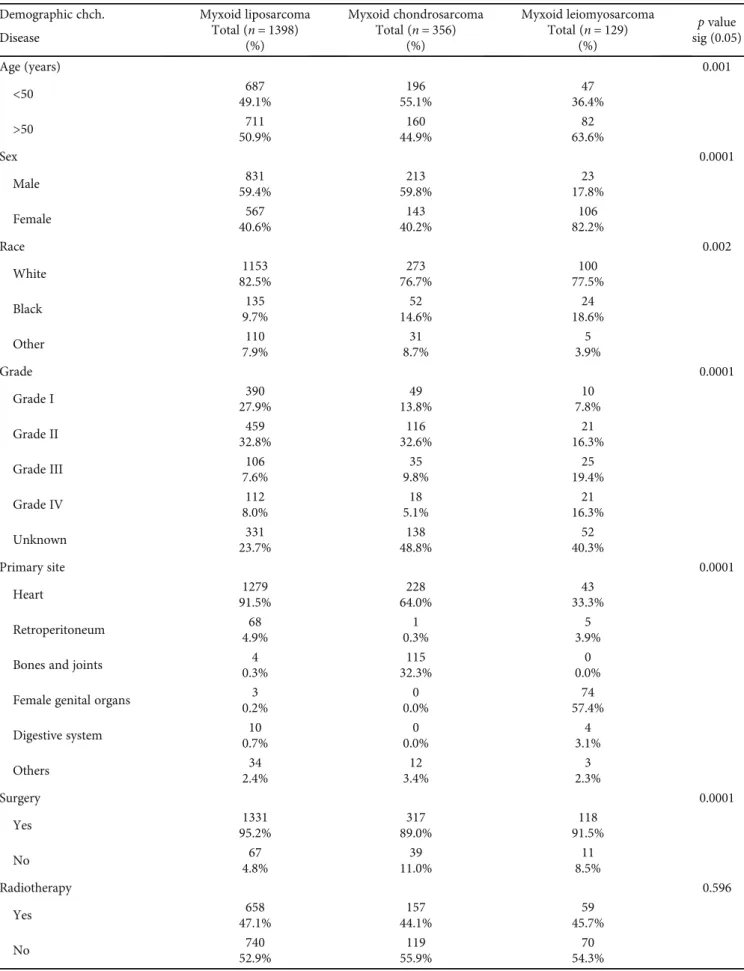
Table 1: Demographic data, tumor characteristics, and treatment modality of locoregional myxoid liposarcoma, locoregional myxoid chondrosarcoma, and locoregional myxoid leiomyosarcoma in the Unites States based on the SEER database.
Demographic chch. Myxoid liposarcoma Total (n= 1398)
(%)
Myxoid chondrosarcoma Total (n= 356)
(%)
Myxoid leiomyosarcoma Total (n= 129)
(%)
pvalue sig (0.05) Disease
Age (years) 0.001
<50 687
49.1%
196 55.1%
47 36.4%
>50 711
50.9%
160 44.9%
82 63.6%
Sex 0.0001
Male 831
59.4%
213 59.8%
23 17.8%
Female 567
40.6%
143 40.2%
106 82.2%
Race 0.002
White 1153
82.5%
273 76.7%
100 77.5%
Black 135
9.7%
52 14.6%
24 18.6%
Other 110
7.9%
31 8.7%
5 3.9%
Grade 0.0001
Grade I 390
27.9%
49 13.8%
10 7.8%
Grade II 459
32.8%
116 32.6%
21 16.3%
Grade III 106
7.6%
35 9.8%
25 19.4%
Grade IV 112
8.0%
18 5.1%
21 16.3%
Unknown 331
23.7%
138 48.8%
52 40.3%
Primary site 0.0001
Heart 1279
91.5%
228 64.0%
43 33.3%
Retroperitoneum 68
4.9%
1 0.3%
5 3.9%
Bones and joints 4
0.3%
115 32.3%
0 0.0%
Female genital organs 3
0.2%
0 0.0%
74 57.4%
Digestive system 10
0.7%
0 0.0%
4 3.1%
Others 34
2.4%
12 3.4%
3 2.3%
Surgery 0.0001
Yes 1331
95.2%
317 89.0%
118 91.5%
No 67
4.8%
39 11.0%
11 8.5%
Radiotherapy 0.596
Yes 658
47.1%
157 44.1%
59 45.7%
No 740
52.9%
119 55.9%
70 54.3%
locoregional metastatic myxoid chondrosarcoma patients than radiation and chemotherapy (Table 1).
3.3. Myxoid Leiomyosarcoma
3.3.1. Demographics.Myxoid leiomyosarcomas with locore- gional metastatic patients were obtained from the SEER data- base in the U.S. during 1975-2016, almost 129 cases. The age variable showed that about 63.6% of cases diagnosed with locoregional myxoid leiomyosarcoma were with age greater than 50 years versus 36.4% of cases with age less than 50 years, indicating that this tumor mostly occurs in the older population. The sex variable of locoregional myxoid leio- myosarcoma patients reported that the female variable was
higher than the male variable with almost 82.2% and 17.8%
of cases. White patients were higher than black and other races, almost 77.5%, 18.6%, and 3.9%, respectively, in this tumor, indicating that the female and white people were more vulnerable to this cancer. The myxoid leiomyosarcoma tumors were higher localized in the female genitals and heart, almost 57.4% and 33.3%, respectively, of the reported cases compared with other sites (Table 1).
3.3.2. Oncology Characteristics. The tumor grading variable of locoregional myxoid leiomyosarcoma reported cases showed that this tumor has higher unknown grading (40.3%) versus grades II, III, and IV (16.3%, 19.4%, and 16.3%, respectively) and less grade I (7.8%). The cause of death of locoregional myxoid leiomyosarcoma cases of almost 72.1% was not attributed to this tumor as reported in SEER data (Table 1).
3.3.3. Treatment.Most of the locoregional myxoid leiomyo- sarcoma patient’s data obtained from the SEER database are performed surgery with almost 91.5% of the total number of patients. And the data of patients with locoregional myx- oid leiomyosarcoma extracted by SEER did not show a big difference between groups receiving and not receiving radio- therapy of almost 45.7% and 54.3%, respectively. And most of the cases did not receive chemotherapy treatments, about 86% in this tumor (Table 1).
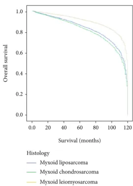
3.4. Survival Analysis.Kaplan-Meier analysis of overall sur- vival showed a statistically significant difference between his- tology subtypes of myxoid liposarcoma, myxoid chondrosarcoma, and myxoid leiomyosarcoma of locoregio- nal metastatic patients with log-rank p< 0:004 (Figure 1).
And locoregional metastatic myxoid leiomyosarcoma patients showed prolonged survival (median 85 months) compared with locoregional metastatic myxoid liposarcoma (median 58 months) and locoregional metastatic myxoid chondrosarcoma (median 60 months) (Figure 1). The Kaplan-Meier study indicated that variables of age more than 50 years, female gender, white race, unknown or missed grade, surgery resection, no radiation, and no chemotherapy variables showed statistical significance of overall survival Table1: Continued.
Demographic chch. Myxoid liposarcoma Total (n= 1398)
(%)
Myxoid chondrosarcoma Total (n= 356)
(%)
Myxoid leiomyosarcoma Total (n= 129)
(%)
pvalue sig (0.05) Disease
Chemotherapy 0.002
Yes 147
10.5%
61 17.1%
18 14.0%
No 1251
89.5%
259 82.9%
111 86.0%
Cause-specific death to cancer 0.0001
Yes 120
8.6%
83 23.3%
36 27.9%
No 1278
91.4%
273 76.7%
93 72.1%
chch: characteristics.
1.0
0.8
0.6
0.4
0.2
0.0
0.0 20 40 60 80 100 120
Overall survival
Survival (months) Histology
Myxoid liposarcoma Myxoid chondrosarcoma Myxoid leiomyosarcoma
Figure 1: Overall survival of patients with locoregional myxoid liposarcoma, locoregional myxoid chondrosarcoma, and locoregional myxoid leiomyosarcoma by histology via Kaplan- Meier analysis with log-rank test and median 58 months, 60 months, and 85 months, respectively,p< 0:004.
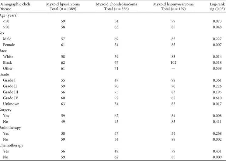
(Table 2). Patients older than 50 showed statistical signifi- cance of survival with a median of 58 months of locoregional myxoid liposarcoma, 63 months of locoregional myxoid chondrosarcoma, and 85 months of locoregional myxoid leiomyosarcoma with log-rank p< 0:008 (Table 2, Figure 2), indicating that patients with locoregional myxoid leiomyosarcoma showed prolonged survival than others.
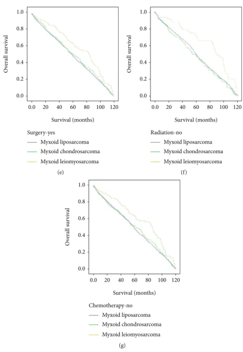
Female patients of locoregional myxoid leiomyosarcoma with a median of 85 months showed significantly prolonged survival time compared with locoregional myxoid liposar- coma and locoregional myxoid chondrosarcoma (61 months and 54 months), p< 0:007 (Table 2, Figure 2). And white patients also showed prolonged survival in locoregional myx- oid leiomyosarcoma (83 months) compared with locoregio- nal myxoid liposarcoma and locoregional myxoid chondrosarcoma (58 months and 59 months, respectively), p< 0:014(Table 2, Figure 2). The tumor grade showed that unknown or missed grading in myxoid leiomyosarcoma has significantly higher overall survival (median 85 months) compared with other tumors (63 and 54 median months, respectively), suggesting the inadequate data collection and poor follow-up of patients to their registries, p< 0:017 (Table 2, Figure 2). Among the treatment modalities, surgery
resection of myxoid leiomyosarcoma tumors in locoregional metastatic presentation results in prolonged survival time with a median of 84 months compared with locoregional myxoid liposarcoma and locoregional myxoid chondrosar- coma,p< 0:008(Table 2, Figure 2). The locoregional myxoid leiomyosarcoma patients who were not receiving radiother- apy and chemotherapy showed a significant difference in overall survival time and lived prolonged than other locore- gional myxoid liposarcoma patients and locoregional myxoid chondrosarcoma patients with log-rank p< 0:002and p<
0:009, respectively (Table 2, Figure 2). Multivariate analysis of histology subtype was also meaningful that showed a sta- tistically significant difference of overall survival between myxoid liposarcoma, myxoid chondrosarcoma, and myxoid leiomyosarcoma of locoregional metastatic patients, p<
0:0001, and the locoregional myxoid leiomyosarcoma cases reported better survival time than other cancers (Figure 3).
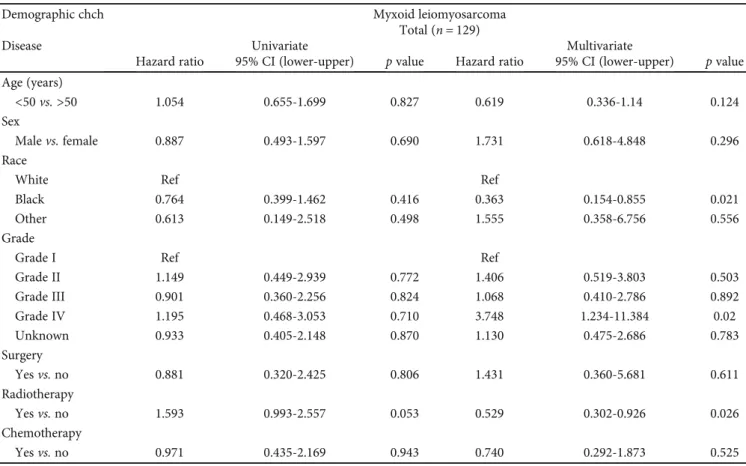
Based on the abovementioned results and comparing these three tumors, we selected the locoregional myxoid leiomyo- sarcoma to perform univariate and multivariate analyses.
And the univariate analysis of locoregional myxoid leiomyo- sarcoma showed no statistically significant difference in over- all survival (Table 3). Moreover, multivariate analysis showed Table2: Overall survival study using Kaplan-Meier analysis on locoregional myxoid liposarcoma, locoregional myxoid chondrosarcoma, and locoregional myxoid leiomyosarcoma in the United States.
Demographic chch Myxoid liposarcoma Total (n= 1389)
Myxoid chondrosarcoma Total (n= 356)
Myxoid leiomyosarcoma Total (n= 129)
Log-rank sig (0.05) Disease
Age (years)
<50 59 54 79 0.073
>50 58 63 85 0.048
Sex
Male 57 69 85 0.227
Female 61 54 85 0.007
Race
White 58 59 83 0.014
Black 62 67 102 0.318
Other 61 71 — 0.538
Grade
Grade I 55 47 98 0.361
Grade II 59 70 70 0.226
Grade III 56 75 83 0.195
Grade IV 60 92 62 0.610
Unknown 63 54 85 0.017
Surgery
Yes 59 62 84 0.008
No 49 45 85 0.411
Radiotherapy
Yes 38 47 54 0.268
No 59 54 89 0.002
Chemotherapy
Yes 56 49 79 0.431
No 59 62 85 0.009
chch: characteristics.
1.0 0.8 0.6 0.4 0.2 0.0
0.0 20 40 60 80 100 120
Overall survival
Survival (months)
Myxoid liposarcoma Myxoid chondrosarcoma Myxoid leiomyosarcoma Age < 50
(a)
1.0 0.8 0.6 0.4 0.2 0.0
0.0 20 40 60 80 100 120
Overall survival
Survival (months)
Myxoid liposarcoma Myxoid chondrosarcoma Myxoid leiomyosarcoma Female
(b)
1.0 0.8 0.6 0.4 0.2 0.0
0.0 20 40 60 80 100 120
Overall survival
Survival (months)
Myxoid liposarcoma Myxoid chondrosarcoma Myxoid leiomyosarcoma Race-white
(c)
1.0 0.8 0.6 0.4 0.2 0.0
0.0 20 40 60 80 100 120
Overall survival
Survival (months)
Myxoid liposarcoma Myxoid chondrosarcoma Myxoid leiomyosarcoma Grade-unknow
(d) Figure2: Continued.
that black race, with a hazard ratio (0.363), 95% CI (0.154- 0.855),p< 0:021; grade IV with a hazard ratio (3.748), 95%
CI (1.234-11.384),p< 0:02; and radiotherapy with a hazard ratio (0.529), 95% CI (0.302-0.926),p< 0:026were indepen- dent prognostic factors for locoregional myxoid leiomyosar- coma patients according to data obtained from SEER (Table 3).
4. Discussion
Myxosarcomas are malignant soft tissue tumors character- ized by abundant mucus [39]. Myxosarcoma was classified
into several subtypes, including myxoid liposarcoma, myx- oid chondrosarcoma, and myxoid leiomyosarcoma [18].
Metastasis presentation of myxoid liposarcomas, myxoid chondrosarcomas, and myxoid leiomyosarcomas was higher with locoregional metastasis in all three abovemen- tioned cancers compared with distant metastasis indicating the rare distant metastasis in these cancers, interpreting that locoregional metastasis may provide a prolonged overall survival rate than distant metastasis [40–43]. The SEER database is an authorized software providing statisti- cal information on cancers among several registries in the U.S. [34]. This study performed statistical analysis of
1.0 0.8 0.6 0.4 0.2 0.0
0.0 20 40 60 80 100 120
Overall survival
Survival (months)
Myxoid liposarcoma Myxoid chondrosarcoma Myxoid leiomyosarcoma Surgery-yes
(e)
1.0 0.8 0.6 0.4 0.2 0.0
0.0 20 40 60 80 100 120
Overall survival
Survival (months)
Myxoid liposarcoma Myxoid chondrosarcoma Myxoid leiomyosarcoma Radiation-no
(f)
1.0 0.8 0.6 0.4 0.2 0.0
0.0 20 40 60 80 100 120
Overall survival
Survival (months)
Myxoid liposarcoma Myxoid chondrosarcoma Myxoid leiomyosarcoma Chemotherapy-no
(g)
Figure2: Overall survival of patients with locoregional myxoid liposarcoma, locoregional myxoid chondrosarcoma, and locoregional myxoid leiomyosarcoma via Kaplan-Meier analysis with log-rank test: (a) age more than 50 years,p< 0:008; (b) female gender,p< 0:007; (c) white race,p< 0:014; (d) unknown grade,p< 0:017; (e) surgery resection,p< 0:008; (f) radiation (no),p< 0:002; (g) chemotherapy (no),p<
0:009.
demographic, tumor study, and survival among patients of locoregional myxoid liposarcoma, locoregional myxoid chondrosarcoma, and locoregional myxoid leiomyosar- coma in the U.S. based on data extracted from the SEER database during 1975-2016 [37].
Our demographic analysis of locoregional myxoid leio- myosarcomas showed that most patients were older than 50 compared with locoregional myxoid liposarcomas, and locoregional myxoid chondrosarcoma patients were show- ing no big difference between age variables according to data reported by SEER, and these results are similar to that reported in literature revealing that myxoid leiomyo- sarcomas occurred mostly in the elderly [44–46]. Myxoid liposarcoma and myxoid chondrosarcoma tumors with locoregional metastasis have mostly male predilection [44, 47, 48], and the locoregional myxoid leiomyosarcoma tumor has commonly female predilection [49]. Our obtained data indicated that cases of myxoid liposarcomas and myxoid chondrosarcomas with locoregional metastasis were mostly male patients. In contrast, locoregional myx- oid leiomyosarcoma patients were mainly female due to myxoid leiomyosarcoma’s histological origin, which com- monly grows in the uterus [50]. White patients were the most patients suffering from the abovementioned cancers compared with other races, indicating that the white race is more vulnerable to these cancers than other races, as reported in several studies [51, 52]. Our data were indicat- ing that the white race represented the most patients suf- fering from these cancers in the U.S., according to the SEER database. Myxosarcoma tumors can be grown in various body locations but mostly grow in the heart [53,
54]. In this study, our analysis indicated that locoregional myxoid liposarcoma and locoregional myxoid chondrosar- coma tumors were primarily located in the heart and locoregional myxoid leiomyosarcoma tumors are located mostly in female genitals and heart too.
Oncology analysis of our data obtained from SEER showed that locoregional myxoid liposarcoma and locoregio- nal myxoid chondrosarcoma patients suffering from tumors have grade II differentiation. In contrast, locoregional myx- oid leiomyosarcoma patients with blank or unknown grading showed a higher percentage compared with another grading, suggesting that the higher unknown cases in locoregional myxoid leiomyosarcomas were due to the less number of patient data collection and poor follow-up to their registries during that time of registration, which provides missing data compared with other well-reported cancers; however, grades II, III, and IV were higher in locoregional myxoid leiomyo- sarcoma compared with grade I.
Surgery is the most common approach of treatment modality used against myxoid liposarcomas, myxoid chon- drosarcomas, and myxoid leiomyosarcomas compared to radiotherapy and chemotherapy [55, 56]; interestingly, our analysis in this study indicated that the most reported cases of myxoid liposarcomas, myxoid chondrosarcomas, and myxoid leiomyosarcomas with locoregional metastasis were treated with surgery. Meanwhile, in radiotherapy modality, our analysis did not show any statistical significance among myxoid liposarcomas, myxoid chondrosarcomas, and myx- oid leiomyosarcomas with locoregional metastasis. And che- motherapy treatment against locoregional metastasis of myxoid liposarcomas, myxoid chondrosarcomas, and myx- oid leiomyosarcomas showed statistical significance with a higher percentage of patients who did not receive chemotherapy.
The overall survival rate was better for locoregional local- ization than distant metastasis, as reported in previous stud- ies [27, 57, 58]. Our study’s overall survival based on Kaplan- Meier analysis showed a statistically significant difference in survival time between patients with locoregional metastasis of myxoid liposarcomas, myxoid chondrosarcomas, and myxoid leiomyosarcomas by using the histology variable.
The locoregional metastasis of myxoid leiomyosarcoma showed prolonged survival than locoregional metastatic myxoid liposarcoma and locoregional metastatic myxoid chondrosarcoma. Kaplan-Meier analysis indicated that age older than 50 years, female gender, white patients, unknown or missed grade, surgery, no radiation, and no chemotherapy variables were independent factors associated with overall survival among locoregional metastasis of myxoid liposarco- mas, myxoid chondrosarcomas, and myxoid leiomyosarco- mas. As reported in the literature, myxoid leiomyosarcomas are associated with a worse prognosis and low overall survival rate [26]; surprisingly, our data extracted from the SEER database of locoregional metastatic myxoid leiomyosarcomas reflected a better prognosis and high survival rate compared with locoregional metastatic myxoid liposarcomas and locor- egional metastatic myxoid chondrosarcomas, suggesting that locoregional metastasis is accompanied with prolonged sur- vival and a better prognosis than distant metastasis.
1.0
0.8
0.6
0.4
0.2
0.0
0.0 20 40 60 80 100 120
Overall survival
Survival (months)
Myxoid liposarcoma Myxoid chondrosarcoma Myxoid leiomyosarcoma Histology
Figure3: Multivariate analysis of patients with locoregional myxoid liposarcoma, locoregional myxoid chondrosarcoma, and locoregional myxoid leiomyosarcoma using Cox hazard proportion study.
Meaningfully, multivariate analysis using Cox hazard pro- portion analysis between the three subtypes of myxoid liposarcomas, myxoid chondrosarcomas, and myxoid leio- myosarcomas with locoregional metastasis showed also a sig- nificant difference in the overall survival rate. However, the locoregional myxoid leiomyosarcoma showed a better sur- vival rate than other abovementioned subtypes, so the uni- variate and multivariate analyses of locoregional myxoid leiomyosarcoma had been performed, and the results of the univariate study did not show statistical significance of over- all survival. The multivariate analysis showed the statistical significance of the black race, tumor grade, and radiotherapy variables of locoregional myxoid leiomyosarcoma, indicating that variables were independent prognostic factors of locore- gional myxoid leiomyosarcoma based on the collected data from the SEER database. By comparing the overall survival of three subtypes of locoregional myxoid liposarcomas, locoregional myxoid chondrosarcomas, and locoregional myxoid leiomyosarcomas, the results illustrated that locore- gional myxoid leiomyosarcomas have better prognostic and overall survival than the other subtypes.
In conclusion, in this study, we analyzed the demographic, oncology, and survival patterns of myxoid liposarcoma, myx- oid chondrosarcoma, and myxoid leiomyosarcoma locore- gional metastasis in the U.S. during 1975-2016 based on the SEER database. Our results were similar to the literature, indicating that white patients suffered more from myxoid liposarcoma, myxoid chondrosarcoma, and myxoid leiomyo-
sarcoma with locoregional metastasis. The heart was the most common organ to develop locoregional metastasis of myxoid liposarcoma, myxoid chondrosarcoma, and myxoid leiomyosarcoma associated with high-grade differentiations.
And surgery was the main approach of treatment modality against these cancers than radiotherapy and chemotherapy.
The survival patterns showed that elderly, female, white patients, unknown or missed grade, surgery, radiation, and chemotherapy were dependent factors of overall survival among these cancers. However, locoregional myxoid leio- myosarcomas showed a better prognosis and better survival rate than locoregional myxoid liposarcoma and locoregional myxoid chondrosarcoma based on Kaplan-Meier and Cox hazard proportion studies. Adjuvant radiation and chemo- therapy against locoregional leiomyosarcoma based on multicenter randomized clinical trials need further study in the future.
Abbreviations
SEER: Surveillance, Epidemiology, and End Results U.S: United States
SRP: Surveillance Research Program NCI: National Cancer Institute
DCCPS: Division of Cancer Control and Population Sciences
ICD-O-3: International classification of disease for oncol- ogy third edition.
Table3: Univariate and multivariate analyses on locoregional myxoid leiomyosarcoma in the United States.
Demographic chch Myxoid leiomyosarcoma
Total (n= 129)
Disease Univariate Multivariate
Hazard ratio 95% CI (lower-upper) pvalue Hazard ratio 95% CI (lower-upper) pvalue Age (years)
<50vs.>50 1.054 0.655-1.699 0.827 0.619 0.336-1.14 0.124
Sex
Malevs.female 0.887 0.493-1.597 0.690 1.731 0.618-4.848 0.296
Race
White Ref Ref
Black 0.764 0.399-1.462 0.416 0.363 0.154-0.855 0.021
Other 0.613 0.149-2.518 0.498 1.555 0.358-6.756 0.556
Grade
Grade I Ref Ref
Grade II 1.149 0.449-2.939 0.772 1.406 0.519-3.803 0.503
Grade III 0.901 0.360-2.256 0.824 1.068 0.410-2.786 0.892
Grade IV 1.195 0.468-3.053 0.710 3.748 1.234-11.384 0.02
Unknown 0.933 0.405-2.148 0.870 1.130 0.475-2.686 0.783
Surgery
Yesvs.no 0.881 0.320-2.425 0.806 1.431 0.360-5.681 0.611
Radiotherapy
Yesvs.no 1.593 0.993-2.557 0.053 0.529 0.302-0.926 0.026
Chemotherapy
Yesvs.no 0.971 0.435-2.169 0.943 0.740 0.292-1.873 0.525
chch: characteristics; ref: reference.
Data Availability
The data used in this study are available in the Surveillance, Epidemiology, and End Results (SEER) database of the National Cancer Institute (http://seer.cancer.gov) and avail- able with the corresponding author.
Conflicts of Interest
The authors declare that there is no conflict of interest.
Authors’Contributions
Marwan Almoiliqy, Abdullah Al-danakh, and Mohammed Alradhi contributed equally to this work.
Acknowledgments
This research was supported by the Applied Basic Research Programs of Science and Technology Department of Sichuan Province (No. 2018JY0495) and Innovation Research team of Yibin University (Nos. 2017TD01 and 2018TD04). The authors acknowledge the SEER database for their technical and statistical supports.
References
[1] N. R. Figueiredo, M. Meena, A. D. Dinkar, and M. M. Khorate,
“Myxosarcoma of the maxilla–a case report,”Journal of Clin- ical and Diagnostic Research, vol. 9, no. 3, pp. ZD13–ZD16, 2015.
[2] “Chapter IX: myxoma and myxosarcoma,”Acta Oto-Laryngo- logica, vol. 26, no. S27, pp. 174–181, 1938.
[3] T. H. Lee, S. C. Huang, T. M. Su, K. Y. Yang, and C. S. Rau,
“Multiple cerebral aneurysms and brain metastasis from pri- mary cardiac myxosarcoma: a case report and literature review,” Chang Gung Medical Journal, vol. 34, no. 3, pp. 315–319, 2011.
[4] R. N. Maitra, D. J. McHaffie, J. S. Wakefield, and B. Delahunt,
“Myxosarcoma of the right ventricle: an immunohistochemi- cal and ultrastructural study,” Anticancer Research, vol. 23, no. 4, pp. 3549–3553, 2003.
[5] L. Finos, A. Righi, T. Frisoni et al.,“Primary extraskeletal myx- oid chondrosarcoma of bone: report of three cases and review of the literature,”Pathology Research and Practice, vol. 213, no. 5, pp. 461–466, 2017.
[6] S. E. Kilpatrick, C. Y. Inwards, C. D. M. Fletcher, M. A. Smith, and S. Gitelis,“Myxoid chondrosarcoma (chordoid sarcoma) of bone: a report of two cases and review of the literature,” Cancer, vol. 79, no. 10, pp. 1903–1910, 1997.
[7] M. G. Hitchcock and W. L. White, “Malicious masquerade:
myxoid melanoma,” Seminars in Diagnostic Pathology, vol. 15, no. 3, pp. 195–202, 1998.
[8] S. D. Newlands, V. Divi, and C. M. Stewart,“Mixed myxoi- d/round cell liposarcoma of the scalp,” American Journal of Otolaryngology, vol. 24, no. 2, pp. 121–127, 2003.
[9] C. Arnaoutoglou, M. G. Lykissas, I. D. Gelalis et al.,“Low grade fibromyxoid sarcoma: a case report and review of the literature,” Journal of Orthopaedic Surgery and Research, vol. 5, no. 1, 2010.
[10] M. Richter, S. Stankeova, B. Hauser, G. Scharf, and B. M.
Spiess,“Myxosarcoma in the eye and brain in a dog,”Veteri- nary Ophthalmology, vol. 6, no. 3, pp. 183–189, 2003.
[11] G. Dell’Aversana Orabona, G. Iaconetta, V. Abbate et al.,
“Head and neck myxofibrosarcoma: a case report and review of the literature,” Journal of Medical Case Reports, vol. 8, no. 1, pp. 2–4, 2014.
[12] A. Baba and C. Câtoi, “Chapter 5, mesenchymal tissue tumors,”inComparative Oncology, The Publishing House of the Romanian Academy, Bucharest, 2007.
[13] K. Singh, U. Blas-Machado, E. J. Cooper, S. L. Caseltine, and R. Nordhausen,“Spontaneous subcutaneous myxosarcoma in a captive European hedgehog (Erinsceus europaeus),”Journal of Veterinary Diagnostic Investigation, vol. 18, no. 6, pp. 627– 631, 2006.
[14] Z. Li, X. Liu, Q. Zhang, J. Zhang, M. Huang, and S. Liu,“Myx- ofibrosarcoma of the mandible: a case report and review of the literature,”BMC Oral Health, vol. 20, no. 1, p. 113, 2020.
[15] J. P. Bray,“Soft tissue sarcoma in the dog–part 1: a current review,” Journal of Small Animal Practice, vol. 57, no. 10, pp. 510–519, 2016.
[16] “Soft tissue sarcoma in dogs | Small Animal Hospital | College of Veterinary Medicine | University of Florida,”https://
smallanimal.vethospital.ufl.edu/clinical-services/oncology/
types-of-cancer-and-treatment/soft-tissue-sarcoma-in-dogs/.
[17] A. D. Baheti, S. H. Tirumani, M. H. Rosenthal et al.,“Myxoid soft-tissue neoplasms: comprehensive update of the taxonomy and MRI features,” American Journal of Roentgenology, vol. 204, no. 2, pp. 374–385, 2015.
[18] J. M. Petscavage-Thomas, E. A. Walker, C. I. Logie, L. E.
Clarke, D. M. Duryea, and M. D. Murphey,“Soft-tissue myxo- matous lesions: review of salient imaging features with pathologic comparison,”Radiographics, vol. 34, no. 4, pp. 964–980, 2014.
[19] F. Muratori, L. Bettini, F. Frenos et al.,“Myxoid liposarcoma:
prognostic factors and metastatic pattern in a series of 148 patients treated at a single institution,”International Journal of Surgical Oncology, vol. 2018, Article ID 8928706, 9 pages, 2018.
[20] T. Assi, J. Kattan, E. el Rassy et al.,“A comprehensive review of the current evidence for trabectedin in advanced myxoid lipo- sarcoma,”Cancer Treatment Reviews, vol. 72, pp. 37–44, 2019.
[21] H. R. Dürr, J. Rauh, A. Baur-Melnyk et al.,“Myxoid liposar- coma: local relapse and metastatic pattern in 43 patients,”
BMC Cancer, vol. 18, no. 1, pp. 304–307, 2018.
[22] P. W. Halcrow, M. Dancer, M. Panteah, C. Walden, and J. E.
Ohm, “Molecular changes associated with tumor initiation and progression of soft tissue sarcomas: targeting the genome and epigenome,”Progress in Molecular Biology and Transla- tional Science, vol. 144, pp. 323–380, 2016.
[23] H. L. Geyer and N. Karlin,“Extraskeletal myxoid chondrosar- coma of the heart and review of current literature,”Current Oncology, vol. 17, no. 5, pp. 58–62, 2010.
[24] K. Ogura, T. Fujiwara, Y. Beppu et al.,“Extraskeletal myxoid chondrosarcoma: a review of 23 patients treated at a single referral center with long-term follow-up,”Archives of Ortho- paedic and Trauma Surgery, vol. 132, no. 10, pp. 1379–1386, 2012.
[25] J. Gootee, E. Voth, C. Curtin, P. Silberstein, and L. Grant,“Epi- demiology and survivorship of myxoid leiomyosarcoma: a national cancer database (Ncdb) review,”SM Journal of Sar- coma Research, vol. 3, no. 1, pp. 1–5, 2019.
[26] C. Parra-Herran, J. K. Schoolmeester, L. Yuan et al.,“Myxoid leiomyosarcoma of the uterus,”American Journal of Surgical Pathology, vol. 40, no. 3, pp. 285–301, 2016.
[27] X. Li, R. Huang, L. Ma, S. Liu, and X. Zong,“Locoregional sur- gical treatment improves the prognosis in primary metastatic breast cancer patients with a single distant metastasis except for brain metastasis,”Breast, vol. 45, pp. 104–112, 2019.
[28] N. Riaz, E. Sherman, and N. Lee,“The importance of locore- gional therapy in metastatic nasopharyngeal cancer,” JAMA Oncology, vol. 6, no. 9, pp. 1353-1354, 2020.
[29] R. You, Y. P. Liu, P. Y. Huang et al.,“Efficacy and safety of locoregional radiotherapy with chemotherapy vs chemother- apy alone in de novo metastatic nasopharyngeal carcinoma: a multicenter phase 3 randomized clinical trial,”JAMA Oncol- ogy, vol. 6, no. 9, pp. 1345–1352, 2020.
[30] M. S. O'Reilly, L. Holmgren, Y. Shing et al.,“Angiostatin: a novel angiogenesis inhibitor that mediates the suppression of metastases by a Lewis lung carcinoma,” Cell, vol. 79, no. 2, pp. 315–328, 1994.
[31] S. Tohme, R. L. Simmons, and A. Tsung,“Surgery for cancer:
A trigger for metastases,” Cancer Research, vol. 77, no. 7, pp. 1548–1552, 2017.
[32] A. Rafii, B. Deval, J. F. Geay et al.,“Treatment of FIGO stage IV ovarian carcinoma: results of primary surgery or interval sur- gery after neoadjuvant chemotherapy: a retrospective study,” International Journal of Gynecological Cancer, vol. 17, no. 4, pp. 777–783, 2007.
[33] S. Z. Lin, H. F. Tong, T. You et al.,“Palliative gastrectomy and chemotherapy for stage IV gastric cancer,”Journal of Cancer Research and Clinical Oncology, vol. 134, no. 2, pp. 187–192, 2008.
[34] “Surveillance, Epidemiology, and End Results Program,” https://seer.cancer.gov/.
[35] “Overview of the SEER Program,”https://seer.cancer.gov/
about/overview.html.
[36] “Surveillance, Epidemiology, and End Results (SEER) Program Contributors | U.S. Cancer Statistics Data Visualizations Tool Technical Notes | CDC,”https://www.cdc.gov/cancer/uscs/
technical_notes/contributors/seer.htm.
[37] “SEER Incidence Database - SEER Data & Software.Dictionary of SEER∗Stat variables November 2018 submission (released April 2019),”https://seer.cancer.gov/data/.
[38] K. Zheng, X. C. Yu, M. Xu, and Y. Yang,“Surgical outcomes and prognostic factors of myxoid liposarcoma in extremities:
a retrospective study,” Orthopaedic Surgery, vol. 11, no. 6, pp. 1020–1028, 2019.
[39] R. Dennis,“Imaging features of orbital myxosarcoma in dogs,” Veterinary Radiology and Ultrasound, vol. 49, no. 3, pp. 256– 263, 2008.
[40] M. J. Wagner, B. Chau, E. T. Loggers et al.,“Long-term out- comes for extraskeletal myxoid chondrosarcoma: a SEER data- base analysis,”Cancer Epidemiology Biomarkers & Prevention, vol. 29, no. 11, pp. 2351–2357, 2020.
[41] D. Tatsis, D. Deligiannidis, S. Papaemmanouil, and K. Vahtsevanos,“Metastatic myxoid liposarcoma to the ton- gue: a unique intraoral metastasis,” Współczesna Onkologia, vol. 24, no. 3, pp. 203–205, 2020.
[42] N. Koizumi, T. Fukuda, Y. Ohnishi et al.,“Pulmonary myxoid leiomyosarcoma,” Pathology International, vol. 45, no. 11, pp. 879–884, 1995.
[43] T. K. Cooper, B. M. Ronnett, D. S. Ruben, and M. C. Zink,
“Uterine myxoid leiomyosarcoma with widespread metastases in a cat,” Veterinary Pathology, vol. 43, no. 4, pp. 552–556, 2006.
[44] “Liposarcoma: myxoid liposarcoma: soft tissue sarcoma: sar- coma surgeon: NYC and NJ,” http://tumorsurgery.org/
tumor-education/soft-tissue-tumors/soft-tissue-tumor-types/
myxoid-liposarcoma.aspx.
[45] B. P. Rubin and C. D. M. Fletcher,“Myxoid leiomyosarcoma of soft tissue, an underrecognized variant,”American Journal of Surgical Pathology, vol. 24, no. 7, pp. 927–936, 2000.
[46] A. D. Drilon, S. Popat, G. Bhuchar et al.,“Extraskeletal myxoid chondrosarcoma: a retrospective review from 2 referral centers emphasizing long-term outcomes with surgery and chemo- therapy,”Cancer, vol. 113, no. 12, pp. 3364–3371, 2008.
[47] Y. Qin, H. B. Zhang, C. S. Ke et al.,“Primary extraskeletal myx- oid chondrosarcoma in cerebellum: a case report with litera- ture review,”Medicine, vol. 96, no. 47, article e8684, 2017.
[48] M. Hisaoka and H. Hashimoto,“Extraskeletal myxoid chon- drosarcoma: updated clinicopathological and molecular genetic characteristics,” Pathology International, vol. 55, no. 8, pp. 453–463, 2005.
[49] A. Busca and C. Parra-Herran,“Myxoid mesenchymal tumors of the uterus: an update on classification, definitions, and dif- ferential diagnosis,”Advances in Anatomic Pathology, vol. 24, no. 6, pp. 354–361, 2017.
[50] L. Zhang, Y. Li, Q. Qu, and K. Wen,“Myxoid leiomyosarcoma of the uterus: a case report and review of the literature,”Euro- pean Journal of Gynaecological Oncology, vol. 37, no. 6, pp. 870–872, 2016.
[51] S. Bock, D. G. Hoffmann, Y. Jiang, H. Chen, and D. Il’yasova,
“Increasing incidence of liposarcoma: a population-based study of national surveillance databases, 2001–2016,”Interna- tional Journal of Environmental Research and Public Health, vol. 17, no. 8, p. 2710, 2020.
[52] K. M. Amer, M. Munn, D. Congiusta, J. A. Abraham, and A. Basu Mallick,“Survival and prognosis of chondrosarcoma subtypes: SEER database analysis,” Journal of Orthopaedic Research, vol. 38, no. 2, pp. 311–319, 2020.
[53] J. E. Morin, D. P. Rahal, and I. Hüttner,“Myxoid leiomyosar- coma of the left atrium: a rare malignancy of the heart and its comparison with atrial myxoma,”Canadian Journal of Cardi- ology, vol. 17, no. 3, pp. 331–336, 2001.
[54] P. Awamleh, M. T. Alberca, C. Gamallo, S. Enrech, and A. Sarraj,“Left atrium myxosarcoma: an exceptional cardiac malignant primary tumor,”Clinical Cardiology, vol. 30, no. 6, pp. 306–308, 2007.
[55] M. Fiore, F. Grosso, S. Lo Vullo et al.,“Myxoid/round cell and pleomorphic liposarcomas: prognostic factors and survival in a series of patients treated at a single institution,” Cancer, vol. 109, no. 12, pp. 2522–2531, 2007.
[56] A. Gronchi and P. G. Casali,“Adjuvant therapy for high-risk soft tissue sarcoma in the adult,”Current Treatment Options in Oncology, vol. 14, no. 3, pp. 415–424, 2013.
[57] “Stage & grade - pancreatic cancer | Johns Hopkins Pathol- ogy,”https://pathology.jhu.edu/pancreas/stage-grade.
[58] B. G. Haffty, Q. Yang, M. Reiss et al.,“Locoregional relapse and distant metastasis in conservatively managed triple negative early-stage breast cancer,” Journal of Clinical Oncology, vol. 24, no. 36, pp. 5652–5657, 2006.