Molecular plant physiology
2019
LECTURE NOTES
UNIVERSITY OF SZEGED
www.u-szeged.hu
EFOP-3.4.3-16-2016-00014
This teaching material has been made at the University of Szeged, and supported by the European Union. Project identity number: EFOP-3.4.3-16-2016-00014
MOLECULAR PLANT PHYSIOLOGY lecture notes
Edited by:
Prof. Dr. Attila Fehér
Written by:
Prof. Dr. Attila Fehér Dr. Jolán Csiszár Dr. Ágnes Szepesi
Dr. Ágnes Gallé Dr. Attila Ördög
© 2019 Prof. Dr. Attila Fehér, Dr. Jolán Csiszár, Dr. Ágnes Szepesi, Dr. Ágnes Gallé, Dr. Attila Ördög
The text can be freely used for research and education purposes but its distribution in any form requires written approval by the authors.
University of Szeged
13. Dugonics sq., 6720 Szeged www.u-szeged.hu
www.szechenyi2020.
Content
Preface ... 5
Expected learning outputs ... 6
Chapter 1. The organization and expression of the plant genome ... 7
1.1. Organization of plant genomes ... 8
1.2.2. Structure of the chromatin ... 9
1.2. The plant genes ... 10
1.3. Regulation of the gene expression ... 11
1.3.1. General features ... 11
1.3.2. Cis-acting regulatory elements ... 12
1.3.3. Transcription factors ... 12
1.3.4. The heterochromatic small interfering RNAs ... 13
1.4. Other mechanisms controlling gene expression ... 14
1.4.1 Post-transcriptional regulations ... 14
1.4.2. Post-translational regulations ... 14
Chapter 2. An introduction to plant cells ... 19
2. 1. Plant cell wall ... 20
2.1.1. The primary cell wall ... 20
2.1.2. Secondary cell wall ... 20
2.2. Membranes of the Cell-Endomembrane System ... 20
2.2.1. Plasmodesmata ... 20
2.2.2. Vacuoles ... 21
2.2.3. Endoplasmic reticulum (ER)... 21
2.2.5. Golgi apparatus ... 22
2.2.6. Trans Golgi network (TGN) ... 23
2.2.7. Microbodies ... 23
2.3. The nucleus ... 23
2.4. Mitochondria ... 24
2.5. Plastids ... 25
2.5.1. Proplastid ... 25
2.5.2. Etioplasts ... 25
2.5.3. Leucoplasts ... 25
2.5.4. Chromoplasts ... 26
2.5.5. Gerontoplasts ... 26
2.5.6. Chloroplasts ... 26
2.6. Plant Cytoskeleton ... 27
Chapter 3. Mineral nutrition ... 31
3.1. Nutrients ... 31
3.2. Liebig’s law of the minimum and Mitscherlich’s law of diminishing returns ... 32
3.3. Mineral content of plant material ... 34
3.4. Soil ... 34
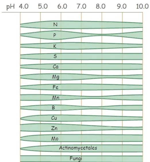
3.5. PH dependence of nutrient uptake ... 35
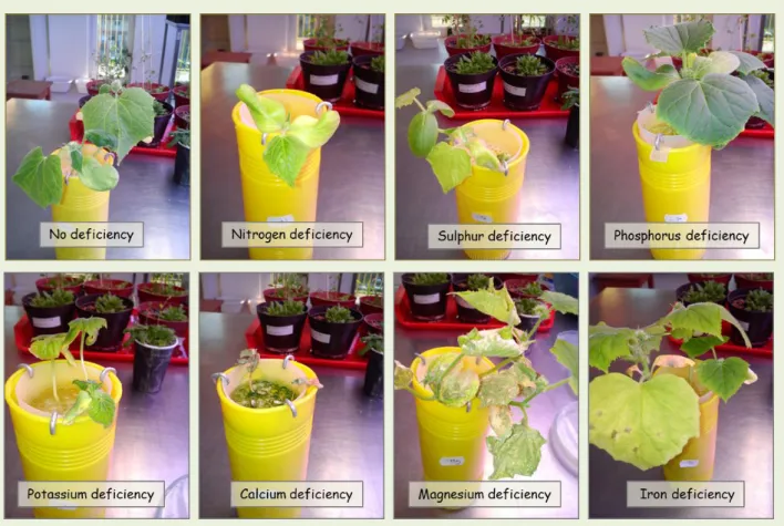
3.6. Nutrient deficiency ... 35
3.7. The most important elements for plants ... 39
3.7.1. Nitrogen ... 39
3.7.2. Sulphur ... 39
3.7.3. Phosphorus ... 39
3.7.4. Potassium ... 40
3.7.5. Calcium ... 40
3.7.6. Magnesium ... 40
3.7.7. Iron ... 41
Chapter 4. Short and long distance transport ... 44
4.1. Chemical potential of water ... 44
4.2. Water potential and water potential gradients ... 45
4.3. Water movement within plant body ... 46
4.4. Chemical potential of solutes ... 48
4.5. Membrane transport: pumps, carriers, channels ... 49
Chapter 5. Photosynthesis, the source of life on the Planet Earth ... 53
5.1. Utilization of light energy, photosynthetic organisms, general equation of photosynthesis .... 54
5.2. Structure of the photosynthetic apparatus: chloroplasts, pigments, light absorption and excitation ... 55
5.3. Operation of photosynthetic electron transport chain ... 58
5.4. Synthesis and export of stable products ... 59
Chapter 6. Plant hormones ... 64
6.1. Definition and general characteristics of plant hormones ... 64
6.2. The processes influencing plant hormone levels ... 66
6.3. Sensing and signalling of plant hormones ... 67
6.4 The biological function of plant hormones ... 69
Chapter 7. Light perception ... 73
7.1. Light and plant development ... 73
7.2. Photoreceptors ... 73
7.2.1. Sensing red and far-red light by phytochromes ... 73
7.2.1.1. The structure of phytochromes ... 73
7.2.1.2 Functioning of phytochromes ... 74
7.2.1.3 Types of phytochromes ... 74
7.2.1.4 Inactivation of phytochromes ... 75
7.2.1.5 Signalling from phytochromes ... 75
7.2.1.6 Classification of phytochrome responses ... 77
7.2.2. The blue light photoreceptors: the cryptochromes and the phototropins ... 78
7.2.2.1 The cryptochromes ... 78
7.2.2.2 The phototropins ... 78
7.2.3. Sensing UV-B by plants ... 79
Chapter 8. The vegetative growth and development of plants ... 82
8.1. Some specificities of plant growth and basic terms of growth and development ... 82
8.2. Shoot morphogenesis ... 83
8.3. Formation of leaves ... 85
8.4. Growth and differentiation of roots ... 85
Chapter 9. Flowering ... 90
9.1. The vegetative-to-reproductive transition ... 90
9.1.1 The flower meristem identity genes ... 91
9.1.2. The inflorescence meristem and its structure ... 91
9.1.3 Regulation of flowering time ... 92
9.1.3.1 The flowering integrator factors ... 93
9.1.3.2 Photoperiodic regulation of flowering ... 93
9.1.3.3 Regulation of flowering by vernalisation ... 96
9.1.3.4 Developmental regulation of flowering ... 98
9.1.3.5 Gibberellin, as flowering hormone and the autonomous pathway of flowering... 99
9.2 Flower morphogenesis ... 99
9.2.1. The ABC model of flower morphogenesis ... 99
9.2.2. The extended ABC model and the quartet model of flower development ... 100
9.2.3. The validity of the ABC model ... 101
9.3. The integrated model for the regulation of flowering time and flower morphogenesis ... 102
Chapter 10. Plant senescence and death ... 107
10.1. Plant senescence ... 107
10.1.1. Programmed cell death (PCD) ... 108
10.1.2. Autolysis and autophagy in plants ... 109
10.1.3. Organ senescence ... 110
10.1.3.1. The leaf senescence syndrome ... 110
10.1.3.2. Leaf senescence and its regulatory network ... 112 10.1.3.3. Leaf abscission ... 115 10.1.4. Whole plant senescence... 116
Preface
Plants are for the benefit of mankind since long as they are widely used as food and feed, raw materials for industry, fossil- or renewable source of energy. They produce oxygen and fix CO
2, clean air and contaminated soil and can be used as source of medically active substances.
Although several basic genetic, biochemical and physiological processes are common in all the living organisms, plants have lots of specificities. They only require light, H
2O, CO
2and mineral nutrients to live, but due to a sessile lifestyle they are forced to continuously adapt to their ever-changing environment.
The
Plant Molecular Physiology textbook is designed to introduce undergraduate studentsinto the life of plants. First, the genetic basis of the growth and development of plants is described. The first chapter explores the molecular regulatory factors with high importance in functioning of the genome and controlling the expression of genes. The second chapter introduces the specific organisation of plant cells and cellular organelles. Following chapters discuss the demand of plants for minerals, the uptake and transport of water and nutrients, and the mechanisms and significance of photosynthesis. The main endogenous factors (plant hormones) and the most important exogenous signal (light) and related signalling events affecting plant life and adaptation are also discussed. The plant-specific aspects of these biological processes, their regulatory and signalling mechanisms are also described. Then, the key points of plant ontogenesis including growth and development, reproduction and senescence are highlighted.
Understanding how plants function internally and how they adapt to their environment
provide deeper insight into their unique and vulnerable life strategy that is essential to
maintain the human civilization and the whole biosphere on this planet.
Expected learning outputs
Students are expected to gain the following knowledge, abilities, attitudes, autonomies and responsibilities learning these lecture notes and completing the related course:
Knowledge Ability Attitude Autonomy/Responsibility
They know the
characteristics of plants at molecular, cellular, physiological, and developmental levels.
They are able to use the obtained knowledge, the basic concepts and modern terminology of plant physiology correctly.
They are open to learn more about plants.
In front of professional or not-professional audiences, they autonomously argue for the essential role of plants in the functioning and maintenance of the Earth’s biosphere.
They know the relationships between the plant's stationary lifestyle, development strategy and
physiological functioning.
They are able to analyse differences between plant and animal development and adaptation strategies.
They do not consider plants as inferior to animals.
They draw others' attention to plants highlighting the uniqueness and
appropriateness of their life strategies.
They know the most important plant growth regulators and their role in plant development and adaptation to the environment.
They are able to use the gained knowledge in scientific research and to generate new scientific results.
They are interested in a deeper
understanding of plant life processes.
They independently discuss issues of plant life,
development and
environmental adaptation in front of a professional audience.
They know the importance of plant sciences in solving the burning problems of the nature and the human civilization.
They are able to use the gained knowledge to solve problems in plant protection, plant breeding, and plant cultivation.
They are open to learn about applied plant sciences.
They autonomously draw the attention of others to the importance of plants in everyday human life. They independently argue for the importance of plant
sciences in solving current problems of the human civilisation and the biosphere.
Chapter 1. The organization and expression of the plant genome
This chapter introduces general and specific features of genetic information of plants that determines their phenotype, growth and functions. At first, basic terms used in molecular plant biology will be defined, followed by the description of the main structural elements of the genome and genes. A more detailed section deals with factors and regulatory mechanisms controlling gene expression. This store of learning supports understanding molecular and signalling mechanisms, processes of the growth and other physiological phenomena.
Learning goals:
Knowledge:
- The students know the specific features of the plant genome
- The students know the functional difference between the heterochromatin and euchromatin and mechanisms responsible for their transition
- The students know the levels of gene expression regulation
Abilities:
- The students can rightly use plant molecular biology terms
- They are able to use the obtained knowledge to understand the role of gene regulatory processes in plant development and adaptation
Attitude:
- The students are open to carry out molecular biology experiments with plants - The students recognise the uniqueness of plants at the molecular level
Autonomy/responsibility
- The students can autonomously study molecular processes in plants
- The students can independently argue why and how plants are similar or different at the genome organisation/gene regulation level as compared to animals
Plants, like all living organisms except certain viruses, contain the information required for their growth and functioning in the form of DNA. The total DNA content of a cell is known as its genome. The genome means the total DNA content of chromosomes in a haploid cell (1n).
Diploid cells contain two sets of homologue chromosomes, two genomes.
Gregor Johann Mendel in the 1850s conducted a series of experiments with pea (Pisum
sativum) and concluded that some physical traits (phenotype) are determined by heritablecomponents. These heritable factors, responsible for e.g. the plant height, shape and colour
of seeds and pods, flower position and petal colour, are called genes. Gene is the DNA region
that contribute to the mature mRNA used to produce a protein (or RNA) and regulatory
regions surrounded by other non-coding sequences and regulatory sections. According to the
present term the regulatory sequences with important roles in the timing and extent of the
transcription are part of the gene, even if their distance from the coding region may be frequently several hundreds or even thousand pairs of nucleotide bases (bp) [1].
1.1. Organization of plant genomes
The plant genome consists of the nuclear genome, which contains majority of the genes necessary for plant development and growth, and the genome of organelles (1-10%).
Mitochondria and plastids have a subset of genes required for their own functions; they have the capacity to synthesis proteins under a strict control of nuclear genome [2]. The nuclear genome of plants is organized into chromosomes, their numbers in higher plants are between 10 and 48. During the evolution occurred several times genome duplication. Multiplication of the chromosomes of the same species resulted in autoployploid plants, while plants bearing chromosomes from closely related species are called allopolyploids (for example Brassicaceae species are allotetraploid, wheat is allohexaploid). Following the ancestral genome duplications, the plants usually became diploid (the DNA contents of the macro- and
microspores became double, 2n → 1n). Genes present in multiple copies might be loss, changetheir function or expression pattern [1]. However, in plant somatic cells frequently occur endoreduplication, thus sometimes 4n, 8n, 16n and even bigger genome number can be found in the older, larger or specified cells of one organism [3].
The plant genome may cover a wide range of size. While Paris japonica has the largest known eukaryotic genome (1.5x10
11bp), the
Genlisea tuberosa (a carnivorous species endemic toBrazil) [4], has the smallest known plant genome (6.1x10
7bp). The human genome lies in the middle of this range with the 3x10
9bp. These are good examples for the so-called C-value paradox, which says that the genome size of an organism does not correlate directly with its complexity (C-value is the DNA content of the haploid cell). Interestingly, the number of genes encoding proteins does not differ so much among plants: their estimated number is between 30 000 and 60 000 [2].
Generally, only 1-10% of the whole genome is transcribed to mRNA and translated to protein.
In the coding sequences can be classified single-copy DNA, large multigene families where genes occur in 20- 50 repeats, or gene cluster and tandem repeats that typically code for gene products in great demand. Furthermore, there are non-protein-coding RNA-coding genes, which are heterologous sequences with usually regulatory role. The first sequenced genome among the higher plants was that of Arabidopsis thaliana, the model plant of molecular plant research [5].
Arabidopsis has 5 pair of chromosomes, 1.19x10⁸bp nuclear genome (totally 1.35x10
8bp). According to the latest data (www.arabidopsis.org) [6], it contains 27655 protein-coding genes, more than 5000 non-protein-coding (they code usually rRNAs, tRNAs or other RNAs with regulatory function) and 4800 non-functional protein coding genes (e.g.
pseudogenes).
Most of the genome sequences are non-coding or with unknown function or highly repetitive
[7] (Biscotti et al. 2015). Repetitive DNA makes up much of the genome in many plants and
they can be responsible for the big differences in the genome size even among the closely
related plant species. Several different types of non-coding or repetitive sequences exist, such
as repeats in chromosome telomeres and centromeres and other, so-called satellite DNA, which can be classified further according to the size and position of the repeated sequences (e.g. simple sequence repeats, tandem repeats, dispersed repeats). Among the dispersed repeats are pseudogenes and non-functional gene sequences (with deletion, insertion or without promoters, etc.) which can be present in large quantities (their number in Arabidopsis
thaliana is ca. 5000). Among the dispersed repeats can be found mobile genetic elements (MGelements) too.
MG elements are the transposable elements (TE), which are DNA-sequence elements that move or transpose from one site of the genome to another. According to the mechanism by which they transpose they are classified as transposons (supposedly originated from mRNAs) and retrotransposon (during the transposition process an RNA intermediate is synthesized;
their origin is presumably viral). TEs were discovered by B. McClintock [8] in Zea mays (Ac/Ds system). Transposons constitute approximately 85% of the maize genome but their number can be also extremely high in some other species [9]. As an example, in rice the Tos 1-20 elements can be found in near 1000 copies. In some plants the TE elements make up the most abundant class of dispersed repetitive sequences. Most of them are inactive or their activity is lowered by different mechanisms in plants, because they may modify the expression of nearby genes [1]. In Arabidopsis 3900 TE genes were identified [6], which can be regarded to relatively low amount among the higher plants.
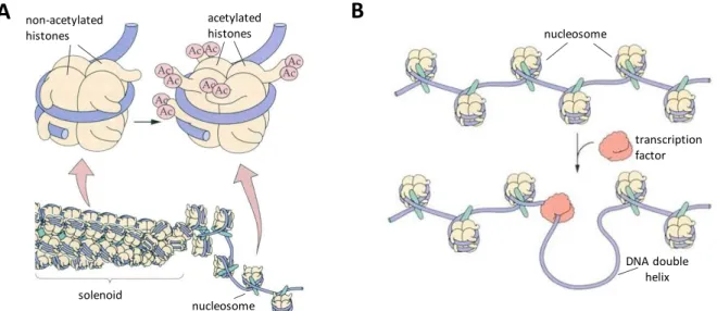
1.2.2. Structure of the chromatin
The long DNA molecule in the nucleus is packed with proteins to form chromatin. The proteins are important both in maintaining the structure of a chromosome and in regulating gene expression. Investigating the chromatin using high resolution microscopy its structure
resembles ’a beads on a string’ [2]. ’Beads’ are the nucleosomes, which contain 166 bp of DNAwrapped around a globular octamer (two tetramers of H2A, H2B, H3 and H4) basic histone proteins (Fig.1.1.). H1 resides outside of the core, it links coils consisting of 6 nucleosomes thus stabilizing the higher-order chromatin structures. Based on cytological observations of how darkly the chromatin is stained, two forms can be distinguished: the regions of heterochromatin and euchromatin. The heterochromatin stains darkly with dyes, is tightly coiled and can be found often near the centromeres of the chromosomes. Generally, it is transcriptionally silent [1]. In contrary, the euchromatin is packed more loosely and characterized by less interacted H1 histones and by the presence of other specific proteins such as High Mobility Group (HMG) proteins. Binding of the HMG proteins may result in the blending of the double-stranded DNA molecule thus facilitates the attachment of other DNA- binding proteins.
Modification of histones in chromatin affects DNA accessibility for transcription. Especially H3
and H4 histones may undergo post-translational modifications which can affect gene
expression both by altering chromatin structure and binding capacity of transcription factors
and RNA polymerases that form the basal transcriptional machinery. (The transcription factors
are proteins that may promote the forming of the transcriptional initiation complex and thus
facilitate the gene expression.) The two major forms of modifications are acetylation and
methylation. During acetylation, an acetyl group is transferred to one or more Lys amino acid
residues of H3 or H4 histones and in this form the nucleosome needs more space (Fig. 1.1.).
The activity of histone acetyl transferases (HATs) eventuates a less compact, while histone deacetylases result in a more condensed chromatin structure, which is less accessible for transcription factors [1]. The histone demethylases also cause looser DNA structure and promote active gene transcriptions. In contrary with this, methylation of histone proteins by histone methyl transferases (HMTases) and DNA cytosines by DNA methyl transferases result in more condensed structure where the gene transcription is inhibited.
Fig. 1.1. Modification of histone proteins by histone acetyl transferases cause less condensed chromatin (A). In the looser chromatin region transcription factors can bind to the regulatory sequences of DNA (B) (modified from Erdei, 2011, with permission).
1.2. The plant genes
A typical plant gene contains several different regulatory sequences beside the region which
is transcribed to RNA and encodes the gene’s protein product (Fig. 1.2.). The DNA sequencessurrounding the coding region contains structural and regulatory elements that have role in gene transcription and protein synthesis (translation). On the 5’ region, some distance from the transcription start site (upstream) can be found the promoter. The core or minimum promoter region involves the most important sequences that are crucial in the transcription, like TATA-box, that has a function in the binding of the basal transcription factors (TFs) of the initiation complex and activating the RNA polymerase, while CAAT-box and GC-box are the
binding sites of other TFs. The 5’ region of the gene (upstream) contains further regulatorysequences (cis-regulatory elements) which can modify the process of transcription. Moreover, regulatory information is present also in the coding region. Intervening sections of protein- coding regions are introns, while the sections encoding protein product are exons (Fig. 1.2.).
Most of the plant genes contain both exons and introns. The number of introns in a typical plant gene is usually low, but it can reach even 40 and their total length may exceed the amino acid-coding exons’ total length [2].
A
acetylatedB
histones non-acetylated
histones
solenoid
nucleosome
nucleosome
transcription factor
DNA double helix
Fig. 1.2. The structure of a typical protein-coding plant gene and the main processes
determining the protein synthesis. The DNA sequences of the gene contain both 5’ and 3’flanking regulatory sequences, such as cis-regulatory elements of the promoter orpolyadenylation signal. The primary transcript (mRNA precursor) is larger than the protein- coding regions. The non-protein-coding intron sequences will be removed during the mRNA maturation process (modified from Erdei, 2011, with permission).
1.3. Regulation of the gene expression
1.3.1. General featuresThe mechanism and regulation of plant’s gene expression basically do not differ from that of
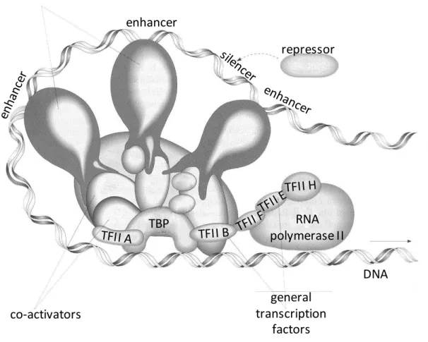
other eukaryotes: transcription takes place in the nucleus, while the translation in the cytoplasm. The transcription is catalysed by RNA polymerases (RNAPs) build-up from 8-12 subunits. Plants have five different nuclear RNAPs, each of them is involved in synthesis of different types of transcripts. The RNAP I is responsible for the transcription of 28S rRNA, 18S rRNA and 5,8S rRNA; the RNAP II for the mRNA precursors of protein-coding genes, but beside those even some other RNA-coding genes, such as microRNA- and small nuclear RNA genes are transcribed by it; the RNAP III catalyses the 5S rRNA and tRNA synthesis. Plants contain another two, plant-specific RNAPs, called RNAP IV and RNAP V, which in their two biggest subunits (RNPD1, RNPD2) differ fundamentally from RNAP II. RNAP IV is involved in endogenous small interfering RNA (siRNA) synthesis and RNAP V in the transcription of repetitive sequences [1]. Binding of RNAPs to DNA requires the presence of several general TFs (e.g. for RNAP II the TFIIA, B, D, E, F, H and the TATA-binding protein vital in recognition of TATA-box are important; see Fig. 1.3.).
Plants, as other organisms require several protein products only in specific cells and tissues or in particular developmental stages. The expression of many plant genes changes during
DNA 5’ 3’
Transcribed region
Protein coding region Minimum promoter
5’ non-translated region
3’ flanking region with regulatory
elements
Exon Exon Exon
Intron Intron TATA-
box CAAT-
box GC-
box 5’ flanking region with
regulatory elements
Start of transcription Start codon for
translation
Stop codon for translation
Poly A signal
mRNA precursor
mRNA
mRNA splicing
m7G cap Poly A tail
AAA…AAA
AAA…AAA Transcription
Polyadenylation Capping
C’ Translation
N’ Functional or regulatory protein
development or in response to environmental factors [1]. The timing and extent of gene expression are controlled by the promoters and other regulatory elements. For example, there are promoters that determine the specific expression of genes in mesophyll cells or others in guard cells of leaves, and similarly, specific promoters are known responsible for root- or tuber-specific gene expression patterns.
1.3.2. Cis-acting regulatory elements
Regulatory elements found in the DNA strand called
cis-acting elements. Cis-elements are usually short sequence regions (˂10 bp) and vary greatly in their base composition [1]. Theycan be either enhancers that increase the efficiency of RNAP II in initiation of transcription (frequently located at a considerable distance from the coding region), or silencers that downregulate the transcription. Several specific cis-acting regulatory elements of promoters were identified defining the specificity of gene expression. The
cis-elements responsible for gene expression regulation of hormones and other signalling molecules are the ‘response elements’. There are enhancer elements that increase the transcription of genes and silencerswhich lower the transcriptional activity. The
cis-elements can occur in several copy in thepromoter region of one gene amplifying their effect, but they can act even individually. In regulatory region of genes encoding proteins with essential function and high demand (like actin, tubulin genes) can be found cis-elements ensuring high level of expression [1].
1.3.3. Transcription factors
The transcriptional initiation complex can interact with several proteins. The cis-elements of promoters and other regulatory regions can modify the gene expression via transcription factors. The TF proteins usually synthesized independently on genes of which transcription they influence and are
trans-acting elements. TF proteins bind to the regulatory sequencesfound in the DNA. Depending on their domains, TFs can be either transcriptional activators or repressors. While the activators increase the expression of the gene(s), repressors block the effects of activators, decrease the rate of transcription, generally via binding to silencers (Fig.
1.3.). The activators and repressors can switch ‘on’ or ‘off’ a gene. Their effect on
transcriptional machinery is mediated by other proteins called co-activators or co-repressors
[10]. Their combination is different at each gene and they may be subjects of different types
of regulation, too. The Plant Transcription Factor Database (http://planttfdb.gao-lab.org)
contains 58 TF families [11]. The TFs can be grouped also by their structural motifs (such as
helix-turn-helix, helix-loop-helix, basic leucine zipper, zinc finger). Several TFs are specific for
plants. As an example for their importance, TFs containing homeobox domains regulate
development and determine cell fates. Genes, that regulate tissue- or organ identity are called
homeotic genes. Their encoded proteins control the expression of other regulatory proteins,
including other TFs, and thereby act as ’master genes’ [2].Fig. 1.3. The transcription apparat in eukaryote cells. Beside the RNA polymerase II (RNAP II), several general transcription factors are essential (TBP, TATA-binding protein, TFIIA-H). Among the proteins of the transcription complex can be found activators and repressors which bind to specific regulatory DNA sequences, which are the enhancer and silencer elements, respectively (modified from Erdei, 2011, with permission).
1.3.4. The heterochromatic small interfering RNAs
Recently, small regulatory RNAs (sRNAs; approximately 20-24 nt in size) have emerged as important gene expression regulators [12]. RNA-mediated transcriptional gene silencing (TGS) is a conserved phenomenon that occurs in fungi, plants and animals [13].
In plants, the predominantly 24-nt long heterochromatic small interfering RNAs (het‐siRNAs) transcriptionally regulate gene expression by RNA‐directed DNA methylation (RdDM).RdDM silences transposable elements and repeat sequences and in this way they have crucial roles in maintaining the genome stability. The number of plant het-siRNAs can be extremely high (more than 10 000). They usually derived from transposable elements and repeats. The plant‐
specific RNA polymerase IV (RNAP IV) produce
single-stranded RNAs from the target sequences, then they are converted to double-stranded RNAs by an RNA-dependent RNA polymerase (RDR2). The double-stranded RNAs are processed by a complex to 24-nt siRNA duplexes. The mature siRNAs then associate with sRNA-binding ARGONAUTE protein (AGO4, -6 or -9) and interacts with scaffold RNAs transcribed from the target loci by the RNA
repressor enhancer
activators
co-activators
TBP RNA
polymerase II
DNA general
transcription
factors
polymerase V (NRAP V). This interaction guides DNA methylation (histone H3K9 methylation) to result in the silencing of the target sequences. As a consequence, heterochromatin formation may occur [13]. The DNA methylation also may result in epigenetic changes, because specific mechanisms ensure the inheritance of DNA methylation patterns both mitotically and meiotically. The epigenetic mechanisms control many cellular and developmental processes during plant development [1].
1.4. Other mechanisms controlling gene expression
1.4.1 Post-transcriptional regulationsDuring the transcription the whole exon-intron region of DNA is transcribed to RNA, that is the mRNA-precursor (Fig. 1.2.). The primary transcript undergoes different modifications such
as splicing (when the introns will be removed), addition of the 5’ „cap” structure and 3’polyadenylic acid end (poly A tail). These modifications promote that the mature mRNA canreach the ribosome and may translated to the entire protein [2]. However, there are known some other posttranscriptional regulations and among them several include sRNAs. The two most important sRNA classes in this process can be the microRNAs (miRNAs) and the plant- specific phased siRNAs (phasiRNAs); the latter group belongs to the secondary siRNAs [14, 15].
The microRNAs are transcribed by RNAP II from the
MIRNA genes and their biogenesis is awidely conserved process in plants. Plant genome typically encodes a hundred to several hundreds of
MIRNA genes which can be located intergenic (between two protein-codinggenes) or intronic [15]. The primary transcript (pri-miRNA) contains a self-complementary foldback (hairpin) structure. After slicing by an RNase III endoribonuclease (DICER-LIKE1), the miRNA/miRNA* duplex is methylated (which is crucial for their stability) and transported to the cytoplasm, where the mature miRNA incorporates into the ARGONAUTE protein (AGO1) forming the RNA-INDUCED SILENCING COMPLEX (RISC). The miRNAs, requiring almost perfect sequence complementary with its target, can trigger the cleavage of the mRNA of target gene (most usual mechanism for plant miRNAs) or inhibit their translation [16, resulting in gene silencing. This mechanism is known as RNA interference (RNAi). The target sequences of miRNA frequently are TFs, and in this way they have a wide range of roles both during normal development and stress responses of plants [2].
Phased secondary siRNAs belong to another class of sRNAs. Their biogenesis relies on the cleavage mediated by sRNAs (mostly miRNAs). There are two typical modes of their biogenesis which eventuate several 21-, 22- or 24-nt siRNAs through sequential miRNA-directed cleavage of one mRNA molecule. These phasiRNAs can function like miRNAs to regulate their target genes in trans (trans- acting siRNAs, tasi-RNAs) or in cis (casiRNAs). The known targets of tasi- RNAs are auxin response factors (ARFs) and having role in the auxin signalling elucidate their involvement also during the entire life of plants [16].
1.4.2. Post-translational regulations
The gene products can be regulated even post-translationally due to different protein
modifications, which may affect their function and lifetime. During or after the completion of
translation, the secondary and tertiary structure will be formed due to protein folding [1]. The
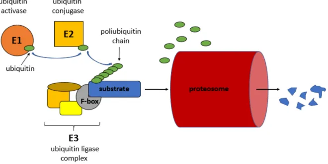
synthesised protein can be transported to membranes or specific organelles and processed e.g. by proteolytic cleavage. The molecule can bind co-factors, intra- and intermolecular disulfide bounds can be formed and additional functional groups or molecules can be attached, such as phosphoryl-, carboxyl-, acetyl groups, glucosides or ubiquitin.
Protein ubiquitination can result in altered function, localization or degradation via the 26S proteasome system. The protein degradation by the 26S proteasome is essential in plant hormonal signalling pathways. Conjugation of an ubiquitin to a substrate is carried out by a multienzyme complex and requires ATP. The enzymes generally involved in the process are called E1 (ubiquitin activating enzyme), E2 (ubiquitin conjugating enzyme) and E3 (ubiqutin ligase) (Figure 1.4). The protein in E3 complex which recognizes the substrate contains F-box motif. The number of F-box proteins in plants are very high because each of them interacts with a different protein or protein family substrates [1]. They regulate diverse cellular processes, including cell cycle transition, transcriptional regulation and signal transduction. In
Arabidopsis thaliana genome the presence of at least 568 F-box protein genes were reported[17].
Fig. 1.4. The mechanism of ubiquitin-ligase complex mediated protein degradation. (Figure
of A. Fehér)Summary
1. The main characteristic parameters of a species are determined by its DNA. The genome is the total DNA content of a haploid cell. Plants have nuclear, mitochondrial and chloroplastic genomes. Most of the higher plants are diploids, although the polyploidy and genome duplication are frequent phenomena.
2.
The size of plant’s genome varies greatly.Plants having bigger genome have more repetitive DNA. In some plants the transposable elements (TE) make up much of the nuclear genome. The activity of repetitive sequences is suppressed by different mechanisms.
3. The nuclear DNA and histone proteins form chromatin The heterochromatin is a more condensed, transcriptionally inactive, while the euchromatin has a loose structure where transcription factors can bind. Modifications of DNA and histone proteins affect the DNA accessibility for transcription. Histone acetylation and -demethylation result in a less compact, while histone de-acetylation and -methylation a more condensed chromatin structure.
4. Plant genes are organized like those of other eukaryotes: the protein-coding region is interspersed and surrounded by regulatory sequences. Regulatory elements found in the DNA strand are
cis-elements. Cis-acting elements responsible for regulation ofhormones and other signalling molecules are called “response elements” The enhancer elements increase transcription, the silencers lower it. Cis-elements modify the gene expression through transcription factor proteins which can be activators or repressors. TFs are trans-acting regulatory elements.
5. The heterochromatic small interfering RNAs are transcriptional regulators promoting heterochromatin form. They silence transposable elements and repeat sequences by
RNA‐directed DNA methylation. In their biogenesis and mechanisms, the plant-specificRNA polymerase IV and RNAP V and the ARGONAUTE proteins are involved. The DNA methylation may result in epigenetic changes too.
6. The microRNAs are post-transcriptional regulators. They are transcribed from the
MIRNA genes by RNAP II. After their processing, they build in the RISC (RNA-inducedsilencing complex) and trigger the cleavage of the mRNA of target gene or inhibit its translation. The resulted gene silencing mechanism is called RNA interference (RNAi).
Their target sequences are frequently TFs.
7. miRNAs also have role in the biogenesis of phased secondary siRNAs. The pha-siRNAs originated from a long dsRNA molecule due to sequential cleavage. The 21-, 22- or 24- nt siRNAs regulate their target genes by mRNA cleavage. One of their group is the plant-specific trans-acting siRNAs (tasi-RNAs). The only known targets of tasi-RNAs are auxin response factors (ARFs), thus they have role in the plant hormone auxin signalling.
8. Protein ubiquitination is post-translational regulatory mechanism. The protein
degradation by the 26S proteasome is essential in plant hormonal signalling pathways.
Questions
1. What is the difference between polyploidy and alloploidy?
2. Why can be big differences in the genome size of plants that are relatives?
3. What kind of sequence types are in the genome?
4. Compare the cis- and trans-acting regulatory elements! What are the main similarities and differences between them?
5. Which mechanisms regulate the gene expression through modification of chromatin?
6. How act the miRNAs?
7. What plant-specific regulatory mechanisms control the amount of a gene product?
Questions to discuss
1. What can be the advantages of the multiplicated genomes in the somatic cells of the plants? Why is rarer it in macro- and microspores?
2. The Paris japonica is a plant with very slow growing and can be found very rarely in the nature. Can it be related with its genome size?
3. If the size of the genome does not correlate with the complexity of the organism than what can be related with the advanced state?
4. What can be the reason that so many heterochromatic siRNAs are in plants?
Suggested reading
Jones R, Ougham H, Thomas H, Waaland S (Eds) The molecular life of plants. Wiley-Blackwell, American Society of Plant Biologists, 2013.
Buchanan BB, Gruissem W, Jones RL (Eds) Biochemistry and molecular biology of plants.
Second Edition. American Society of Plant Biologists, 2015.
References
[1] B.B. Buchanan, W. Gruissem and R.L. Jones (Eds), Biochemistry and molecular biology of plants. Second Edition. American Society of Plant Biologists, pp. 401-437, 2015.
[2] R. Jones, H. Ougham, H. Thomas and S. Waaland (Eds), The molecular life of plants.
Wiley-Blackwell, American Society of Plant Biologists, pp. 74-111, 2013.
[3]
J. Joubèsand C. Chevalier, Endoreduplication in higher plants.
Plant Mol Biol. 43(5-6):735-45, 2000.
[4] F. Rivadavia, P.M. Gonella and A. Fleischmann, A new and tuberous species of Genlisea (Lentibulariaceae) from the Campos Rupestres of Brazil. Syst Bot, 38(2):464-470, 2013.
[5] Arabidopsis Genome Initiative, Analysis of the genome sequence of the flowering plant
Arabidopsis thaliana. Nature. 408(6814):796-815, 2000.[6] C.Y. Cheng, V. Krishnakumar, A.P. Chan, F. Thibaud-Nissen, S. Schobel and C.D. Town, Araport11: a complete reannotation of the Arabidopsis thaliana reference genome.
Plant J.89(4):789-804, 2017.
[7] M.A. Biscotti, E. Olmo and J.S. Heslop-Harrison, Repetitive DNA in eukaryotic genomes.
Chromosome Res. 23(3):415-420, 2015.
[8] B. McClintock, The origin and behavior of mutable loci in maize. Proc Natl Acad Sci USA.
36(6):344–355, 1950.
[9] S. Ravindran, Barbara McClintock and the discovery of jumping genes. Proc Natl Acad
Sci USA. 109 (50): 20198-20199, 2012.[10] H.W. Heldt, Pflanzenbiochemie. Spektrum Akademischer Verlag Gmbh Heidelberg.
Berlin, 2. Auflage, 1999.
[11] J.P. Jin, F. Tian, D.C. Yang, Y.Q. Meng, L. Kong, J.C. Luo and G. Gao, PlantTFDB 4.0:
toward a central hub for transcription factors and regulatory interactions in plants.
Nucleic Acids Res. 45(D1):D1040-D1045, 2017.[12] S. Li, C. Castillo-Gonzalez, B. Yu and X. Zhang, The functions of plant small RNAs in development and in stress responses. Plant J. 90: 654-670, 2017.
[13] H. Zhang, X. He and J.K. Zhu, RNA-directed DNA methylation in plants. Where to start?
RNA Biol. 10:10, 1593-1596, 2013.
[14] F. Borges and R.A. Martienssen, The expanding world of small RNAs in plants. Nat Rev
Mol Cell Biol. 16: 727-741, 2015.[15] J. Wang, J. Mei and G. Ren, Plant microRNAs: Biogenesis, homeostasis, and degradation. Front Plant Sci. 10, 360, pp. 12, 2019
[16] C. Chen, Z. Zeng, Z. Liu and R. Xia, Small RNAs, emerging regulators critical for the development of horticultural traits. Hortic Res. 5:63. pp. 14, 2018.
[17] H. Kuroda, N. Takahashi, H. Shimada, M. Seki, K. Shinozaki and M. Matsui, Classification and expression analysis of Arabidopsis F-box-containing protein genes. Plant Cell Physiol.
43(10):1073-85. 2002.
Chapter 2. An introduction to plant cells
This chapter introduces the organelles of plant cells (see Fig. 1). It starts with a discussion of the distinctions among cell wall types. This is followed by a short introduction of plant organelles and their roles in growth and development. The chapter details the structures of the endomembrane system. The functions of plastid types are also briefly surveyed.
Learning goals:
Knowledge:
- The students know the plant cell organelles
- The students can distinguish among the organelles of the various plant cell types - The students know the differences between animal and plant cells
Abilities:
- The students can recognise plant cell organelles in microscopic images
- The students are able to use the obtained knowledge to understand the role of cell types/organelles in plant development and adaptation
Attitude:
- The students are open to carry out cell biology experiments with plants - The students recognise the uniqueness of plants at the cellular level
Autonomy/responsibility
- The students can autonomously study cellular processes in plants
- The students can independently argue why and how plants are similar or different at the cellular level as compared to animals
Fig. 2.1. An idealized plant cell and its components. (Source: Wikimedia Commons. This work has been released into the public domain by its author, LadyofHats. This applies worldwide.
Link: https://commons.wikimedia.org/wiki/File:Plant_cell_structure-en.svg)
2. 1. Plant cell wall
The main difference between plant and animal cells is the presence of the cell wall. Their rigid cell walls take part in the development, which depends on the cell wall-mediated cell division and cell enlargement. Cell wall structure is diverse containing a variety of compounds. There are two main types of plant cell wall, the primary and the secondary cell wall. The major component of the fundamental framework of primary cell walls is the cellulose. These linear cellulose molecules can bond to each other by hydrogen bonds in order to form microfibrils.
A microfibril can contain about 36 cellulose molecules. These cellulose rods can have connected to each other by cross-linking glycans and all this network is embedded in a matrix of pectic polysaccharides. Structural proteins or phenylpropanoids can be found as a third, independent and non-polysaccharide network. Callose is a compound which can be found in cell walls of pollen grains, pollen tubes or in the cell plates of dividing cells.
2.1.1. The primary cell wall
Cellulose microfibrils in the primary cell wall are relatively short and thin, compared to those of the secondary cell wall and hemicellulose is composed of xyloglucan. This type of cell wall is rich of pectin. Type I cell wall is specific for eudicot species and half of the monocots.
On the contrary, the type II cell wall is the main type of commelinoid monocots (bromelias, palms, sedges, grasses). During cell expansion, the biosynthesis and assembly of primary cell walls can occur. pH and other cell-wall loosening factors can contribute to the cell enlargement processes.
2.1.2. Secondary cell wall
The secondary cell wall is deposited between the primary cell wall and the plasma membrane.
This type of cell wall contains relatively long and thick cellulose microfibrils, hemicellulosic xylan, and lignin. Among lignin, other depositions also occur like phenylpropanoids, suberin, cutin, or waxes. The secondary cell wall modifications have a lot of different functions as in the case of cotton fiber, pear fruit stone cells, collenchyma cell in the cell corners or guard cell pairs.
Middle lamella is a thin layer of material, consisting mainly of pectins, that binds together the walls of adjacent plant cells.
Apoplast is the name of the cell walls of the plant cell. It has important function in sensing the outer environment.
2.2. Membranes of the Cell-Endomembrane System
Plant cell and cell organelles are bounded by lipid bilayer membranes. Protoplast is the living part of the cell surrounded by plasma membrane without cell wall, which can separate it from external environment. Plant cells contain around 14 different membrane types.
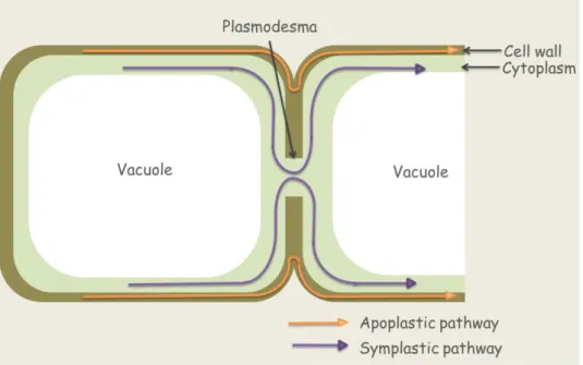
2.2.1. Plasmodesmata
Plasmodesmata are tubular extensions of the plasma membrane, 40 to 50 nm in diameter,
that traverse the cell wall and connect the cytoplasms of adjacent cells. Most plant cells are
interconnected by this structure, so their cytoplasms form a continuum referred to as the symplast. Symplastic transport is the way with which solutes can transport between cells.
Primary and secondary plasmodesmata help to maintain tissue developmental gradient primary plasmodesmata can form between clonally derived cells by cytoplasmic connections.
There is a size exclusion limit which can restrict the transported molecules by size. This size is adjusted by the width of the cytoplasmic sleeve that surrounds the ER tubule, or desmotubule, which is the centre of the plasmodesmata. Globular proteins within the sleeve generate spiralling microchannels through the plasmodesmata. There is little information about the transport of solutes through the cytoplasmic sleeve and the desmotubule itself. Actin and myosin are located in the plasmodesmata. Viruses can also go across the plasmodesmata by movement proteins. These proteins can form a transport tubule within the plasmodesmatal pore that facilitates the mature viruses through the plasmodesmata.
Symplastic transport can also occur between non-clonally related cells through the formation of secondary plasmodesmata. In these connections, the plasma membrane of the adjacent cells can fuse and the ER network became connected. These facts can describe the importance of symplast in developmental signalling and nutrition.
2.2.2. Vacuoles
Vacuole is a membrane-enclosed compartment which contains vacuolar sap composed of water and other solutes. Large vacuoles which can located in central position can occupy up to 95 % of the total cell volume, so it can take role in cell expansion. The number of vacuoles varies between cell types, as in the case of flower petals with vacuoles. Vacuoles can differ in size and appearance; some stress types can change their size. The level of maturation also a factor which can determine the size of the vacuoles, e.g. there is no large central vacuole in meristematic cells, but they have many small vacuoles.
Tonoplast is the name of vacuolar membrane, which contains proteins and lipids that are synthesized initially in the ER. Vacuole is a storage compartment of the cell full with plant secondary metabolites involved in plant defence against herbivores and pathogens.
There are two types of vacuoles in plant cells. Lytic vacuoles are large, water-containing vacuoles, playing a role in water and ion balance. Variety of specific membrane transporters can contribute to the accumulation of inorganic ions, sugars, organic acids, and pigments or toxins. In seeds, there are protein bodies, which can accumulate proteins. Protein storage vacuoles are smaller and filled with proteins or lipids and they can be found in seeds.
2.2.3. Endoplasmic reticulum (ER)
ER is a network of internal membranes and the place of protein and lipid synthesis. In ER,
proteins can be synthesized and delivered to the plasma membrane, or apoplast, vacuoles. ER
has a quality control system. By multiple processing steps it can supervise and conduct the
secreted proteins. ER is one of the largest calcium stores participating in the intracellular
calcium signalling. It can identify and transfer the misfolded proteins to the ERAD machinery
(ER-associated degradation).
ER is composed of tubules which can form flattened saccules called cisternae. Tubular and cisternal forms of ER rapidly transform to each other. This transition is regulated by a class of proteins called reticulons. Reticulons (RTNs) are membrane-spanning proteins sharing a typical domain named reticulon homology domain (RHD). RTN genes have been identified in all eukaryotes examined so far, in plants as well. RTZNs are involved in numerous cellular processes such as apoptosis, cell division and intracellular trafficking.
The region of the ER that contain many membrane-bound ribosomes is called rough ER, as it has rough appearance in electron micrographs. The ER without bound ribosomes is called smooth ER. Secretion of the proteins from cells begins with the rough ER. ER provides the building stones of membranes and protein cargo for other compartments in the endosomal system. Many proteins are synthesized on the rough endoplasmic reticulum. ER is the major source of membrane phospholipids. Flippases are enzymes which can counteract the membrane asymmetry by flipping newly synthesized phospholipids across the bilayer. ER and plastids are capable to add new membrane directly through lipid and protein synthesis.
Smooth ER participates in fatty acid modification, lipid synthesis and the production of oil bodies.
2.2.5. Golgi apparatus
The Golgi apparatus processes and packages newly synthesized macromolecules.
Glycoproteins and polysaccharides destined for secretion are processes in the Golgi apparatus.
Golgi apparatus has another name dictyosome. This organelle is a group of polarized stack of cisternae, with fatter cisternae on the cis side or forming face, which connect to the ER. The opposite face is the trans side of Golgi body is more flattened, thinner cisternae and include a tubular network called the trans-Golgi-network or TGN. Meristematic cells can contain up to hundreds of Golgi bodes, while other cells differ in their number of the Golgi bodies. Cisternae can be different in one Golgi body with different enzymes. There are some glycoprotein modification enzymes. Membrane and its content can be delivered to the Golgi from the ER at specialized site called ER exit sites (ERES). This site is determined by the presence of a coat protein called COP II. This can associate with the transmembrane receptors which bind the specific cargo destined for the Golgi. These membrane regions then bud off forming coated vesicles losing their COP II coats. Anterograde (forward) movement is the pathway out of the ER to the Golgi, within the Golgi from the cis to trans face, followed by transport to the plasma membrane or to the prevacuolar structures via vesicles. Contrarily, retrograde or backward movement is the way of recycling membrane vesicles from the Golgi to the ER or from the trans to cis face of Golgi
The main function of Golgi apparatus is the processes and packages polysaccharides and proteins for secretion.
Nowadays, the distribution of the Golgi stacks in a variety of plant cell types can be examined
by immunohistochemical studies and by live cell imaging after expression of Golgi targeted
fluorescent protein constructs, so the structure can be examined.
2.2.6. Trans Golgi network (TGN)
TGN is an important hub site in plant cells. TGN is essential for the assembly of cell walls, including the cell plate, and organizes traffic or cargoes not only to but also from the plasma membrane. TGN is a membrane compartment on the trans-side of Golgi stacks responsible for the sorting and packaging of cargo molecules for delivery to the plasma membrane and vacuoles. TGN is a distinct organelle and not just a tubular reticulum on the trans-side of the Golgi.
There are two forms of TGN, 1, the Golgi-associated TGN (GA-TGN) cisternae attached to the trans-side of the Golgi; and 2, the detached, free TGN cisternae.
2.2.7. Microbodies
Microbodies are the site of specific biochemical pathways.
Some organelles grow and proliferate independently from the endomembrane system even though they can form from that. These organelles are oil bodies, peroxisomes and glyoxysomes.
During seed development, many plants store large amounts of oil, which can be accumulated in organelles called oil bodies, lipid bodies, or spherosomes. They are surrounded by a half- unit membrane, a phospholipid monolayer. Oil bodies are formed within the ER. They can store triglycerides, so their lumen is hydrophobic.
When oil bodies can degrade during seed germination, they can associate with other organelles that contain the enzymes for lipid peroxidation, so the glyoxysomes.
Microbodies are spherical organelles are surrounded by single membrane and they are specialized for one special functions.
Peroxisomes and glyoxysomes are microbodies that are specialized for the B-oxidation of fatty acids and the metabolism of glyoxylate, a two-carbon acid aldehyde. Microbodies lack DNA and they can associate with other organelles in order to share intermediate metabolites. The glyoxysome is associated with mitochondria and oil bodies, while the peroxisome is associated with mitochondria and chloroplasts.
Peroxisomes can develop directly from glyoxysomes, at least in greening cotyledons.
In the peroxisomes, glyoxylate, a two-carbon oxidation product of the photorespiratory cycle is oxidized to the acid aldehyde glyoxylate. In this reaction, hydrogen peroxide can generate.
The most abundant enzyme located in this organelle is the catalase, which can degrade the hydrogen peroxide. Catalase enzyme can exist in crystalline forms.
Most proteins can enter the peroxisomes from cytosol posttranslationally by means of a specific targeting signal, consisting of serine-lysine-leucine at the carboxyl terminus.
2.3. The nucleus
The nucleus is the organelle which contains the genetic information responsible for regulating
the metabolism, growth, and differentiation of the cell. Nuclear genome refers to the genes
and their intervening sequences. The size of the nuclear genome is very different between
plants.
Arabidopsis thaliana has a genome size around 1.2x10⁸base pairs, while the lily
Fritillaria assyriaca has 1x10¹¹base pairs. Chloroplasts and mitochondria have own genetic information.
Nucleus has double membrane called nuclear envelope, this is a subdomain of the endoplasmic reticulum (ER). There are some selective channels across both membranes which connect the nucleoplasm with cytoplasm called nuclear pores. The number of these types of pores can vary between hundreds to thousands. These nuclear pores can be composed of more hundred different proteins called nucleoporins in order to form a 105-nm long nuclear pore complex. The NPC can form a 40 nm long channel and act as a supramolecular sieve. The nuclear localization signal is a specific amino acid sequence which is required for a protein to enter into the nucleus.
Chromosomes can store and replicate in the nucleus. Chromosomes can compose of DNA and its associated proteins, this DNA-protein complex is called chromatin. Segments of the linear double helix of DNA are coiled twice around a solid cylinder of eight histone protein molecules, and this structure is called nucleosome. At interphase, two types of chromatin can be distinguished on different condensation: heterochromatin and euchromatin.
Heterochromatin is a highly compact and transcriptionally inactive form of chromatin which can count of about 10 % of DNA. Most of the heterochromatin is concentrated along the periphery of the nuclear membrane and is associated with the regions of chromosomes containing few genes, such as telomeres and centromeres. The rest of the DNA consists of the euchromatin which is dispersed and transcriptionally active form. At any given time just the 10% of euchromatin is active transcriptionally. Dynamic structural changes with chromatin during the cell cycle. Transient local changes are required for transcription and heterochromatic regions can be converted to euchromatic regions and vice versa by addition and removal of functional groups on the histone proteins. Such enormous changes globally the whole genome can result to stable changes in gene expression. Stable changes in gene expression can occur without changes in the DNAS sequence called epigenetic regulation.
Nuclei contain a dense region called nucleolus, which is the site of ribosome synthesis. Typical cells have one nucleolus while others have more. The nucleolus includes some chromosomes where ribosomal RNA (rRNA) genes are clustered to form a structure called the nucleolar organizer region. The nucleolus assembles the proteins and rRNA of the ribosome into a large and small subunit, which unite in the cytoplasm to a complete ribosome. These ribosomes are protein synthesizing factories.
2.4. Mitochondria
Mitochondria synthesize ATP and small carbon skeletons.
Mitochondrium is an energy-producing, semiautonomous and independently dividing organelle. It is separated from the cytosol by a double membrane (an outer and inner membrane) and contain their own DNA and ribosomes. Mitochondria are the site of cellular respiration, a process which can produce energy from sugar metabolism for ATP synthesis.
Mitochondria can always undergo fission and fusion. The inner membrane contains proton-
pumping ATP synthase which uses a proton gradient to synthesize ATP for cells. The
generation of proton gradient is the work of the electron transport chain, a massive assembly of electron transporters, which are located in the inner membrane.
Cristae are the enfolded inner membranes in plant cells. The inner membrane can mean a border for the mitochondrial matrix, the place where the enzymes of Krebs cycle are located.
2.5. Plastids
Plastids are a diverse family of anabolic organelles. Plastids cannot be formed de novo in the cytoplasm. Therefore, these types of organelles can be inherited to the offspring and have a developmental continuity within the organism. The metabolism and thus the structure of plastids vary along with the differentiation of the organs, tissues and specific cells of the plant body. This fluctuation can continue during the entire life cycle as part of the developmental program of the host cell that harbours them. However, plastid differentiation is also strongly influenced by changes in the environmental conditions.
2.5.1. Proplastid
Plastids are found in high number in plant cells. There are many types in the plant cell.
Chromoplasts are pigment-containing plastids, while colourless plastids are called leucoplasts.
These plastids can be changed from one to other. Originally, they develop from proplastids.
Proplastids are colourless bodies in meristematic or immature cells.
2.5.2. Etioplasts
Etioplasts, another type of plastids, are found in stems, but not in roots. They are a long-lasting intermediate stage in the way of differentiation from proplastid to chloroplast when there is very low intensity of light or it is dark. Etioplasts restart the differentiation toward chloroplasts as soon as the light is intense enough.
2.5.3. Leucoplasts
Leucoplasts are colourless plastids (without pigments), that function as storage organelles.
Leucoplasts comprise amyloplasts, elaioplasts (or oleoplasts), and proteinoplasts. They store starch, lipids and proteins, respectively.
Amyloplast is an organelle in plants that stores starch. Amyloplasts are often found in non- photosynthetic tissue, such as roots and storage tubers. In plant cells, amyloplasts synthesize starch, and all the stored starch in a cell can be found in these organelles as starch granules.
Amyloplasts, apart from storing starch, are gravity sensors in root cells. Starch granules are denser than water so that amyloplasts fall to the bottom of the cell. These organelles interact with microtubules of the meristematic cells in a way that during cell mitosis the division plane is perpendicular to the gravity vector (which points to the Earth centre). Amyloplasts are also involved in the metabolism of nitrogen.
Elaioplasts are a type of leucoplasts specialized for lipid storage in plant cells. They have been demonstrated to be involved in the biosynthesis of terpenes in essential oil, which are then exported into the secretory pocket, greatly affecting the aroma and the taste of fruit in citrus species which means the importance of these organelles. Elaioplasts are small size plastids containing oil and lipids in fat drops. In plant cells, there are two synthetic pathways for lipids.
The eukaryotic pathway depends on the smooth endoplasmic reticulum, whereas the so-
called prokaryotic pathway involves plastids. These two pathways produce different types of lipids. Elaioplast are also involved in pollen maturation. Some plants are able to store lipids in other organelles known as elaiosomes, which are derived from endoplasmic reticulum.
Proteinoplasts contain a high concentration of proteins, so high that proteins form crystals or very dense amorphous material. However, it is not clear yet if there is a type of plastids dedicated exclusively to protein storage in plant cells.
2.5.4. Chromoplasts
Chromoplasts contain carotenoid pigments that give the red, orange and yellow colours to the plant structure where they are present. These plastids are abundant in flowers, fruits, old leaves, and some roots. It is thought that one of their main functions is to attract animals for pollination or for spreading the seeds. Chromoplasts are metabolically active, though they contain fewer DNA copies than chloroplasts.
Chromoplasts have lipid drops containing carotenoids and complex molecules known as fibrils, which also contain a core with carotenoids. Chromoplasts are differentiated from chloroplasts, as well as from proplastids. During differentiation from chloroplasts, the photosynthetic machinery, mostly in thylakoids, is degraded and carotenoids are synthesized along with the compartments where they are going to be included. These compartments, known as plastoglobules, are lipid drops made up of mainly triacylglycerides that are located in the stroma. Carotenoids, mainly xanthophylls, are stored inside the plastoglobules, so concentrated that they can form filaments or crystals. Plastoglobules may be also found in other plastids. Chromoplasts develop an internal membrane system in the outer part of the stroma. These new membranes arise from the inner membranes and do not from the old degraded thylakoids. Carotenoids like lutein, beta-carotenoid, and others, may be also associated to these new membranes. Occasionally, the internal membranes show a reticular arrangement. Chromoplasts are non-photosynthetic plastids that accumulate carotenoids.
They derive from other plastid forms, mostly chloroplasts. The entire set of Calvin cycle and of the oxidative pentose phosphate pathway persist after the transition from chloroplast to chromoplast.
2.5.5. Gerontoplasts
Chlorophyll and its degradation products are dangerous and lethal for cells because of their photoreactive features. Chloroplasts can be transformed to gerontoplasts, which resemble to chromoplasts. In the gerontoplasts, grana are unstacked, thylakoid membranes are lost and lipid-like plastoglobuli can accumulate. The efficiency of photochemical reactions and Calvin- Benson cycle is declined. There is a possibility the gerontoplast to transform back chloroplast till the terminal phase of cell death.
2.5.6. Chloroplasts