Research Article
Tumor cell and carcinoma-associated fi broblast interaction
regulates matrix metalloproteinases and their inhibitors in oral squamous cell carcinoma
Alexandra Fullár
a,b, Ilona Kovalszky
a, Mario Bitsche
b, Angela Romani
b,
Volker Hans Schartinger
b, Georg Mathias Sprinzl
b, Herbert Riechelmann
b, József Dudás
b,⁎
a1st Department of Pathology and Experimental Cancer Research, Semmelweis University, Üllői út 26, 1085 Budapest, Hungary
bDepartment of Otorhinolaryngology, Medical University Innsbruck, Anichstrasse 35, A-6020 Innsbruck, Austria
A R T I C L E I N F O R M A T I O N A B S T R A C T
Article Chronology:
Received 21 December 2011 Revised version received 19 March 2012 Accepted 23 March 2012 Available online 1 April 2012
Co-culture of periodontal ligament (PDL) fibroblasts and SCC-25 oral squamous carcinoma cells (OSCC), results in conversion of PDLs into carcinoma-associated fibroblasts (CAFs). Paracrin circuits between CAFs and OSCC cells were hypothesized to regulate the gene expression of matrix remodeling enzymes in their co-culture, which was performed for 7 days, followed by analysis of the mRNA/protein expression and activity of metalloproteinases (MMPs), their tissue inhibitors (TIMPs) and other relevant genes. Interleukin1-β, transforming growth factor-β1, fibronectin and αvβ6 integrin have shown to be involved in the regulation of the MMP and TIMP gene expression in co-culture of CAFs and tumor cells. In addition, these cells also cooperated in activation of MMP pro-enzymes. It is particularly interesting that the fibroblast-produced inactive MMP-2 has been activated by the tumor-cell-produced membrane-type 1 matrix metalloproteinase (MT1-MMP).
The crosstalk between cancer- and the surrounding fibroblast stromal-cells is essential for the fine tuning of cancer cells invasivity.
© 2012 Elsevier Inc.
Keywords:
HNSCC
Co-culture insert Metastasis Matrix remodeling
Introduction
One of the most predictive factors of poor clinical outcome of head and neck squamous cell carcinoma (HNSCC) is the presence of regional lymph node metastasis, and nodal status of the neck plays a decisive role in the choice of treatment[1]. Hensen et al. recently reported an independent gene expression analysis of metastasized
versus non-metastasized HNSCC. This analysis revealed differential- ly expressed gene sets involved in the progression of HNSCC, including extracellular matrix (ECM) remodeling- (i.e. matrix metalloproteinases, (MMPs)), hypoxia- and angiogenesis-related genes[2]. Interestingly, a similar gene profiling assay performed over 10 years ago by Villaret et al. also showed overexpressed matrix metalloproteinases in head and neck squamous cell carcino-
⁎Corresponding author.Fax: + 43 512 504 23175.
E-mail addresses:fullarsz@gmail.com(A. Fullár),koval@korb1.sote.hu(I. Kovalszky),mario.bitsche@i-med.ac.at(M. Bitsche), angela.romani@i-med.ac.at(A. Romani),volker.schartinger@i-med.ac.at(V.H. Schartinger),georg.sprinzl@i-med.ac.at(G.M. Sprinzl), herbert.riechelmann@i-med.ac.at(H. Riechelmann),jozsef.dudas@i-med.ac.at(J. Dudás).
Abbreviations: CAFs, carcinoma-associated fibroblasts; COX-2, prostaglandin-endoperoxide synthase 2; DEX, dexamethasone; ECM, extracellular matrix; EMT, epithelial-to-mesenchymal transition; FBS, foetal bovine serum; FN,fibronectin; HNSCC, head and neck squamous cell carcinoma; IL, interleukin; IMVD, intratumoral microvessel density; LTBP-1, latent-transforming growth factor beta-binding protein; 1MMP, matrix metalloproteinase; MT1-MMP, membrane-type 1 matrix metalloproteinase; OSCC, oral squamous cell carcinoma; PDL, periodontal ligament (PDL)fibroblasts; TGF-β1, transforming growth factor-β1; TIMP, tissue inhibitor of metalloproteinases.
0014-4827 © 2012 Elsevier Inc.
doi:10.1016/j.yexcr.2012.03.023
A v a i l a b l e o n l i n e a t w w w . s c i e n c e d i r e c t . c o m
w w w . e l s e v i e r . c o m / l o c a t e / y e x c r
Open access under CC BY-NC-ND license.
Open access under CC BY-NC-ND license.
ma tumor tissues [3]. By regulating matrix metalloproteinase (MMP) activity and controlling the breakdown of ECM components, also tissue inhibitors of metalloproteinases (TIMPs) play an important role in the process of tumor invasion and metastasis[4].
TIMPs not only inhibit the catalytic activity of MMPs, but also are able to act as growth factors and are involved in the activation or inactivation of MMPs[5]. The signaling pathways and circles that regulate MMPs and TIMPs are not fully understood.
Carcinoma-associated fibroblasts (CAFs) are able to promote the growth of carcinoma cells[6]. CAFs induce an epithelial-to- mesenchymal transition (EMT) in epithelial tumor cells, which is a major biological process in invasion of squamous cell carcinoma [7], progression and metastasis. During this process invasive tumor cells tend to lose their epithelial antigens [8], their epithelial cell polarity and morphology, and acquire mesenchymal and stemness-related features[9,10]. In our recent reports we have described a co-culture model of periodontal ligament (PDL) fibroblasts and SCC-25 oral squamous carcinoma cells (OSCC), which resulted in conversion of normal fibroblasts into CAFs. In the same model EMT occurred in SCC-25 cells[11]. Moreover, we have described that SCC-25 cells produce active, processed IL-1β, and PDL fibroblasts possess receptor for it, whose expression is increased in the presence of SCC-25 tumor cells. Upon interaction with SCC-25 cells active IL-1β signaling occurs in co-cultured fibroblasts leading to induction of several genes involved in tumor progression, including interleukin-6 (IL-6) and prostaglandin- endoperoxide synthase 2 (COX-2) [12]. As we reported before, dynamic interaction between CAFs and tumor cells of HNSCC dictates gene expression changes in the interacting cells, which covers major events of tumor progression. Here, we hypothesize that the paracrine interplay between SCC-25 carcinoma cells and CAFs provides a mechanistic background for the gene regulation of MMPs and TIMPs, which contributes to poor clinical outcome of HNSCC. Traces of several regulatory pathways in the paracrine interplay between SCC-25 carcinoma cells and CAFs were acknowledged to determine the gene expression and the activa- tion of matrix remodeling enzymes and their inhibitors.
Materials and methods
Cell linesPDL fibroblasts [11–13] were isolated from periodontal ligament (PDL) and received from Prof. Dr. Miosge (Department of Prostho- dontics, Georg-August-University, Göttingen, Germany) [13], they were routinely cultured in DMEM-low glucose (PAA, Pasching, Austria) supplemented with 10% foetal bovine serum (FBS) (PAA), 2 mML-glutamine, 100 units/ml penicillin, and 100μg/ml streptomy- cin. SCC-25 cells were purchased from the German Collection of Microorganisms and Cell Cultures (DSMZ, Braunschweig, Germany, ref. ACC 617), and were routinely cultured in DMEM/F12 (PAA) supplemented with 10% FBS (PAA), 2 mML-glutamine, 1 mM sodium pyruvate, 100 units/ml penicillin, and 100μg/ml streptomycin[11,12].
Co-culture
Co-culture between SCC-25 cells and PDL fibroblasts was de- scribed in details before[11,12]. PDL fibroblasts were plated into cell culture inserts and SCC-25 cells into the bottom of 6-well
plates[11,12], they were cultured in 1:1 mixed medium of DMEM- low glucose and DMEM/F12, supplemented with 10% FBS, and also the single cultured control cells were cultured in the same medium in order to have the same glucose concentration and glycemic conditions in control and co-cultured conditions. The medium in the co-cultures and in the controls was changed after 3 days. After letting them grow in co-culture for 7 days, the cells in the inserts and in the wells were used for RNA isolation[11]or for protein fractionation [12]. For gelatinase zymogram assay, the media in the inserts and in the six-well-plates were replaced with low-serum (0.3% FBS)-containing medium (a 1:1 mixed medium of DMEM-low glucose and DMEM/F12 containing 0.3% FBS) for the last 24 h. The conditioned medium was collected from the control and co-cultured cells for gelatinase zymogram assay and for combined immunoprecipitation–western blot[12], the potential disturbing effect of serum-metalloproteinases was minimized in this way. In a further run of experiments, co-cultured cells were treated with 10−6mol/L dexamethasone (Sigma, Vienna, Austria) as previously reported[12,14].
Treatment of the cells with IL-1β
2 × 10⁵/ml PDLs or SCC-25 cells were plated in 10% FBS- supplemented-DMEM-low-glucose or in 10% FBS-supplemented- DMEM/F12 (PAA) in 10 cm culture plates (Unilab, Innsbruck, Austria). After 48 h, medium was replaced by 0.3% FBS-containing- medium. Cells were treated with IL-1βat 0.015–1.5 ng/ml concen- tration range for 4, 8 and 24 h as described before[12,15].
RNA extraction, reverse transcription and real-time RT-PCR
Total RNA was isolated and reverse transcribed from control and co-cultured cells as described before, real-time RT-PCR was done as referenced [11,16]. Several human PCR primers were published previously: β-actin [17], MMP-1 [18], MMP-3 [19], MMP-7 [20], MMP-9 [21], MMP-13[22], TIMP-1 [23], TIMP-2 [24], TIMP-3[25], TGF-β1[26], Integrinαv[27]; fibronectin (FN) and latent-transforming growth factor beta-binding protein-1 (LTBP-1) primers were taken from the NCI, Primer Viewer Database, ref. nos. 11092, and 10108 respectively. Other primers are listed in Supplementary Document 1.β-Actin functioned well as a housekeeping gene [11], and did not show significant changes in co-cultured conditions compared to controls. The relative gene expression was calculated as previously reported [16].
Protein expression and activity measurements
For the analysis of integrinαv, non-nuclear protein fractions were subjected to western blot analysis [11]as described previously [12,28]. Identical equal protein containing 20μl samples were loaded for western blot, which was performed either with the JLA20 β-actin antibody (in 1:100 dilution), purchased from Developmental Studies Hybridoma Bank (Iowa city, Iowa, USA) as a loading control[29], or with rabbit polyclonal integrinαv antibody (#4705, Cell Signaling Technology, Danvers, MA, USA, in 1:1000 dilution).
Conditioned media were collected from both the fibroblast and the SCC-25 side of the co-culture and also from the control cells.
From the supernatant (conditioned media) of the control cells and from both sides of the co-culture 20μl medium was used from all samples for gelatinase zymogram, and 200μl samples were used for immunoprecipitation[12]with 2μl of rabbit poly- or monoclonal antibodies (anti-MMP-2 #4022; anti-TIMP-1
#8946; anti-TIMP-3 #5673, Cell Signaling Technology). The immunprecipitated samples were taken in 50μl Laemmli- sample-buffer [30] and 20μl/well was loaded to 9 or 12%
acrylamide gels, followed by western blot[11]with the above- mentioned antibodies. Comparable intracellular protein extracts were reacted with the JLA20 β-actin antibody as loading controls. Cells after co-culture and controls were scraped into 500μl lysis buffer[31](50 mM TRIS HCl (pH 7.5), 500 mM NaCl, 5 mM CaCl2)/well or /insert. Protein concentration was deter- mined in the cell lysates, and 10μg proteins were loaded to the gelatin-containing gels from the lysates. The gelatinase assay was performed as described previously [31]. Supernatants represented conditioned medium of the cells, which was pipetted off directly after completion of culture, lysates repre- sented cells lysed after removal of conditioned medium, which contained molecules bound on the cell surface, membrane-, cytoplasmatic- and cell nuclear-fractions inclusive. Both super- natants and lysates were subjected to polyacrylamide gel electrophoresis using gelatin-containing 10% polyacrylamide gels[31]. Gelatinase activity is recognized as white bands in colloidal Coomassie Brilliant Blue G-250 (Roth, Karlsruhe Germany)-stained gels. Density of the detected bands was measured by the Image J software[11,16].
Immunocytochemistry and confocal microscopy
IBidi Dishes (Martinsried, Germany) with control and co-cultured PDLs were used for fibronectin (primary antibody 1:40 diluted (5μg/ml); Santa Cruz Biotech., Santa Cruz, CA, USA, Cat. No.
sc59826, clone IST-9) immunocytochemistry after fixation with methanol for 20 min at −20 °C, as described previously [12].
Immmunostained Ibidi dishes were evaluated by confocal micros- copy[12].
ELISA
Conditioned media were collected from both the fibroblast and the SCC-25 side of the co-culture and also from the control cells.
From the supernatant (conditioned media) of the control cells and from both sides of the co-culture 100μl medium was used from all samples for quantitative TGF-β1 detection using the RayBio human TGF-β1 ELISA Kit (RayBiotech, Norcross, GA, USA).
Statistical analysis
Each experiment was performed in three independent sets contain- ing at least three biological repeats/set. The relative gene-expression results were tested for normal distribution by D'Agostino and Pearson omnibus normality test using the Graphpad Prism 4.03 (Graphpad Software Inc). Significance of changes in co-culture vs.
controls was tested by non-parametric tests (Mann–Whitney) and Students' t-tests depending on the distribution of the data. The independent experimental sets were then compared for reproduc- ibility. Only reproducible changes with a p < 0.05 level[11,12]were considered as significant.
Results
Model of the regulation of the paracrine interplay between SCC-25 cells and CAFs
A continuous interaction between tumor cells and fibroblasts was hypothesized in the regulation of MMP and TIMP expression and activity. The expression of MMPs and TIMPs was expected in the PDL fibroblasts, whose regulation was hypothesized to occur via inflammatory cytokines, including IL1-β[32], produced by SCC-25
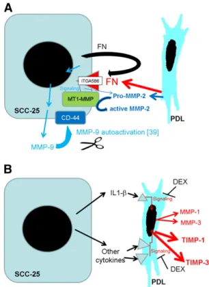
Fig. 1–Summary of the suggested mechanism for the regulation of MMPs and TIMPs in the paracrine interplay between SCC-25 cells and fibroblasts. MMP-9 showed a tumor specific expression, regulated presumably by the fibronectin ITGA5B6 pathway. The ITGA5 was inducible in both SCC-25 and PDL fibroblasts in co-culture, but ITGB6 expression was tumor (SCC-25) specific. Based on a previous report[44], MMP-9 might be activated in interaction with CD-44, and according to our gelatinase assay results, it remains bound with the tumor cells (A). The results of this study suggest that MMP-2 is secreted in its pro- (inactive-) form by CAFs surrounding the tumor cells, and at a lower extent also by the tumor cells themselves. Activation of MMP-2 either requires MT1-MMP localized on the SCC-25 cancer cells[34], or integrins, where the involvement ofαv integrins (ITGA5) is expected (A).MMPs-1, -3 and TIMPs-1, -3 are produced in the PDL fibroblasts, and their expression might be regulated by inflammatory cytokines, including IL1-βproduced by SCC-25 cells. The gene expression of MMP-1, MMP-2, TIMP-1 and TIMP-3 was reduced by dexamethasone (DEX) (B).
cells. In the regulation of MMP-9 gene expression the fibronectin– integrin αvβ6 (ITGA5B6) pathway was hypothesized. Another possible pathway, especially in regulation of TIMP expression, is the TGF-β1-pathway [31,32]; the expression of TGF-β1 was expected in the fibroblasts and in the tumor cells (Fig. 1).
Fibroblast contribution to the MMP and TIMP expression in co-culture of SCC-25 cells and CAFs
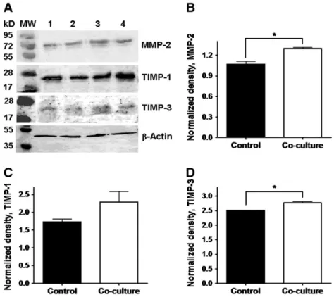
Gene expression of MMPs-1, -2, -3, -7, -9 and -13 and TIMPs 1–3 was investigated in fibroblasts (PDLs) in control and co-cultured Fig. 2–mRNA expression of MMP-1 (A), MMP-2 (B), MMP-3 (C), TIMP-1 (D), TIMP-3 (E) and MMP-9 (F) in PDL fibroblasts and SCC-25 cells in control and co-cultured conditions. *: p < 0.05; **: p < 0.01, ***: p < 0.001.
conditions. MMPs-1–3 and TIMPs-1 and -3 were constitutively expressed in PDLs, and their gene expression was significantly upregulated in co-cultured PDLs (Figs. 2A–E; Table 1). The upregulation of MMP-2, TIMP-1 and TIMP-3 gene expression in co-cultured PDLs has also been confirmed at protein level (Figs. 3A–D). MMPs-7, -9 (Table 1) and -13 were not detected in PDLs. TIMP-2 was also constitutively expressed in PDLs, and in co- culture it was not significantly regulated (Table 1).
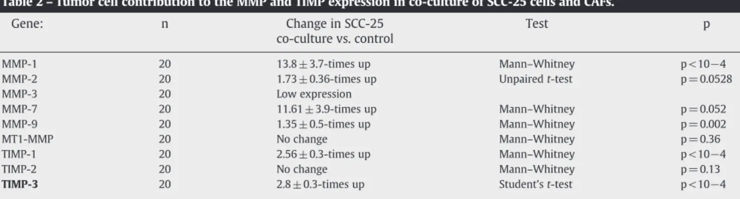
Tumor cell contribution to the MMP and TIMP expres- sion in co-culture of SCC-25 cells and CAFs
MMP-1 at low levels and MMP-9 at higher levels were constitu- tively expressed in SSC-25 cells. In co-culture, their gene expression was significantly upregulated compared to controls (Figs. 2A, F;Table 2). Membrane-type 1 matrix metalloproteinase (MT1-MMP) was also constitutively expressed in SSC-25 cells, in co-culture, no significant regulation was observed in its gene expression (Table 2). Although the expression compared to PDLs in SCC-25 cells was low, TIMP-1 and TIMP-3 showed significant upregulation in co-cultured SCC-25 cells (Figs. 2D–E,Table 2).
Regulation of MMPs and TIMPs expression in co-cultured fibroblasts
Based on our previous study, we investigated, if IL-1β [12,15]
contributed to the upregulation of MMPs-1–3 in PDLs. 24 h treatment of PDLs with IL-1βat 1.5 ng/ml induced a significant upregulation of MMP-1 (8.8± 1.8-times; p = 0.003 using unpaired t-test with Welch's correction) and of MMP-3 (2.1± 0.3-times, p = 0.012 using unpaired t-test with Welch's correction), while MMP-2 was not upregulated. Similar to the gene expression of MMPs, that of TIMPs was also investigated in 4–24 h treatment of PDLs with IL-1βat 0.015–1.5 ng/ml. IL-1βdid not change signifi- cantly the gene expression of TIMP-1 and TIMP-3 in fibroblasts (p= 0.8 and 0.7 respectively, by one way analysis of variance). In a further run of experiments, co-cultures were treated with
Fig. 3–Western blot analysis of MMP-2 (A, B), TIMP-1 (A, C) and TIMP-3 (A, D) related to loading control (β-actin) in PDL fibroblasts in control (1–2) and co-cultured (3–4) conditions. Densitometry of the detected bands normalized to theβ-actin band densities.
MMP-2 (B), TIMP-1 (C), TIMP-3 (D), *: p < 0.05.
Table 1–Fibroblast contribution to the MMP and TIMP expression in co-culture of SCC-25 cells and CAFs.
Gene: n Change infibroblast co-culture related tofibroblast control
Test p
MMP-1 20 4 ± 1-times up Mann–Whitney p = 0.012 MMP-2 20 2.3 ± 0.4-times up Mann–Whitney p = 0.0047 MMP-3 20 3.15 ± 0.6-times up Unpaired t test with
Welch's correction
p = 0.02
MMP-7 20 Very low expression MMP-9 20 Not detected MT1-
MMP
20 Not detected
TIMP-1 20 3.15 ± 0.6-times up Mann–Whitney p = 0.014 TIMP-2 20 No change Mann–Whitney p = 0.4 TIMP-3 20 5 ± 0.8-times up Unpaired t test with
Welch's correction
p = 0.008
10−6mol/L dexamethasone (DEX) as previously reported [12,14]. The gene expression of both TIMP-1 and TIMP-3 showed significant (p < 0.01 using unpairedt-test with Welch's correc- tion) decrease in DEX-treated co-culture compared to normal one (0.4 ± 0.1 (TIMP-1) and 0.3 ± 0.1 TIMP-3 of the normal co- culture after DEX treatment).
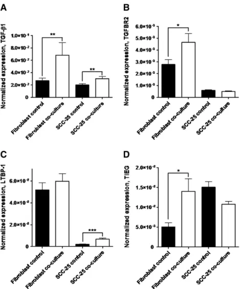
Another possible regulatory way of TIMPs expression is the TGF-β1-pathway[31,32]. At mRNA level in PDLs and SCC-25 cells TGF-β1 was expressed at comparable levels (Fig. 4A), and it was significantly (p < 0.002, using Mann Whitney test) upregulated in co-culture in both of them (2.4 ± 0.6-times in fibroblasts and 1.6 ± 0.1-times in SCC-25 cells) (Fig. 4A). At protein level the TGF-β1 expression in PDLs and SCC-25 cells was also comparable (8.5– 37.8 pg/ml). In co-culture, there was no significant regulation either in supernatants of fibroblasts or SCC-25 cells compared with controls (p > 0.05 with non-parametric tests), and a sustained expression of TGF-β1 was detectable in co-culture using ELISA. The receptor TGFBR2 was constitutively expressed in PDLs, in co-culture its gene expression was significantly (p < 0.05, using Student's t-test), 1.7 ± 0.3-times increased in them (Fig. 4B). For the activation of TGF-β1: latent TGFβ-binding protein-1 (LTBP-1) is required [33], which was constitutively expressed in high levels in PDLs (Fig. 4C), in co-culture, its gene expression has not changed significantly (p = 0.26, using Mann– Whitney test) in them. Nevertheless, in SCC-25 cells there is a significant (p< 10−4, using Mann–Whitney test), 3.3 ± 0.5-times increase (Fig. 4C) in co-culture. TIEG (TGF-β1-inducible early response gene) is used as a marker for the efficacy and activity of TGF-β1[31]. Constitutive TIEG expression was detected in both PDLs and SCC-25 cells, and PDLs showed a significant (p< 0.05, using Mann–Whitney test), 2.8 ± 0.6-times upregulation in co-culture (Fig. 4D). The gene expression of TIMPs-1–3 and MMP-2 was compared with that of TGFBR2 and TIEG in co-cultured vs. control PDLs, where both TGFBR2 and TIEG showed highly significant correlation with TIMPs-1–3 and MMP-2 (correlation coefficients over 0.66, p < 10−3, Supplementary Document 2).
Regulation of MMP and TIMP expression in co-cultured tumor cells
MMP-1 and MMP-9 were constitutively expressed in SCC-25 cells, and their gene expression was upregulated in co-cultured SCC-25 cells (Figs. 2A and F). Previously[12], we have shown that the IL-1 receptor is much lower expressed in SCC-25 cells than in PDL
fibroblasts. Nevertheless, we have investigated if MMP-1 and MMP-9 are regulated by IL-1βin SCC-25 cells. Using the same time and concentration ranges for the IL-1β-treatment as for PDLs, there were no significant changes compared to control in SCC-25 cells (p= 0.7 for MMP-1, p =0.24 for MMP-9 using non-parametric tests).
It was previously reported that fibronectin, via theαvβ6 integrin receptor (ITGA5B6) might upregulate MMP-9 gene expression[32].
Since IL-1β-treatment did not significantly contribute to MMP-9 gene expression in SCC-25 cells, it was investigated, if fibronectin, via ITGA5B6 receptor might have a contribution to MMP-9 regulation. Integrin αv (ITGA5) was constitutively expressed in both PDLs and SCC-25 cells (Fig. 5A). In co-culture, it was significantly upregulated in both of them (2.9± 1-times in fibro- blasts and 2 ± 0.3-times in SCC-25 cells, using Mann–Whitney test) (Fig. 5A), which has been confirmed at the protein level as well (Fig. 5D). Integrinβ6 (ITGB6) was expressed only in SCC-25 cells, and its gene expression remained unchanged in co-culture (p= 0.1, using Mann–Whitney test,Fig. 5B). In SCC-25 cells bothαv andβ6 integrins were present, their expression levels were comparable with the one of MMP-9 (Figs. 2F,5A–B), and the SCC-25-specific expression of ITGB6 was also comparable with the SCC-25-specific expression of MMP-9.
Fibronectin, the ligand, was constitutively highly expressed in PDLs (Figs. 5C, E), and 29.9 ± 0.3-times lower in SCC-25 cells, in co-culture its gene expression did not change significantly (Fig. 5C, using parametric and non parametric tests). At the protein level using an antibody specific for the EDA-domain of fibronectin it was detected only in the PDL fibroblasts (Fig. 5E).
MMP-2 and -9 gelatinase activity in the co-culture of SCC-25 cells andfibroblasts
MMP-2 and -9 were functionally analysed in gelatinase zymo- graphy. Both supernatants and cell lysates of controls and co- cultures (of both the SCC-25- and PDL-side) were used in this assay. Active and inactive bands of MMP-9 and MMP-2 were recognized, and active bands of these enzymes were found at lower molecular weight. 5μl of fetal bovine serum was used as positive control (Figs. 6A–B, FBS lane).
In the cell lysates of PDL controls and co-culture only the inactive pro-MMP-2 was recognized, which showed comparable bands (Fig. 6A, lanes 1–2 and 5–6). In the cell lysates of SCC-25 control (Fig. 6A, lanes 3–4), three bands were recognized, the inactive pro-MMP-9, the functional active MMP-9 and the inactive,
Table 2–Tumor cell contribution to the MMP and TIMP expression in co-culture of SCC-25 cells and CAFs.
Gene: n Change in SCC-25
co-culture vs. control
Test p
MMP-1 20 13.8 ± 3.7-times up Mann–Whitney p < 10−4
MMP-2 20 1.73 ± 0.36-times up Unpairedt-test p = 0.0528
MMP-3 20 Low expression
MMP-7 20 11.61 ± 3.9-times up Mann–Whitney p = 0.052
MMP-9 20 1.35 ± 0.5-times up Mann–Whitney p = 0.002
MT1-MMP 20 No change Mann–Whitney p = 0.36
TIMP-1 20 2.56 ± 0.3-times up Mann–Whitney p < 10−4
TIMP-2 20 No change Mann–Whitney p = 0.13
TIMP-3 20 2.8 ± 0.3-times up Student'st-test p < 10−4
pro-MMP-2. In co-culture both the pro- and the active MMP-9 band intensity increased (1.57 ±0.04 and 2.72 ±0.3-times respectively;
Fig. 6A, lanes 7–8,Figs. 6C, D), while pro-MMP-2-inactive showed comparable bands in the cell lysate of control and co-cultured SCC-25 (Fig. 6A lanes 3–4 and 7–8). In the supernatants of PDL controls only the inactive pro-MMP-2 was recognized (Fig. 6B, lanes 1–2). In co- cultured PDLs inactive pro-MMP-9 appeared and the inactive pro- MMP-2 bands decreased (Fig. 6B, lanes 5–6, Fig. 6E). In the supernatants of SCC-25 control three bands were recognized, pro- MMP-9 inactive, pro-MMP-2 inactive and MMP-2 active (Fig. 6B, lanes 3–4). In co-culture the inactive pro-MMP-9 band intensity increased (1.34± 0.01-times;Fig. 6B, lanes 7–8), both pro-MMP-2- inactive and MMP-2 active bands intensity increased significantly (p <10−3using Mann–Whitney test) (1.38±0.02-times and 1.27±
0.02-timesFig. 6B, lanes 7–8,Figs. 6E, F).
Taken together, MMP-9 showed tumor cell specific function, its active form was only detected in/on the SCC-25 cells. MMP-2 showed fibroblast and tumor cell production (Fig. 6A), its active form was secreted, not found in cell lysates, and was present only at the side of tumor cells. A co-culture led to increased representation of active MMP-9 and MMP-2 either directly at the tumor cells, or in their vicinity.
Discussion
MMPs play a complex role in tumor progression and metastasis. They facilitate invasion by degrading components of the ECM and there is also evidence that they are involved in angiogenesis. Tokumaru et al.
earlier showed that MMP-2 is activated by MT1-MMP, which is localized on the cancer cells (Fig. 1A)[34]. MT1-MMP is constitutively expressed in SCC-25 cells, not regulated in co-culture with PDLs, and is not expressed in PDLs in control and co-cultured conditions. MMP- 2[35,36]and -9 are involved in the invasion process of oral cancer, and MMP-9 is related to poor prognosis in the subset of patients without neck node metastasis [37]. However, MMPs are not only produced by tumor cells but also by stromal cells and tumor infiltrating leucocytes, especially along the invasive front of the tumor[37–39]. Tumors with strong MMP-2 expression in stromal fibroblasts showed a significantly higher intratumoral microvessel density (IMVD). In addition, postoperative prognosis of strong stromal MMP-2 patients was significantly poorer than that of weak stromal MMP-2 patients[40].
The results of this study suggest that MMP-2 is secreted in its pro- (inactive-) form mainly by CAFs surrounding the tumor cells, Fig. 4–mRNA expression of TGF-β1 (A), TGFBR2 (B), LTBP-1 (C) and TIEG (D) in fibroblasts and SCC-25 cells in control and co-cultured conditions. *: p < 0.05; **: p < 0.01, ***: p < 0.001.
and at a lower extent also by the tumor cells themselves (Figs. 6A, 1A). Activation of MMP-2 either requires MT1-MMP localized on the cancer cells (Table 2)[34], or integrins, where the involve- ment ofαv integrins (ITGA5,Figs. 5A,1A) was reported in more papers[41,42]. Our results show that the main contribution to the activation of MMP-2 resides in the tumor cells, and might be due to the constitutive expressed tumor-specific MT1-MMP. Never- theless, CAFs play an essential role not only in the production of pro-MMP-2, but also in influencing the activation of MMP-2 by tumor cells. On the one hand ITGA5 is upregulated in CAFs, on the other hand, the interplay between CAFs and tumor cells contrib- utes to the upregulation of ITGA5 also in the tumor cells (Fig. 5A).
Interestingly, pro-MMP-2 is highly expressed in fibroblasts, but its activation only occurred in the presence of tumor cells (Figs. 6A– B). For the upregulation of MMP-2 also the interaction between
fibroblasts and carcinoma cells was required, where the involve- ment of the TGF-β1-pathway was recognized (Supplementary Document 2) [43]. The paracrine interaction between oral SCC cells and CAFs leads to the upregulation of other MMPs as of MMP- 1 and MMP-3. The clear involvement of tumor-produced IL-1βin this process has been evidenced by the results of this work (Fig. 1B), where the induction effect was reproduced in IL-1β- treatments of fibroblasts. Several lines of evidence indicate that fibroblasts might facilitate the invasion of SCC cells by expressing MMPs on their own in response to tumor-cell-produced cytokines (i.e. IL-1β), to TGF-β1 or to integrin–fibronectin interactions[41].
The tumor cells are capable of activating pro-MMPs.
In contrast to pro-MMP-2, pro-MMP-9 is produced by oral carcinoma cells, not by fibroblasts. Pro-MMP-9 is activated on the surface of tumor cells (Fig. 1A). The paracrine interaction between Fig. 5–mRNA expression of ITGA5 (A), ITGB6 (B) and fibronectin (C), in PDL fibroblasts and SCC-25 cells in control and co-cultured conditions. *: p < 0.05; **: p < 0.01, ***: p < 0.001. Western blot of ITGA5 and loading controlβ-actin (D) in nuclear-free extracts of control (1) and co-cultured (2) fibroblasts, control (3) and co-cultured (4) SCC-25 cells. Confocal microscopy of EDA-fibronectin (red detected by Alexa fluor 647–conjugated anti-mouse IgG) in PDLs, bar: 100μm.
CAFs and tumor cells also resulted in the upregulation of MMP-9 in oral carcinoma cells, where fibronectin, and its receptor,αvβ6 integrin [32] as well as CD-44[44] could have been involved (Fig. 1A).
Interestingly, besides their MMPs expression, fibroblasts pro- duce several-fold higher amounts of inhibitors of metalloprotei- nases (i.e. TIMPs) whose gene expression (especially of TIMP-1 and TIMP-3) is also similarly upregulated in response to the paracrine
circuits between CAFs and oral carcinoma cells (Fig. 1B). In this regulation, the inhibitory effect of dexamethasone suggests involve- ment of inflammatory cytokines (Fig. 1B) (dexamethasone was also able to inhibit the upregulation of MMPs-1–2 in co-cultured fibroblasts, not shown), although, treatment of PDLs with IL-1βdid not change significantly the gene expression of TIMP-1 and TIMP-3.
In this regard, IL-1β might induce MMPs, but TIMPs are not upregulated at the same time, which might lead to excess of matrix Fig. 6–Detection of MMP-2 and MMP-9 gelatinase activity in cell lysate (A) and supernatant (B) of fibroblasts and SCC-25 cells by combined gelatinase zymography. 1–2: fibroblast control, 3–4: SCC-25 control, 5–6: fibroblast co-culture, 7–8: SCC-25 co-culture.
Densitometry of MMP-2 and MMP-9 gelatinase zymography (C–F). C and D represent the densitometry of pro-MMP-9 (C) and active MMP-9 (D) bands in cell lysates. E and F show the densitometry of pro-MMP-2 (E) and active MMP-2 (F) form from supernatants.
degrading enzymes over their inhibitors. In fact, all TIMPs inhibit active forms of all MMPs[45], in addition, they might contribute to activation of pro-MMPs, as TIMP-1 to the activation of MMP-9[46]
and TIMP-2 to the activation of MMP-2[47].
Inflammatory cytokines could be, as mentioned before, selective in activation of MMPs and TIMPs, in addition, the TGF-β1-pathway is also involved in the regulation of TIMP expression in CAFs (Supplementary Document 2). A tissue of progressive tumor cells and CAFs is characterized by high expression of TIMPs in fibroblasts, and also by a significant TIMP expression in tumor cells. In fact, in co- cultured SCC-25 cells MMP-1, MMP-2 and MMP-9 were higher expressed than TIMP-1 or TIMP-2. These results allow the assumption that; matrix in tumor tissue is probably not digested by CAFs. In contrast, MMPs are also produced by tumor cells;
moreover, pro-MMP-9 was tumor-cell-specific, not found in fibroblasts or CAFs. At the same time, tumor cells possess the tools for activation of pro-MMPs, either as MT-MMPs, or TIMPs (as TIMP- 2 for MMP-2[47]). The MMP activation might occur at the surface of tumor cells or nearby individual tumor cells. The relation of MMP/
TIMP expression at this area allows activation of MMPs at the surface of tumor cells, which has been also seen experimentally using gelatinase assays, finding active MMPs at the side of tumor cells in co-culture. The tumor cell expression of TIMP-3 is relatively high, but this TIMP is sequestered bound to extracellular matrix[45], and will probably not be located close to the surface of tumor cells. In co- culture, the diffusion of released MMPs is freely allowed by the 0.4μm cell-culture inserts, but the tumor-cell-surface-associated activation provides a continuous excess concentration of active MMPs by the direct surface of tumor cells.
A coordination of the MMP–TIMP expression and MMP activation between progressing OSCC tumor cells and CAFs leads to local invasion potential of tumor cells, without a serious impact on whole organ tissue remodeling.
Conclusion
Several lines of evidence indicate that fibroblasts might facilitate the invasion of OSCC cells, in part by paracrine regulatory circuits, or by increasing the proteolytic activity of latent metalloprotei- nases on the surfaces of OSCC cells.
Supplementary data to this article can be found online at doi:10.1016/j.yexcr.2012.03.023.
Role of the funding source
The funding source has no influence on the direction and the outcome of the study, it is an independent granting.
Acknowledgments
This work was supported by the Austrian Science Fund [FWF P 22287-B13]; the Hungarian National Scientific Research Founda- tion [OTKA 100904]; and the Austrian–Hungarian Action Founda- tion [78öu1].
R E F E R E N C E S
[1] G. Mamelle, J. Pampurik, B. Luboinski, R. Lancar, A. Lusinchi, J.
Bosq, Lymph node prognostic factors in head and neck squamous cell carcinomas, Am. J. Surg. 168 (1994) 494–498.
[2] E.F. Hensen, M.J. De Herdt, J.J. Goeman, J. Oosting, V.T. Smit, C.J.
Cornelisse, R.J. Baatenburg de Jong, Gene-expression of metastasized versus non-metastasized primary head and neck squamous cell carcinomas: a pathway-based analysis, BMC Cancer 8 (2008) 168.
[3] D.B. Villaret, T. Wang, D. Dillon, J. Xu, D. Sivam, M.A. Cheever, S.G.
Reed, Identification of genes overexpressed in head and neck squamous cell carcinoma using a combination of complementary DNA subtraction and microarray analysis, Laryngoscope 110 (2000) 374–381.
[4] A.A. El Badry, A.A. El-Fadle, A.L. El-Balshy, Tissue inhibitor of matrix metalloproteinase-2 in nasopharyngeal carcinoma, MedGenMed 9 (2007) 3.
[5] H. Ruokolainen, P. Paakko, T. Turpeenniemi-Hujanen, Tissue inhibitor of matrix metalloproteinase-1 is prognostic in head and neck squamous cell carcinoma: comparison of the circulating and tissue immunoreactive protein, Clin. Cancer Res. 11 (2005) 3257–3264.
[6] A. Orimo, R.A. Weinberg, Stromal fibroblasts in cancer: a novel tumor-promoting cell type, Cell Cycle 5 (2006) 1597–1601.
[7] K. Higashikawa, S. Yoneda, M. Taki, H. Shigeishi, S. Ono, K.
Tobiume, N. Kamata, Gene expression profiling to identify genes associated with high-invasiveness in human squamous cell carcinoma with epithelial-to-mesenchymal transition, Cancer Lett. 264 (2008) 256–264.
[8] P. Paterlini-Brechot, N.L. Benali, Circulating tumor cells (CTC) detection: clinical impact and future directions, Cancer Lett. 253 (2007) 180–204.
[9] K.E. Hoot, J. Lighthall, G. Han, S.L. Lu, A. Li, W. Ju, M. Kulesz- Martin, E. Bottinger, X.J. Wang, Keratinocyte-specific Smad2 ablation results in increased epithelial–mesenchymal transition during skin cancer formation and progression, J. Clin. Invest. 118 (2008) 2722–2732.
[10] C. Casarsa, N. Bassani, F. Ambrogi, G. Zabucchi, P. Boracchi, E.
Biganzoli, D. Coradini, Epithelial-to-mesenchymal transition, cell polarity and stemness-associated features in malignant pleural mesothelioma, Cancer Lett. 302 (2011) 136–143.
[11] J. Dudas, M. Bitsche, V. Schartinger, C. Falkeis, G.M. Sprinzl, H.
Riechelmann, Fibroblasts produce brain-derived neurotrophic factor and induce mesenchymal transition of oral tumor cells, Oral Oncol. 47 (2) (Feb. 2011) 98–103.
[12] J. Dudas, A. Fullar, M. Bitsche, V. Schartinger, I. Kovalszky, G.M.
Sprinzl, H. Riechelmann, Tumor-produced, active Interleukin-1 beta regulates gene expression in carcinoma-associated fibroblasts, Exp. Cell Res. 317 (15) (Sep. 10 2011) 2222–2229.
[13] D. Docheva, D. Padula, C. Popov, P. Weishaupt, M. Pragert, N.
Miosge, R. Hickel, W. Bocker, H. Clausen-Schaumann, M. Schieker, Establishment of immortalized periodontal ligament progenitor cell line and its behavioural analysis on smooth and rough titanium surface, Eur. Cell. Mater. 19 (2010) 228–241.
[14] K. Miyazawa, A. Mori, H. Okudaira, Regulation of
interleukin-1beta-induced interleukin-6 gene expression in human fibroblast-like synoviocytes by glucocorticoids, J. Biochem. 124 (1998) 1130–1137.
[15] C.M. Yang, S.F. Luo, H.L. Hsieh, P.L. Chi, C.C. Lin, C.C. Wu, L.D.
Hsiao, Interleukin-1beta induces ICAM-1 expression enhancing leukocyte adhesion in human rheumatoid arthritis synovial fibroblasts: involvement of ERK, JNK, AP-1, and NF-kappaB, J. Cell.
Physiol. 224 (2010) 516–526.
[16] J. Dudas, T. Mansuroglu, F. Moriconi, F. Haller, J. Wilting, T. Lorf, L.
Fuzesi, G. Ramadori, Altered regulation of Prox1-gene-expression in liver tumors, BMC Cancer 8 (2008) 92.
[17] F. Haller, B. Kulle, S. Schwager, B. Gunawan, A. von Heydebreck, H. Sultmann, L. Fuzesi, Equivalence test in quantitative reverse
transcription polymerase chain reaction: confirmation of reference genes suitable for normalization, Anal. Biochem. 335 (2004) 1–9.
[18] S.F. Elliott, C.I. Coon, E. Hays, T.A. Stadheim, M.P. Vincenti, Bcl-3 is an interleukin-1-responsive gene in chondrocytes and synovial fibroblasts that activates transcription of the matrix
metalloproteinase 1 gene, Arthritis Rheum. 46 (2002) 3230–3239.
[19] A. Moran, P. Iniesta, C. de Juan, R. Gonzalez-Quevedo, A. Sanchez-Pernaute, E. Diaz-Rubio, S. Cajal, A. Torres, J.L.
Balibrea, M. Benito, Stromelysin-1 promoter mutations impair gelatinase B activation in high microsatellite instability sporadic colorectal tumors, Cancer Res. 62 (2002) 3855–3860.
[20] H. Sasaki, H. Yukiue, S. Moiriyama, Y. Kobayashi, Y. Nakashima, M.
Kaji, M. Kiriyama, I. Fukai, Y. Yamakawa, Y. Fujii, Clinical significance of matrix metalloproteinase-7 and Ets-1 gene expression in patients with lung cancer, J. Surg. Res. 101 (2001) 242–247.
[21] M. Ueda, Y. Yamashita, M. Takehara, Y. Terai, K. Kumagai, K. Ueki, K. Kanda, Y.C. Hung, M. Ueki, Gene expression of adhesion molecules and matrix metalloproteinases in endometriosis, Gynecol. Endocrinol. 16 (2002) 391–402.
[22] S. Winter, A. Kohl, A. Huppertz, C. Herold-Mende, T. Wiest, G.
Komposch, P. Tomakidi, Expression of mRNAs encoding for growth factors, ECM molecules, and MMP13 in mono-cultures and co-cultures of human periodontal ligament fibroblasts and alveolar bone cells, Cell Tissue Res. 319 (2005) 467–478.
[23] K. Sasaki, M. Takagi, J. Mandelin, I. Takei, S. Santavirta, H. Ida, T.
Ogino, Y.T. Konttinen, Quantitative analysis of mRNA expression of TIMPs in the periprosthetic interface tissue of loose hips by real-time PCR system, J. Biomed. Mater. Res. 58 (2001) 605–612.
[24] Y. Yanai, M.J. Micallef, S. Yamamoto, K. Yamamoto, H. Yamauchi, H. Ikegami, M. Kurimoto, Expression profiling of tumor necrosis factor alpha-induced apoptosis-associated genes in human solid tumor cell lines, Anticancer Res. 23 (2003) 2339–2348.
[25] C.D. Hough, K.R. Cho, A.B. Zonderman, D.R. Schwartz, P.J. Morin, Coordinately up-regulated genes in ovarian cancer, Cancer Res.
61 (2001) 3869–3876.
[26] M. Wenghoefer, A. Pantelis, T. Najafi, J. Deschner, J.P. Allam, N.
Novak, R. Reich, M. Martini, S. Berge, H.P. Fischer, S. Jepsen, J.
Winter, Gene expression of oncogenes, antimicrobial peptides, and cytokines in the development of oral leukoplakia, Oral Surg.
Oral Med. Oral Pathol. Oral Radiol. Endod. 110 (2010) 351–356.
[27] A.T. Rogojina, W.E. Orr, B.K. Song, E.E. Geisert Jr., Comparing the use of Affymetrix to spotted oligonucleotide microarrays using two retinal pigment epithelium cell lines, Mol. Vis. 9 (2003) 482–496.
[28] J. Dudas, G. Ramadori, T. Knittel, K. Neubauer, D. Raddatz, K.
Egedy, I. Kovalszky, Effect of heparin and liver heparan sulphate on interaction of HepG2-derived transcription factors and their cis-acting elements: altered potential of hepatocellular carcinoma heparan sulphate, Biochem. J. 350 (Pt 1) (2000) 245–251.
[29] W.K. Tse, D.W. Au, C.K. Wong, Characterization of ion channel and transporter mRNA expressions in isolated gill chloride and pavement cells of seawater acclimating eels, Biochem. Biophys.
Res. Commun. 346 (2006) 1181–1190.
[30] U.K. Laemmli, Cleavage of structural proteins during the assembly of the head of bacteriophage T4, Nature 227 (1970) 680–685.
[31] K. Baghy, K. Dezso, V. Laszlo, A. Fullar, B. Peterfia, S. Paku, P. Nagy, Z. Schaff, R.V. Iozzo, I. Kovalszky, Ablation of the decorin gene
enhances experimental hepatic fibrosis and impairs hepatic healing in mice, Lab. Invest. 91 (2011) 439–451.
[32] G.T. Thomas, M.P. Lewis, P.M. Speight, Matrix metalloproteinases and oral cancer, Oral Oncol. 35 (1999) 227–233.
[33] G. Ge, D.S. Greenspan, BMP1 controls TGFbeta1 activation via cleavage of latent TGFbeta-binding protein, J. Cell Biol. 175 (2006) 111–120.
[34] Y. Tokumaru, M. Fujii, Y. Otani, K. Kameyama, Y. Imanishi, N.
Igarashi, J. Kanzaki, Activation of matrix metalloproteinase-2 in head and neck squamous cell carcinoma: studies of clinical samples and in vitro cell lines co-cultured with fibroblasts, Cancer Lett. 150 (2000) 15–21.
[35] J. Kusukawa, Y. Sasaguri, I. Shima, T. Kameyama, M. Morimatsu, Expression of matrix metalloproteinase-2 related to lymph node metastasis of oral squamous cell carcinoma. A clinicopathologic study, Am. J. Clin. Pathol. 99 (1993) 18–23.
[36] Y. Imanishi, M. Fujii, Y. Tokumaru, T. Tomita, M. Kanke, J. Kanzaki, K. Kameyama, Y. Otani, H. Sato, Clinical significance of expression of membrane type 1 matrix metalloproteinase and matrix metalloproteinase-2 in human head and neck squamous cell carcinoma, Hum. Pathol. 31 (2000) 895–904.
[37] J.C. de Vicente, M.F. Fresno, L. Villalain, J.A. Vega, V.G. Hernandez, Expression and clinical significance of matrix metalloproteinase-2 and matrix metalloproteinase-9 in oral squamous cell carcinoma, Oral Oncol. 41 (2005) 283–293.
[38] E.I. Deryugina, J.P. Quigley, Matrix metalloproteinases and tumor metastasis, Cancer Metastasis Rev. 25 (2006) 9–34.
[39] S.C. Lin, S.S. Lo, C.J. Liu, M.Y. Chung, J.W. Huang, K.W. Chang, Functional genotype in matrix metalloproteinases-2 promoter is a risk factor for oral carcinogenesis, J. Oral Pathol. Med. 33 (2004) 405–409.
[40] S. Ishikawa, K. Takenaka, K. Yanagihara, R. Miyahara, Y. Kawano, Y. Otake, S. Hasegawa, H. Wada, F. Tanaka, Matrix
metalloproteinase-2 status in stromal fibroblasts, not in tumor cells, is a significant prognostic factor in non-small-cell lung cancer, Clin. Cancer Res. 10 (2004) 6579–6585.
[41] Y. Hayashido, K. Urabe, Y. Yoshioka, H. Kitano, T. Okamoto, T.
Matsuya, Participation of fibroblasts in MMP-2 binding and activation on the surface of oral squamous cell carcinoma cells, Int. J. Oncol. 22 (2003) 657–662.
[42] Y. Yang, D. Dang, A. Atakilit, B. Schmidt, J. Regezi, X. Li, D. Eisele, D.
Ellis, D.M. Ramos, Specific alpha v integrin receptors modulate K1735 murine melanoma cell behavior, Biochem. Biophys. Res.
Commun. 308 (2003) 814–819.
[43] S.W. Lin, F.C. Ke, P.W. Hsiao, P.P. Lee, M.T. Lee, J.J. Hwang, Critical involvement of ILK in TGFbeta1-stimulated invasion/migration of human ovarian cancer cells is associated with urokinase plasminogen activator system, Exp. Cell Res. 313 (2007) 602–613.
[44] B. Desai, M.J. Rogers, M.A. Chellaiah, Mechanisms of osteopontin and CD44 as metastatic principles in prostate cancer cells, Mol.
Cancer 6 (2007) 18.
[45] A.H. Baker, D.R. Edwards, G. Murphy, Metalloproteinase inhibitors:
biological actions and therapeutic opportunities, J. Cell Sci. 115 (2002) 3719–3727.
[46] R. Visse, H. Nagase, Matrix metalloproteinases and tissue inhibitors of metalloproteinases: structure, function, and biochemistry, Circ.
Res. 92 (2003) 827–839.
[47] H. Nagase, Cell surface activation of progelatinase A (proMMP-2) and cell migration, Cell Res. 8 (1998) 179–186.