Development of therapeutically applicable S-nitrosoglutathione formulation
Ph.D. Thesis
Istvan Hornyak
Semmelweis University Doctoral School of Basic Medicine
Supervisor: Zsombor Lacza, Ph.D
Official Reviewers: István Antal, Ph. D
Andrea Kerényi, Ph. D
Head of Comprehensive Exam Committee: Imre Klebovich, Ph.D Members of Comprehensive Exam Committee: Zoltán Zádori, Ph.D
Zoltán Hell, Ph.D
Budapest 2012
1
Introduction
Since the 1980s there have been important discoveries based on a simple compound:
nitric oxide (NO). NO takes part in the vascular tone regulation as an antagonist of the adrenergic regulation. It inhibits platelet aggregation and vascular platelet adhesion. Besides this function NO causes smooth muscle relaxation in the vascular and gastrointestinal system. NO also affects the central and peripheral nervous system and has influence on the respiratory, gastrointestinal and reproductive system. These discoveries were so significant that the Nobel prize was awarded for investigating the role of NO in 1998.
The research regarding this field was so extensive that NO became a new biological topic. The investigation in NO was separated to chemical, biological and practical fields. The present study investigates the therapeutic application of NO involving its role in increasing local blood circulation. The stability and the optional topical therapeutic application of the well known endogenic NO donor S-nitrosoglutathione (GSNO) is also discussed.
The inappropriate endothel function can lead to insufficient local blood circulation. In the lack of treatment vasoconstriction, diabetes mellitus and so the corresponding organ failures can occur.
One of the major reasons that can lead to insufficient endothel function is the inadequate operation of the NO producing system. In these cases, when the NO producing enzyme system is not functional, e.g. diabetic foot syndrome, only those compounds can be used, from which NO can directly be liberated.
The NO donor compounds can be divided into five groups, the most simple members are the organic nitrates (e.g. glyceryl trinitrate) organic nitrites (e.g. amyl nitrite). Besides these there are inorganic nitroso compounds (e.g. sodium nitroprusside), organic imines (e.g. molsidomine) and nitrosated thiols (e.g. GSNO).
All these NO donors have systemic effect except for GSNO, so the major advantage of GSNO compared to the other NO donors is that it can be used as a local topical vasodilator.
Besides this the metabolism of GSNO is well known as it is endogenic and has an important role in the human endothel vasodilator system.
2
S-nitrosothiols (RSNOs) are generally synthesized by the addition of a NO source and a thiol, e.g. to synthesize GSNO a NO source and glutathione (GSH) is used. The major obstruct in the application of GSNO is its fast decomposition. The stability of GSNO in solid form can be maintained by freezing, so it needs to be stored at -20°C or below. The stability can be increased using micronisation, salt formation or storage under inert gas (e.g. CO2, N2).
Therapeutically the most important formulations are aqueous compositions the stability of GSNO in these formulations was thoroughly investigated. GSNO can be present is two tautomer structures and the distribution and the concentration of the tautomer structures affect stability. The effect of metal ions and the pH dependent effect of ascorbic acid and hydrogen sulphide was also measured.
In summary the GSNO formulations developed for NO release are mainly hidrogels and films, which were either used immediately after preparation or kept frozen. Storability and the connection between stability and efficacy was not examined. We investigated gels, films and freeze dried matrices as potencially applicable formulations.
NO liberation was measured spectrophotometrically. The used primary RSNOs are red coloured and have a characteristic absorption interval at 540-545 nm in the visible range and a characteristic peak at 333 nm in the UV range. At these wavelenghts the absorption of the –S- NO bond is the most intensive. As GSNO decomposes to oxidised glutathione and NO gas, the – S-NO bond cleaves so decomposition is inversely proportional to the absorption 333 nm or at 545 nm.
From prior experimental GSNO applications we can conclude that GSNO formulations with concentrations that do not interfere with blood pressure can effectively reduce embolisation and platelet aggregation. These effects were observed mainly in acute interventions. Higher GSNO dosages are applicable to decrease blood pressure e.g. in cases were other vasodilators are not effective. Another important advantage is that GSNO can be applied locally on skin or mucus as a local vasodilator without causing systemic disturbances.
3
Aims
1. We were investigating the effect of the chemical environment more precisely the effect of secondary bonds, pH, inert gas, radical scavenging enzymes on decomposition. We examined if the modification of these parameters can reduce decomposition and we wanted to find out the possible mechanism of the stabilization/destabilization.
2. As a therapeutically applicable formulation it is important to know if GSNO itself is the therapeutic agent, and if it is that how does it work? There are two possibilities according to this theory. One option is that GSNO acts after penetrating the skin (in the enzymatic GSNO cycle or as an NO donor). The other possibility is that GSNO does not penetrate the skin, only the decomposition byproduct, NO does. We were trying to find out if GSNO does not penetrate the skin than is the amount of diffusing NO effective enough?
3. In order to develop an effective formulation we need to find out the mechanism of action and we need a controlled release system. To achieve this we have to unite the optimal results of the chemical environment, stability and efficacy in a pharmaceutically applicable formulation. The final aim of the present research is to convert the scientifically proved stability results to a product that can be used in the pharmaceutical practice. In summary the final question is can we develop a therapeutically applicable topical local vasodilator based on our results?
4
Materials and methods
Production of polymer matrices (hydrogel, film, freeze dried matrix)
The following additives, polymers were used in the GSNO based formulations according to prior publications:
• PVA (polyvinyl-alcohol)
• PEG (polyethylene-glycol)
• chitosan (Chi)
• Na-alginate
• dextrin
• dextrane
• HPC (hydroxypropyl cellulose)
• HPMC (hydroxypropyl methylcellulose)
• galactan
All gels were prepared by mixing distilled water with different concentrations of polyvinyl alcohol (PVA, 0-12 m/m %) and polyethylene glycol (PEG, 0-55 m/m %) and pharmaceutically acceptable additives (e.g. Chi). The gelling agents were stirred until the solutions were homogenous, between 20-60°C. The initial GSNO concentration for the stability measurements was 80 mM we used this stock solution in a ratio of 1:1 with the aqueous polymer solutions, so the used GSNO gels contained 40 mM active component.
The remaining GSNO amounts were expressed as percentages compared to the starting concentration. In details the absorbance of the freshly prepared GSNO solutions (200 µl in a 96 well plate) were measured and were compared to the absorbances measured after the storage phase. Stability was expressed as the remaining active component after the storage interval in percentage. With the gel matrixes that were appropriate for the purpose, we produced films by simply evaporating the water content and we also measured the stabilizing effect of films.
Besides film preparation we also tried to increase stability by preparing freeze dried matrices.
5
The solutions were kept at 4°C for various time periods, in closed wells to prevent evaporation, light was excluded.
The effect of enzymes and N2 atmosphere
The stability of the GSNO solution was examined in the presence of enzymes. The used enzymes were superoxide dismutase (SOD, EC 1.15.1.1) and catalase (Cat, EC 1.11.1.6) the used concentration was 500 U/ml with both enzymes. The stability was also measured for samples where the solvents were bubbled with N2 before preparing the solutions in both acetic and alkaline solutions. The decomposition was assessed using a spectrophotometer, monitoring the 540 nm wavelength. The solutions were kept at 4°C for 5 days, in closed wells to prevent evaporation, light was excluded.
Effect of pH
The stabilizing effect of pH modification was examined using GSNO and two further RSNOs: S-nitroso-N-acetylcysteine (SNAC) and S-nitroso-3-mercaptopropionic acid (SN3MPA). The RSNO solutions were produced at different formation pH values from 0.3 to 12.6. The solutions were stored at 4°C, in closed wells to prevent evaporation, light was excluded.
Examination of ionic mechanism using UV spectroscopy
UV spectroscopy was used to assess ionic mechanism in aqueous solutions, because if changes occur on the UV spectra it means that the electron structure of a compound’s atom orbital or molecule orbital has also changed. In order to examine the characteristics of the decomposition we tested the pH induced changes in the spectra of GSNO along with thiols (e.g.
GSH, sodium hydrosulphide) and nitroso compounds (e.g. SNAC, SN3MPA) having similar structure or absorption. For these measurements the concentrations of the used compounds were in the 20-80 mM interval.
6
Examination of radical mechanism in the presence of the spin trapping agents TEMPOL and DEPMPO
When we applied spin trapping agents, the GSNO concentration was 80 mM. As spin trapping agents 1mM 4-hydroxy-2,2,6,6-tetramethylpiperidine-1-oxyl (TEMPOL) and 20mM 5- (diethoxyphosphoryl)-5-methyl-1-pyrroline N-oxide (DEPMPO) were used in aqueous solutions.
We used electron spin resonance (ESR) spectroscopy to test the decrease of TEMPOL signal intensity, which is proportional with the radicals formed from the decomposing GSNO. Four type of solutions were examined: the pH of the control solution was 4.2 the pH of the modified solution was 8.8, both type of solutions were examined under N2 gas and atmospheric conditions.
Maximal signal amplitude of the central line of the free-radical TEMPOL was registered at 20°C for 300 seconds. The other method was to test the increase of DEPMPO signal intensity using ESR spectroscopy, the signal increase is proportional to the formation of free radicals formed from the decomposing GSNO. Two type of solutions were examined: the pH of the control solution was 4.2 under atmospheric conditions and the pH of the modified solution was 8.8 under N2 gas.
Measurement of dermal blood flow
The changes in dermal blood flow were measured using laser Speckle contrast analysis (PERIMED Pericam PSI). 20 µl 40 mM GSNO solution was applied on a 0,8 mm x 0,8 mm area.
The scanner measured every 25 msec on the specified area. Each site was characterized with a minimum, maximum and mean perfusion value. Based on these data we calculated the maximal increase in blood flow (Qmax), the time to reach the maximal blood flow (tpeak) and the time till the blood flow decreases to 50% (t1/2) of the maximal increase. The experiment was performed on human subjects.
7 Statistical analysis
All data were expressed as means ± SD (n=1 or n=2) the samples were duplicated. Data were analyzed using Student’s t test, simple analysis of variance (ANOVA) or 2-way ANOVA with Bonferroni’s multiple comparison post-hoc test where appropriate (Graphpad, Prism 4).
Differences were considered significant when p < 0.05, this significance level was marked with
„*”, p < 0.01 was marked with „**”, p < 0.001 was marked with „***”.
Results
Production of polymer matrices (hydrogel, film, freeze dried matrix)
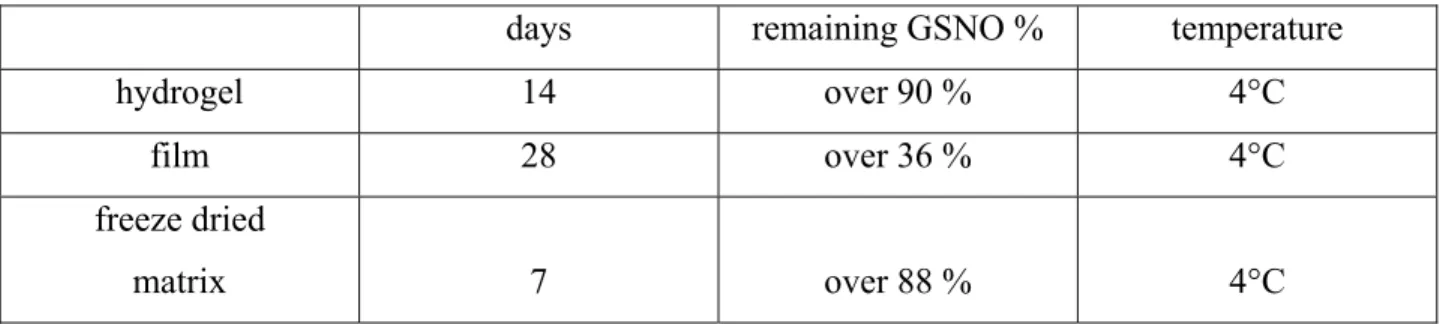
The stabilizing effect of the three type of formulations had significant differences. The best results with the optimal formulations:
days remaining GSNO % temperature
hydrogel 14 over 90 % 4°C
film 28 over 36 % 4°C
freeze dried
matrix 7 over 88 % 4°C
Table 1.: Best results of GSNO storage experiments using three different formulations
According to the results we chose hydrogel as the best type of formulation so we based the further research and development on gels. The storage times were chosen in a manner that we could compare samples prepared by the same method.
8 The effect of enzymes and N2 atmosphere
We found that the stabilizing effect of the N2 gas was significant; the combined usage of Cat and SOD enzymes did not have significant stabilizing effect.
Effect of pH
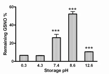
pH modification had influence on both formation and stability of GSNO. First we adjusted the pH in the interval between 0.3 and 12.6. We measured the starting absorbances and compared to the absorbances after the storage period (7 days 4°C, light exclusion). We found that the formation ratio was best at highly acidic pH, but the stability was very low only 6.7±0.1
% GSNO remained at the pH value of 0.3 after 7 days. Thus we found that mildly basic had the best stabilizing effect (even better than under physiologic pH) but strongly basic pH only enhanced decomposition. According to our results it is best to store the GSNO solution at the pH value of 8.6 with this adjustment 52.17±2.72 % of the starting GSNO amount remained (Fig. 1).
Fig 1.: The effect of pH on GSNO decomposition,
Samples were stored in simple aqueous solutions for 7 days in the pH range of 0.3-12.6, (n=4). All data were expressed as means + SD. *** p <0,001 between the solution at pH=8.6 and
all other solutions. *** p <0,001 between the solution at pH=7.4 and all other solutions. *** p
<0,001 between the solution at pH=12.6 and all other solutions.
9
The effect of enzymes and N2 gas was extended with pH modifications; we found that mildly basic pH had significant stabilizing effect, neither N2 nor the enzymes decreased decomposition significantly, the effect of pH was the most important.
We found that pH adjustment is a significant stability modifying factor so we combined this factor with pharmaceutically acceptable gel forming additives, which (probably through secondary bonds) decrease GSNO decomposition.
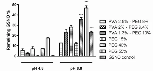
Our results showed that in gels without pH adjustment (pH=4.8) only 0-25 % of the starting GSNO remained after 25 days. It is important to note, that there were no physical changes in the gels, all samples were homogenous and transparent. The best result was achieved using the gel that contained 55% PEG in this composition 45.3 % of the starting GSNO remained. All samples were stored at 4°C, light was excluded (Fig. 2.).
Fig. 2.: Effect of gelling agents in gels without pH adjustment (pH=4.8) and with adjusted pH (pH=8.8), (n=4). All data were expressed as means + SD. *** p <0,001 between the solution containing 40 % PEG, 55 % PEG at pH=8.8 and the other solutions
with pH=4.8. *** p <0,001 between the solutions at the pH value of 8.8 and the other solutions with pH=4.8
10
Examination of ionic mechanism using UV spectroscopy
We examined the changes on the UV spectra of GSNO due to pH adjustment we found that new peaks appeared when we adjusted the pH to mildly basic values.
Examination of radical mechanism in the presence of the spin trapping agents TEMPOL and DEPMPO
We examined TEMPOL signal intensity in acidic and basic GSNO solutions under atmospheric conditions and under N2 gas. According to the results radical formation (which is directly proportional to the reduction of TEMPOL) decreased at mildly basic pH (pH=8.8) compared to acidic pH.
The difference was two orders of a magnitude between the fastest and the slowest decomposition rate. The basic solution under N2 was the most stable. N2 reduced decomposition compared to those kept under atmospheric conditions but pH adjustment was more effective. The results of the tests applying DEPMPO showed that the pH adjustment does not change the decomposition path of GSNO only reduces the rate of decomposition probably through polarization so the formation of complexes seem to be the main reason.
Measurement of dermal blood flow
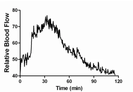
The increased relative blood flow was measured after applying the optimized GSNO containing hydrogel that was stored for 25 days. The test was performed using laser Speckle contrast analysis. The measured maximum relative blood flow intensity was 96.20±35.29 with the starting relative intensity of 36.29±10.16 (Fig. 3.). According to these experiments maximum blood flow intensity was reached 24.16±9.38 minutes after applying the GSNO solution and decreased to 50 % of the maximum value 33.75±15.06 minutes later (n=8).
11
Fig. 3: Effect of 40 mM GSNO solution on blood flow using laser Speckle contrast analysis (n=8).
Discussion
According to our results the production of RSNOs (GSNO and SNAC) in strongly acidic (pH=1.6) NaNO2 solutions increased thiol conversion compared to alkaline NaNO2 solutions with 20.04 and 23.18 %, respectively; compared to basic pH. The original NO donor concentration dropped slower, if we adjusted the pH to mildly basic values (pH=8.4-8.8) for the storage period. We were able to further decrease the rate of decomposition with the application of PVA/PEG polymer matrices. This combined method allowed us to sustain 45.3% of the original GSNO concentration after a 25 day storage period. This composition when applied topically increased blood flow intensity with more than 100%, proving that the stabilization steps did not influence the biological effect.
We concluded that the role of polymers is important in increasing stability, although our results prove that pH has a larger influence on stability. Structural analysis is very limited in aqueous solutions, we only relied on our results using UV and ESR spectroscopy. We can conclude - from the changes in the UV spectra and from the continuous decrease of pH during the storage phase – that an acid is formed during degradation, potentially because of GSSG formation. We did not see new peaks on the spectra after the storage phase, thus the logical
12
explanation is that the pH modification didn’t introduce new degradation pathways, because there were no unknown degradation products. The degradation intermedier of GSNO is GS- (the degradation product of RSNO is generally the corresponding RS-) during decomposition and the final product of degradation is GSSG, which we could not convert into any other compound after further pH changes. Our results show that the increased stability that occurs due to changes in the pH is caused by the changes in the electron structure of the sulfide ion; presumably because of the formation of an intermedier ionic complex.
The difference in the rate of degradation was nearly two orders of magnitude, as analyzed by ESR spectroscopy. These results also point to the fact that the radical degradation slowed down, most likely due to the formation of polarizing and ionic complex, possibly by shifting the RSNO tautomeric distribution to the ionic form and stabilizing it. In addition and comparison to the stabilizing effect of gelling agents we investigated the stabilizing effect in unmodified and basic aqueous environment to further increase the stability of GSNO.
After numerous experiments the best gelling agents were PEG and PVA, we used various concentrations to examine the effects. With using high amounts of PEG almost 20% of the starting GSNO remained after 25 days in the composition even at acidic pH. Our experiment also proved that GSNO stability increases in gel matrices, however, according to our experiments the pH adjustment was more effective than only the addition of PEG. We confirmed that polymers increase the stability, even in relatively small concentrations (Fig 2.) but combined with pH adjustment the stability increasement is significant; so we found that the effect was highly pH dependent. In the best composition, using 55 w/w% PEG, nearly half of the starting GSNO remained after 25 days. Taken together, some polymers, especially PEG, probably stabilizes through intermolecular bonds and with the additional pH adjustment, the stabilizing effect is further increased via our hypothesized sulphide complex formation.
To test the biological effectiveness of the decreased concentration of GSNO we investigated the vasodilatory effect on human subjects with the concentration that was still available after 25 days. We have found that this concentration was still capable of increasing the blood flow with more than 100% for a prolonged period. Our results confirmed the potential of GSNO as a topical vasodilator.
13
Conclusions
The decomposition of S-nitrosothiols happen indeterminably fast. This is a serious impediment to use these compounds as NO donors in therapeutic practice. It is a clinically justified need to reach and maintain a higher RSNO concentration; and to create a new formula that sustains the biological activity from the time of production until reaching the end user.
Our experiments proved that RSNO solutions can be produced in nearly stoichiometrical quantities, from the corresponding thiols using strongly acidic NaNO2 solution, which is then best stored under mildly basic pH in the presence of polymer matrixes.
The mildly basic pH slowed down radical decomposition with nearly two orders of magnitude (95.9 %) compared to the solution with unmodified pH. According to the ESR measurements we can explain these results with the formation of a more stable ionic complex.
pH modification results in RSNO structural changes, which is confirmed by the appearance of new absorption bands in the UV spectra. The modified pH allows long term storage and the interval of pH=8.4-8.8 is still in the physiologically acceptable range, thus the solution is suitable for therapeutic application.
The optimized components and the pH modification helped us to preserve 45.3 % of the starting GSNO concentration after 25 days; and this composition enhanced the blood flow with more than 100 %. This discovery gives a new opportunity to generate a stable but still biologically active and topically applicable RSNO solution or gel formula. This can be used in a wide range of medical applications for increasing local blood flow; from microcirculatory disorders in diabetes to the enhancement of wound healing.
14
Publcations
Publications closely related to the thesis:
Scientific articles:
Hornyak, I.; Pankotai, E.; Kiss, L.; Lacza, Z. Current Developments in the Therapeutic Potential of S-Nitrosoglutathione, an Endogenous NO-donor Molecule. Curr Pharm Biotechnol. 12:1368- 1374; 2011.
IF:3,35
Dongó, E.; Hornyak, I.; Benkő, Z.; Kiss, L. The cardioprotective potential of hydrogen suphide in myocardial ischemia/reperfusion injury. Acta Physiol Hung. 98:369-381; 2011.
IF:1,2
Hornyak, I.; Marosi, K.; Kiss, L.; Gróf, P.; Lacza, Z. Increased stability of S-nitrosothiol solutions via pH modulations. Free Radis Res. 46:214-225; 2012.
IF:2,8
Patents:
Lacza, Z.; Hornyak, I. Pharmaceutical Composition Comprising S-nitrosoglutathione and polisaccharide. 2009. WO2009050527A1
Lacza, Z.; Hornyak, I. Method for stabilitation of S-nitrosoglutathione and composition prepared by the same. 2009. WO2009090439A2
15 Publications not related to thesis:
Niesz, K., Hornyak, I., Borcsek, B., Darvas, F., Nanoparticle synthesis completed with in situ catalyst preparation performed on a high-pressure high-temperature continuous flow reactor, Microfluid Nanofluidics, 2008, 1613-4982
IF:3,314