Changes of CB1 cannabinoid receptor distribution in temporal lobe epilepsy
PhD Dissertation
Mária Rita Karlócai
János Szentágothai School of Neurosciences Semmelweis University
Supervisor: Dr. Zsófia Maglóczky, Ph.D.
Official reviewers: Prof. Tibor Wenger, MD, Ph.D., D.Sc.
Dr. József Takács, MD, Ph.D.
Members of the Final Examination Committee:
Prof. József Kiss, MD, Ph.D., D.Sc. -chair Dr. Andrea Székely, MD, Ph.D.
Dr. Hajnalka Ábrahám, MD, Ph.D.
Budapest, 2012
Hungarian Academy of Sciences, Institute of Experimental Medicine
Table of contents:
1. Abbreviations
………....42. Introduction
………...………62.1. Temporal Lobe Epilepsy………..………..……6
2.2. Anatomy of the mouse hippocampus………..……….…….8
2.3. Cell types of the hippocampus……….….………9
2.3.1. Perisomatic targeting interneurons………...10
2.3.2. Dendritic targeting interneurons………..……….…10
2.3.3. Interneuron-selective (IS) interneurons………....11
2.4. Animal models of epilepsy……….………..……15
2.5. The endocannabinoid system………..…...……….17
3. Aims
………...194. Materials and methods
………...….……....214.1. The pilocarpine model of epilepsy………..…………..…..21
4.2. In vivo electrophysiology………..………...22
4.3. Analysis of the EEG recordings……….….…………..…..23
4.4. Slice preparation……….….………....23
4.5. Tissue preparation for morphological examination……….……....24
4.6. Human tissue………..………..…24
4.7. Immunocytochemistry……….…….…...25
4.8. CB1-R Antibodies………..………26
4.9. Gallyas silver impregnation………..……….…….28
4.10. Nissl staining………...28
4.11. Determination of principal cell loss……….….28
4.12. Quantitative electron microscopic analysis………...30 4.12.1. Ratio and number of symmetric and asymmetric synapses………30 4.13. Target distribution of CB1-R-positive terminals………....30
5. Results
……….………...31 5.1. Reorganization of CB1 receptor expressing GABAergic fibers in temporal lobe epileptic patients……….……….31 5.1.1. Pathological classification of the epileptic samples………...31 5.1.2. Distribution of CB1-R-stained inhibitory synapses in control human hippocampus...32 5.1.3. Distribution of CB1-Rs in epileptic human hippocampus…...…...335.1.4. Qualitative analysis of immunopositive fiber density in human sclerotic tissue ……….……….…..…………..35 5.1.5. Ultrastructural analysis of CB1-R distribution in control and sclerotic human tissue………..35 5.1.6. Target distribution of CB1-immunopositive elements in human TLE.………...35 5.2. Changes of CB1-receptor immunostained inhibitory fibers in a model of temporal lobe epilepsy……….………...37
5.2.1. Target distribution of CB1-immunopositive elements in sclerotic mouse tissue………..38 5.3.Description of the pilocarpine model of epilepsy………..……….40
5.3.1. The pattern of cell loss in pilocarpine induced epilepsy…………...40 5.3.2. In vivo electrophysiological recordings………..………...46 5.3.3. Anatomical changes in the acute phase of epilepsy (2 hours)....………....50 5.3.4. Morphological changes in the latent phase of epilepsy (1, 3 days post pilo)………..……….50 5.4. Analysis of CB1-R distribution in different phases of pilocarpine-induced epilepsy.………..…..60
5.4.1.Distribution of CB1 receptors in control tissue………...60
5.4.2.Distribution of CB1 receptors in the acute phase of epilepsy (2 hours)……… ………...62
5.4.3. Reversible changes in CB1-R expression in acute slices (2 hours post pilo)……… ……….……….64
5.4.4.Increased mortality after seizures in CB1 KO animals………66
5.4.5.Ultrastructural changes of CB1-R expression in the acute phase of epilepsy………..……..……….……...….68
5.4.6. Distribution of CB1 receptors in the latent phase of epilepsy (1 and 3 days post pilo)………....…73
5.4.7.Distribution of CB1 receptors in the chronic phase of epilepsy (1 and 2 months) in mice……….74
5.4.8. Ultrastructural analysis of CB1-R distribution in the chronic phase (in mice)………..…………..….76
6. Discussion
……….………836.1.Changes in the acute phase of epilepsy………...………84
6.2.Changes in the chronic phase of epilepsy………...……….……85
6.2.1. Sprouting of excitatory fibers………....…85
6.2.3. Sprouting of inhibitory fibers, changes in perisomatic inhibition...86
6.3. Preserved target distribution in human TLE and in the animal model of epilepsy……….………88
6.4. Therapeutic implications….………....89
7. Conclusions
………..908. Summary
……….…….919. Összefoglalás
………...9210. References
……….……….9311. List of publications underlying the thesis
………...…..10812. Acknowledgements
………...…1091. Abbreviations:
a: alveus
ABC: avidin-biotin-horseradish peroxidase complex ACSF: artificial cerebrospinal fluid
BSA: bovine serum-albumin CA: Cornu Ammonis
CB1-R: Type 1 cannabinoid receptor CCK: cholecystokinin
CR: calretinin
DAB: 3,3’-diaminobenzidine-4HCl deg: degeneration
EEG: electroencephalogram ff: fimbria fornix
GABA: γ-amino-butric-acid GD: gyrus dentatus
h: hilus
HSP72: Heat shock protein 72 KO: knock-out
NeuN: Neuronal Nuclear antigen NGS: Normal goat serum
PB: phosphate buffer Pilo: Pilocarpine
s.g.: stratum granulosum
s.lm.: stratum lacunosum-moleculare s.m.: stratum moleculare
s.o.: stratum oriens s.p.: stratum pyramidale s.r.: stratum radiatum SE: status epilepticus str.: stratum
TB: TRIS buffer
TBS: Physiological saline buffered with TRIS TLE: Temporal Lobe Epilepsy
WT: wild type
2. Introduction
2.1. Temporal Lobe Epilepsy
According to the definition of WHO epilepsy is a chronic neurological disorder with various etiologies that affects people of all ages and is characterized by recurrent seizures.
Approximately 5 million people have epilepsy worldwide (1-3% of the population (Corsellis & Meldrum, 1976; Houser, 1990). Seizures can vary from the briefest lapses of attention or muscle jerks, to severe and prolonged convulsions (i.e. violent and involuntary contractions, or a series of contractions, of the muscles). Seizures can also vary in frequency, from less than one per year to several per day.
Epilepsy is one of the world's oldest recognized diseases. Fear, misunderstanding, discrimination and social stigma have surrounded epilepsy for centuries. In many countries this condition has a great impact and can impact on the quality of life for people with the disorder and their families. Moreover, it increases a person's risk of premature death by about two to three times compared to the general population.
One seizure does not necessarily indicate epilepsy (up to 10% of people worldwide have one seizure during their lifetimes), thus, epilepsy is defined by two or more spontaneously recurrent seizures (Halász & Rajna, 1990). At the neuronal network level it manifests as states of pathological hyperexcitability and hypersynchronous activity.
Imbalanced synaptic input may cause excessive neuronal activity, eventually leading to neuronal death and synaptic reorganization (Engel, 1996; Green, 1991; McNamara, 1999).
Characteristics of seizures vary and depend on where in the brain the disturbance first starts, and how far it spreads later on. Temporary symptoms can occur, such as loss of awareness or consciousness, and disturbances of movement, sensation (including vision, hearing and taste), mood or mental function.
Epilepsy syndromes used to be classified based on the classification system accepted by the International League Against Epilepsy (ILAE) in 1989. This classification was based on seizure phenomenology and EEG alterations, it differentiated localized (partial) and generalized forms, and moreover, based on their origin genetic, symptomatic and cryptogenic forms were distinguished as well. However, the development of numerous techniques (electrophysiological techniques, Magnetic resonance imaging (MRI)) and the improvement of our knowledge regarding genetics (mapping of the human genome and uncovering the genetic origin of certain epilepsies) have pointed out the need for a new classification (Berg & Cross, 2010).
According to the most widely accepted concept, the reasons for epilepsies are genes (Salzmann et al, 2008) disposing the network to generate epilepsy under certain circumstances. This notion is strengthened by the fact that epileptic patients often have a history of accidents, febrile seizures, or perinatal disorders (Rocca et al, 1987a; Rocca et al, 1987b). These events do not trigger epilepsy by them selves; however, they may increase the risk of generating seizures in certain people. Thus, most probably the interaction of the entire genome and environmental factors is responsible for this disease.
In adulthood the most common form is Temporal Lobe Epilepsy. The epileptogenic area is mostly found in the hippocampus and quite frequently hippocampal sclerosis occurs.
Though recently a high variety of antiepileptic drugs have been available a significant number of patients are pharmacoresistant (cannot be cured with medication). In case of therapy resistant patients with severe clinical consequences where the epileptic focus can be precisely localized epilepsy surgery is a possible solution (Falconer & Taylor, 1968; Spencer & Spencer, 1994). The aim of the surgery is blocking seizures by lesioning the epileptogenic focus. 85% of partial epilepsies originate from the temporal lobe, thus the surgery most often carried out is anterior temporal lobectomy. After surgery, global cognitive decline can rarely be seen, however certain special functions (like memory) may be modestly impaired. Still, in most cases the quality of life improves following the surgery.
2.2. Anatomy of the mouse hippocampus
The hippocampus is a part of the filogenetically most ancient cortical region, the archicortex. It has a major role in learning and memory and their modifications by emotions. In case of its injury processing of new information may be impaired.
It is a curved elaboration of the edge of cerebral cortex in the floor of the inferior horn of the lateral ventricle; a functional component of the limbic system. The hippocampal formation can be divided into two regions: the cornu Ammonis (CA) and the dentate gyrus (DG), forming two U-shaped semicircles. Based on the morphology, distribution and connectivity of principal cells, the cornu Ammonis can be divided into CA1, CA2 and CA3 (a,b,c) regions (Duvernoy, 1998; Nó, 1934; Seress, 1988) (Figs 1 and 2). These regions (except CA3c) form typical layers: the somata of pyramidal cells are located in str.
(stratum) pyramidale. Their basal dendrites can be found in str. oriens, whereas, their apical dendrites localize in str. radiatum and arborize in sr. lacunosum-moleculare (Cajal; Cajal, 1968). The axons of pyramidal cells can be seen in str. oriens and run in the alveus; in the CA3 region axons form recurrent collaterals as well, innervating each other. In the CA3 the region between str. pyramidale and str. radiatum, where mossy fibers terminate is termed str. lucidum (Cajal; Cajal, 1968). In the dentate gyrus granule cell are located in str.
granulosum, while their dendrites can be found in str. moleculare. Under the densely packed layer of granule cells the hilus can be seen, where mossy cells are the principal cells.
The human hippocampus differs from rodent hippocampus in certain connectivity patterns; however, the cellular structure is rather similar.
In the hippocampal formation the network of principal cells form a so called trisynaptic loop (Amaral DG, 1990). The perforant path, this glutamatergic pathway originates from the entorhinal cortex, and synapses with apical dendrites of CA1 and CA3 pyramidal cells in str. lacunosum-moleculare, and the dendrites of granule cells in the outer part of str. moleculare (first synapse). Axons of the granule cells (mossy fibers) form
synapses with CA3 dendrites on special complex spines, so called, thorny excrescences in str. lucidum (second synapse). Axons of CA3 pyramidal cells innervate apical dendrites of CA1 pyramidal cells in str. radiatum (third synapse). In addition CA3 pyramidal cells strongly innervate each other forming recurrent collaterals. CA1 pyramidal cells project to the subiculum (Amaral et al, 1991), finally subicular pyramidal cells send axons to the entorhinal cortex (Other fibers lave the hippocampus via fimbria hippocampi).
2.3. Cell types of the hippocampus
The two types of neurons that can be found in the hippocampus are principal and non-principal cells. Principal cells are glutamatergic, pyramidal cells of the cornu Ammonis, whereas, in the dentate gyrus they are granule and mossy cells. Axons of CA1 pyramidal cells project to the subiculum, and these subicular cells innervate the entorhinal cortex forming the main output of the hippocampus. The vast majority of non-principal cells are interneurons with local axonal arborisation (Freund & Buzsaki, 1996).
Though, interneurons form a small portion of hippocampal cells, functionally they are quite important. One single interneuron by its extended axonal tree may innervate numerous pyramidal cells, thus they have a significant role in modifying the firing properties of pyramidal cells.
The functional classification of interneurons is based on their input and output
characteristics which can be identified with tracing studies. In addition, interneurons can be categorized based on their neurochemical marker content
(calcium binding proteins, neuropeptides, transmitter receptor-content or neurotroph factors) According to most studies, these cells can be classified into three major groups;
-Perisomatic targeting interneurons -Dendritic targeting interneurons -Interneuron specific interneurons
The first two populations target mostly principal cells, perisomatic targeting interneurons (basket and axo-axonic cells) (Halasy et al, 1996; Handelmann et al, 1981; Katsumaru et al, 1988; Kosaka et al, 1987; Kosaka et al, 1985; Li et al, 1992) innervate the perisomatic region, thus, they may control the output of principal cells, whereas dendritic targeting interneurons innervate dendrites, therefore, they can modulate synaptic plasticity and dendritic electrogenesis (Arai et al, 1995; Freund & Buzsaki, 1996; Gulyas et al, 1992;
Gulyas et al, 1993; Halasy et al, 1996; Han et al, 1993; Kawaguchi & Hama, 1988; McBain et al, 1994; Miles et al, 1996).
The third population selectively targets interneurons (Freund & Buzsaki, 1996; Gulyas et al, 1996)
2.3.1. Perisomatic targeting interneurons
Interneurons containing the calcium-binding protein parvalbumin (PV) are either
perisomatic targeting interneurons (Kosaka et al, 1987) with two distinct subpopulations:
basket cells innervating the soma and proximal dendrites and axo-axonic cells innervating the axon-initial segments of principal cells (Katsumaru et al, 1988; Kosaka et al, 1987) . Another basket cell type is known which does not contain parvalbumin, instead, another calcium-binding protein, cholecystokinin (CCK) can be found in it. (Acsady et al, 1996a;
Gulyas et al, 1991)
2.3.2. Dendritic targeting interneurons
Dendritic targeting interneurons form a more heterogeneous class compared to perisomatic targeting interneurons. In the dentate gyrus we may distinguish HICAP cells (HIlar
Commissural-Associational Pathway-related), where the soma is located in the hilus and the axon in the inner part of str.moleculare, where the input from the commissural and perforant pathways arrive (Freund & Buzsaki, 1996; Han et al, 1993; Sik et al, 1997). Other groups are HIPP (HIlar Perforant Path-associated) and MOPP
(MOlecular layer Perforant Path-associated) cells, where the soma is located in the hilus (HIPP) or in str. moleculare (MOPP) and they innervate the outer part of stratum
moleculare where the input from the perforant path can be found (Hampson & Deadwyler,
1992; Sik et al, 1997). HIPP cells can be identified based on their neurochemical marker- content, by containing somatostatin and neuropeptid Y (Katona et al, 1999a; Sik et al, 1997). In addition, certain hilar interneurons innervate CA1 and CA3 regions (Buckmaster
& Schwartzkroin, 1995; Sik et al, 1997). In the cornu Ammonis O-LM (oriens-lacunosum- moleculare targeting cells) can be found as well with their soma in str. oriens and axons in stratum lacunosum-molaculare (Cajal, 1968; Nó, 1934). Most probably, these cells have a function similar to HIPP cells by providing feedback-inhibition, since in case of both cell types, the somata and dendrites are located at the output region of principal cells, moreover, they project to the same layer of the entorhinal cortex (Gulyas et al, 1993; McBain et al, 1994; Sik et al, 1995). Moreover, both O-LM cells and HIPP cells contain somatostatin (SOM) and occasionally neuropeptid Y as well (Katona et al, 1999a; Sik et al, 1995) Trilaminar and bistratified cells innervate the more proximal dendritic regions of principal cells. Trilaminar cells innervate strata oriens, pyramidale and radiatum, whereas,
bistratified cells send axons only to strata oriens and radiatum (Halasy et al, 1996; Miles et al, 1996; Sik et al, 1995) their somata can be found in the same regions. Dendritic targeting interneurons can be classified according to their neurochemical marker content as well;
bistratified cells were shown to be either calbindin-containing (CB)(Sik et al, 1995) or parvalbumin-containing (Klausberger et al, 2004), whereas numerous other interneurons contained Calbindin (Gulyas & Freund, 1996; Sloviter, 1989; Toth & Freund, 1992)or CB1-R (Katona et al, 1999b).
Additionally other interneurons can be distinguished with soma, dendrites and axons located in str. radiatum (Cajal, 1968; Gulyas et al, 1993; Kawaguchi & Hama, 1988; Miles et al, 1996 or in str. lacunosum-moleculare (Nó, 1934 #3164)]. Finally back-projection interunerons located at the border of str. oriens and the alveus of CA1 are dendritic targeting interneurons as well, besides their local axonal arbor, they project back to CA3/hilus (Sik et al, 1995; Sik et al, 1994).
2.3.3. Interneuron-selective (IS) interneurons
These interneurons exclusively innervate other interneurons. Based on their connectivity and morphology, they maybe divided into three groups.
IS-1 cells can be found in all hippocampal regions and contain calretinin (CR)
(dendro-dendritic connections are quite frequent between these cells). Their somata are often located in the proximal dendritic region of principal cells. These cells innervate interneurons in the str. radiatum and dentate gyrus (Acsady et al, 1996b; Gulyas et al, 1996).
IS-2 cells were shown to be VIP-positive, with somata and dendrites in str. lacunosum- moleculare, axons in str. radiatum (Acsady et al, 1996a; Acsady et al, 1996b).
IS-3 cells contain VIP as well, however, their axons innervate interneurons in str. oriens and in the hilus (Acsady et al, 1996a; Acsady et al, 1996b).
In the rodent hippocampus an additional interneuron group was identified projecting to the septum, the so-called (Gulyas et al, 2003; Jinno & Kosaka, 2002), HS (hippocampo-septal) cells, with local and projecting axons innervating GABAergic interneurons (Gulyas et al, 2003). These interneurons mostly contain SOM and CB, moreover, spiny, CR-positive interneurons are part of this group as well (Gulyas et al, 2003; Gulyas et al, 1992).
CA1 pyramidal cell CA3 pyramidal cell
Mossy cell Granule cell
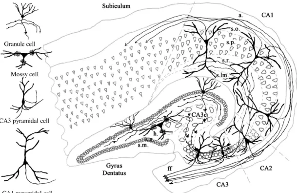
Figure 1: Schematic of human hippocampal formation.
Dentate gyrus and Cornu Ammonis, the two parts of the hippocampal formation, form two U-shaped curves looping into each other. The Cornu Ammonis can be further divided to four subregions: CA1, CA2, CA3 and CA3c (Lorente de Nó, 1934, Seress, 1988). These subregions can be separated by the morphology and connectivity of principal cells (pyramidal cells and mossy cells) found there. Granule cells forming stratum granulosum are the principal cells of the Dentate Gyrus, above this area, stratum moleculare can be found, whereas under it the hilus. The region enclosing the hilus and the CA3c is often mentioned as the endfolium. Abbreviations: CA: Cornu Ammonis, a: alveus, s.p.: stratum pyramidale, s.r.: stratum radiatum, s.lm.: stratum lacunosum-moleculare, ff: fimbria fornix, Figure by Lucia Wittner (PhD thesis 2004)
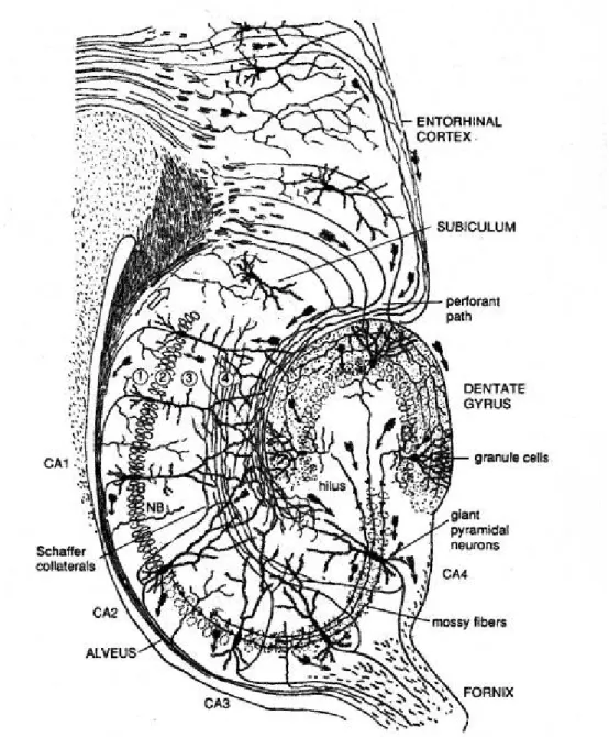
Figure 2: Schematic of rodent hippocampal formation
Similar to the human hippocampus, the rodent hippocampal formation contains two U shaped regions, the Cornu Ammonis and the Dentate Gyrus. The Cornu Ammonis can be divided to CA1, CA2 (not significant in mice), CA3 and CA3c (indicated as CA4 in the figure) subregions. Densely packed granule cells form stratum granulosum in the Dentate Gyrus, with their dendrites in stratum moleculare above, and the hilus underneath them.
Figure by Ramon y Cajal (arrows in CA str. oriens point to the opposite direction of real projection).
2.4. Animal models of epilepsy
Numerous existing animal models demonstrate features typical for human epilepsy. In these models either genetic alterations can be seen, or an acute lesion induces seizures which are followed by a silent period and later by spontaneously occurring recurrent seizures.
-genetic models
A significant proportion of epilepsies have a genetic component. Thus genetic models can provide valuable information regarding the mechanisms underlying neuronal hyperexcitability. The seizure predisposition in these animal models is inherited by the disfunction of certain ion channels, neurotransmitter or neuromodulatory systems.
For example, in the model of genetic absence epilepsy spontaneous seizures occur with age in animals, characterized by motor arrest and head drops that are correlated with generalized spike-wave on the electroencephalogram (EEG). The seizure generating mechanism appears to be located in the lateral thalamic nuclei (Crunelli & Leresche, 2002;
Fisher, 1989; Polack & Charpier, 2006).
-kindling models
In the kindling models the development of seizures and epilepsy are acquired by repeated stimulation of the brain (either by electric or chemical stimuli). The stimuli is set at a subthreshold level, however after a certain time, the threshold lowers and acute seizures begin (McNamara, 1986). After the first seizure no further stimulation is required, since a seizure increases the likelihood that more (recurrent) seizures will occur. (Seizures beget seizures) (Brown & Stone, 1973; Lothman et al, 1989; Sutula & Steward, 1986)
-excitatory amino acid analogs
The overexcitation of cells is considered a basic feature of epilepsy. Increase of glutamate release has an important role in causing this overexcitation. Thus by adding additional glutamate or aspartate analogues it is possible to exert toxic effects on the nervous system under certain conditions (Brown & Stone, 1973).
-substances altering the function of ion channels
In epileptic specimens changes in the number and/or function of ion channels can often be seen leading to abnormal extra and intracellular ion concentrations and thus impaired membrane potential. Consequently, by altering the function of ion channels it is possible to mimic abnormal ion concentrations and cause seizure activity (Karnup & Stelzer, 2001;
Panuccio et al, 2010). This model is suitable to study the acute phase of epilepsy exclusively, since neither network reorganization nor recurrent seizures occur.
-substances altering the concentration of chations or anions
Similarly to the previous model, the ion balance and thus the membrane potential can be manipulated by changing ion concentrations themselves. Thus, by increasing or decreasing chations and/or anions, the balance of excitation and inhibition can be altered effectively (Gutnick et al, 1982; Heinemann et al, 1986)
In the current studies alterations were examined in pilocarpine induced epilepsy model.
Pilocarpine is cholinergic agonist acting on each type of muscarinic acetylcholine receptors (Turski et al, 1986). By activating these receptors it induces status epilepticus (acute seizures) which is followed by a seizures-free latent phase and finally the chronic phase where spontaneously emerging seizures occur (Turski et al, 1984).
2.5. The endocannabinoid system
Physiological and psychological effects of compounds deriving from the plant Cannabis Sativa have been known since the ancient times. The main psychoactive molecule, the terpenoid derivative 9-tetrahydrocannabinol (Northcutt) (Gaoni & Mechoulam, 1971) was described in the 60s, however the site of activation remained hidden. Finally in 1990 the CB1 cannabinoid receptor was cloned from rat brain by Matsuda and co (Felder et al, 1992; Matsuda et al, 1990) and three years later its immune-system counterpart, the CB2 receptor as well (Munro et al, 1993). These findings enlightened the mechanisms of action of THC, and a search began for the brain-derived cannabinoid ligands.
The first ligand described was N-arachidonoyl ethanolamide (anandamide)(Devane et al, 1992), the second 2-arachidonoyl-glycerol (2-AG) (Mechoulam et al, 1995; Sugiura et al, 1995), both extracted from animal tissue (Pertwee, 1997) Even though anandamide was the endocannabinoid described first (Devane et al, 1992), it seems more possible that 2-AG is the one effectively activating the receptor (Sugiura et al, 2006). After identified these two ligands, their synthesizing and degrading enzymes were described as well. Synthesizing (or metabolic) enzyme for anandamide is N-acyl phosphatidylethanolamine phospholipase D (NAPE-PLD) (Okamoto et al, 2004), whereas diacylgycerol lipase- α (DGL-α) (Bisogno et al, 2003) for 2-AG. For degrading anandamide, the enzime is fatty acid amide hydrolase (FAAH) (Egertova et al, 1998), and monoacylglycerol lipase (mice) (Dinh et al, 2002) for 2-AG.
Activation of the endocannabinoid system is involved in numerous physiological functions like food intake, pain sensation and memory formation. In the brain, the endocannabinoid system is responsible for retrograde synaptic signaling via CB1-R (Type 1 cannabinoid receptor) (Freund et al, 2003; Mackie, 2008; Mackie & Stella, 2006).
Endocannabinoids are released from the postsynaptic neurons in an activity-dependent manner, and bind to presynaptic CB1-Rs, thereby suppressing transmitter release from presynaptic terminals (both in excitatory and inhibitory synapses) (Hajos et al, 2000; Hajos et al, 2001; Kathmann et al, 1999; Katona et al, 1999b; Mackie, 2005; Misner & Sullivan, 1999; Ohno-Shosaku et al, 2002; Sullivan, 1999; Varma et al, 2001).
In addition to its physiological roles, this system was found to be affected in pathological processes as well. Controversial data were published regarding the effects of cannabinoids in epilepsy. On one hand, in an animal model of TLE (temporal lobe epilepsy), CB1-R agonists displayed anti-epileptic effects (Wallace et al, 2003), in addition, CB1-Rs on glutamatergic axon terminals were shown to mediate anticonvulsant effect, by attenuating glutamate release (Azad et al, 2003; Monory et al, 2006). On the other hand, proconvulsive effects of CB1-R agonists were described as well (Gordon & Devinsky, 2001; Keeler & Reifler, 1967; Lutz, 2004). Moreover, a CB1-R antagonist was shown to prevent the long-term increase in seizure susceptibility when applied in a certain time- window (Chen et al, 2007; Echegoyen et al, 2009).
Human studies showed that recurrent seizures may lead to an adverse reorganization of the endocannabinoid system and to the impairment of its protective effect (Goffin et al, 2011; Ludanyi et al, 2008) which may be accomplished by the downregulation of CB1-Rs located at excitatory synapses occurring in the inner molecular layer of the dentate stratum moleculare (Ludanyi et al, 2008).
Since more numerous CB1-R-positive asymmetric synapses can be found in the hippocampus compared to stained symmetric synapses, previous ultrastructural studies have not demonstrated, what happens to inhibition regulated by the endocannabinoid system at the ultrastructural level. Moreover, the target distribution of CB1-R-positive terminals was not examined either.
Regarding CB1-Rs located at inhibitory synapses, results are restricted to animal models and are contradictory reporting both up and downregulation of the receptor (Falenski et al, 2009; Wyeth et al); (Wallace et al, 2003).
3. Aims
In the present study we wished to uncover anatomical changes affecting the endocannabinoid system in patients with TLE and in pilocarpine treated mice. We focused on two phases of epilepsy in the mouse model (acute and chronic) and to the chronic phase in human TLE, since robust and controversial changes occur at early and later time points post epileptogenesis. (the acute phase obviously cannot be studied in human samples) The imbalanced ratio of dendritic and perisomatic inhibition has long been regarded as an important feature linked to epilepsy (Magloczky & Freund, 2005; Wittner et al, 2005) , causing cell death and hippocampal reorganization. Increased excitation was shown to originate from mossy fiber sprouting (Houser et al, 1990; Sutula et al, 1989a) and an increased input from the supramammilary nucleus (Abdulla & Campbell, 1993; Magloczky et al, 1994; Magloczky et al, 2000). Although a decreased inhibition was proposed to be associated to the strong excitation, several studies show, that increased inhibition occurs, leading to hyperexcitability through several mechanisms (hypersynchronization, excitatory GABA, etc) (Bernard et al, 2000; Cossart et al, 2001; Karlocai et al, 2011; Wittner et al, 2005; Zhang et al, 2009). The mechanism leading to hypersynchronization can vary; most often the reorganization of excitatory or inhibitory fibers is one of them. Sprouting can often be characterized by changes in target distribution, increase in fiber density or the number of terminals, leading to altered transmitter release. Cannabinoids act as retrograde transmitters, decreasing transmitter release when activating their receptors, thus they are in the position of effectively regulating network activity. Several studies have examined functional and anatomical changes; however no detailed investigation was carried out to quantify the exact changes in number and target distribution of CB1-R-positive fibers.
In the present set of experiments we wished to examine the following issues:
• The correlation between pathological changes and CB1-R distribution in human TLE samples (chronic phase) and in the acute, latent and chronic phases of pilocarpine induced epilepsy.
• Alterations in the target distribution of CB1-R-positive elements in epileptic mice and human patients.
• The correlation between behavioral signs of acute seizures and later emerging cell death and reorganization in the epileptic mice.
• The difference of the seizure susceptibility in CB1-R knock out animals compared to controls.
• We assumed that by understanding the dynamics of changes in CB1-R expression during different phases of epilepsy we could suggest whether CB1-R agonists or antagonists would be suitable candidates for antiepileptic treatment.
4. Materials and methods
4.1. The pilocarpine model of epilepsy
Animals were kept under standard conditions with 12 h dark-light cycle; food and water were supplied ad libitum. Experiments were performed according to the guidelines of the Institutional Ethical Codex & the Hungarian Act of Animal Care & Experimentation (1998, XXVIII, Section 243 ⁄ 1998), The Animal Care and Experimentation Committee of the Institute of Experimental Medicine of Hungarian Academy of Sciences and the Animal Health and Food Control Station, Budapest, has approved the experimental design under the number of 2303/003/FŐV/2006. The experiments are in accordance with 86/609/EEC/2 Directives of European Community, which is in full agreement with the regulation of animal experiments in the European Union. All efforts were made to reduce the number of animals used and to minimize pain and suffering. For this animal model 20-30 g male CD1 mice (Harlan, Italy) and CB1-R knock-out CD1 mice were used (Ledent et al, 1999).
Animals were assigned to control and experimental groups. Age-matched control mice were injected with physiological saline (12 animals, 0.1 ml/30g) or scopolamine (12 animals 5 mg/kg). Since no difference was found between the two control groups, during the following experiments, control mice were injected with physiological saline.
Experimental mice were injected with intraperitoneal Pilocarpine hydrochloride (340 mg/kg, Sigma) to induce status epilepticus (SE). Scopolamine methyl nitrate (5 mg/kg, Sigma) was injected 30 minutes in advance to prevent peripheral cholinergic effects of pilocarpine. No benzodiazepine treatment was used to stop seizures. By omitting antiepileptic drugs we were able to examine seizure-induced changes 2 hours post SE, without further alteration of the GABAergic function by additional drugs.
Animals were observed for two hours after the pilocarpine injection, and the behavioral signs of seizures were monitored and scored. At every 5 minutes or when behavioral changes occurred a score was determined. Acute seizures started 5-15 minutes after the pilocarpine administration (post pilo). Seizures were classified by using the modified Racine’s scale (Racine, 1972) (1-5), animals were separated into “weak” and
“strong” groups according to their seizure activity (Magloczky et al). Seizure activity was
defined for every animal by using the maximal value of Racine -scale reached more than once.
In the weak group mice developed only a few mild seizures characterized by few ictal seizure period in the EEG accompanied with startling and shaking, whereas in the strong group the seizures were frequent and EEG showed several ictal seizure periods behaviorally represented by intense motor symptoms including jaw movements, salivation, and forelimb clonus with rearing and tonic-clonic seizures (Magloczky et al; Racine, 1972;
Turski et al, 1984).
Acute phase was examined 2 hours after pilocarpine injection. The period examined 1-3 days after the injection was regarded as the latent phase. At day three mass cell loss was observed, therefore this time point was considered as the end of the latent phase. EEG recordings were carried out 1 month after pilocarpine treatment in the chronic phase, at this time point recurrent seizures occurred in most of the strong epileptic animals.
We examined the expression pattern of CB1-Rs in the hippocampus at different survival times: 2 hours after the treatment in the acute phase, 1 and 3 days after the treatment in the latent phase and 1-2 months after the treatment in the chronic phase.
4.2. In vivo electrophysiology
We proved the occurrence of recurrent electrographic seizures with EEG recordings in the chronic phase. Mice were anesthetized with a 1-1.5 % halothane-air mixture (Narcotan, Leciva, Praha, Czech Republic) and secured in a stereotaxic frame (David Kopf, 900 USA, equipped with a SUPERTECH Ltd. made mouse adaptor HU).
Five holes were drilled into the skull above the frontal and parietal lobe bilaterally and above the cerebellum (reference). Stainless steel wire electrodes (MEDWIRE SST1 ) were placed on the skull and covered with conductive paste (Ten20, USA) to decrease the impedance. The electrodes and the connector were embedded in dental acrylic cement (GS, Japan). This way the electrodes were firmly implanted to the skull but the dura was not pierced. The EEG activity was recorded by a Grass EEG 8B model (Grass Instruments, Quincy, MA, USA), filtered at 1 Hz to 70 Hz and amplified. Data were recorded with a CED 1401 system using SPIKE2 v2.1 software (Cambridge Electronic Design Limited,
Cambridge, UK). Sampling rate was 500 Hz , amplification 20 µV/mm, filter: LP: 70Hz, HP: 1Hz.
Recordings were made for 3 days, in the morning (9 AM) and in the afternoon (4PM) for 1.5 hours. The behavior of the animal was observed during EEG recording. To answer the question whether animals had different diurnal and nocturnal epileptic activity, 24-hour continuous EEG monitoring was also carried out in 4 animals (2 controls, 2 chronically epileptic mice).
4.3. Analysis of the EEG recordings
All EEG recordings were evaluated by three independent and experienced researchers to determine the occurrence of seizures or interictal spikes (Litt et al, 2001).
EEG recordings were carried out in 16 mice (6 controls, 4 weak and 6 strong epileptic) in the chronic phase. In 4 animals (2 controls, 2 chronically epileptic ones) 24-hours EEG monitoring was carried out.
Epileptic activity was considered as interictal burst when the amplitude of occasionally appearing EEG spike activity was three times higher than the normal resting control EEG waves. The ictal EEG was determined so that the appearance of large amplitude EEG spikes was continuous and that activity pattern lasted for at least 5 seconds.
Occasionally, an increase of frequency and decrease of amplitude of large spikes were seen prior to the ictal event. That phase was considered as interictal activity. Data were analyzed with SPIKE2 v2.1 software (Cambridge Electronic Design Limited, Cambridge, UK).
4.4. Slice preparation
Two hours after pilocarpine injection, control and “strong” epileptic mice (two of each) were deeply anesthetized with isoflurane (Abbott Labs, USA) After decapitation, the brain was quickly removed and placed into ice-cold artificial CSF containing (in mM):
sucrose, 252; KCl, 2.5; NaHCO3, 26; CaCl2, 0.5; MgCl2, 5; NaH2PO4, 1.25; glucose, 10, and bubbled with 95% O2 and 5% CO2 (carbogen gas). We prepared 400- µm-thick coronal slices using a Leica (Nussloch, Germany) VT1000S microtome. Slices containing the hippocampal formation were trimmed from other brain regions. 2 slices of each animal
were immediately transferred to fixative containing 0.05% glutaraldehyde (TAAB, UK), 4% paraformaldehyde (TAAB, UK) and 15% picric acid in 0.1 M phosphate buffer (PB) , whereas other slices were kept in an interface-type holding chamber at room temperature for 2 hours, before fixation. After overnight fixation, CB1-immunostaining was carried out.
4.5. Tissue preparation for morphological examination
Animals were sacrificed at different survival times including 2 hours (n=22), 1 day, 3 days (n=48), 1 month and 2 months (n=105) after pilocarpine administration. Mice were perfused under equithesine anesthesia (chlornembutal 0.3 mL ⁄ 100 g), first with physiological saline (1 min) and then with a fixative containing 0.05% glutaraldehyde (TAAB, UK), 4% paraformaldehyde (TAAB, UK) and 15% picric acid in 0.1 M phosphate buffer (PB) for 30 min.
4.6. Human tissue
Hippocampal samples were obtained from patients (n=34) with therapy-resistant temporal lobe epilepsy. The seizure focus was identified by multimodal studies including video-EEG monitoring, MRI, single photon emission computed tomography and/or positron emission tomography. Patients with intractable temporal lobe epilepsy underwent surgery in the National Institute of Neuroscience in Budapest, Hungary within the framework of the Hungarian epilepsy Surgery Program. A written informed consent for the study was obtained from every patient before surgery. Standard anterior temporal lobectomies were performed (Spencer and Spencer, 1985); the anterior third of the temporal lobe was removed together with the temporomedial structures. The study was approved by the ethics committee at the Regional and Institutional Committee of Science and Research Ethics of Scientific Council of Health (TUKEB 5-1/1996, further extended in 2005) and performed in accordance with the Declaration of Helsinki.
Four control post-mortem human hippocampi were used in this study, courtesy of the Lenhossék program. Control brain tissue was obtained from 53-65 years old subjects 2- 4 hours post mortem delays. None of the control subjects had any record of neurological
disorders. Brains were removed 2 hours after death, the dissection was performed in the Forensic Pathology Department of the Semmelweis University Medical School.
After surgical removal, the epileptic tissue was immediately dissected to 3-4 mm thick blocks, and immersed into a fixative containing 4% paraformaldehyde, 0.1%
glutaraldehyde and 0.2% picric acid in 0.1 M PB (pH=7,2-7,4) (Magloczky et al, 1997).
Fixative was changed every 30 minutes to a fresh solution during constant agitation for 6 h, and the blocks were then post-fixed overnight, in the same fixative without glutaraldehyde.
In case of control samples the procedure was similar.
4.7. Immunocytochemistry
Brains were removed from the skull and 60 µm thick vibrotome sections were cut from the blocks. Following washing in PB (6x 10 minutes), sections were cryoprotected in 30% sucrose for 1–2 days, followed by freezing three times over liquid nitrogen. Sections were processed for immunostaining with the use of two different antibodies against CB1- Rs (shall be described ). Other antibodies used in the study were Neuronal Specific Nuclear Protein (NeuN) and Heat Shock protein 72 (HSP72) as follows. Sections were transferred to Tris-buffered saline (TBS, pH 7.4), then endogenous peroxidase was blocked by 1%
H2O2 in TBS for 10 min. TBS was used for all the washes (3x10 min between each step) and dilution of the antisera. Non-specific immunostaining was blocked by 5% normal goat serum, 0.1g/ml glycine, 0.1g/ml lysine. A polyclonal guinea pig antibody against CB1-R (1:1000), monoclonal mouse antibody against NeuN (1:4000 Chemicon) and a monoclonal mouse antibody against HSP72 (1:800, Calbiochem) were used for 2 days at 4oC. The specificity of the antibody has been thoroughly tested by the manufacturer and in our laboratory using CB1 knock out mice (Katona et al, 2006). For visualization of immunopositive elements, biotinylated anti-guinea pig or anti-mouse immunoglobulin G (IgG) (1:250, Vector, 2 hours) was applied as secondary antiserum followed by avidin- biotinylated horseradish peroxidase complex (ABC; 1:250, Vector, 1.5 hours). The immunoperoxidase reaction was developed by 3,3’-diaminobenzidine tetrahydrochloride (DAB; Sigma) as a chromogen dissolved in TRIS buffer (TB, pH 7.6). Sections were then
treated with 1% OsO4 in PB (40 min) and dehydrated in ethanol (1% uranyl acetate was added at the 70% ethanol stage for 40 min) and mounted in Durcupan (ACM, Fluka). The control sections were processed in the same way. For immunogold staining against CB1-R, the sections were blocked in a solution containing 5% normal goat serum, 0.1g/ml glycine and 0.1g/ml lysine (GE Healthcare UK Limited). Incubation in anti-CB1 guinea pig antiserum (1:1000, 2 days) was followed by a second blocking step with 5% normal goat serum, 0.1g/ml glycine, 1g/ml lysine and 0.1% fish gelatin. Secondary antiserum was ultra small gold conjugated goat anti-guinea pig (1:50, overnight incubation, Aurion). Gold labeling was intensified using the R-Gent silver intensification kit (Aurion, Wageningen, The Netherlands). Sections were then osmicated (0.5% OsO4, 30 min, 4 oC), dehydrated and embedded in Durcupan. The control hippocampi were processed in the same way.
After light microscopic examination, areas of interest were re-embedded and sectioned for electron microscopy. Ultrathin serial sections were collected on Formvar- coated single slot grids, stained with lead citrate, and examined with a Hitachi 7100 electron microscope.
4.8. CB1-R antibodies
In the first set of experiments (human and mouse target distribution) our antibody against CB1-R (from Prof. Ken Mackie) visualized CB1-Rs only on terminals forming symmetric synapses. Our alternative CB1-R antibody. (from Prof. M. Watanabe, Hokkaido University, Sapporo, Japan) (described by Fukudome (Fukudome et al, 2004)) (Fig. 3) stained both symmetric and asymmetric synapses formed by terminals positive for CB1-Rs.
This antibody was used in the detailed investigation of CB1-R expression in the pilocarpine model of epilepsy in mice (second part of the study).
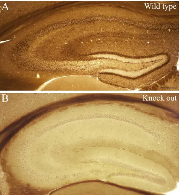
Figure 3: Lack of CB1-R staining in knock out animals
Staining was carried out with the antibody staining CB1R-positive terminals giving symmetric and the ones giving asymmetric synapses as well. In wild type animals (A) CB1- R staining was found in all hippocampal regions as shall be described later. In contrast, in CB1-R knock out animals (B) no specific staining was found, only light background staining occurred in the somatic regions. Lack of staining indicates the specificity of the antibody. Scale: 200 µm
4.9. Gallyas silver impregnation
In the mouse tissue degenerated neuronal elements were visualized with the staining procedure of Gallyas et al. (Gallyas et al, 1980). The steps of this staining are the following: 2x5min in the pretreating solution (2% NaOH and 2.5% NH4OH), 10 min in the impregnating solution (0–0.8% NaOH, 2.5% NH4OH, 0.5% AgNO3), 3x5 min in washing solution (0.5% Na2CO3 and 0.01% NH4NO3 in 30% ethanol), 1 min in developing solution (0.4–0.6% formaldehyde and 0.01% citric acid in 10% ethanol, pH 5.0 –5.5), 3x10 min wash in 0.5% acetic acid. Afterwards sections were mounted on gelatin-coated slides, dehydrated in xylene and covered with XAM neutral medium (BDH, Poole, UK).
4.10. Nissl staining
Cell loss could be visualized with Cresyil violet staining as well. Sections were mounted in chrome-gelatine and air-dried on slides. Afterwards the following steps were carried out: 2 mins in xylene, 3 mins in ethanol abs., 3 mins in 90% ethanol, 3 mins in 70%
ethanol, 3 mins in 50% ethanol, 5 mins in cresyle violet (1% in tap water), rinse in 70%
ethanol, rinse in 70% ethanol+drops of acetic acid for fixation, rinse in 90% ethanol, 3 mins in ethanol abs., 2x3 mins of xylene, and mounting with DePeX (SERVA Electrophoresis GmbH, Heidelberg, Germay) finally.
4.11. Determination of principal cell loss in epileptic mice
In the chronic phase, the severity of the acute seizures (using the Racine-scale, seizures from 1 to 5) was correlated with the cell-loss found in CA1, CA3 and hilus. Cell loss was determined in case of a large number of animals using a well defined semiquantitative scale by two independent examiners as shown previously (Magloczky &
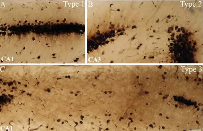
Freund, 1993; Magloczky & Freund, 1995; Wittner et al, 2005). Each region was classified as type 1-3 according to the severity of cell death (type1: cell-loss under 10 %, type2: cell- loss between 10% and 50%, type3: cell-loss above 50%) (Fig.4). Pearson correlation was used to calculate the relationship between cell-loss and seizure strength. Seizure strength was defined for every animal by using the highest number of Racine scale reached during
acute seizures. This number, representative for each animal was correlated with the severity of cell death (types 1-3) in each subregion.
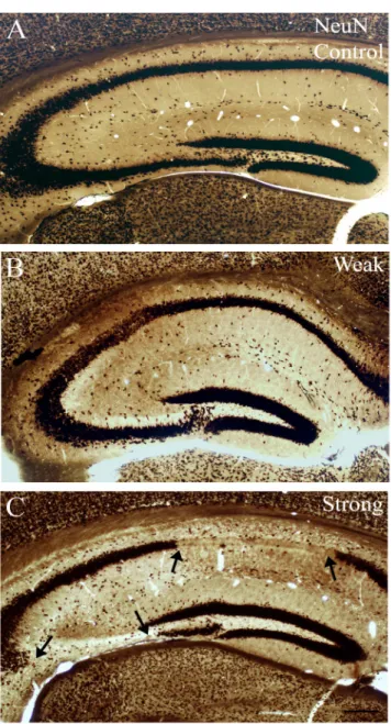
Figure 4: NeuN-staining shows different types of cell loss in the epileptic hippocampus Based on the severity of principal cell loss, distinct regions were classified as type 1, 2 or 3.
In case of type 1 (A) cell loss is quite mild, with only few missing cells (<10% of principal cells are missing in the given region). Type 2 (B) indicated cell loss in patches, affecting more numerous cells (11-50% of principal cells are missing in the given region). Type 3 (C) indicates sclerosis in the given region with more than 51% of principal cells missing.
Scale: 50 µm
4.12. Quantitative electron microscopic analysis
4.12.1. Ratio and number of symmetric and asymmetric synapses
Electron microscopic analysis was carried out in the dentate gyrus, since this area is affected by seizure induced changes, however granule cells are well-preserved even in case of robust sclerosis in the CA1 (Houser & Esclapez, 1996; Peng & Houser, 2005) therefore it is possible to examine the surviving elements which may not be the case in CA1.
The quantity of symmetric and asymmetric synapses established by CB1-R-positive axon terminals was examined throughout the str. moleculare of 3 control, 3 chronic epileptic and 3 acute epileptic hippocampi. Serial sections were made from the blocks reembedded from stratum moleculare and examined in the electron microscope. CB1-R stained terminals were analyzed in every 10th section in order, following the rules of systematic random sampling, to avoid sampling of the same axon terminals. Photographs were taken of all terminals in a given area, and the ratio of immunostained symmetric versus asymmetric synapses was determined.
To quantify the exact changes in the number of stained terminals in the chronic and acute phase, we further examined the str. moleculare. The number of CB1-R immunostained terminals forming symmetric and asymmetric synapses was calculated, both in control and in epileptic samples and normalized. The studied areas were measured with NIH ImageJ (U.S. National Institutes of Health, Bethesda, MD) program and were normalized to unit area (40 000 µm2).
To assess any changes occurring in the density of CB1-Rs per terminals, the number of gold particles located in the membrane of axon terminals was counted and normalized to a unit length of the terminal’s perimeter (perimeter length was measured with NIH ImageJ, number of particles was normalized to 1 µm). Significance was tested using Mann-Whitney U-test (Statistica 6.0).
4.13. Target distribution of CB1-R-positive terminals forming symmetric synapses In the human and mouse tissue target distribution of CB1-R immunopositive elements was studied in the stratum moleculare of the dentate gyrus in control and sclerotic subjects, since in sclerotic samples the highest fiber density can be observed in this area.
Both in controls (N=105, two subjects) and in epileptic patients (N=175, three sclerotic subjects) the postsynaptic target of CB1-immunopositive terminals establishing symmetric synapses were examined (photographed and quantified) at the electron microscopic level.
5. Results
5.1. Reorganization of CB1 receptor expressing GABAergic fibers in temporal lobe epileptic patients
The expression pattern of CB1-receptors associated with inhibitory synapses was studied by immunocytochemistry in epileptic hippocampal tissue derived from intractable TLE patients (N=44). A previous study by Ludanyi et al. has demonstrated a decrease of CB1-Rs associated with asymmetric synapses, thus, we examined changes affecting the number of CB1-R-positive symmetric synapses.
In the hippocampi of human TLE patients the pattern of cell loss was analyzed by light microscope in sections immunostained for different neurochemical markers labeling principal and non-principal cells (Magloczky & Freund, 2005; Toth et al, 2010; Wittner et al, 2005). Based on anatomical alteration observed in the hippocampus patients could be classified into different groups.
5.1.1. Pathological classification of the epileptic samples
All patients examined in the present study had therapy resistant epilepsy of temporal lobe origin. The patients had different degrees of hippocampal atrophy and/or sclerosis. All the examined sections derived from the anterior part of the hippocampal body. Similarly to our previous studies (Clemens et al, 2007; Toth et al, 2010; Wittner et al, 2005; Wittner et al, 2002) and other results (de Lanerolle et al, 2003), patients with epilepsy were classified based on principal cell loss and interneuronal changes examined at the light microscopic level as follows.
(i) Epileptic Type 1 (mild) (n = 6): similar to control, no considerable cell loss in the CA1 region, pyramidal cells present, layers are visible and intact, their borders are
clearly identified. There is a slight loss in certain interneuron types, mostly in the hilus and the stratum oriens of the CA1 region.
(ii) Epileptic Type 2 (patchy) (n = 16): pyramidal cell loss in patches in the CA1 pyramidal cell layer but the CA1 region is not atrophic. Interneuron loss is more pronounced.
(iii) Epileptic Type 3 (sclerotic) (n = 22): the CA1 region is shrunken, atrophic, more than 90% of principal cells are missing. Only the stratum lacunosum-moleculare is present as a distinct layer in the CA1 region, the others could not be separated from each other due to the lack of pyramidal cells and their dendrites and the shrinkage of the tissue. This remaining part should contain the layers identified in the control as stratum oriens, stratum pyramidale and stratum radiatum. Mossy fiber sprouting and considerable changes in the distribution and morphology of interneurons (not shown) can be observed in the samples of this group.
The number, morphology and distribution of cells were similar in patients in the same pathological group, but differed between the groups. The control samples with short (2–4 h) post-mortem delay were similar to each other and differed from the epileptic groups.
Therefore, we concluded that the differences found between control and epileptic tissues in the present study are likely to be caused by epilepsy. (Toth et al, 2010; Toth et al, 2007;
Wittner et al, 2005).
In the present study the distribution and localization of CB1-immunoreactive elements was studied in controls, in non-sclerotic (Type 1 and Type 2) and sclerotic cases (Type 3)
5.1.2. Distribution of CB1-R-stained inhibitory synapses in control human hippocampus In the following set of experiments the antibody used to visualize CB1-Rs stained receptors found in symmetric synapses, but did not stain receptors located at asymmetric synapses.
Immunostaining revealed numerous CB1-positive cell bodies of interneurons scattered in all hippocampal subfields, these cells have been shown to express the neuropeptide CCK (Katona et al, 1999b) and were either perisomatic or dendritic targeting interneurons (Freund & Buzsaki, 1996). Parts of interneuron dendrites were stained, and a dense meshwork of CB1-immunoreactive axons covered the entire hippocampal formation.
The strongest axonal labeling was found in stratum moleculare of the dentate gyrus, mostly in the inner part of str. moleculare. Strong staining appeared also in stratum pyramidale of CA1-CA3 containing numerous axon terminals forming basket –like structures surrounding pyramidal cells (Katona et al, 2000). In contrast no labeling occurred in the neuropil in the hilus and str. lucidum, moreover, granule cells were immunonegative.
5.1.3. Distribution of CB1-Rs in epileptic human hippocampus
In the non-sclerotic cases, the distribution of CB1 receptors in the dentate gyrus did not show any major changes compared to the normal post mortem controls. In contrast, a strong increase in CB1 receptor immunostaining was found in the dentate gyrus of epileptic patients with CA1 sclerosis, similar to what we have seen in the mouse model.
Immunopositive interneuron somata were preserved both in the dentate gyrus and in the CA1 area. The density of immunostained fibers increased in the dentate molecular layer (Fig. 5) and became inhomogeneous in the hilus forming dense arrays of boutons around the surviving mossy cells and interneurons.
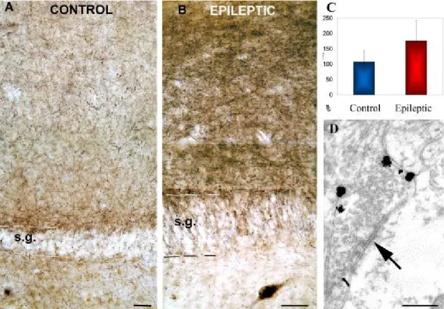
Figure 5. Distribution of CB1-R associated with symmetric synapses at light and electron microscopic level in human dentate gyrus
A) Low power light micrograph showing the distribution of CB1-positive profiles in the control human dentate gyrus. Granule cells are always negative for CB1. B) Tissue from sclerotic epileptic patients remarkably differs from controls. The density and staining intensity of CB1-positive fibers is considerably increased in the stratum moleculare. C) Density of CB1 receptor immunopositive fibers in control (N=3) and sclerotic epileptic patients (N=10) revealed by confocal laeser scanning microscope. The intensity of CB1 receptor staining is elevated in epileptic samples. The difference between control and epileptic samples was highly significant (p<0,05; Student t test). D) A CB1-R-positive terminal from the stratum moleculare of an epileptic patient labeled by immunogold technique establishes symmetric synapse on a dendrite. All of the terminals stained by this CB1 antibody established symmetric synapses. Scales: A,B:50 µm; D:0,5 µm
5.1.4. Qualitative analysis of immunopositive fiber density in human sclerotic tissue Qualitative analysis revealed an increased density of immunostained axonal meshworks in the sclerotic stratum moleculare compared to the control or non-sclerotic cases. To quantify this increase, sections with fluorescent immunostaining were processed in 3 control and 10 sclerotic cases, and the density of immunolabeling was measured by confocal laser scanning microscopy. The results showed that the density of fibers had significantly increased in epileptic cases (type 3) (Fig. 5).
5.1.5. Ultrastructural analysis of CB1-R distribution in control and sclerotic human tissue
Electron microscopic examination confirmed that the antibody used in this study visualized CB1-Rs only on terminals giving symmetric synapses (Fig. 5, 6). The cellular and subcellular localization of CB1-Rs was similar in the epileptic and control cases. CB1- R-positive terminals form synapses mostly on dendrites and less frequently on spines and somata of granule cells in control and epileptic samples. However, we wished to uncover any potential changes occurring in the target distribution of CB1-R containing terminals in epileptic the tissue.
5.1.6. Target distribution of CB1-immunopositive elements in human TLE
Target distribution of CB1-immunopositive elements was studied in the stratum moleculare of the dentate gyrus in control and sclerotic TLE subjects, where the highest fiber density was observed. Examination of immunogold terminals showed that CB1 receptors were localized in the membrane, outside the synaptic active zone in epileptic patients, as it was shown previously in the control human hippocampus (Katona et al, 2000) (Graph 1). Both in controls (N=105, two subjects) and in epileptic patients (N=175, three subjects) the CB1-immunopositive terminals established symmetric synapses mostly on dendrites (75 v.72,5 %, spines 13,2 v. 15,5 %, and cell bodies 11,8 v. 13 %, in control and epileptic subjects, respectively).
Therefore, we can conclude that the target distribution was not changed, although the density of fibers has been elevated greatly.
Target distribution of CB1-R-positive terminals in human
0 20 40 60 80
dendrite spine soma
% control
sclerotic
Graph 1: Target distribution of CB1-R-positive terminals establishing symmetric synapses in human TLE samnples
The ratio of CB1-R-positive fibers synapsing on distinct neuronal elements does not change in the epileptic tissue compared to controls.
A previous study by Ludanyi et al (Ludanyi et al, 2008) has demonstrated a downregulation of CB1-Rs linked to asymmetric synapses, whereas, our results show an upregulation in CB1-Rs related to symmetric synapses. These results are quite surprising and indicate that epileptic reorganization affects CB1-R-positive excitatory and inhibitory networks in a different way. To understand the time course of the dynamics of CB1-R expression, we established an animal model of TLE (pilocarpine model, we shall describe it
later) showing similar pathological changes to human TLE. First we characterized changes affecting CB1-Rs linked to symmetric synapses and studied alterations in the target distribution of immunopositive terminals in the mouse model with the same antibody as we used for human tissue (Antibody staining CB1-R-containing terminals establishing symmetric synapses).
5.2. Changes of CB1-receptor immunostained inhibitory fibers in a model of temporal lobe epilepsy
Similar to human TLE tissue, upregulation of CB1-Rs linked to symmetric synapses could be seen in the mouse model as well (using the same antibody as in case of human tissue). The expression pattern of CB1-Rs in the hippocampi of both strong and weak CD1 mice was studied at a survival time of 4-8 weeks (chronic phase).
In control samples numerous immunostained cell bodies were seen (previously shown to be CCK-positive (Katona, 1999 #3054) and could be either dendritic or perisomatic targeting interneurons (Freund & Buzsaki, 1996)). The most intense staining of CB1-positive fibers was found in the molecular layer of the dentate gyrus (DG), in stratum pyramidale of the cornu Ammonis (CA), and in the subiculum. In contrast, faint labeling was observed in the strata radiatum and granulosum (Fig. 3). CB1-immunostaining showed no change in the hippocampi of weak animals compared to controls. In the sclerotic samples CB1-R-positive cell bodies were preserved, however, the general CB1- immunostaining was much stronger throughout the hippocampus. The most striking difference was found in the strata moleculare and granulosum of the DG (Fig. 6), in the strata pyramidale and radiatum of the CA1.)
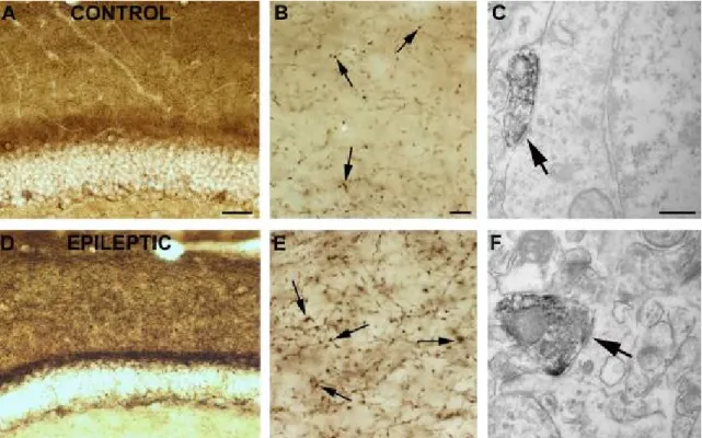
Figure 6: Demonstration of CB1-Rs at light and electron microscopic level in mice
CB1 immunostaining in the dentate gyrus of a control (A, B, C) and a “strong” epileptic (D, E, F) mouse. In control samples (A,B), dense staining of CB1-positive fibers can be found in the molecular layer of the DG. In the stratum moleculare of sclerotic animals (D, E) the intensity of CB1 immunostaining is elevated. The most frequent postsynaptic targets of CB1 receptor-positive terminals establishing symmetric synapses (arrows) are cell bodies (C) and dendrites (F) Scales: A,D: 200 µm; B,E: 50 µm; C,F:1 µm.
5.2.1. Target distribution of CB1-immunopositive elements in sclerotic mouse tissue Quantification of CB1-immunopositive element’s target distribution was carried out in the mouse model as well to examine whether the unchanged target distribution is human specific or not. Stratum moleculare of the dentate gyrus was studied in control and sclerotic animals, since the highest fiber density was observed in this area. Similar to controls and human tissue, CB1 receptors were localized in the membrane, outside the synaptic active zone (Nyiri et al, 2005). Both in controls (N=53) and sclerotic cases (N=48) the target
distribution of CB1-R immunopositive fibers was examined. Stained terminals established symmetric synapses mostly on dendrites (77.4 v.77.1%, spines 22.6 v.20.8% and cell bodies 0 v. 2.1 %) (Graph 2). Thus, target distribution was not changed, similar to the human tissue.
Target distribution of CB1-R-positive terminals in mouse tissue
0 20 40 60 80 100
dendrite spine soma
% control
sclerotic
Graph 2: Target distribution of CB1-R-positive terminals establishing symmetric synapses in mouse TLE samples
The ratio of CB1-R-positive fibers synapsing on distinct neuronal elements does not change in the epileptic tissue compared to controls.
As the next step we carried out a more detailed investigation to reveal changes in the receptor protein expression in different phases of pilocarpine induced epilepsy (acute, latent and chronic). For this set of experiments we used an antibody staining CB1-R- positive symmetric and asymmetric synapses as well (see materials and methods).
5.3. Description of the pilocarpine model of epilepsy
5.3.1. The pattern of cell loss in pilocarpine induced epilepsy
To clarify the alterations of the hippocampal circuits on the pilocarpine model of epilepsy using CD1 mice, we carried out detailed investigation. Based on the behavioral signs during the acute seizures, animals were classified as “weak” or “strong” epileptic using the modified Racine scale (Magloczky et al, 2010; Racine, 1972; Turski et al, 1984).
Animals showing seizure severity from Racine 1 to Racine 4 (shaking, chewing, nodding, forelimb clonus, rearing but no tonic-clonic seizures) were assigned to the weak group.
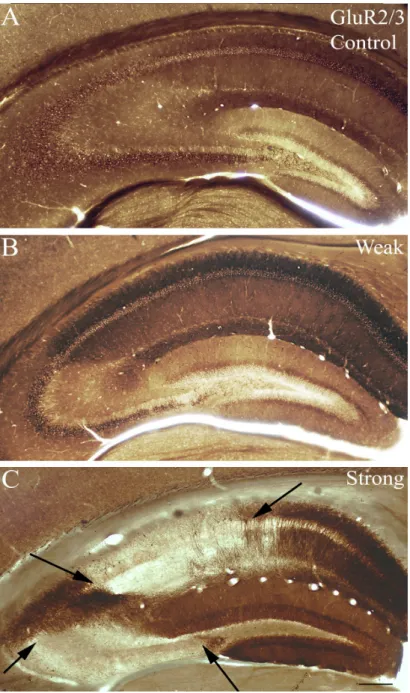
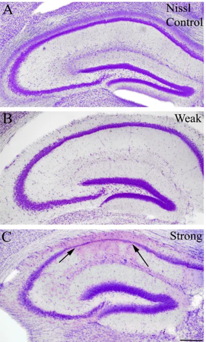
However, in the strong group all animals showed tonic-clonic seizures (Racine 5) with other less severe seizure manifestations (Fig. 7). We examined the cell loss pattern in the hippocampi at different survival times using NeuN-immunostaining (Fig. 8), GluR2/3- immunostaining (Fig. 9) Nissl staining (Fig. 10). Since cell loss observed was similar in case of all three staining further on in the study, NeuN staining was used to determine cell- loss.
In the acute phase (2 hours after the treatment) cell loss was not observed neither in the weak nor in the strong animals, nor did signs of hippocampal reorganization occur (data not shown).
In the latent phase, (1-3 days post-pilocarpine) loss of sensitive interneurons (calretinin positive) and few principal cells could already be seen in the CA1 and CA3 regions (3 days) (not shown). Three days post pilocarpine, cell loss was usually patchy in the CA1-3; occasionally pyramidal cells disappeared in long segments of areas.
In the chronic phase, patchy cell loss was found infrequently in “weak” animals, but none of their hippocampi showed sclerosis (Fig. 8B, 9B, 10B). In contrast, hippocampi of the “strong” mice showed sclerosis in most cases (70%) meaning that CA1 and CA3 regions were shrunken, atrophic and more than half of the cells were missing from the CA1 region. Loss of vulnerable interneurons was also observed in all regions (Fig. 7) (Fig. 8C, 9c, 10C). In our model calretinin-containing cells were used as a marker to monitor early cell death, since these cells were the most sensitive to epileptic insult (not shown) (Magloczky & Freund, 2005; Toth et al, 2010) .
Based on the severity of cell death, the degree of damage affecting different subregions (CA1, CA3, dentate gyrus) was classified as type 1 (mild cell loss), type 2 (patchy cell loss) and type 3 (sclerotic), defined according to the following semiquantitative scale: type 1, up to 10% of the cells are damaged; type 2, 11-50% of the cells are missing;
type 3: more than 50% of the cells are missing. CA1 and CA3 were considered sclerotic when cell loss exceeded 50 %, especially when strata oriens, pyramidale and radiatum could not be separated any more (Magloczky & Freund, 1993; Magloczky & Freund, 1995) (Fig 4).
Statistical analysis revealed a significant correlation (Pearson correlation) between the degree of cell-loss and seizure strength in Racine scale values (p<0.05) (105 animals) (Fig. 7). Thus, behavioral signs of the acute seizures could be used to predict the degree of cell loss in the chronic phase.