American Journal of Otolaryngology and Head and Neck Surgery
Hemipharyngo-Laryngectomy for Treatment of T1-2 Hypopharyngeal Tumours
OPEN ACCESS
*Correspondence:
Miklós Csanády, Department of Otorhinolaryngology and Head & Neck Surgery, University of Szeged, Tisza Lajos bld. 111, Szeged, 6725, Hungary, Tel: 3662545310; Fax: 3662545848;
E-mail: mcsanady@freemail.hu Received Date: 24 Apr 2019 Accepted Date: 15 May 2019 Published Date: 22 May 2019
Citation:
Szabó D, Csanády M, Rovó L.
Hemipharyngo-Laryngectomy for Treatment of T1-2 Hypopharyngeal Tumours. Am J Otolaryngol Head Neck Surg. 2019; 2(5): 1052.
Copyright © 2019 Miklós Csanády.
This is an open access article distributed under the Creative Commons Attribution License, which permits unrestricted use, distribution, and reproduction in any medium, provided the original work is properly cited.
Published: 22 May, 2019
Abstract
Authors report the larynx function sparing surgical technique: supraglottic hemipharyngo- laryngectomy in 44 cases of T1 and T2 early pyriform sinus tumours infiltrating the supraglottic area and the medial hypopharyngeal wall. Surgical technique, preoperative diagnostics, oncological and functional results are demonstrated. Between 2000 to 2011, 44 patients underwent function sparing supraglottic hemipharyngo-laryngectomy with neck dissection. In 34 cases the therapy was completed with radiotherapy according to the pathological stage of the primary tumour and the lymph nodes.
During the period of following in case of 10 patients (23%) did not occur recurrent tumour. In 4 (9%) patients total laryngectomy was performed due to local recurrents, 7 (16%) patients underwent radical neck dissection due to late cervical metastases. Second primary tumour occurred in case of 4 (9%) patients and 12 (27%) patients were diagnosed with distant metastases. 12 (27%) of our patients died because of distant recurrences, and 6 (14%) because of other reasons. The Kaplan- Maier survival rate shows the followings: 1 year 76.5%, 3 year 41.9%, and 5 year 36.4%.
The authors find that the supraglottic hemipharyngo-laryngectomy combined with neck dissection is an appropriate technique in the clinical practice for the removal of early hypopharyngeal tumours invading the medial wall of pyriform sinus not only for function sparing but from oncological aspect as well. The phonation of the patients after surgery is good, the quality of life is significantly better than after total laryngectomy even in spite of the remaining minimal aspiration.
Keywords: Supraglottic hemipharyngo-laryngectomy; Lateral pharyngotomy; T1-2 pyriform sinus tumours; Larynx function sparing; Neck dissection
Diána Szabó, Miklós Csanády* and László Rovó
Department of Otorhinolaryngology and Head & Neck Surgery, University of Szeged, Hungary
Introduction
The incidence of hypopharyngeal tumours show a growing tendency, which is on one hand due to the well known risk factors (smoking, alcohol consumption) and on the other hand due to the change in the eating habits, the inappropriate nourishment and poor oral hygiene. The efforts for preventing the development of hypopharyngeal tumours were proved ineffective according to the survival statistics thus the early diagnosis of these tumours can be the key to the more effective treatment. The early diagnosis of tumours invading the lateral or the medial wall of the pyriform sinus not only improves the survival rates but enables the preservation of laryngeal function.
In case of early T1 selected hypopharyngeal (marginal and posterior wall) tumours endoscopic laser resection completed with neck dissection can be performed on a selected group of patients (1), of T1 and T2 hypopharyngeal tumours infiltrating the medial wall of pyriform sinus we consider that the partial pharyngo-laryngectomy with neck dissection is safer from the oncological point of view.
In case of T3 and T4 hypopharyngeal tumours total laryngectomy and partial pharynx resection with neck dissection is the proper treatment according to the oncological principles. According to the principals of the complex oncological treatment the complementary radio and/or chemotherapy is necessary in every case depending on the postoperative histological result.
Ambulant endoscopic diagnostic and imaging techniques are essential in order to set the diagnosis. Directoscopy performed in intratracheal narcosis and appropriate biopsy, are necessary for setting the surgical plan. The type of the surgery and the indication of other oncological treatments are determined after the assessment of the tumor’s extension and its laryngeal infiltration by endoscopy and imaging.
In our department we have introduced the laryngeal function sparing technique, since 20
years supraglottic hemipharyngo-laryngectomy for the removal of T1 and T2 tumours infiltrating the medial wall of the pyriform sinus and reaching into the supraglottic and marginal regions in the ’90-s. In selective cases of T1 marginal hypopharyngeal tumours endoscopic laser resection, neck dissection and radiotherapy can be a treatment option [1], but this technique depends on the extension and localization of tumour.
As for the Hungarian results Balatoni et al. [2], Répássy et al. [3,4]
demonstrated the supracricoid hemipharyngo-laryngectomy; both of them had promising oncological results. Gödény [5] told about the imaging diagnostics of laryngeal and pharyngeal tumours and emphasized the importance of CT and MRI.
In the international literature Makeieff let al. [6] demonstrated 60% 5-year-survival after performed supraglottic hemipharyngo- laryngectomy with neck dissection and postoperative radiotherapy in patients with T1 and T2 (N1-N3) tumours infiltrating the medial wall of pyriform sinus and marginal laryngeal tumours with cervical metastases. Local recurrence occurred in 20%, second primary tumour occurred in 24% and late cervical metastasis occurred in 28%. Jacob et al. [7] find the hemipharyngo-laryngectomy performed by Lacourreye et al. [8] a good function sparing method, they have performed it on 26 patients and beside the good long-term oncological results they have found the postoperative swallowing and phonation results proper as well. Hagen et al. [9] demonstrates the extended function sparing hemipharyngo-laryngectomy in case of advanced T3, T4 hypopharyngeal tumours, where a free tissue flap taken from the underarm was applied with microvascular anastomosis for the reconstruction of the pharynx and the vocal chords. Chantrain et al. [10]
used such a flap for the laryngeal reconstruction after hemipharyngo- laryngectomy, which enables a more radical tumour resection in their opinion. Chevalier et al. [11] found the supraglottic hemipharyngo- laryngectomy and postoperative radiotherapy a good choice of function sparing therapy in 49 patients with early marginal pyriform sinus tumours. They have demonstrated 2% local recurrence and 47%
5-year-survival. Gehanno et al. [12] performed lateral hemipharyngo- laryngectomy with arytenoidectomy in case of marginal T1-T3 larynx and hypopharyngeal tumours and achieved good functional and as good oncological results as Chevalier’s patients had. 15% patients had locoregional recurrences, 5-year survival rates were 51%. From the oncological point of view Holsinger et al. [13] achieved promising results by performing lateral pharyngotomy, primary reconstruction and complementary radiotherapy in case of tumours infiltrating the lateral wall of the pyriform sinus; the 5-year-survival was 80%. Spector et al. [14] analyzed the treatment results of 408 patients with pyriform sinus tumour after radiotherapy, hemipharyngo-laryngectomy and combined therapy. 207 patients received partial surgical treatment, pharynx reconstruction and postoperative irradiation. This group of patients with tumours infiltrating the medial wall of the pyriform sinus showed 63% 5-year-survival. The 5-year-survival was better only in case of one region infiltrating lateral wall involving hypopharynx tumours. In case of tumours affecting more regions the 5-year- survival was 49%. Regional metastases significantly decrease the survival rate: 26% by N1 stage, 38% by N2 and N3. Further survival influencing factors are the occurrence of a second primary tumour, distant metastases and the other following diseases of the patient. The results of Wang et al. [15] show that survival results after function sparing partial surgical treatment of hypopharyngeal tumours with or without complementary radiotherapy and after laryngectomy are similar (46% vs. 44%), but the quality of life is significantly better if the phonation is preserved.
We demonstrate the surgical technique of supraglottic hemipharyngo-laryngectomy applied at the Department of Otorhinolaryngology and Head & Neck Surgery in Szeged, the oncological and functional results in the aspects of quality of life in case of T1-T2, N1-N2 tumours infiltrating of the medial wall from pyriform sinus.
Methods
In the Department of Otorhinolaryngology and Head & Neck
Figure 1: Preoperative endoscopic picture of pyriform sinus tumour infiltrating the medial hypopharyngeal wall.
Figure 2: Schematic drawing of the hemipharyngo-laryngectomy.
Parameter No. of patients %
Age
< 60 26 59
˃60 18 41
Sex
male 9 20.5
female 35 79.5
T classification T1 15 34.1
T2 29 65.9
Cervical metastasis
yes 35 79.5
no 9 20.5
Postoperativ irradiation
yes 34 77.3
no 10 22.7
Postoperativ chemotherapy irradiatirradiation
yes 16 36.4
no 28 63.6
Table 1: Demografic profile of patients.
Surgery of University of Szeged between 2000 and 2011 we performed 44 (Table 1) supraglottic hemipharyngo-laryngectomy in case of T1, T2 tumours infiltrating the medial wall of the pyriform sinus and spreading to the supraglottic area. Our patients underwent preoperative endoscopy (Figure 1), video (voice) record was made, and directoscopy and biopsy in intratracheal narcosis and in case of cervical metastasis a fine needle aspiration cytology. Cervical ultrasound examination and CT was performed, but some MRI has been made as well. In some cases we recommended 3 dimension color CT examination in order to the better assessment of the tumor’s extension (3D Slicer).
In general we applied directoscopy and laryngoscopy to define the extension of tumour to the hypopharyngeal and laryngeal structures in case hypopharyngeal tumours (aryepiglottic fold, ary region ventricular fold). In 40 cases we performed supraglottic hemipharyngo-laryngectomy with neck dissection and tracheotomy from a unilateral incision, so called a “big “J” shaped incision line”.
Bilateral neck dissection was carried out in 4 cases in order to remove the bilateral cervical metastases.
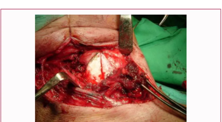
After performing the neck dissection (Figures 2 and 3) our surgical method includes dissection of the prelaryngeal muscles, freeing up and removing the half of the hyoid bone. After this came the resection of the tumour together with the lateral part of the thyroid cartilage straight from the inferior horn up to medial part of the upper edge (Figure 4), but the perichondrium of the resected thyroid cartilage must be preserved for the reconstruction of the pharynx. During lateral pharyngotomy (Figures 5 and 6) we opened the pharynx so the epiglottis and the pyriform sinus became visible, then we ran the resection line laterally in the pyriform sinus, medially removed a part of the epiglottis then further went through the preepiglottic area (or removed some part of it) we resected the aryepiglottic fold, the ventricular fold (in some cases the ary-region as well) together with the medial wall of the pyriform sinus (Figure 7). The preserved hypopharyngeal mucosa, the vallecula and the radix of the tongue were closed with running stitches the preserved perichondrium would be used to the reconstruction of the larynx and pharynx (Figure 8).
At the end of the reconstruction the prelaryngeal muscles would be sutured to the radix of the tongue (Figure 9). The skin was closed with intracutaneous stitches. During the surgery temporary tracheostomy was prepared and inserted a nasogastric tube (except in a patient with gastrostomy). We did not prepare any preoperative gastrostomy, only one patient required gastrostomy after the surgery because of long- term swallowing difficulty in the postoperative period.
We completed the therapy with radiotherapy in 34 cases according to the primary tumour and the pathological stage of cervical lymph nodes due to the decision of the Head and Neck Surgery Oncoteam of University of Szeged.
Ten patients did not receive any postoperative oncological
Figure 3: Modified radical neck dissection.
Figure 4: Exploratio of lateral part of the thyroid cartilage straight from the inferior horn up to medial part of the upper edge.
Figure 5: Neck dissection and tracheotomy from unilateral incision, lateral pharyngotomy, exploration of larynx (thyroid cartilage).
Figure 6. The pyriform sinus tumour’s resection.
Figure 7: Tumour specimen with removed lateral part of thyroid cartilage.
treatment: 2 patients had N0 histological results (decision of the oncoteam) and the other 8 patients either did not accept the complementary treatment or it could not be performed (due to early death).
The changes of the patient’s voice were evaluated by postoperative voice analysis: by analyzing the Mean Phonation Time (MPT), Mean Fundamental Frequency (MFF), jitter (%), shimmer (%) and Harmonics-to-Noise Ratio (HNR). The voice samples were recorded with a high sensitivity (40 Hz to 16 kHz) condenser head microphone (Audio-Technica ATM75) at a sampling frequency of 96 kHz, 24 bit (Tascam US 122MkII external soundcard) and analyzed by Praat 5.3.2.9. Software (www.praat.org).
Life quality was evaluated with the help of the Voice Handicap Index as well as with the Monnier’s Quality Of Life questionnaire.
Results
Function sparing supraglottic hemipharyngo-laryngectomy with
Figure 8: The preserved hypopharyngeal mucosa, perichondrium be used for the reconstruction of the larynx and pharynx.
Figure 9: Laryngeal end pharyngeal reconstruction.
Figure 10: Postoperativ endoscopic documentation.
neck dissection was performed in 44 cases because of early (T1, T2) hypopharyngeal tumour infiltrating the medial wall of the pyriform sinus. Ten (23%) patients had no recurrence, second primary tumour (2 oesophageal, 1 radix of the tongue, 1 soft palate) occurred in 4 (9%) patients. These patients received radio- and/or chemotherapy according to the decision of the oncoteam. Late (regional) cervical metastasis (2 ipsy and 5 contralateral) occurred in 7 (16%) patients, thus we performed I-IV. level radical neck dissection. Distant metastases were diagnosed in 8 (18%) patients: 5 pulmonary, 1 vertebral, 1 inguinal and 1 mediastinal tumour. 12 (27%) of our patients died because of distant metastases and 6 (14%) patients died because of general health condition problems (pneumonia, pulmonary oedema and exsiccation). Laryngeal function was successfully preserved in 89%. 73% of our patients were locally tumour free after salvage surgery.
The result of the histological examination was squamous cell carcinoma in all cases. In 2 cases we did not find cervical metastasis during neck dissection. Postoperative complications were the followings: 1 case of subcutaneous emphysema, 3 cases of bleeding, 5 cases of pharyngocutaneous fistula and 3 cases of wound fluid discharge. Medium-high grade aspiration occurred after surgery, which improved gradually. The patients were decannulated between 5 to 10 postoperative days; the nasogastric tube was removed after 2 to 3 weeks. We did not prepare any preoperative gastrostomy, but 1 patient (decannulated, preserved phonation) required it temporarily because of chronic swallowing problems. In spite of the remaining postoperative minor residual aspiration the patients are able to feed properly. Control video endoscopies (Figure 10) demonstrated the mobility of the affected half of the larynx was reduced or in most cases
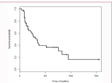
Figure 11: Kaplan-Meier disease-specific survival curve.
Patient 1. Patient 2. Normal Values
Mean Phonation Time (s) 10.18 9.37 15-20
Mean fundamental frequency [Hz] 111.636 145.035 85-196
Jitter (%) 2.313 0.683 <1.040
Shimmer (%) 11.239 13.436 <3.81
Harmonics-to-noise ratio (dB) 13.506 13.240 20 dB<
Voice Handicap Index 46 52 0-120
Quality Of Life 12 10 6-25
Table 2: The postoperativ recorded acoustic parameters in two representative patients.
ceased because of the removal of pyriform sinus and the resection in the ary region, furthermore the degree of aspiration correlated with the previous facts.
The survival according to Kaplan-Meier with 95% of confidence interval is demonstrated: 1 year survival 76.5%, 3 year survival 41.9%, and 5 year survival 36.4% (Figure 11).
Summary of the postoperative acoustic data are shown in Table 2. Voice outcome in patients who underwent hemipharygo- laryngectomy was good.
These results indicate that the preservation of laryngeal function after hemipharyngo-laryngectomy in early pyriform sinus tumours improved the postoperative quality of patient’s life and beside this the long-term oncological results are satisfying as well.
Discussion
Organ sparing supraglottic hemipharyngo-laryngectomy combined with neck dissection and tracheotomy and introduction of nasogastric tube were performed in our department for the treatment of T1, T2 tumours infiltrating the medial wall of pyriform sinus. We found the patient’s postoperative aspiration a medium-high degree, which generally moderated in 2 to 3 weeks, but in most cases we were able to remove the nasogastric tube after 10 to 14 days because the improved deglutition. In case of T1, T2 hypopharyngeal tumours this method caused a smaller surgical stress for the patients because of the unilateral exploration. Due to the intact contralateral half of the pharynx and larynx and prelaryngeal muscles the regeneration of swallowing and the laryngeal functions were faster than in case of bilateral pharyngo-laryngeal exploration. The preservation of vocal chords results in the proper closure of the glottis thus the degree of aspiration can be decreased and the phonation remains appropriate as well. We successfully decannulated all of our patients, the degree of aspiration became minimal or ceased in most cases, but in case of a low degree aspiration the oral alimentation remained appropriate as well.
The supraglottic hemipharyngo-laryngectomy for the treatment of T1, T2 tumours infiltrating the medial wall of pyriform sinus proved to be a good organ sparing alternative vs. total laryngectomy.
According to the data the survival rate is similar to the patients who underwent total laryngectomy, but the quality of life is significantly better because the preservation of the phonation, in spite of the probable swallowing problems as well. The side effects of the postoperative radiotherapy, promotes the patient’s swallowing complaints (dryness of the mouth, local oedema).
References
1. Csanády M, Kiss JG, Iván L, Jóri J, Czigner J. 5-aminolevulinic acid (ALA) induced protoporphyrin IX fluorescence in the endoscopic diagnostic and control of pharyngo-laryngeal cancer. Eur Arch Otorhinoplaryngol.
2004;261(5):262-6.
2. Balatoni Zs. Supracricoid hemilaryngopharyngectomia. Fül-orr- gégegyógyászat. 2002;46(4):219-27.
3. Répássy G, Hirschberg A, Rezek Ö, Kisely M, Tóth A, Juhász A.
Supracricoid lateralis gégeresectio a recessus piriformis rák kezelésére.
Fül-orr-gégegyógyászat. 2000;46(2):155-62.
4. Répássy G, Hirschberg A, Jókay I, Tóth L, Rezek Ö, Juhász A, et al.
Supracricoid horisontalis laryngectomia. Fül-orr-gégegyógyászat.
2000;46(2):91-8.
5. Gödény M. Role and responsibility of multimodal imaging in head and neck cancer. Magy Onkol. 2013;57(3):182-202.
6. Makeieff M, Mercante G, Jouzdani E, Garrel R, Crampette L, Guerrier B. Supraglottic hemipharyngolaryngectomy for the treatment of T1 and T2 carcinomas of laryngeal margin and piriform sinus. Head Neck.
2004;26(8):701-5.
7. Jacob R, Zorowka P, Welkoborsky HJ, Mann WJ. Long-term functional outcome of Laccourreye hemipharyngectomy-hemilaryngectomy with reference to oncologic outcome. Laryngorhinootologie. 1998;77(2):93-9.
8. Laccourreye O, Laccourreye L, Crevier-Buchman L, Brasnu D, Weinstein GS. Supracricoid hemilaryngo-pharyngectomy conversion to Pearson's near-total laryngectomy: A case report. Head Neck. 1997;19(3):232-4.
9. Hagen R. Functional long-term results following hemipharyngo- hemilaryngectomy and microvascular reconstruction using the radial forearm flap. Laryngorhinootologie. 2002;81(3):233-42.
10. Chantrain G, Deraemaecker R, Andry G, Dor P. Wide vertical hemipharyngolaryngectomy: reconstruction with the radial forearm free flap. Laryngoscope. 1991;101(8):869-75.
11. Chevalier D, Watelet JB, Darras JA, Piquet JJ. Supraglottic hemilaryngopharyngectomy plus radiation for the treatment of early lateral margin and pyriform sinus carcinoma. Head Neck. 1997;19(1):1-5.
12. Gehanno P, Barry B, Guedon C, Depondt J. Lateral supraglottic pharyngo- laryngectomy with arytenoidectomy. Head Neck. 1996;18(6):494-500.
13. Holsinger FC, Motamed M, Garcia D, Brasnu D, Ménard M, Laccourreye O. Resection of selected invasive squamous cell carcinoma of the pyriform sinus by means of the lateral pharyngotomy approach: the partial lateral pharyngectomy. Head Neck. 2006;28(8):705-11.
14. Spector JG, Sessions DG, Emami B, Simpson J, Haughey B, Harvey J, et al. Squamous cell carcinoma of the pyriform sinus: a nonrandomized comparison of therapeutic modalities and long-term results. Laryngoscope.
1995;105(4 Pt 1):397-406.
15. Wang XL, Xu ZG, Tang PZ. Surgical treatment on primary lesion of advanced pyriform sinus cancer. Zhongguo Yi Xue Ke Xue Yuan Xue Bao.
2006;28(4):534-7.